EM · ABCDE approach
ABCDE approach to the deteriorating patient
A structured, prioritised approach to the recognition and resuscitation of the deteriorating emergency department patient — airway, breathing, circulation, disability, exposure — built on oxygen-delivery physiology, early-warning scoring, the pathophysiology of compensated shock, and time-critical sepsis care. ACEM-primary, globally tagged.
On this page & tools
Your progress
Saved locally on this device.
Target exams
Red flags
The ABCDE approach is a prioritised, repeatable framework for assessing and resuscitating the acutely ill or deteriorating patient. It treats problems in the order in which they threaten life — airway first, then breathing, then circulation, then disability, then exposure — so that the clinician corrects the most immediately fatal derangement before moving to the next step.[1] Its purpose is not to reach a diagnosis but to keep the patient alive, to buy time, and to reveal the diagnosis as resuscitation proceeds. In the emergency department it is applied to every undifferentiated, potentially unstable patient: the collapsed patient in the resus bay, the breathless patient pulled forward from the waiting room because of abnormal observations, and the patient who is stable by numbers but quietly deteriorating. The assessment is rapid, performed in parallel with treatment and investigation, and repeated as a loop after every intervention.
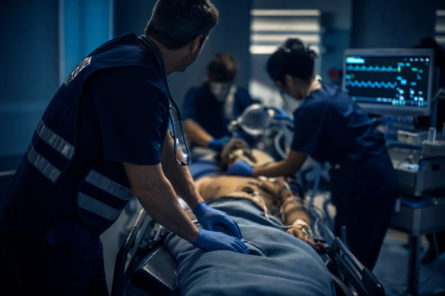
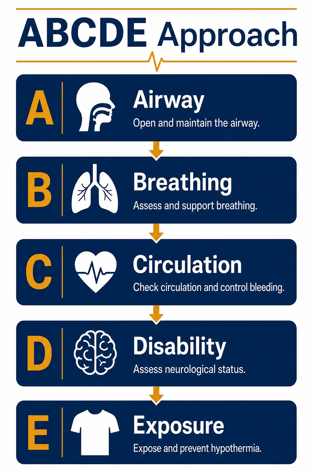
Three principles govern the whole approach. First, treat first what kills first: a blocked airway kills the patient long before the cause of the blockage is known, so it is managed immediately. Second, resuscitate and investigate simultaneously: intravenous access, bloods, a gas and an electrocardiogram are obtained while the airway and breathing are secured, not deferred until afterwards. Third, call for help early: a deteriorating patient is managed by a team, and senior, anaesthetic and critical-care assistance should be summoned at the first sign of instability rather than when resuscitation has failed. [1]
The physiology that underpins resuscitation
Every step of ABCDE exists to preserve oxygen delivery to the tissues, and a Fellowship candidate must be able to defend the approach from first principles. Global oxygen delivery is the product of cardiac output and arterial oxygen content: DO2 = CO × CaO2. The arterial oxygen content is CaO2 = (1.34 × Hb × SaO2) + (0.003 × PaO2), so it depends overwhelmingly on haemoglobin and saturation, with dissolved oxygen contributing negligibly. Cardiac output is CO = stroke volume × heart rate, and stroke volume is determined by preload, afterload and contractility. Mean arterial pressure approximates to MAP = CO × systemic vascular resistance (SVR), which is why a falling output can be masked for a time by rising SVR (vasoconstriction). [1]
This physiology dictates both the order and the content of the assessment. The airway and breathing exist to maintain the SaO2 term; the circulation exists to maintain the CO term; together they protect DO2 and therefore cerebral and myocardial oxygenation. A blocked airway removes SaO2 within minutes; a failing circulation removes CO. The same framework explains why a patient can look well while dying: healthy adults compensate for a falling output through sympathetic tachycardia, vasoconstriction and tachypnoea, holding MAP and cerebral perfusion until reserve is exhausted. Decompensation is then abrupt — the blood pressure falls, the conscious level drops, and the patient arrests. [1]
Three clinical consequences follow, and they are the heart of safe emergency practice. A normal blood pressure does not exclude shock, particularly in the young, the pregnant, the athlete and the chronically hypertensive, who may hold a normal pressure until a substantial fraction of circulating volume is lost. A rising heart rate, a rising respiratory rate and cold, clammy peripheries are the footprints of compensation and may precede hypotension by minutes or hours. A raised lactate signals that DO2 has fallen behind demand and anaerobic metabolism has begun, and it may be the first objective marker of occult shock. The resuscitation team must therefore act on the trend and the compensatory signs, not wait for the blood pressure to collapse. [1]
Recognising deterioration: track-and-trigger and early-warning scores
In-hospital cardiac arrest is rarely sudden; it is usually preceded by measurable physiological deterioration over hours, much of it documented but not acted upon. Systematic reviews of physiological track-and-trigger warning systems show that deviations in routine observations reliably identify patients at risk of cardiorespiratory arrest on the ward.[1] Aggregating those observations into a single early-warning score improves the consistency and reproducibility of risk identification across observers and shifts.[3]
The clinical value of early identification is not merely theoretical. Introducing a Modified Early Warning Score into acute medical admissions was associated with a reduction in cardiopulmonary arrests and in intensive care utilisation, suggesting that earlier recognition changes outcome rather than merely predicting it.[2] The modern standard, the Royal College of Physicians' National Early Warning Score (NEWS2), scores seven parameters — respiratory rate, oxygen saturation (with a second scale, scale 2, for patients at risk of hypercapnic respiratory failure), whether supplemental oxygen is required, systolic blood pressure, pulse rate, conscious level (using ACVPU — Alert, new Confusion, Voice, Pain, Unresponsive) and temperature — into a weighted aggregate score.[1] The aggregate score drives a graded response: a total of zero is low-risk and needs routine monitoring; a score of 1 to 4 (or any single parameter scoring 3) prompts urgent ward-based review by a competent clinician; a score of 5 to 6 is a key threshold requiring urgent review and consideration of a critical-care referral; and a score of 7 or more is an emergency demanding immediate assessment, usually escalation to a critical-care team and consideration of transfer to higher-level care. Its performance has been re-examined in contemporary cohorts, including in patients with COVID-19, where it retained discriminative value for deterioration.[4]
The preventable failure is not the abnormal observation itself but the failure to respond to it. Retrospective study of in-hospital deterioration shows that a failure to escalate in the face of deranged physiology — rather than the absence of the abnormality — is the recurring system error that precedes avoidable arrest.[5] National guidance therefore requires that acutely ill adults are monitored with a track-and-trigger system and that a graded, agreed response is triggered automatically by the physiology, not by a subjective judgment that the patient "looks well".[1] In the emergency department the same logic applies at the front door: an abnormal observation set on arrival, or a worsening trend while the patient waits, is an instruction to reassess immediately, not a number to be charted. The single most important — and most consistently under-recorded — vital sign is the respiratory rate, which is often the earliest marker of deterioration across sepsis, metabolic acidosis, shock and neurological injury and carries disproportionate prognostic weight.
A — Airway
The first question is whether the airway is patent, protected, and maintainable, because an obstructed airway will kill the patient before any other problem matters. Assessment is rapid and clinical: look for chest movement and abdominal breathing, listen for obstructive noises, and feel for airflow at the mouth. Stridor, a harsh sound on inspiration, indicates partial upper-airway obstruction and is an emergency; gurgling suggests fluid — blood, vomit or secretions — that must be cleared; snoring implies the tongue is obstructing the oropharynx in an obtunded patient; and a silent chest in a patient making respiratory effort is the most ominous sign of all, suggesting complete obstruction. [1]
The causes of upper-airway obstruction group logically: the relaxed tongue in the obtunded or arrested patient; fluid and foreign material — blood, vomit or an inhaled foreign body; soft-tissue swelling from anaphylaxis, burns, or infection such as epiglottitis, retropharyngeal abscess or Ludwig's angina; laryngospasm and trauma; and tumour. Management proceeds from the simple to the definitive. Suction the airway; perform a head tilt and chin lift, or a jaw thrust if cervical-spine injury is suspected; and insert an airway adjunct. An oropharyngeal (Guedel) airway suits the deeply unconscious patient and is sized by matching the device from the corner of the mouth to the angle of the jaw or the earlobe; it is poorly tolerated and may provoke vomiting or laryngospasm in a patient with an intact gag reflex, in whom a nasopharyngeal airway (sized from nostril to earlobe) is preferable, though it is contraindicated in suspected basal skull fracture. The patient who cannot protect their own airway is placed in the lateral recovery position. High-flow oxygen is given to the spontaneously breathing patient throughout. [1]
If the airway cannot be maintained or protected, a definitive cuffed airway is required, typically by rapid sequence intubation, and the patient is bag-valve-mask ventilated with oxygen while the skilled operator and equipment are assembled. In the small minority in whom intubation is impossible and oxygenation cannot be maintained — the can't-intubate, can't-oxygenate situation — a surgical airway is the final option: in the adult this is a scalpel-bougie-tube cricothyroidotomy, a horizontal incision through the cricothyroid membrane with a tracheal tube passed over a bougie. Throughout airway management the cervical spine is protected in any patient with a mechanism compatible with spinal injury, using manual in-line stabilisation rather than rigid immobilisation during airway manoeuvres. [1]
B — Breathing
Breathing is assessed for the work, the effectiveness, and the symmetry of ventilation. The respiratory rate is measured over a full minute; the effort is assessed by inspecting for accessory-muscle use, intercostal recession, tracheal tug, abdominal breathing and the ability to speak in full sentences; and the chest is examined for symmetry of movement, expansion, percussion and auscultation. Oxygen saturation is recorded and a capillary refill time bridges the breathing and circulation assessments. [1]
Two patterns of respiratory failure are distinguished on blood gas. Type 1 is hypoxaemic — a low PaO2 with a normal or low PaCO2 — and arises from ventilation-perfusion mismatch, shunt, diffusion impairment, or a low inspired oxygen. Type 2 is hypercapnic — a high PaCO2, with hypoxaemia — and reflects alveolar hypoventilation from reduced respiratory drive, neuromuscular weakness, chest-wall failure or airway obstruction, and it carries the risk of CO2 narcosis and acidosis. The pattern guides therapy: type 1 is treated with oxygen and treatment of the cause; type 2 requires attention to ventilation, often with non-invasive ventilation. [1]
The immediately life-threatening breathing problems are treated at once, before the examination is completed. A tension pneumothorax is decompressed by needle thoracocentesis (typically the fourth or fifth intercostal space, anterior axillary line, or the second intercostal space mid-clavicular line) followed by a definitive intercostal drain; massive airway haemorrhage is managed with suction, positioning and a definitive airway; a severe asthma or chronic obstructive pulmonary disease exacerbation receives bronchodilators, steroids and, in the exhausted patient, ventilatory support; and cardiac pulmonary oedema is treated with oxygen, nitrates and positive pressure. [1]
Oxygen is a treatment for hypoxaemia and is titrated to a target saturation, not prescribed indiscriminately. The devices deliver a graded inspired fraction: low-flow nasal cannulae give roughly 24 to 35 percent at 2 to 4 litres per minute; a simple face mask gives 35 to 50 percent; a non-rebreather reservoir mask at 10 to 15 litres per minute delivers the highest concentration available in the emergency department, up to about 90 percent; and a Venturi mask delivers a precise, fixed FiO2 through colour-coded valves (24 to 60 percent), useful when control matters, as in the hypercapnic patient. The target saturation is 94 to 98 percent for most patients, but 88 to 92 percent for those at risk of hypercapnic respiratory failure — typically the patient with chronic obstructive pulmonary disease reliant on hypoxic drive — with an early arterial blood gas to confirm. A rising respiratory rate with falling saturation, increasing work of breathing, fatigue, or a rising PaCO2 on blood gas are the markers of impending ventilatory failure; the threshold to escalate to non-invasive ventilation or to intermittent positive-pressure ventilation is a clinical one based on exhaustion and should not be delayed until arrest. Capnography (waveform end-tidal CO2) is mandatory once any airway adjunct or positive-pressure ventilation is in place: it confirms tracheal tube placement, monitors the adequacy of ventilation, and detects circuit disconnection or oesophageal intubation immediately. [1]
C — Circulation
Circulatory assessment determines whether tissue perfusion is adequate and, if not, why. The heart rate, blood pressure, capillary refill time, peripheral temperature and pulse character are measured, the skin is examined for pallor and sweating, and any obvious external haemorrhage is sought and controlled with direct pressure. Two large-bore intravenous cannulae are sited, blood is drawn for a full blood count, electrolytes, a venous or arterial blood gas, a lactate, coagulation and a group-and-save or crossmatch, and an electrocardiogram and rhythm strip are obtained. A balanced crystalloid bolus is given to the hypotensive or poorly perfused patient and the response is assessed. [1]
Shock is the state in which the circulation fails to deliver enough oxygen to meet metabolic demand, and contemporary guidance stresses early recognition of the inadequately perfused patient and a structured, monitored resuscitation strategy.[6] Because MAP = CO × SVR, shock is usefully classified by which determinant fails. Hypovolaemic shock (low preload, from haemorrhage or fluid loss) presents with cold peripheries, a narrow pulse pressure and a low jugular venous pressure. Distributive shock (low SVR, from sepsis or anaphylaxis) classically presents with warm peripheries and a bounding pulse early, becoming cold and shutdown as it progresses. Cardiogenic shock (low contractility, from infarction, arrhythmia or failure) presents with cold peripheries, a raised venous pressure and pulmonary oedema. Obstructive shock (impaired filling or outflow, from tension pneumothorax, cardiac tamponade or massive pulmonary embolism) presents with signs of right-heart strain and impaired venous return. The lactate is a practical marker of impaired perfusion and anaerobic metabolism: a raised or rising lactate indicates ongoing tissue hypoxia and a falling lactate confirms that resuscitation is working.
Resuscitation is targeted and reassessed, not given by rote. Modern guidance favours dynamic assessment of fluid responsiveness — the passive leg-raise, pulse-pressure or stroke-volume variation, and inferior vena cava variability — over static pressures such as the central venous pressure, which do not reliably predict the response to fluid.[6] The hypovolaemic patient is given a balanced crystalloid bolus of 250 to 500 millilitres and reassessed for an improvement in perfusion, blood pressure and conscious level, with blood products considered early in haemorrhagic shock and damage-control principles applied. The arrhythmic patient is treated by the peri-arrest algorithm. The specific vasopressor or inotrope follows the cause: noradrenaline for vasodilatory (distributive) shock to restore SVR, with adrenaline an alternative; dobutamine or milrinone for primary pump failure; and vasopressin considered in refractory vasodilatory shock. The response to every bolus and every infusion is judged against predefined targets.
Anaphylaxis is the archetype of a time-critical circulatory emergency and its algorithm must be automatic. At the bedside, recognise the rapidly evolving multi-system reaction — airway swelling, bronchospasm, hypotension, urticaria, abdominal symptoms — often with a clear trigger. The first-line treatment is intramuscular adrenaline into the anterolateral thigh: 500 micrograms (0.5 mg of 1:1000) for an adult, repeated after five minutes if there is no response, and further doses as needed. The patient is laid flat with the legs elevated (or kept sitting up if breathless), given high-flow oxygen and an intravenous fluid bolus, and the airway and breathing are managed as they deteriorate. Refractory anaphylaxis — a poor response to repeated IM doses — calls for an intravenous adrenaline infusion titrated to effect, and glucagon is considered in the patient on a beta-blocker. Every patient is observed after resolution for the biphasic reaction and reviewed for the trigger and an adrenaline auto-injector.[1]
D — Disability
The disability assessment evaluates central neurological function and several rapidly reversible causes of altered consciousness. A Glasgow Coma Score is recorded: the eye response (1 to 4), the verbal response (1 to 5) and the motor response (1 to 6), for a maximum of 15, with the best motor response the most informative single component (obeying commands, localising pain, withdrawing, flexing, extending, or none). The intubated patient is recorded as "E_Vt M" with a "t" for tube. The pupils are examined for size, symmetry and reactivity: a unilateral fixed dilated pupil suggests third-nerve palsy and impending herniation; pin-point pupils suggest an opioid or pontine lesion; and fixed, mid-position pupils suggest brainstem failure. [1]
The blood glucose is checked at the bedside on every obtunded, agitated or fitting patient, because hypoglycaemia is immediately and completely reversible and is missed at peril. A new alteration in conscious level is itself a marker of deterioration that warrants escalation. The quick Sequential Organ Failure Assessment, from the Sepsis-3 consensus, uses three bedside criteria — an altered mentation, a systolic blood pressure of 100 mmHg or less, and a respiratory rate of 22 per minute or more — as a prompt to consider possible sepsis in a patient not already identified as such.[10] Beyond sepsis, the reversible contributors to a reduced conscious level are hypoglycaemia, hypoxia, opioid or other drug toxicity (suggested by pin-point pupils and a brisk response to titrated naloxone), a post-ictal state, and rapidly correctable metabolic or toxic derangement. Naloxone is titrated in small increments to restore adequate respiration rather than full consciousness, to avoid precipitating acute withdrawal. Signs of raised intracranial pressure — a falling conscious level, a unilateral dilated pupil, abnormal posturing, and Cushing's triad of hypertension, bradycardia and irregular respiration — demand immediate reduction of intracranial pressure and neurosurgical referral.
E — Exposure
Full exposure allows a complete head-to-toe examination while preserving the patient's dignity and preventing heat loss, because a cold patient deteriorates across every system already assessed — coagulation fails, arrhythmia is provoked, and drug metabolism slows. The examination seeks the clue that explains the presentation: a petechial or purpuric rash in meningococcal disease, surgical emphysema in a pneumothorax or oesophageal rupture, hidden external haemorrhage, an obvious source of infection such as cellulitis or a septic joint, the track marks of intravenous drug use, the bruising of non-accidental injury, or the surgical scars and indwelling devices of comorbidity. The core temperature is measured: hypothermia is corrected actively and hyperthermia is recognised as a feature of sepsis, heat illness and certain drug toxicities. [1]
Investigations in parallel with resuscitation
Resuscitation and investigation proceed together. A venous or arterial blood gas gives an immediate read on oxygenation, ventilation, the lactate and a rapid haemoglobin, and is the single most useful early test in the unstable patient; the difference between the venous and arterial sample is understood when interpreting it. Bloods are sent for a full blood count, electrolytes, liver and renal function, coagulation, a lactate and, where relevant, troponin, lipase, beta-hCG or a drug screen. Blood cultures are drawn before antibiotics in the septic patient when this does not delay administration. An electrocardiogram screens for the arrhythmia and the ischaemia that may underlie shock, and bedside ultrasound — FAST in trauma, and focused cardiac examination in shock — identifies free fluid, a failing, hyperdynamic or underfilled heart, a pericardial effusion, and a proximal aortic aneurysm. Targeted imaging, typically computed tomography once the patient is stable enough to leave the department, defines the diagnosis, but it follows, never precedes, the resuscitation of the unstable patient. [1]
The time-critical septic patient
Sepsis is among the most common and most time-critical causes of deterioration in the emergency department. Sepsis is defined as a life-threatening organ dysfunction caused by a dysregulated host response to infection,[7] and septic shock is the subset whose circulatory, cellular and metabolic abnormalities are severe enough to markedly increase mortality — clinically, persisting hypotension requiring vasopressors to maintain an adequate mean arterial pressure together with a serum lactate above 2 mmol per litre despite adequate fluid resuscitation.[8]
Time is the modifiable determinant of outcome. In a large cohort studied under mandated emergency sepsis care, each additional hour to completion of the sepsis bundle — above all antibiotic administration — was associated with an increase in mortality, with the relationship apparent from the very first hour.[9] The practical consequence is that recognition of possible sepsis triggers a simultaneous, time-stamped resuscitation bundle rather than a sequential workup: blood cultures are drawn before antibiotics where this does not delay them; broad-spectrum intravenous antibiotics are administered within the first hour; a balanced crystalloid is given as 250 to 500 millilitre boluses to the hypotensive or lactate-raised patient, with reassessment after each; vasopressors, typically noradrenaline, are commenced for fluid-refractory hypotension to restore an adequate mean arterial pressure; a source of infection is identified and controlled; and the lactate is measured serially to confirm that perfusion is improving. The antibiotics must not wait for cultures, imaging or a bed.
Reassessment, targets and the loop
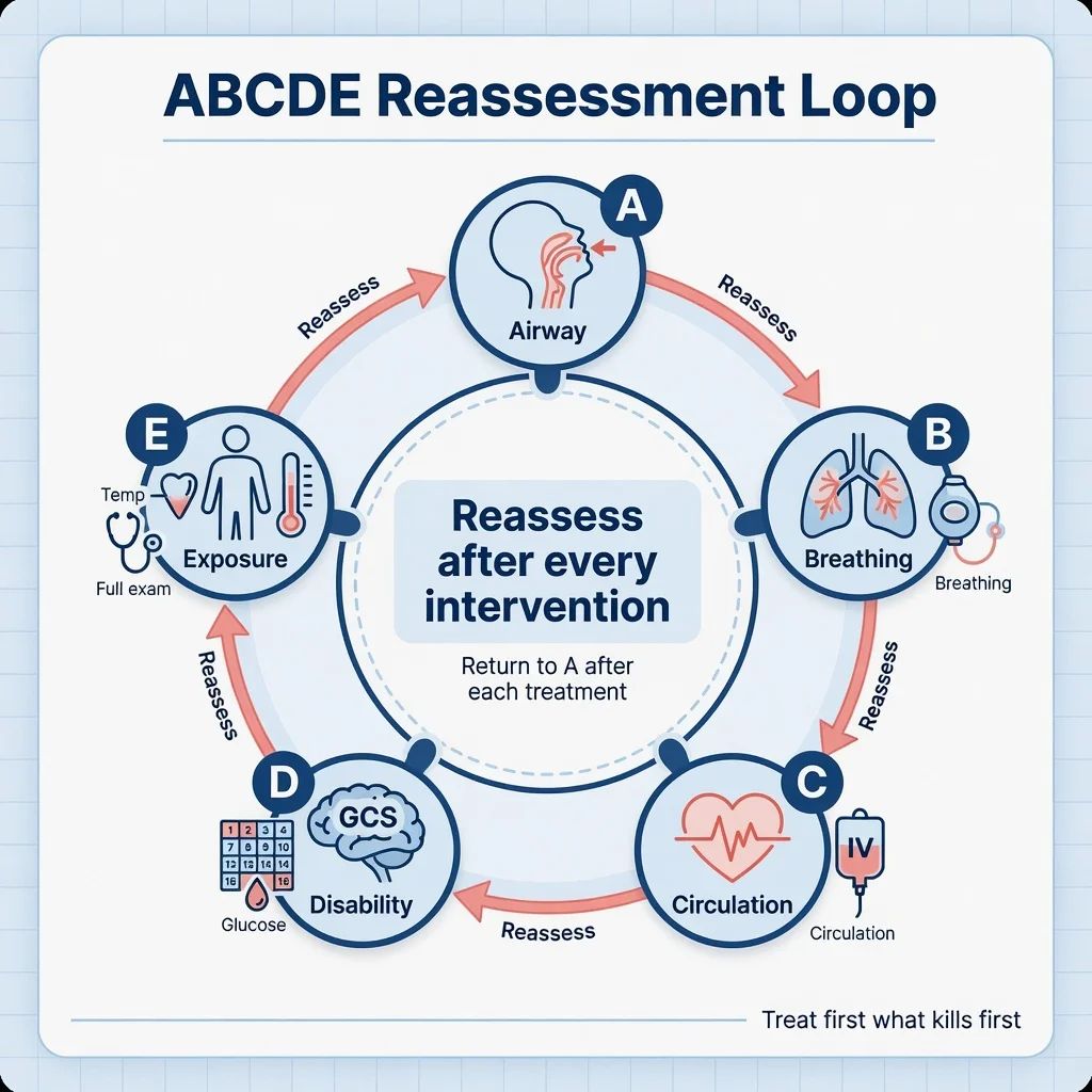
ABCDE is an iterative cycle, not a single pass. After every intervention — placing an airway adjunct, giving a fluid bolus, decompressing a pneumothorax, delivering antibiotics — the clinician returns to the airway and reassesses. Resuscitation is guided by physiological targets: a patent and protected airway, an oxygen saturation at target, an adequate work of breathing, a blood pressure and perfusion consistent with end-organ function, a falling lactate, and a stable or improving conscious level. Persisting derangement despite appropriate treatment is an indication to escalate — to a senior decision-maker, to anaesthetic assistance for a definitive airway, or to critical care for ongoing support — rather than to repeat the same measure and hope for a different result. [1]
Differential diagnosis — the time-critical causes identified at each step
- Airway obstruction — foreign body, anaphylaxis, infection (epiglottitis), burns — each managed with a specific intervention (removal, adrenaline 500 mcg IM, intubation).
- Tension pneumothorax — unilateral decreased breath sounds, tracheal deviation, hypoxia — needle decompression at the 2nd intercostal space mid-clavicular line.
- Hypovolaemic shock — haemorrhage, sepsis, dehydration — balanced crystalloid 500 mL bolus, noradrenaline 0.05 to 0.5 mcg/kg/min if fluid-refractory.
- Anaphylaxis — urticaria, angioedema, hypotension after a trigger — adrenaline 500 mcg IM, chlorphenamine 10 mg IV, hydrocortisone 200 mg IV.
- Hypoglycaemia — glucose below 3 mmol/L — glucose 25 g IV (50 mL of 50 per cent dextrose), glucagon 1 mg IM. [1]
Management — the emergency drug doses at each step
The ABCDE approach identifies the problem and triggers the drug: A — adrenaline 500 mcg IM for anaphylactic airway obstruction; B — high-flow oxygen 15 L/min, salbutamol 5 mg nebulised for bronchospasm; C — balanced crystalloid 500 mL bolus, noradrenaline 0.05 to 0.5 mcg/kg/min for vasodilatory shock, tranexamic acid 1 g IV for trauma haemorrhage; D — glucose 25 g IV for hypoglycaemia, naloxone 400 mcg IV for opioid overdose; E — paracetamol 1 g for fever, potassium chloride 10 mmol in 100 mL for hypokalaemia.
Teamwork, human factors and communication [1]
The deteriorating patient is managed by a team, and effective crisis resource management is as important as clinical knowledge. Clear leadership, an explicit allocation of roles, closed-loop communication in which instructions are repeated back and confirmed, and a willingness to call for help early are the components of good team function. Fixation error — the failure to revise a working diagnosis in the face of contradictory information, such as persisting with a diagnosis of gastroenteritis in a patient who is actually in septic shock — is a recognised cause of failed resuscitation, and the structured ABCDE reassessment is itself a defence against it. When help arrives, a structured handover such as SBAR (Situation, Background, Assessment, Recommendation) or ISBAR conveys the situation, the working diagnosis, what has been done and what is needed, so the responding team inherits an accurate picture without having to reconstruct it. [1]
Special populations
Children deteriorate physiologically before they decompensate and compensate well until reserve is exhausted, after which collapse is sudden. Age-specific vital signs and weight-based drug dosing are essential — the dose calculated from a weight, a length-based tape, or a formula such as weight (kg) ≈ (age + 4) × 2 — and a balanced crystalloid bolus is typically 10 to 20 mL per kg in shock, with adrenaline for anaphylaxis at 10 micrograms per kg intramuscularly. Bradycardia in a sick child is a pre-terminal sign, almost always reflecting hypoxia, and demands immediate ventilation and oxygenation rather than cardiac drugs. The paediatric early-warning scores extend the track-and-trigger principle to age-specific thresholds. [1]
In pregnancy, the supine position causes aortocaval compression by the gravid uterus that worsens any shock from around 20 weeks; the patient is resuscitated with a left lateral tilt or manual uterine displacement. Normal pregnancy physiology — a mild tachycardia, an increased minute ventilation with a respiratory alkalosis, a relative anaemia, and a mild rise in cardiac output — must not be mistaken for deterioration, but it also means the pregnant patient decompensates more quickly when she does. Older patients often present atypically — with confusion or a fall rather than classical symptoms — and have reduced reserve alongside comorbidity and polypharmacy, so a subtle change may represent advanced disease. The immunocompromised patient may mount a blunted inflammatory response and present with an occult, rapidly progressive infection. [1]
Common pitfalls
The recurring errors are well described. Treating abnormal observations in isolation, or recording them without acting, ignores the central lesson of the early-warning-score evidence.[5] Delaying antibiotics or fluids in sepsis while awaiting investigations costs lives hour by hour.[9] Prescribing a fixed volume of fluid without reassessing the response risks both under-resuscitation of the shocked patient and pulmonary oedema in the failing heart. Omitting a bedside glucose in the obtunded patient risks missing the most easily reversible cause of coma. Failing to reassess after each intervention abandons the loop on which the whole approach depends. And under-recognition of deterioration in the elderly, the young and the chronically ill — populations in whom the blood pressure may be preserved until late — rests on the false reassurance of a single normal reading.
SAQ — ABCDE primary survey in multi-trauma
10 minutes · 10 marks
A 24-year-old man is brought to the resuscitation bay after a high-speed motorcycle collision. He speaks with slurred words, the respiratory rate is 32 per minute, the oxygen saturation is 90 per cent on a non-rebreather mask, the blood pressure is 76 over 40, and he has a swollen deformed left thigh with active external haemorrhage.
SAQ — The Disability step and the reversible causes of coma
10 minutes · 10 marks
A 78-year-old woman is found unresponsive on the floor at home. In the resuscitation bay the airway is patent and the breathing is adequate, the blood pressure is 150 over 90, and she does not open her eyes, makes no sound, and withdraws to pain. The pupils are 2 mm and reactive, the bedside glucose is 2.1 mmol per litre, and the temperature is 35.2 degrees Celsius.
Red flags
The following features identify the patient at immediate risk, in whom resuscitation must begin now and the cause be sought in parallel: [1]
[1]References
- [1]Gao H, McDonnell A, Harrison DA, et al. Systematic review and evaluation of physiological track and trigger warning systems for identifying at-risk patients on the ward Intensive Care Med, 2007.PMID 17318499
- [2]Subbe CP, Davies RG, Williams E, et al. Effect of introducing the Modified Early Warning score on clinical outcomes, cardio-pulmonary arrests and intensive care utilisation in acute medical admissions Anaesthesia, 2003.PMID 12859475
- [3]Subbe CP, Gao H, Harrison DA. Reproducibility of physiological track-and-trigger warning systems for identifying at-risk patients on the ward Intensive Care Med, 2007.PMID 17235508
- [4]Kostakis I, Smith GB, Prytherch D, et al. The performance of the National Early Warning Score and National Early Warning Score 2 in hospitalised patients infected by the severe acute respiratory syndrome coronavirus 2 (SARS-CoV-2) Resuscitation, 2021.PMID 33176170
- [5]Smith GB, Redfern O, Maruotti A, et al. The association between nurse staffing levels and a failure to respond to patients with deranged physiology: A retrospective observational study in the UK Resuscitation, 2020.PMID 31945427
- [6]Monnet X, Messina A, Greco M, et al. ESICM guidelines on circulatory shock and hemodynamic monitoring 2025 Intensive Care Med, 2025.PMID 41236566
- [7]Singer M, Deutschman CS, Seymour CW, et al. The Third International Consensus Definitions for Sepsis and Septic Shock (Sepsis-3) JAMA, 2016.PMID 26903338
- [8]Shankar-Hari M, Phillips GS, Levy ML, et al. Developing a New Definition and Assessing New Clinical Criteria for Septic Shock: For the Third International Consensus Definitions for Sepsis and Septic Shock (Sepsis-3) JAMA, 2016.PMID 26903336
- [9]Seymour CW, Gesten F, Prescott HC, et al. Time to Treatment and Mortality during Mandated Emergency Care for Sepsis N Engl J Med, 2017.PMID 28528569
- [10]Seymour CW, Liu VX, Iwashyna TJ, et al. Assessment of Clinical Criteria for Sepsis: For the Third International Consensus Definitions for Sepsis and Septic Shock (Sepsis-3) JAMA, 2016.PMID 26903335