EM · Cardiac arrest & ALS
Cardiac arrest and advanced life support
The full advanced life support of the arrested adult: the chain of survival, high-quality CPR, the universal ALS algorithm with the shockable and non-shockable loops, adrenaline and amiodarone, defibrillation, the four Hs and four Ts, the other arrest drugs, the team and human factors, the immediate post-ROSC period with the targeted-temperature evidence, the special arrests, and the regional algorithm differences.
On this page & tools
Your progress
Saved locally on this device.
Target exams
Red flags
Cardiac arrest is the sudden cessation of effective cardiac mechanical activity, and its management — advanced life support — is the archetype of the time-critical, team-based emergency. Survival to a good neurological outcome depends on a small number of things done well and without delay: the immediate initiation of high-quality chest compressions, the rapid defibrillation of a shockable rhythm, the identification and reversal of the cause, and the high-quality post-resuscitation care. The algorithm is universal, it is rehearsed, and it is built to be executed under stress; the Fellowship candidate must know it cold, including the regional variations and the evidence behind each drug. [1]
The chain of survival and high-quality CPR
The chain of survival — early recognition and call for help, early high-quality cardiopulmonary resuscitation, early defibrillation, and high-quality post-resuscitation care — frames the whole effort, and the two pre-hospital links (CPR and defibrillation) are the ones that determine good survival. The quality of the CPR is decisive. The compressions are delivered at a rate of 100 to 120 per minute, to a depth of 5 to 6 centimetres in the adult, with full chest recoil between compressions and minimal interruption (pauses kept below 5 seconds and confined to the rhythm check and the shock). The compressor is rotated every two minutes to prevent fatigue, and excessive ventilation is avoided because it raises intrathoracic pressure and reduces coronary and cerebral perfusion. The compression-to-ventilation ratio for the adult with an unprotected airway is 30 to 2; once the airway is secured with a cuffed tracheal tube, compressions continue uninterrupted at 100 to 120 per minute with around 10 breaths per minute. High-quality CPR is the foundation on which everything else is built, and no intervention should be allowed to compromise it.[1]
The ALS algorithm and rhythm analysis
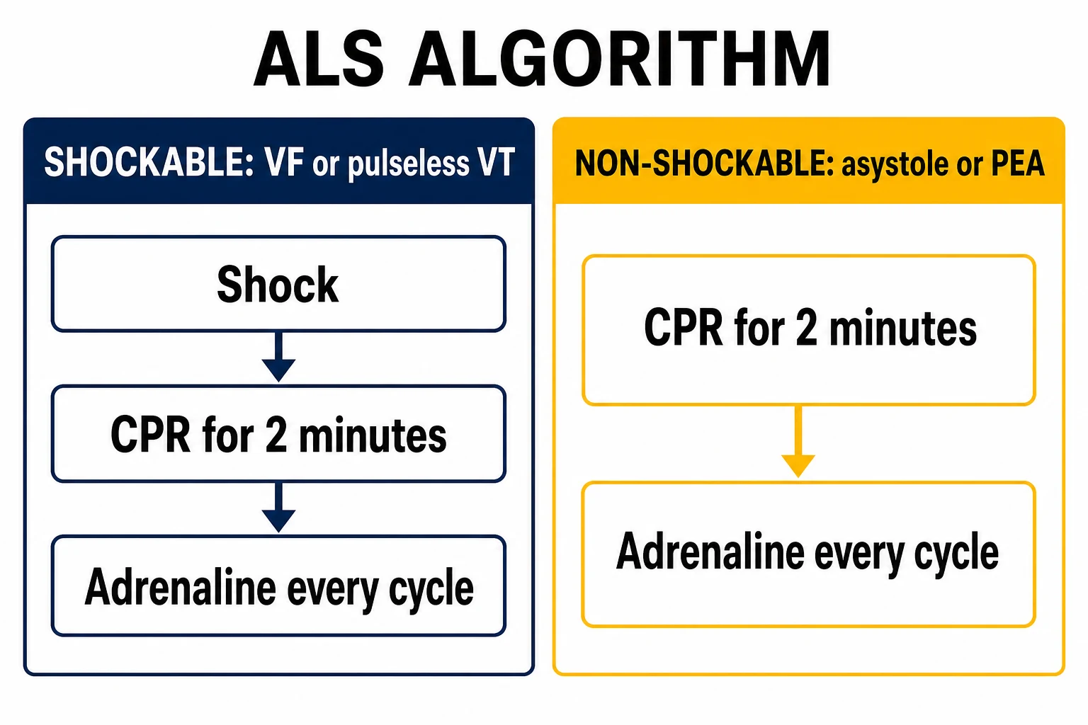
On recognising the arrest, help is summoned, CPR is begun, and the defibrillator is attached. The rhythm is analysed at the two-minute mark (the cycle length) and classified as shockable — ventricular fibrillation (VF) or pulseless ventricular tachycardia (pVT) — or non-shockable — asystole or pulseless electrical activity (PEA). The two rhythms are managed in two parallel loops, each built around two-minute cycles of CPR with a rhythm analysis and a drug at each cycle, and each interrupted by the search for and treatment of a reversible cause.[1][1]
Shockable rhythms: VF and pulseless VT
VF and pVT are the rhythms for which defibrillation is the definitive treatment, and the sooner the shock the better. The shock is delivered (a biphasic shock at the manufacturer-recommended energy, commonly 150 to 200 joules, escalating for subsequent shocks), CPR is resumed immediately for two minutes without pausing to check a rhythm or a pulse, and the rhythm is then reassessed. If VF or pVT persists, a second shock is given and CPR resumed. After the third shock, adrenaline 1 milligram intravenously or intraosseously is given every three to five minutes (every cycle) thereafter, and amiodarone 300 milligrams is given after the third shock with a further 150 milligrams after the fifth shock if needed (lidocaine is an alternative). The cycle of CPR, rhythm check and shock continues, with the drugs, until a stable rhythm returns or the decision is made to stop. The presence of a shockable rhythm is a good prognostic sign, and the energy of the resuscitation goes into minimising the time to the shock.[1][1]
Non-shockable rhythms: asystole and PEA
Asystole and PEA are not treated with a shock; they are managed with continuous CPR and adrenaline, and an unremitting search for a reversible cause. Adrenaline 1 milligram is given intravenously or intraosseously as soon as IV or IO access is available, and repeated every three to five minutes (every cycle). PEA — organised electrical activity without a pulse — is almost always a manifestation of a reversible cause (a tamponade, a tension pneumothorax, a massive thrombosis, hypovolaemia, or a toxin), and the survival of the PEA arrest turns on identifying and treating that cause; bedside ultrasound during the rhythm check is invaluable. Asystole carries the worst prognosis, but it too may reflect a reversible cause or a correctable profound bradycardia, and atropine is no longer recommended routinely.[1][1]
Differential diagnosis — the reversible causes
- Hypoxia — the asphyxial arrest, the drowning, the hanging; give the oxygen, the ventilation.
- Hypovolaemia — the haemorrhage, the sepsis; give the fluid, the blood.
- Hypo- or hyperkalaemia — the renal failure, the dialysis; give the calcium chloride 10 mL of 10 per cent, the insulin-dextrose, the bicarbonate.
- Hypothermia — the exposure, the immersion; rewarm slowly, do not declare death until warm and dead.
- Thrombosis (coronary) — the STEMI; consider the thrombolysis or the PCI.
- Thrombosis (pulmonary) — the massive PE; give the thrombolysis, continue the CPR for 60 minutes.
- Tension pneumothorax — the trauma; the needle decompression, the finger thoracostomy.
- Tamponade — the trauma, the post-MI; the pericardiocentesis, the resuscitative thoracotomy.
- Toxins — the TCA, the opioid, the beta-blocker; give the specific antidote (the sodium bicarbonate, the naloxone, the glucagon).
Adrenaline [1]
Adrenaline is the vasoconstrictor that raises the aortic and the coronary perfusion pressure during CPR, improving the return of spontaneous circulation. It is given at 1 milligram intravenously or intraosseously every three to five minutes, in both the shockable and the non-shockable loops, the timing of the first dose differing slightly between the regional guidelines (after the third shock in the United Kingdom algorithm, after the second in the Australasian). The large PARAMEDIC2 trial established that adrenaline improves survival to hospital discharge after out-of-hospital cardiac arrest — but it did not improve the rate of survival with a favourable neurological outcome, because some of the additional survivors survived with neurological injury.[1][4] The practical message is that adrenaline is given, but it is not allowed to delay or interrupt the high-quality CPR and the early defibrillation on which good outcome truly depends.
Antiarrhythmics and defibrillation
The antiarrhythmic is given for the refractory shockable rhythm. Amiodarone 300 milligrams after the third shock, with 150 milligrams after the fifth, is the standard; lidocaine (1 to 1.5 milligrams per kilogram) is an alternative. The ROC-ALPS trial found that neither amiodarone nor lidocaine produced a significant improvement in survival to discharge compared with placebo for out-of-hospital cardiac arrest overall, though a signal of benefit in the witnessed, monitored subgroup has kept the drugs in the algorithm.[3] Defibrillation itself is delivered with self-adhesive pads placed in the anterolateral (and, where available, anteroposterior) position, with a brief safety check and immediate resumption of CPR; the modern practice is a single shock followed by CPR rather than stacked shocks, because the heart is refractory and the CPR is more valuable in the immediate post-shock period. Hands-on defibrillation (with the compressor pausing only for the shock) is an emerging technique to reduce interruptions.[1]
The shockable versus non-shockable decision — the fork in the algorithm
The first rhythm analysis is the fork in the algorithm: the shockable rhythm (VF or pulseless VT) enters the defibrillation loop, and the non-shockable rhythm (asystole or PEA) enters the adrenaline-and-search loop. The distinction is made on the monitor within seconds and is re-checked every two-minute cycle, because the rhythm may change — the VF may degenerate to the asystole, and a fine VF masquerading as the asystole may be revealed by turning the gain up. The Fellowship candidate must recognise both the canonical rhythms and the pitfalls (the fine VF, the artefact, the disconnected lead).[1][1]
VF / pulseless VT (shockable)
- Defibrillation is the definitive treatment — shock ASAP, every cycle while the rhythm persists
- Biphasic 150-200 J initially, escalating for the subsequent shocks (follow the manufacturer recommendation)
- Adrenaline 1 mg IV/IO after the 3rd shock, then every 3-5 min (every cycle)
- Amiodarone 300 mg after the 3rd shock; 150 mg after the 5th shock if the VF/pVT persists
- Best prognosis of the arrest rhythms; the outcome is time-to-shock dependent
Asystole (non-shockable)
- No shock; continuous CPR with the adrenaline 1 mg IV/IO q3-5 min from the first cycle
- Confirm the true asystole: the leads connected, the gain turned up, a second lead — exclude the fine VF
- Worst prognosis; search relentlessly for a reversible cause (the 4 Hs and the 4 Ts)
- Atropine is NO LONGER recommended routinely
- Pacing considered if a correctable profound bradycardia is the bridge
PEA (non-shockable)
- Organised electrical activity WITHOUT a pulse — no shock
- Adrenaline 1 mg IV/IO q3-5 min from the first cycle
- Almost always a reversible cause — the ultrasound during the rhythm check is invaluable
- Narrow-complex PEA: think the tamponade, the tension PTX, the massive PE, the hypovolaemia
- Wide-complex PEA: think the hyperkalaemia, the severe hypoxia, the severe acidosis, the sodium-channel-blocker toxin
The ALS drug doses — the adult arrest card
The reversible causes: the four Hs and four Ts
The search for a reversible cause runs through every arrest and is the key to the resuscitation that is failing. The four Hs are hypoxia (give oxygen, ventilate), hypovolaemia (give fluid or blood), hypo- or hyperkalaemia and the other metabolic derangements (treat with glucose, bicarbonate or electrolyte correction), and hypo- or hyperthermia. The four Ts are thrombosis (coronary or pulmonary — consider thrombolysis or thrombectomy), tension pneumothorax (decompress), tamponade (drain), and toxins (give the antidote).[1]
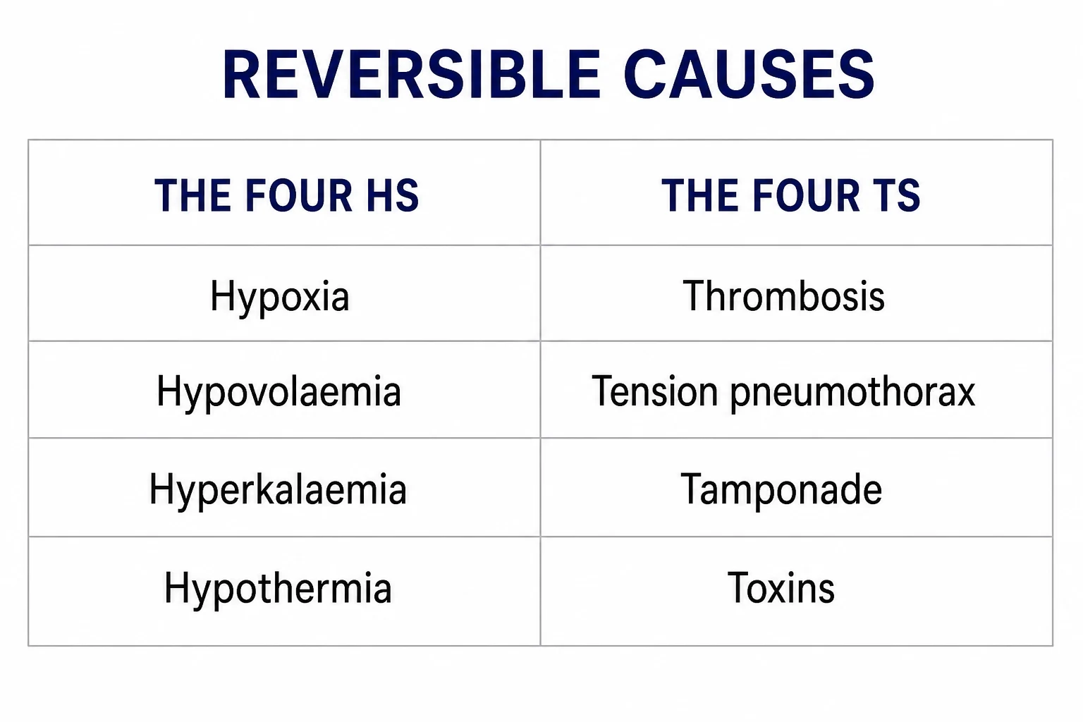
The Fellowship candidate must not only recall the list but know the bedside clue, the rapid diagnostic test and the immediate corrective action for each cause — this is the substance of the reversible-cause viva.[1][1]
Hypoxia
- The asphyxial arrest: the drowning, the hanging, the obstructed airway, the displaced tube
- Bedside clue: the cyanosis, the low SpO2 pre-arrest, the witnessed asphyxial event
- Action: the high-flow oxygen, confirm and secure the airway, the bilateral equal air entry and the chest rise
Hypovolaemia
- The haemorrhage, the sepsis, the GI bleed, the ruptured AAA, the trauma
- Bedside clue: the flat IVC on the ultrasound, the empty RV, the history of the blood loss
- Action: the IV fluid bolus, the blood and the massive transfusion protocol, identify and control the source
Hypo-/Hyperkalaemia (and the other metabolic)
- The renal failure, the dialysis, the dialysis-dependent patient; also the Na, the Ca, the Mg, the acid-base
- Bedside clue: the dialysis fistula, the wide QRS, the peaked T waves (hyperK); the long QT (hypoK, hypoMg, hypoCa)
- Action: the calcium chloride 10 mL of 10%, the insulin-dextrose, the bicarbonate (hyperK); correct the specific electrolyte
Hypothermia
- The exposure, the immersion, the avalanche; the patient is cold to the central core
- Bedside clue: the low core temperature, the Osborne J wave, the slow and refractory arrest
- Action: the active rewarming; do NOT declare the death until warm and dead; the prolonged resuscitation is justified; consider the ECPR
Thrombosis (coronary / pulmonary)
- The STEMI / the acute coronary occlusion; the massive pulmonary embolism
- Bedside clue: the preceding chest pain, the ECG changes; on the ultrasound a dilated RV with the D-shape (PE) or the regional wall motion abnormality
- Action: the thrombolysis (alteplase 50 mg for the PE, continue the CPR 60-90 min) or the PCI after the ROSC; the thrombectomy if available
Tension pneumothorax
- The trauma, the ventilated asthmatic, the central-line complication
- Bedside clue: the unilateral absent breath sounds, the tracheal deviation, the distended neck veins; on the ultrasound the absent lung sliding with the lung point
- Action: the immediate needle decompression (5th ICS mid-axillary, or 2nd ICS mid-clavicular), then the finger thoracostomy or the chest tube
Tamponade (cardiac)
- The trauma, the post-MI rupture, the malignancy, the uraemia, the post-cardiac-surgery
- Bedside clue: on the ultrasound the echo-free pericardial space with the RV diastolic collapse; the muffled heart sounds, the distended neck veins
- Action: the pericardiocentesis (or the resuscitative thoracotomy for the traumatic tamponade)
Toxins
- The TCA, the beta-blocker, the calcium-channel blocker, the opioid, the digoxin, the local anaesthetic, the recreational drug
- Bedside clue: the history, the wide QRS (TCA), the bradycardia (BB/CCB), the pinpoint pupils (opioid)
- Action: the specific antidote — bicarbonate (TCA), glucagon/high-dose insulin (BB/CCB), naloxone (opioid), digoxin Fab (digoxin), lipid emulsion (LA)
Other arrest drugs
Beyond adrenaline and amiodarone, the other drugs are situation-specific. Sodium bicarbonate 50 mL of 8.4 per cent (50 mmol) IV is given for the arrest caused by hyperkalaemia or tricyclic antidepressant toxicity and in the prolonged arrest with a profound acidosis, but it is not given routinely. Magnesium 2 g (10 mL of 20 per cent) IV is given for torsades de pointes, hypomagnesaemia, and polymorphic VT. Calcium chloride 10 mL of 10 per cent IV is given in hyperkalaemia, hypocalcaemia, and calcium-channel-blocker toxicity. A thrombolytic (alteplase 50 mg IV or tenecteplase as weight-based) is given when a pulmonary embolism is the suspected cause of the arrest, with CPR continued for up to an hour. Glucose 25 g IV (50 mL of 50 per cent) is given for hypoglycaemia. The bedside blood gas, the electrolytes and the glucose taken early guide the targeted therapy. [1]
The team and human factors
The arrest is managed by a team with allocated roles — the team leader, the compressor, the airway operator, the person at the defibrillator, the drugs, the timekeeper and the scribe — and the quality of the resuscitation depends on the leadership and the communication as much as on the algorithm. Closed-loop communication, the structured handover, and the willingness to call for senior or critical-care help early are the components. The cognitive aids (the algorithm itself, the reversible-causes list) are displayed and followed. Fixation on the rhythm at the expense of the reversible-cause search, and the failure to rotate the compressor, are the common human-factor failures. [1]
The immediate post-ROSC period
The return of spontaneous circulation is not the end of the resuscitation; the post-arrest care determines the neurological outcome, and it begins in the emergency department. The airway and the breathing are managed with a target oxygen saturation of 94 to 98 per cent (avoiding both hypoxia and hyperoxia) and a normocapnic ventilation. The circulation is supported to a mean arterial pressure of 65 mmHg or more, with the cause investigated and, where cardiac, reperfused. The targeted temperature is managed at normothermia, actively avoiding fever — the TTM2 trial found that targeting hypothermia at 33 degrees did not improve outcome over normothermia, and the contemporary practice is to maintain a normothermic, fever-free state. Seizures are treated, glucose is controlled, and the prognostication is deferred. These elements are explored in the post-cardiac-arrest-care topic.[2][1]
Special arrests
The cause and the context modify the resuscitation. The asphyxial arrest (drowning, hanging) prioritises oxygenation and ventilation. The anaphylactic arrest is treated with intramuscular adrenaline and fluid. The asthma arrest benefits from ventilation with a small tidal volume and a prolonged expiratory time, and from bronchodilators. The pulmonary embolism arrest is treated with thrombolysis. The traumatic arrest is managed with simultaneous resuscitation and the control of catastrophic haemorrhage, and adrenaline is not routinely effective in the hypovolaemic traumatic arrest.[4] The pregnant arrest is resuscitated with manual left uterine displacement, and a perimortem caesarean section is begun at four minutes of arrest in the pregnancy beyond 20 weeks to relieve aortocaval compression and to resuscitate mother and fetus. The hypothermic arrest is prolonged and resistant, and is resuscitated to normothermia with active rewarming (and, where available, extracorporeal) before the arrest is called. The drug-overdose arrest is treated with the antidote and the prolonged resuscitation that the reversibility warrants. Paediatric arrest is managed with age-appropriate compressions and ventilations (15 to 2 with two rescuers), a lower defibrillation energy, and an emphasis on the respiratory cause.
Termination, the refractory arrest, and regional practice
The decision to terminate the resuscitation is clinical, made by the team leader, and considers the downtime, the rhythm, the reversible causes, the comorbidity, and the response. The refractory arrest — VF or pVT that will not terminate despite the full algorithm — is an indication, in a suitable patient, for extracorporeal CPR as a bridge to recovery or to a definitive intervention. The guidelines are regional: the ARC/NZCOR guidelines govern the Australasian practice, the Resuscitation Council UK guidelines the United Kingdom practice, and the ILCOR Consensus on Science integrates the global evidence; the algorithm is the same in outline and differs in small details (the timing of the first adrenaline, the antiarrhythmic), which the candidate knows for the local exam.[1][1][1]
Post-ROSC care — the resuscitation that begins with the pulse
The return of spontaneous circulation is the beginning, not the end, of the resuscitation that determines the neurological outcome. The post-arrest patient is admitted to the ICU for the systematic bundle — the oxygenation, the ventilation, the haemodynamics, the temperature, the cause, the seizure, the glucose, the prognostication — and the emergency department initiates every element of it.[2][1]
The post-ROSC bundle — the first hour in the ED
Airway: secure the definitive airway if not already intubated; confirm the tube placement with the waveform capnography (the ETCO2 35-40 mmHg).
Breathing: titrate the FiO2 to the lowest value that holds the SpO2 at 94-98 per cent — avoid the hyperoxia (the free-radical injury) and the hypoxia; target a normocapnic PaCO2 of 35-45 mmHg (4.6-6.0 kPa).
Circulation: support the MAP to 65 mmHg or more with the fluid, the vasopressors (noradrenaline) and the inotropes (dobutamine) as the cause dictates; treat the cause — the STEMI goes to the catheter lab.
Disability: the targeted temperature management — maintain the normothermia (36-37.5 degrees C) and actively prevent the fever (37.7 degrees C or more) with the cooling devices; treat the seizures; control the glucose (target 6-10 mmol/L).
Expose / Environment: the 12-lead ECG for the STEMI, the chest X-ray for the tube positions and the cause, the bloods (the gas, the lactate, the electrolytes, the troponin, the beta-HCG), the targeted temperature probe.
Investigate the cause: the coronary angiography for the suspected cardiac cause; the CT brain and the CT pulmonary angiogram as the history directs; the tox screen if the overdose is suspected.
Prognosticate late: no early prognostication; the multimodal assessment (the examination, the EEG, the imaging, the biomarkers) is deferred to at least 72 hours after the ROSC, longer if the sedation and the TTM are ongoing.
Targeted temperature management (TTM)
The TTM2 trial (Dankiewicz, 2021) settled the long debate: targeting the hypothermia at 33 degrees C did not improve the outcome over the normothermia in the comatose survivor of the out-of-hospital cardiac arrest.[2] The contemporary practice is to maintain a normothermic, fever-free state — a target temperature of 36 to 37.5 degrees C with the active prevention of the fever (37.7 degrees C or more in the first 72 hours) using the cooling devices, the antipyretics and the sedation. The hypothermia at 32 to 36 degrees C is retained for the specific indications (the refractory status epilepticus, the malignant hyperthermia) and is not the routine post-arrest target. The Hyperion trial (the non-shockable arrest) is the exception — it showed a benefit of the 33-degree-C hypothermia in the non-shockable subgroup, and the cautious candidate keeps the option open for that context. The hypothermia itself has the predictable physiological effects — the bradycardia, the coagulopathy, the impaired clearance of the sedatives, the insulin resistance — and these are weighed when the lower target is chosen.
Coronary angiography after the ROSC
The early coronary angiography — and the percutaneous coronary intervention of the culprit lesion — improves the outcome of the comatose survivor of the out-of-hospital cardiac arrest with the suspected cardiac cause, regardless of the ST elevation on the post-ROSC ECG. The ST-elevation post-arrest patient goes to the angiography as the emergency; the non-ST-elevation patient with the high clinical suspicion (the witnessed collapse, the initial shockable rhythm, the older patient) is also referred for the early angiography. The decision is made jointly with the interventional cardiology in the first hours. The shockable-rhythm arrest without an obvious non-cardiac cause is, by default, a cardiac cause until proven otherwise. [1]
When to stop the resuscitation
The decision to terminate the resuscitation is clinical, made by the senior team leader, and it weighs the downtime, the rhythm, the reversible causes, the comorbidity, the baseline function and the response to the full algorithm. The termination is not the failure — it is the recognition that the resuscitation will not restore a meaningful life. The Fellowship candidate must know the criteria that support, but do not mandate, the termination.[1][1][1]
Asystole over 20 min despite the full ALS
- The unwitnessed or the refractory asystolic arrest that has not responded to the full algorithm (the CPR, the adrenaline, the reversible-cause search and treatment) for over 20 minutes
- A reasonable basis for the termination — but not absolute; the reversible cause and the special situation (the hypothermia, the toxin, the pregnancy) override it
- Confirm the rhythm is the genuine asystole (the leads, the gain, the second lead) before the decision
PEA with the reversible cause excluded
- The PEA arrest that has been searched and treated for the four Hs and the four Ts, and that has not responded, supports the termination
- The refractory PEA without an identifiable reversible cause has a very poor prognosis
- The exclusion is active — the ultrasound, the blood gas, the history — not the absence of the looking
Unwitnessed arrest, asystole over 30 min
- The unwitnessed out-of-hospital arrest presenting in the asystole, with the downtime estimated over 30 minutes and the full algorithm without response, carries a negligible survival
- A reasonable basis for the early termination on the scene (the ALS guidelines allow the termination on the scene in this scenario)
- The exception is the hypothermic arrest — "not dead until warm and dead" — which is resuscitated far longer
Futility — the pre-existing directive
- The known advance directive, the documented "not for resuscitation", the terminal illness with the documented comfort care — the resuscitation is not begun or is terminated
- The decision is documented in the record with the time, the rationale and the team
- The family is informed and supported; the bereavement care is begun
ECPR — the extracorporeal cardiopulmonary resuscitation in the ED
The extracorporeal CPR is the veno-arterial extracorporeal membrane oxygenation (the VA-ECMO) instituted during the ongoing CPR as the bridge to the recovery or the definitive intervention. It is the salvage therapy for the refractory in-hospital or the out-of-hospital arrest in the selected patient, and its role is expanding as the ED-based cannulation programmes mature. The candidate must know the indications, the contraindications and the timing.[1]
ECPR — the indications
- The refractory VF/pVT or the PEA arrest that is not terminating despite the full ALS algorithm
- A witnessed arrest, an immediate bystander CPR, an initial shockable rhythm, a short no-flow and a low-flow time
- A reversible cause (the acute coronary occlusion, the massive PE, the profound hypothermia, the drug toxicity) or a bridge to the definitive therapy
- The availability of the ECPR programme and the rapid cannulation team
ECPR — the contraindications
- The prolonged downtime (the unwitnessed arrest with the estimated no-flow over 10 minutes — the devastating neurological injury)
- The severe comorbidity or the limited life expectancy (the advanced malignancy, the end-stage organ failure)
- The aortic regurgitation, the severe peripheral vascular disease, the unfixable catastrophic cause
- The unwitnessed asystolic arrest — the futility
ECPR — the ED considerations
- The cannulation during the ongoing CPR (the femoral vein and artery, the percutaneous Seldinger technique) by the trained ED or the ECMO team
- The activated clotting time is maintained; the blood is cross-matched for the circuit prime
- The transition to the ECMO flow — the mean arterial pressure and the cerebral perfusion improve as the circuit supports the circulation
- The definitive therapy follows: the PCI for the coronary occlusion, the thrombectomy or the thrombolysis for the PE, the rewarming for the hypothermia, the toxin clearance for the overdose
Clinical pearls — the high-yield viva answers
[1] [1] [1] [1] [1] [1] [1]The landmark arrest trials — the evidence the candidate must know
The pivotal cardiac-arrest trials
PARAMEDIC2 — adrenaline for the out-of-hospital cardiac arrest
New England Journal of Medicine
PMID 29859184
Key finding
The randomised placebo-controlled trial of the adrenaline vs placebo in 8014 out-of-hospital arrests; the adrenaline increased the survival to discharge (3.2% vs 2.4%, p=0.01) but the rate of the favourable neurological outcome (the modified Rankin 0-3) was not significantly different — the additional survivors included more with the severe neurological disability.
Practice change
The adrenaline is given — it improves the survival — but not at the expense of the high-quality CPR and the early defibrillation on which the good neurological outcome depends.
TTM2 — hypothermia vs normothermia after the OHCA
New England Journal of Medicine
PMID 34133859
Key finding
The randomised trial of the targeted hypothermia at 33 degrees C vs the normothermia (with the active fever prevention) in 1900 comatose survivors of the out-of-hospital cardiac arrest; no difference in the death or the poor neurological outcome at 180 days.
Practice change
The routine post-arrest target is the normothermia with the active fever prevention; the hypothermia at 33 degrees C is reserved for the specific indications.
ROC-ALPS — amiodarone, lidocaine or placebo for the VF/pVT
New England Journal of Medicine
PMID 27557314
Key finding
The randomised trial in 3026 out-of-hospital arrests with the shockable rhythm; neither the amiodarone nor the lidocaine significantly improved the survival to discharge vs the placebo overall; the witnessed/monitored subgroup showed a benefit of the active drug.
Practice change
The antiarrhythmic remains in the algorithm after the third shock, with the understanding that the survival benefit is modest and concentrated in the witnessed subgroup.
SAQ — Shockable-rhythm arrest and the arrest drugs
10 minutes · 10 marks
A 62-year-old man collapses in the waiting room and is found in cardiac arrest. The monitor shows a coarse ventricular fibrillation. The chest compressions begin at once and the defibrillator is attached.
SAQ — Pulseless electrical activity and the reversible-cause search
10 minutes · 10 marks
A 55-year-old woman who is two weeks post-operative from a hip replacement collapses in the department with a brief seizure and then a loss of output. The monitor shows a narrow-complex organised rhythm at 70 per minute but there is no pulse.
Red flags
The following features identify the arrest at risk of a poor outcome or the resuscitation that is failing, in which the high-yield interventions are intensified: [1]
[1]References
- [1]Drennan IR, Berg KM, Böttiger BW, et al. Advanced Life Support: 2025 International Liaison Committee on Resuscitation Consensus on Science With Treatment Recommendations Resuscitation, 2025.PMID 41117578
- [2]Dankiewicz J, Cronberg T, Lilja G, et al. Hypothermia versus Normothermia after Out-of-Hospital Cardiac Arrest N Engl J Med, 2021.PMID 34133859
- [3]Kudenchuk PJ, Daya M, Dorian P, et al. Amiodarone, Lidocaine, or Placebo in Out-of-Hospital Cardiac Arrest N Engl J Med, 2016.PMID 27557314
- [4]Ji C, Pocock H, Deakin CD, et al. Adrenaline for traumatic cardiac arrest: A post hoc analysis of the PARAMEDIC2 trial Resusc Plus, 2025.PMID 40026713