EM · ED airway management & RSI
Airway management and rapid sequence intubation
The emergency airway end-to-end: indications for a definitive airway, predicting the difficult airway (LEMON), preoxygenation and apnoeic oxygenation, the rapid sequence intubation sequence, induction and paralytic agents with doses and scenario-specific choice, capnographic confirmation, video versus direct laryngoscopy, the bougie, the unanticipated difficult-airway algorithms (DAS, Vortex, AIDAA) and front-of-neck access, the common and dangerous peri-intubation complications, and special situations.
On this page & tools
Your progress
Saved locally on this device.
Target exams
Red flags
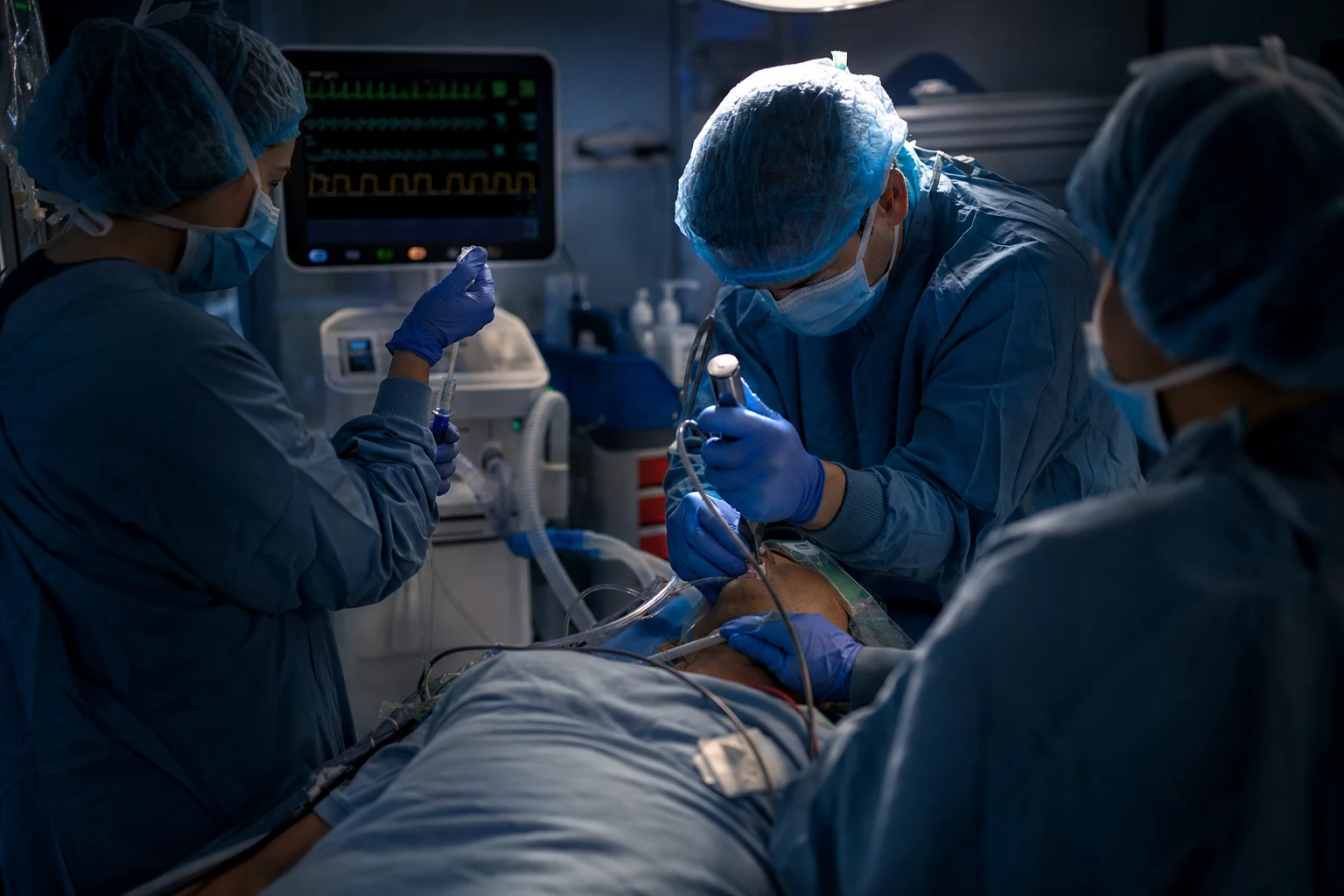
The airway is the first letter of the primary survey for a reason: without a patent, protected airway and effective oxygenation, no other resuscitation matters. In the emergency department the definitive airway — a cuffed tracheal tube — is required whenever the patient cannot maintain or protect their own airway, cannot be oxygenated or ventilated, or is expected to follow a course that will compromise these. Rapid sequence intubation is the technique used to achieve that tube swiftly while minimising the risk of aspiration in a patient who has not fasted and whose physiology is precarious. It is one of the highest-stakes procedures in emergency medicine: it is performed frequently, it is time-critical, and its complications — hypoxia, hypotension, oesophageal intubation, and peri-intubation cardiac arrest — are common and dangerous. [1]
Indications for a definitive airway
A definitive airway — a cuffed tube in the trachea, secured and ventilated — is indicated when the patient fails one of three tests. Failure to maintain the airway: the obtunded patient whose tongue or secretions obstruct, or the patient with soft-tissue swelling from anaphylaxis, burns or infection. Failure to protect the airway: the patient with a reduced conscious level who cannot guard against aspiration, classically the patient with a Glasgow Coma Score of 8 or below. Failure to oxygenate or ventilate: the hypoxaemic patient despite supplemental oxygen, the tiring patient in respiratory failure, or the patient who cannot be ventilated with a bag-valve-mask. A fourth indication is the anticipated course: the patient who will predictably lose the airway, such as the rapidly progressing airway burn or the patient about to undergo a time-critical procedure for which a secure airway is a prerequisite. The decision is clinical and made early, before the patient arrests. [1]
Predicting the difficult airway
A structured bedside assessment predicts many difficult intubations, and the difficult-airway guidelines assume it has been done.[1] The LEMON score is the emergency-department standard: Look externally (receding chin, beard, facial trauma, obesity); Evaluate the 3-3-2 (mouth opening of three finger-breadths, hyoid-to-mental distance of three, hyoid-to-thyroid notch of two); Mallampati (the higher the class, the more difficult); Obstruction (stridor, infection, mass); Neck mobility. The companion assessments are MOANS for difficult mask ventilation (Mask seal, Obesity, Age, No teeth, Stiffness) and RODS for a difficult supraglottic airway (Restricted mouth opening, Obstruction, Disrupted or Distorted anatomy, Stiffness). No score is perfectly sensitive, and the anticipated difficult airway — a high LEMON score, known anatomical difficulty, or a disrupted airway — changes the plan from a crash rapid sequence to an awake technique or a double-set-up. The key examiner point is that prediction changes strategy.
Differential diagnosis
Not every emergency airway encounter is the same, and the operator must distinguish the clinical scenario at the bedside — because each dictates a different preparation, a different drug strategy, and a different algorithm. The four airway scenarios to distinguish are: [1]
- Anticipated difficult airway: identified before induction by a positive LEMON score, difficult mask ventilation (MOANS), difficult supraglottic airway (RODS), or known anatomical distortion from burns, infection, trauma or tumour. The response is a "double set-up" — a senior operator, a video laryngoscope ready, a supraglottic airway drawn up, and a front-of-neck access kit open — and, where time and cooperation allow, an awake look.
- Crash airway: the periarrest or arrested patient who cannot be preoxygenated or assessed and in whom there is no time for a full rapid sequence. The operator proceeds directly to laryngoscopy with no or a minimal induction dose, paralysis if any circulation remains, and an immediate plan for surgical airway if the first look fails.
- Failed intubation: the unanticipated difficult airway — the best optimised attempt at laryngoscopy (position, blade, bougie, video laryngoscope, reduced cricoid) has not secured the tube. The response is to declare failure early and ascend the Difficult Airway Society ladder (Plan B supraglottic airway, Plan C face-mask ventilation) rather than make repeated, worsening attempts.
- Can't-intubate, can't-oxygenate (CICO): the ultimate airway emergency — neither the tracheal tube nor a supraglottic airway succeeds and face-mask ventilation cannot oxygenate. This is the single trigger for immediate scalpel-bougie-tube cricothyroidotomy (Plan D); hesitation, not technique, is the dominant cause of death. [1]
Preparation: equipment, team and checklist
Most failed intubations fail at preparation. The equipment is laid out before induction and checked: a working suction (the single most important and most often absent item), the laryngoscope (direct and video) with a chosen blade and a spare, the endotracheal tube of the predicted size with its cuff tested and a smaller tube available, a bougie or stylet, a supraglottic airway as a rescue device, tape and a tube tie, and the means to ventilate (a bag-valve-mask with a mask that fits). The drugs are drawn up and labelled — the induction agent and the paralytic, in weight-appropriate doses — with a flush attached, and vasopressors drawn up for the hypotensive patient. The team is briefed in roles (intubator, assistant, drugs, cricoid if used, in-charge) and a pre-induction checklist is run. Capnography is connected and ready. A "double set-up" — preparing simultaneously for the anticipated difficult airway, with the front-of-neck access kit open and a senior present — is used when difficulty is expected. [1]
Preoxygenation and apnoeic oxygenation
Preoxygenation buys the safe apnoeic time during which the intubation must occur. The aim is to denitrogenate the functional residual capacity, creating an oxygen reservoir that maintains saturation during the apnoeic period of laryngoscopy. Effective preoxygenation is a tight-fitting mask delivering a high inspired oxygen for three minutes, or eight vital-capacity breaths over one minute in the urgent case, aiming for an end-tidal oxygen approaching 90 per cent. Apnoeic oxygenation extends this window: high-flow nasal oxygen at 30 to 70 litres per minute (transnasal humidified rapid-insufflation ventilatory exchange) continues to deliver oxygen to the alveoli during apnoea by the pressure gradient, and substantially prolongs the time to desaturation. Inadequate preoxygenation is one of the commonest and most preventable causes of peri-intubation hypoxia. [1]
The rapid sequence intubation sequence
The sequence is a choreographed, time-pressured series of steps in which the induction and paralytic agents are given in close succession without a pause for mask ventilation, in order to minimise the window during which the airway is unprotected. The steps are: prepare and preoxygenate as above; give the induction agent followed immediately by the paralytic (a true push-push technique, with no interval); do not routinely mask-ventilate; perform laryngoscopy once the jaw relaxes (typically 45 to 60 seconds after suxamethonium, or around 60 seconds after rocuronium); pass the tube through the cords under direct or video vision, ideally over a bougie; confirm placement with capnography before releasing cricoid pressure; inflate the cuff, secure the tube, and ventilate. [1]
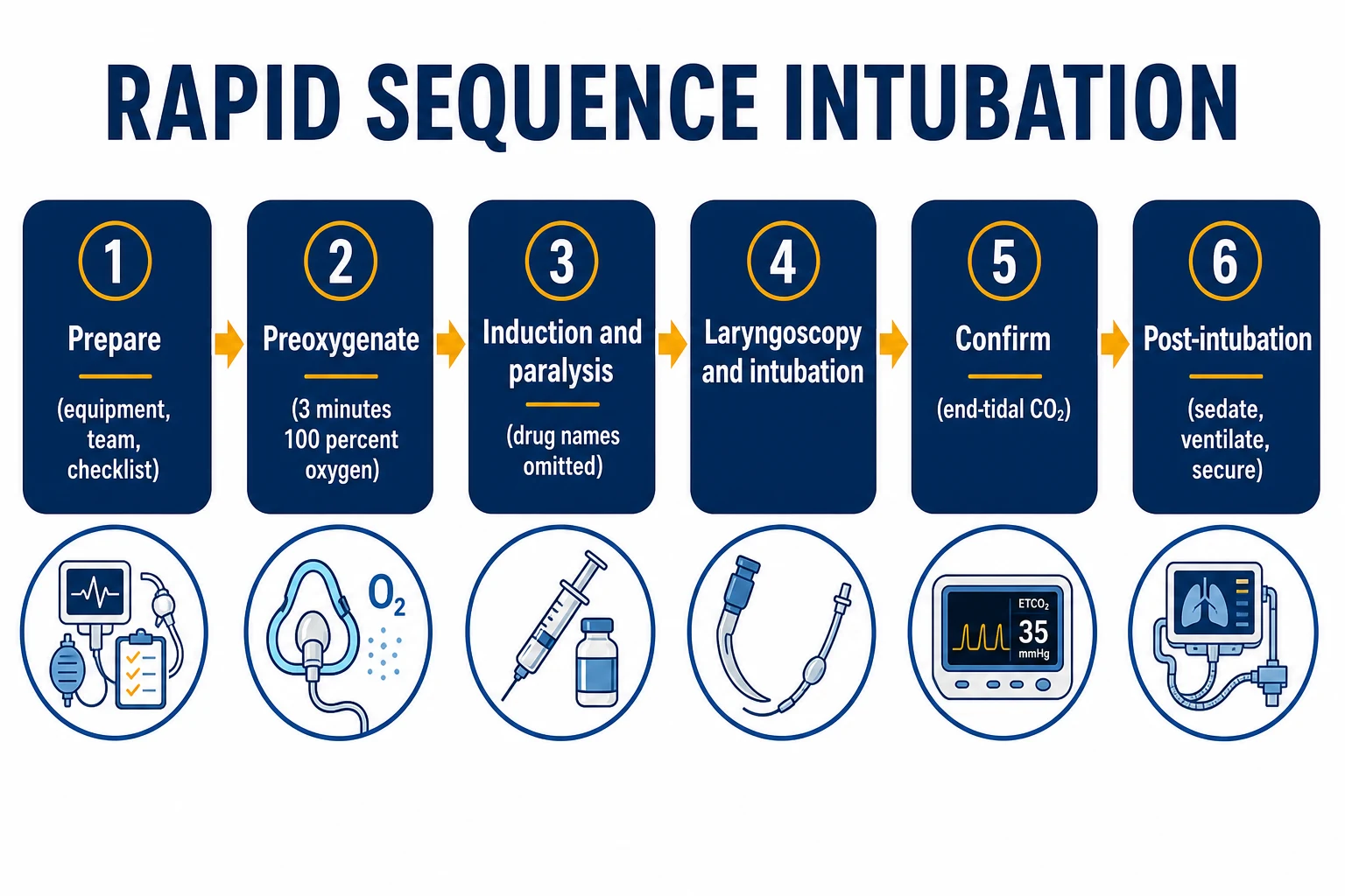
Two traditional elements have been substantially abandoned. Cricoid pressure (the Sellick manoeuvre) was once routine to occlude the oesophagus and prevent regurgitation, but it is unreliable, may worsen the laryngoscopic view, and contemporary practice applies it selectively or not at all. Routine pretreatment — the historical "three minutes of pre-treatment" with atropine, fentanyl or a defasciculating dose — is no longer recommended, although atropine retains a role to blunt suxamethonium-induced bradycardia in young children. [1]
Drug doses and choice: induction agents
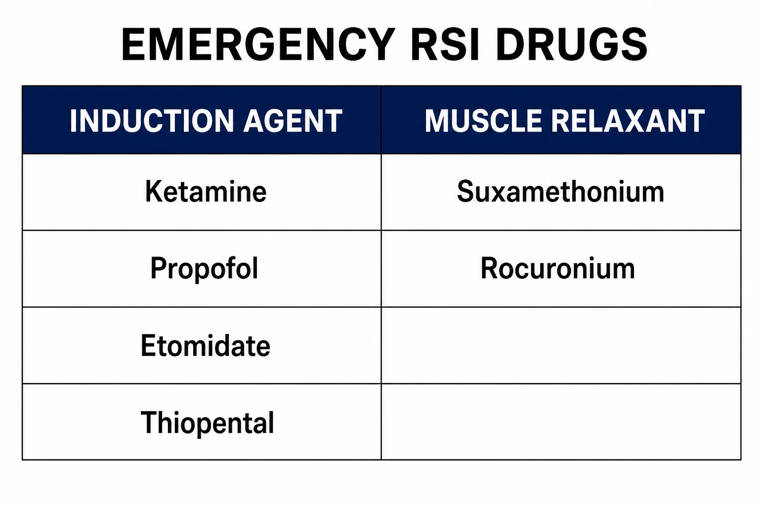
The induction agent is chosen for the patient's physiology, and the wrong choice — a full dose of a cardiovascular depressant in a shocked patient — is a frequent cause of peri-intubation collapse. Ketamine, 1 to 2 mg per kilogram intravenously, preserves sympathetic tone and bronchial tone and is the agent of choice in hypotension or shock, severe asthma, and the head-injured patient (it does not raise intracranial pressure to a clinically meaningful extent and it maintains cerebral perfusion pressure by preserving blood pressure). Propofol, 1 to 2.5 mg per kilogram, is smooth and antiemetic but a potent cardiovascular depressant; it is unsuitable for the shocked patient. Etomidate, 0.3 mg per kilogram, is haemodynamically stable and the debate around its transient adrenal suppression has not displaced it from the shocked or septic patient in many departments. Thiopental, 3 to 5 mg per kilogram, is the agent of choice for status epilepticus and for induction in raised intracranial pressure but is a cardiovascular depressant and is caustic if extravasated. Midazolam is a weak, slow and unreliable induction agent for rapid sequence intubation. The principle: match the agent to the haemodynamics, dose conservatively in the shocked or elderly, and have a vasopressor drawn up. [1]
Drug doses and choice: neuromuscular blocking agents
The paralytic must produce profound, rapid relaxation so that the cords are open and immobile on laryngoscopy. Suxamethonium (succinylcholine), 1 to 1.5 mg per kilogram, is the fastest and shortest-acting: it depolarises the motor endplate, produces fasciculations then paralysis within 30 to 60 seconds, and recovers in 5 to 10 minutes. Its problems are its complications: a predictable rise in serum potassium (dangerous in the patient with pre-existing hyperkalaemia, renal failure, burns beyond 24 hours, or denervation injury), malignant hyperthermia in the susceptible, bradycardia (especially with repeated doses and in children), and prolonged apnoea in the patient with atypical plasma cholinesterase. Rocuronium, 1.0 to 1.2 mg per kilogram for rapid sequence, is a non-depolariser with a slightly slower onset than suxamethonium and a longer duration, but its decisive advantage is that it is rapidly and reliably reversible by sugammadex, which means a "can't intubate, can't oxygenate" situation after rocuronium has a pharmacological rescue. The Cochrane review concluded that suxamethonium provides superior intubating conditions to rocuronium overall, but the margin is small and the ability to reverse rocuronium makes it the preferred agent in many emergency departments, especially where suxamethonium is contraindicated.[2][5]
Confirming tube placement: capnography
Unrecognised oesophageal intubation is lethal, and confirmation of tracheal placement is therefore mandatory and immediate. The gold standard is continuous waveform capnography: a tracheal tube in the airway shows a carbon-dioxide trace with each breath, while an oesophageal tube does not. Capnography also monitors the adequacy of ventilation thereafter and detects disconnection or tube displacement at once. Clinical signs — chest movement, condensation in the tube, auscultation — are adjuncts, not confirmations, and the oesophageal detector device is a useful adjunct in the absence of capnography. The tube is then secured and its position confirmed on an early chest radiograph. [1]
Video versus direct laryngoscopy
Video laryngoscopy improves the glottic view, particularly in the predicted difficult airway and the anterior larynx, and is now first-line in many emergency departments.[6] The Cochrane review found that video laryngoscopy improves the view of the cords, though the effect on first-pass success and time is more nuanced across devices and operators. Direct laryngoscopy with the Macintosh blade remains a core skill and may be faster in the easy airway; the bougie is the natural partner of direct laryngoscopy and improves first-pass success in the difficult airway, where the BEAM trial demonstrated a clear benefit — first-attempt success of 96 per cent with a bougie against 82 per cent with a stylet-and-tube in patients with a difficult airway.[4]
The unanticipated difficult airway and front-of-neck access
When the first attempt at intubation fails, the response is structured, not improvised, and the difficult-airway guidelines exist to be followed under stress.[1][1] The Difficult Airway Society algorithm proceeds as a ladder: Plan A — optimise the intubation attempt (position, blade, bougie, video laryngoscope, reduce cricoid); Plan B — place a supraglottic airway as a rescue or intubate through it; Plan C — re-establish face-mask ventilation, optimising with a two-person technique, an oropharyngeal airway and full paralysis; and Plan D — the can't-intubate, can't-oxygenate emergency, which is the front-of-neck access. The Vortex approach frames the same three rescue steps (supraglottic, supraglottic device, invasive) as concentric rings around the failed intubation, with the decision to cut to the surgical airway made when the "best attempt" at each is exhausted.

In the adult, the front-of-neck access of choice is the scalpel-bougie-tube cricothyroidotomy: a transverse incision through the cricothyroid membrane, a bougie passed into the trachea, and a cuffed tube railroaded over the bougie. It is a last-resort, life-saving procedure, and its performance is degraded by hesitation; rehearsing the steps and having the kit open in the anticipated difficult airway (the double set-up) is what makes it succeed. Needle cricothyrotomy with jet ventilation is unreliable in the adult and is not the recommended technique. [1]
Peri-intubation complications and the post-intubation patient
The peri-intubation period is dangerous, and the complications are commoner than is often appreciated. The international INTUBE study of critically ill patients reported cardiovascular instability in 42.6 per cent of intubations, with severe hypoxaemia in 9.3 per cent and cardiac arrest in 3.1 per cent — a reminder that the act of intubating can precipitate the collapse the resuscitation was intended to prevent.[3] Repeated intubation attempts are a key driver: a national emergency airway registry study found that multiple attempts are strongly associated with complications, including hypoxaemia, regurgitation, and cardiac arrest.[7] The principles that reduce these events are preoxygenation, a haemodynamically stable induction agent and a vasopressor ready, limiting intubation attempts (the operator who fails twice should step back and escalate or change approach), and the disciplined difficult-airway algorithm. After the tube is secured the patient is sedated and ventilated to target (avoiding both hyperoxia and permissive hypercapnia extremes unless indicated), reassessed, and prepared for definitive imaging or disposition.
Special situations
In the shocked patient, the induction dose is reduced (often by half) because the cardiovascular reserve is gone and a full dose of any agent will precipitate collapse; ketamine is preferred, and noradrenaline is drawn up. In the head-injured patient, the goals are a definitive airway, normoxia (saturation of 94 per cent or more) and normocapnia, while avoiding hypotension — every episode of hypotension worsens secondary brain injury — so ketamine with careful haemodynamics is standard, with attention to the possibility of raised intracranial pressure. In status epilepticus, the patient is intubated to terminate the seizure and protect the airway, with a rapid agent and a thiopental or propofol infusion for seizure control. In the agitated or hypoxaemic patient who cannot tolerate preoxygenation, delayed sequence intubation — a dissociative dose of ketamine to calm the patient and allow effective preoxygenation before the formal induction and paralysis — is a useful technique. [1]
Special populations and regional guidelines
In children, the airway is anatomically different (a larger tongue, a higher larynx, a narrowest point at the cricoid), the endotracheal tube is sized by age (uncuffed, internal diameter roughly age in years divided by four plus four; cuffed, plus three-and-a-half), and suxamethonium is avoided in the undiagnosed myopathy of the young child; bradycardia from suxamethonium is prevented with atropine in the youngest. In pregnancy, the airway is more difficult (oedema, breast engorgement, reduced functional residual capacity and faster desaturation), aspiration risk is higher, and a smaller tube and experienced operator are the rule. The guidelines differ by region: the Difficult Airway Society 2015 algorithm is the United Kingdom standard, the AIDAA 2016 algorithm is the Indian standard, the Vortex (developed in Australasia) frames the rescue steps, and the ARC/NZRC guidelines govern the Australasian context.[1][1][1] The principles are universal; the operator must know the local algorithm.
SAQ — Rapid sequence intubation in the shocked patient
10 minutes · 10 marks
A 60-year-old man with the severe sepsis and a lobar pneumonia is hypoxaemic and tiring. The oxygen saturation is 88 per cent on a non-rebreather, the respiratory rate is 36, the blood pressure is 85 over 50, and the decision is made to intubate.
SAQ — Preoxygenation and the paralytic choice
10 minutes · 10 marks
A 45-year-old man with a 15 per cent burn sustained three weeks ago, now ventilated on the intensive care unit for an evolving inhalation injury, requires the intubation for the worsening respiratory failure. He is obese and the LEMON score is high.
Red flags
The following features identify the airway at immediate risk or the intubation likely to be dangerous, in which a senior, a full set-up and a low threshold for the difficult-airway algorithm are required: [1]
[1]ED airway management — the exam-exhaustive deep dive
This section consolidates the operator-level detail an examiner probes: the preoxygenation physics, the drug-by-drug decision matrix with explicit contraindications, the LEMON/MOANS/RODS discriminators as a side-by-side, the bougie and video-laryngoscopy trials that have reshaped modern practice, the can't-intubate-can't-oxygenate surgical airway as a rehearsed sequence, and the disciplined post-intubation bundle. Each block below is a discrete, citable point a Fellowship examiner can ask about. [1]
Preoxygenation — buying and extending the safe-apnoea window
Preoxygenation — maximising the apnoeic time
The goal — denitrogenate the functional residual capacity
Preoxygenation replaces the nitrogen-rich functional residual capacity (about 2.5 L in the adult) with an oxygen reservoir. Done well it buys 5 to 8 minutes of safe apnoea in a healthy adult before desaturation; done poorly the patient falls below 90 per cent within a minute. The endpoint is an end-tidal oxygen at or above 80 to 90 per cent, signalling the alveoli are nearly pure oxygen.
Standard technique — 3 minutes of 100% oxygen
A tight-fitting mask sealed to the face delivering 100 per cent oxygen (a non-rebreather or a Mapleson C / anaesthetic circuit at 15 L per minute or more) for three minutes with the patient taking tidal breaths. A mask leak is the commonest cause of failure — hold it on firmly, two hands if needed.
The urgent alternative — 8 vital-capacity breaths
When three minutes is too long, eight vital-capacity breaths of 100 per cent oxygen over roughly one minute achieves near-equivalent denitrogenation, because each deep breath flushes alveolar nitrogen far faster than tidal breathing.
Apnoeic oxygenation — keep the flow running
Leave high-flow nasal oxygen at 30 to 70 L per minute running through induction and laryngoscopy (THRIVE — transnasal humidified rapid-insufflation ventilatory exchange). During apnoea the alveoli take up oxygen faster than carbon dioxide leaves, generating a sub-atmospheric gradient that draws oxygen in and can extend safe apnoea to 20 to 30 minutes in selected patients.
The patients who desaturate fast
The shocked, septic, obese, pregnant, and small child, and anyone with a low FRC or high shunt (pneumonia, pulmonary oedema, ARDS), desaturates in under a minute regardless of technique. In these patients preoxygenation is non-negotiable, head-up positioning is mandatory, and delayed-sequence intubation may be needed to permit it.
The RSI protocol as a timed, choreographed sequence
The rapid sequence — second by second
Prepare, brief, checklist (minus 5 min)
Equipment laid out and checked — suction on and working (the most often absent item), laryngoscope (direct and video) with chosen blade and a spare, the ETT with a tested cuff and a smaller tube available, a bougie, a supraglottic airway, capnography connected. Drugs drawn up and labelled in weight-appropriate doses with a flush attached; vasopressors drawn up for the hypotensive. Roles assigned; a pre-induction checklist run aloud.
Preoxygenase (minus 3 min)
Three minutes of 100 per cent oxygen by a sealed mask, or eight vital-capacity breaths if urgent; high-flow nasal oxygen running for apnoeic oxygenation; patient head-up if possible.
Induce and paralyse — true push-push (T0)
The induction agent followed immediately by the paralytic in one continuous flush, with no interval and no pause. The two drugs go in as a single push-push, not push-pause.
No routine mask ventilation
The airway is left unprotected only for the briefest window; routine bagging is omitted to avoid gastric insufflation and regurgitation. Gentle mask ventilation may be used in the patient at high risk of hypoxaemia (the infant, the critically ill) — a judgement call.
Wait for paralysis, then intubate (T+45 to 60 s)
The jaw relaxes at around 45 to 60 seconds after suxamethonium, or around 60 seconds after rocuronium at 1.2 mg per kilogram. Laryngoscope once, pass the tube through the cords under vision, ideally over a bougie.
Confirm with capnography before releasing cricoid
A sustained carbon-dioxide waveform over six breaths confirms tracheal placement. Only then inflate the cuff, secure the tube, and ventilate.
Propofol
- Dose 1 to 2.5 mg per kilogram IV
- Smooth, antiemetic, fast offset; the default for the haemodynamically well patient
- POTENT cardiovascular depressant — avoid in shock, hypovolaemia, the elderly, sepsis
- Pain on injection; reduces blood pressure by vasodilation and myocardial depression
Ketamine
- Dose 1 to 2 mg per kilogram IV (reduce to 0.5 to 1 mg per kilogram in shock)
- Preserves sympathetic tone and bronchial tone; first-line in hypotension, sepsis, severe asthma, raised ICP
- Dissociative emergence (rare in the sedated ICU patient); modest sialorrhoea
- The ICP concern is historical — at modern doses ketamine maintains cerebral perfusion pressure by preserving blood pressure
Etomidate
- Dose 0.3 mg per kilogram IV
- The most haemodynamically stable induction agent; favoured in septic and shocked patients
- Transient dose-dependent adrenal suppression (11-beta-hydroxylase) — debated but has not displaced it in many departments
- Consider a stress-dose steroid in septic shock if etomidate is used and steroids are otherwise indicated
Thiopental
- Dose 3 to 5 mg per kilogram IV
- Cerebroprotective; the agent of choice for status epilepticus and raised intracranial pressure
- Cardiovascular depressant — reduce the dose in shock; caustic if extravasated
- Careful in porphyria (contraindicated in acute intermittent porphyria)
Midazolam
- Dose 0.1 to 0.3 mg per kilogram — unreliable
- Weak, slow and unpredictable onset; NOT recommended as a sole RSI induction agent
- Marked hypotension in the shocked patient despite being "cardiovascularly gentle" in health
- Reserve for sedation, not induction
Suxamethonium (succinylcholine)
- Dose 1 to 1.5 mg per kilogram IV; onset 30 to 60 s; recovery 5 to 10 min
- The fastest, shortest depolariser — gold standard for the crash and the anticipated rapid recovery
- Predictable hyperkalaemia: DANGEROUS in known hyperkalaemia, renal failure, burns 24 h to 6 months, denervation/crush/immobilisation
- Malignant hyperthermia in the susceptible; bradycardia (especially repeat doses and children — premedicate with atropine); prolonged apnoea in atypical plasma cholinesterase
Rocuronium
- Dose 1.0 to 1.2 mg per kilogram IV for RSI; onset around 60 s; duration 30 to 60 min
- Non-depolariser with a slightly slower onset than suxamethonium but no hyperkalaemia, no malignant hyperthermia
- Fully and rapidly reversible by sugammadex (16 mg per kilogram for immediate reversal) — the decisive advantage in CICO planning
- Use when suxamethonium is contraindicated and when the operator wants a pharmacological escape from the paralysed airway
Predicting the difficult airway — LEMON, MOANS, RODS side by side
LEMON — difficult intubation
- L — Look externally (receding chin, overbite, beard, facial trauma, obesity, pregnancy)
- E — Evaluate 3-3-2 (mouth opening 3 finger-breadths, hyoid-to-mental 3, hyoid-to-thyroid 2)
- M — Mallampati class (I to IV; higher is harder)
- O — Obstruction (stridor, infection such as epiglottitis, mass, haematoma)
- N — Neck mobility (cervical collar, arthritis, ankylosing spondylitis, obesity)
MOANS — difficult mask ventilation
- M — Mask seal (beard, facial trauma, oedema)
- O — Obesity (low FRC, fast desaturation, soft tissue)
- A — Age 55 and over (loss of tissue tone)
- N — No teeth (poor seal; edentulous patients are easier to mask if the cheeks are inflated)
- S — Stiffness (resistance to ventilation — asthma, COPD, pulmonary oedema, ARDS, obesity)
RODS — difficult supraglottic airway
- R — Restricted mouth opening (less than 2 to 3 finger-breadths)
- O — Obstruction at or below the larynx
- D — Disrupted or Distorted upper-airway anatomy (trauma, infection, tumour, oedema)
- S — Stiffness (high airway pressures prevent the SGA from sealing and ventilating)
The bougie and video laryngoscopy — the trials that shaped practice
BEAM trial — Driver 2018 (JAMA): bougie vs stylet in the difficult ED airway
Design
Single-centre randomised clinical trial; 757 patients, Hennepin County Medical Center ED
Intervention
Bougie vs endotracheal tube with stylet for emergency RSI
Primary result
First-attempt intubation success: bougie 96 per cent vs stylet 82 per cent in the difficult-airway subgroup (98 per cent vs 87 per cent overall)
Caveat
Single-centre, at an institution where bougie use was already the default — generalisability questioned
Clinical bottom line
Established the bougie as first-line for the difficult emergency airway with direct laryngoscopy; the single best first-attempt success benefit attributed to the device.
BOUGIE trial — Driver 2021 (JAMA): the multicentre bougie vs stylet test
Design
Pragmatic multicentre randomised clinical trial; 1102 adults across 7 EDs and 8 ICUs in the USA
Intervention
Bougie vs endotracheal tube with stylet for tracheal intubation of critically ill adults
Primary result
First-attempt intubation success: bougie 80.4 per cent vs stylet 83.0 per cent — NO statistically significant difference
Key caveat
Did not replicate the large BEAM benefit; bougie advantage is operator- and setting-dependent and most evident in the genuinely difficult airway
Clinical bottom line
Routine bougie use does not guarantee better first-pass success across all comers, but the bougie remains a low-cost, high-value tool for the difficult view and the partial view — reach for it when the cords are not fully seen.
MACMAN trial — Lascarrou 2017 (JAMA): McGrath MAC video vs Macintosh direct laryngoscopy in ICU
Design
Multicentre randomised clinical trial; 371 patients across 7 French ICUs
Intervention
McGrath MAC video laryngoscopy vs direct Macintosh laryngoscopy
Primary result
First-pass intubation success: video 67.7 per cent vs direct 70.3 per cent — NO significant difference
Key caveat
Video laryngoscopy improved the glottic view (better Cormack-Lehane grade) but this did NOT translate into better first-pass success; some signal to more life-threatening complications
Clinical bottom line
A better view does not automatically mean an easier intubation — passing the tube around the hyperangulated blade is a separate skill. Device-specific competence matters; video laryngoscopy is not a panacea.
INTUBE study — Russotto 2021 (JAMA): peri-intubation adverse events in 29 countries
Design
Prospective international cohort; 2964 intubations across 29 countries, 197 ICUs
Population
Critically ill adults undergoing tracheal intubation
Primary result
At least one major peri-intubation adverse event in 45.2 per cent: cardiovascular instability 42.6 per cent, severe hypoxaemia 9.3 per cent, cardiac arrest 3.1 per cent
Risk factors
Absence of preoxygenation, no checklist, no haemodynamic preparation, multiple attempts, operator inexperience
Clinical bottom line
The act of intubating can precipitate the collapse it was meant to prevent. Preoxygenation, a stable induction agent, vasopressors ready, and limiting attempts are the modifiable preventers — know them.
Can't-intubate, can't-oxygenate (CICO) and emergency front-of-neck access
Scalpel-bougie-tube cricothyroidotomy (eFONA) — the adult CICO rescue
Declare CICO — the single trigger
Neither the tracheal tube nor a supraglottic airway succeeds AND face-mask ventilation cannot oxygenate. Declare it aloud, call for the scalpel, and stop fruitless laryngoscopy. Hesitation — not technique — is the dominant cause of death in CICO.
Identify the cricothyroid membrane
Laryngeal handshake: stabilise the larynx with the non-dominant hand, palpate the thyroid prominence and drop down to the depression between the cricoid and thyroid cartilages. In the obese or distorted neck, ultrasound confirms the membrane before induction if time allows (a planned double set-up).
Transverse stab incision through the membrane
A single decisive transverse scalpel incision (number 10 or 20 blade) through skin and the cricothyroid membrane at the membrane, keeping the scalpel in situ. A vertical skin incision with a horizontal membrane cut is an accepted alternative when landmarks are hard.
Turn the blade and pass the bougie
Rotate the scalpel 90 degrees to open the airway, slide the bougie through the incision caudally into the trachea, and confirm tracheal passage by the absence of resistance.
Railroad a cuffed tube over the bougie
Pass a cuffed tube (a size 6.0 cuffed ETT or a dedicated cuffed cricothyrotomy tube) over the bougie into the trachea, remove the bougie, inflate the cuff, and ventilate. Confirm with capnography. Needle cricothyrotomy with jet ventilation is unreliable in the adult and is not the recommended technique.
Post-intubation care — confirm, sedate, ventilate, reassess
The post-intubation bundle — the first 30 minutes
Confirm the tube — capnography first
A sustained carbon-dioxide waveform over six breaths is mandatory and confirms tracheal placement. Clinical signs (chest rise, condensation, auscultation) are adjuncts, not confirmation. An absent or fading trace means oesophageal intubation, tube displacement, or circuit disconnection — investigate immediately.
Secure the tube and check the depth
Tie or tape the tube at the right incisor; adult depth at the teeth is roughly three times the internal diameter in millimetres (a size 8.0 tube sits at about 21 to 23 cm). Too deep = right mainstem intubation (unilateral breath sounds, left-side collapse); too shallow = accidental extubation or cuff leak.
Confirm position on chest radiograph
A portable chest film confirms the tube tip 3 to 5 cm above the carina, excludes mainstem intubation and pneumothorax, and documents the underlying pathology. Do not delay other resuscitation for the film, but obtain it early.
Sedate and analgesise — never leave a paralysed patient undersedated
Start a sedation infusion (propofol or midazolam) plus an opioid (fentanyl or morphine) as soon as the tube is confirmed. A patient paralysed but unsedated experiences awareness and pain — never let a paralytic run without sedation. Target a RASS of 0 to minus 2 unless deeper sedation is indicated (raised ICP, severe ARDS).
Set the ventilator and re-check physiology
Tidal volume 6 to 8 mL per kilogram predicted body weight; a respiratory rate to achieve normocapnia (or permissive hypercapnia in ARDS/raised ICP as appropriate); FiO2 titrated to a saturation of 92 to 96 per cent (avoid hyperoxia in the non-hypoxic, target 94 per cent in the head-injured). Recheck blood pressure — positive-pressure ventilation unmasks hypovolaemia.
Prepare for disposition
Reassess the patient end to end, obtain the definitive imaging or specialty input needed, and arrange the bed (ICU, theatre, or inter-hospital transfer with a secured tube and monitored transport).
Clinical pearls — rapid recall
[1] [1] [1] [1] [1] [1]Additional red flags
[1]References
- [1]Frerk C, Mitchell VS, McNarry AF, et al. Difficult Airway Society 2015 guidelines for management of unanticipated difficult intubation in adults Br J Anaesth, 2015.PMID 26556848
- [2]Tran DT, Newton EK, Mount VA, et al. Rocuronium versus succinylcholine for rapid sequence induction intubation Cochrane Database Syst Rev, 2015.PMID 26512948
- [3]Russotto V, Myatra SN, Laffey JG, et al. Intubation Practices and Adverse Peri-intubation Events in Critically Ill Patients From 29 Countries JAMA, 2021.PMID 33755076
- [4]Driver BE, Prekker ME, Klein LR, et al. Effect of Use of a Bougie vs Endotracheal Tube and Stylet on First-Attempt Intubation Success Among Patients With Difficult Airways Undergoing Emergency Intubation: A Randomized Clinical Trial JAMA, 2018.PMID 29800096
- [5]Hristovska AM, Duch P, Allingstrup M, et al. Efficacy and safety of sugammadex versus neostigmine in reversing neuromuscular blockade in adults Cochrane Database Syst Rev, 2017.PMID 28806470
- [6]Hansel J, Rogers AM, Lewis SR, et al. Videolaryngoscopy versus direct laryngoscopy for adults undergoing tracheal intubation Cochrane Database Syst Rev, 2022.PMID 35373840
- [7]April MD, Schauer SG, Nikolla DA, et al. Association between multiple intubation attempts and complications during emergency department airway management: A national emergency airway registry study Am J Emerg Med, 2024.PMID 39288499
- [8]Driver BE, Semler MW, Self WH, et al. Effect of Use of a Bougie vs Endotracheal Tube With Stylet on Successful Intubation on the First Attempt Among Critically Ill Patients Undergoing Tracheal Intubation: A Randomized Clinical Trial JAMA, 2021.PMID 34879143
- [9]Lascarrou JB, Boisrame-Helms J, Bailly A, et al. Video Laryngoscopy vs Direct Laryngoscopy on Successful First-Pass Orotracheal Intubation Among ICU Patients: A Randomized Clinical Trial JAMA, 2017.PMID 28118659