EM · Fluid resuscitation
Fluid resuscitation
Fluid resuscitation in the emergency department: the choice between balanced crystalloid and saline and the evidence (SMART), the fluid bolus and the concept of fluid responsiveness (dynamic over static), the ROSE model of fluid phases, the 4 D's of fluid therapy, the harm of fluid overload, the specific scenarios from sepsis to burns, and the monitoring of the response.
On this page & tools
Your progress
Saved locally on this device.
Target exams
Red flags
Intravenous fluid is one of the commonest interventions in the emergency department, and like any drug it has an indication, a choice of agent, a dose, a duration, and adverse effects. The goal of fluid resuscitation is to restore the intravascular volume and the venous return, so that the stroke volume and the cardiac output rise and the tissue perfusion is improved — but only the patient who is on the ascending limb of the Frank-Starling curve, the fluid-responsive patient, will benefit. The Fellowship-level understanding rests on the evidence for the fluid choice (balanced over saline), the concept and the testing of fluid responsiveness (dynamic over static), the phases of fluid therapy (the ROSE model), and the recognition that fluid, given injudiciously, is as harmful as its absence. [1]
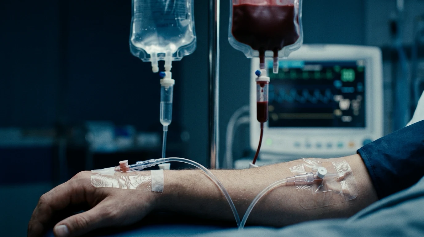
Crystalloid types: balanced versus saline
The resuscitation fluid of the modern emergency department is a balanced crystalloid — Hartmann's (Ringer's lactate) or Plasma-Lyte — rather than normal saline. The evidence is strong: the SMART trial, in a large critically ill population, found that balanced crystalloids led to fewer composite outcomes of death, new dialysis or persistent renal dysfunction than saline, and the SALT-ED trial found the same in the emergency department.[1] The mechanism is that saline, with its supraphysiological chloride concentration (154 mmol per litre), causes a hyperchloremic metabolic acidosis, renal vasoconstriction, and an increased risk of acute kidney injury. The balanced crystalloids, with their chloride concentration closer to plasma, avoid this. Dextrose-containing fluids are free-water replacements, not resuscitation fluids; hypertonic saline has a specific role in traumatic brain injury and hyponatraemia but is not a general resuscitation agent.[5]

Colloids and the evidence
The colloid-versus-crystalloid debate is largely settled, and the answer is crystalloid. Albumin is an option in the selected septic patient (the SAFE trial found it equivalent to crystalloid), and it has a role in the burns and the hypoalbuminaemic patient. The synthetic colloids — the hydroxyethyl starches — are harmful (the CHEST, the 6S and the Crystalloid MaSS trials showed they increase the risk of renal failure and mortality), and they are no longer recommended for resuscitation. The crystalloid is the default, the balanced crystalloid is the preferred crystalloid, and the colloid is the exception.[4]
The fluid bolus and the fluid challenge
The fluid bolus — a discrete amount of fluid given over a short period to test and to improve the stroke volume — is the operational unit of fluid resuscitation. A 250 to 500 millilitre bolus of balanced crystalloid is given over 15 to 30 minutes, and the response is assessed against the markers of perfusion (the blood pressure, the heart rate, the capillary refill, the conscious level, the urine output, the lactate). A bolus that improves these markers is a positive response; the patient is on the ascending limb of the Starling curve and may benefit from further fluid. A bolus that does not improve them is a negative response; the patient is not fluid-responsive and further fluid will cause harm. The fluid challenge — a smaller, faster bolus designed to test the Starling curve rather than to resuscitate — is a related concept used at the bedside with the dynamic monitoring. [1]
Fluid responsiveness: dynamic over static
The critical insight of modern fluid therapy is that only about half of the critically ill patients are fluid-responsive, and that the decision to give fluid must be guided by the likelihood of a response. The static markers of volume status — the central venous pressure, the single blood pressure, the heart rate — do not reliably predict the response to a fluid bolus, as the systematic review of the central venous pressure literature confirmed.[3] The dynamic markers do: the passive leg raise (a reversible endogenous fluid bolus, with the stroke-volume response measured by the echocardiography or the arterial line), the pulse-pressure or the stroke-volume variation in the ventilated patient, and the variation of the inferior vena cava with respiration. The patient who is fluid-responsive by these dynamic tests may benefit from a bolus; the patient who is not needs a vasopressor or an inotrope, not more crystalloid.[4]

The ROSE model and the 4 D's
Fluid therapy is not a single intervention but a process with phases, and the ROSE model — Resuscitation, Optimisation, Stabilisation, Evacuation — frames it. In the resuscitation phase the fluid is given to restore the perfusion. In the optimisation phase the fluid is titrated to the ongoing need, guided by the dynamic markers and the perfusion. In the stabilisation phase the fluid is minimal — only the maintenance and the ongoing losses. In the evacuation (or the de-resuscitation) phase the accumulated fluid is mobilised and removed, sometimes actively with the diuretics or the renal replacement therapy, because the fluid overload is itself a cause of harm. The 4 D's — the Drug (which fluid), the Dose (how much), the Duration (how long), and the De-escalation (when to stop) — are the practical questions at every phase. [1]
The harm of fluid overload
Fluid, given beyond the point of responsiveness, is harmful. The tissue oedema impairs the oxygen diffusion and the wound healing; the pulmonary oedema worsens the gas exchange and the work of breathing; the abdominal compartment syndrome from the gut oedema compresses the venous return and the renal perfusion; the coagulopathy dilution from the large-volume crystalloid lowers the haematocrit and the clotting factors. The patient who has received a large volume of crystalloid and is now in positive fluid balance is a patient at risk, and the de-resuscitation is begun as soon as the perfusion is restored.[4]
Specific scenarios
The fluid therapy is adapted to the cause. In sepsis, the Surviving Sepsis Campaign recommends an initial 30 millilitres per kilogram of balanced crystalloid for hypotension or a lactate of 4 mmol per litre or more, given rapidly with reassessment.[2] In haemorrhagic shock, the resuscitation is with blood products (the 1-to-1-to-1 ratio) and not crystalloid, which dilutes the clotting factors. In diabetic ketoacidosis, the fluid is given cautiously (the first litre over the first hour, then a slower rate), with attention to the risk of cerebral oedema, particularly in the child and the young adult. In burns, the Parkland formula (2 to 4 millilitres per kilogram per per cent of body surface area, half in the first 8 hours) guides the first 24 hours, titrated to the urine output. In heat stroke, the rapid cooling is the priority, and the fluid is given to support the perfusion without overloading. In heart failure, the fluid is given with extreme caution and with the early addition of the vasopressor or the inotrope.
Maintenance fluids and monitoring
The maintenance fluid, for the patient who is not eating or drinking, is a balanced crystalloid with the appropriate sodium and the potassium, given at a rate determined by the weight (the classic 4-2-1 rule: 4 mL per kg for the first 10 kg, 2 mL for the next 10 kg, 1 mL for each subsequent kg, per hour), and adjusted for the fever, the losses and the comorbidity. The prolonged use of saline as the maintenance fluid causes the hypernatraemia and the acidosis, and the balanced crystalloid is preferred. The fluid response is monitored clinically — the perfusion, the urine output, the lactate, the conscious level — and by the dynamic markers and the serial laboratory tests (the electrolytes, the creatinine, the haemoglobin). The elderly, the cardiac and the renal-failure patient is monitored with extra caution. [1]
Common pitfalls
The recurring errors are: using saline rather than the balanced crystalloid for resuscitation; giving fluid by rote without assessing the response; relying on the central venous pressure or the heart rate as the markers of the volume status; over-resuscitating the non-responsive patient; not recognising the fluid overload; giving crystalloid rather than blood to the bleeding patient; not de-escalating the fluid once the perfusion is restored; and using the dextrose-containing fluid as a resuscitation agent. [1]
The evidence for the fluid choice — the crystalloid trials in detail
The shift from the saline to the balanced crystalloid rests on three landmark trials. The SMART trial (the Saline versus Plasma-Lyte in the ICU, NEJM 2018) randomised 15,752 critically ill adults in the ICU to the balanced crystalloid (Plasma-Lyte 148) versus the saline for ALL the fluid needs; the primary composite of the death, the new dialysis, or the persistent renal dysfunction at 30 days was lower in the balanced group (14.3 per cent versus 15.4 per cent).[1] The SALT-ED trial (NEJM 2018, the ED sister of SMART) randomised 13,347 adults receiving the isotonic fluid in the emergency department; the 30-day MAKE30 outcome (the Major Adverse Kidney Event — the death, the new dialysis, the persistent renal dysfunction) was 4.7 per cent versus 5.6 per cent favouring the balanced crystalloid, with the effect largest in the septic subgroup.[6] The SPLIT trial (JAMA 2015, the NZ precursor) — 2,278 ICU patients, the balanced versus the saline — found NO difference in the AKI, but it used the lactated Ringer's, the median fluid volume was small (under 2 L), and it was almost certainly underpowered.[7]
SMART — balanced vs saline in the ICU (NEJM 2018)
PMID 29467594
Key finding
The pragmatic, the cluster-crossover, the single-centre trial at Vanderbilt; 15,752 ICU adults. The composite of the death, the new RRT, or the persistent renal dysfunction at 30 days: 14.3 per cent (balanced) vs 15.4 per cent (saline); OR 0.91 (P = 0.04). The effect was larger in the sepsis subgroup (OR 0.80).
Practice change
The largest fluid trial to date; the balanced crystalloid is the ICU default. The Number Needed to Treat is 94 for the composite; for the septic patient it is 20.
SALT-ED — balanced vs saline in the ED (NEJM 2018)
PMID 29467593
Key finding
The 13,347-patient ED companion to the SMART; the 30-day MAKE30 was 4.7 per cent (balanced) vs 5.6 per cent (saline); OR 0.82 (P = 0.01). The hospital-free days did not differ; the septic subgroup drove the benefit.
Practice change
The ED-level evidence for the balanced crystalloid. Every patient resuscitated in the ED should receive the balanced crystalloid, not the saline.
SPLIT — the smaller NZ precursor (JAMA 2015)
PMID 26441092
Key finding
The 2,278-patient double-blind NZ ICU trial of the buffered crystalloid (Plasma-Lyte) vs the saline. NO difference in the AKI (9.6 vs 9.2 per cent) or the RRT — likely underpowered, and the median fluid volume was small (under 2 L).
Practice change
The negative precursor that was superseded by the larger SMART and SALT-ED. The lesson: the effect of the saline harm is small per patient but cumulative across the populations.
0.9% Saline
- Na 154, Cl 154 mmol/L — the supraphysiological chloride
- Strong-ion-difference acidosis; the hyperchloremic metabolic acidosis
- The tubuloglomerular feedback activation; the afferent arteriolar vasoconstriction; the AKI
- Osmolarity 308; pH 5.0 (acidic)
- The fluid to AVOID for the large-volume resuscitation; reserve for the hyponatraemia, the hypercalcaemia, the chloride-deficient metabolic alkalosis, and the dilution of the blood products (the citrate compatibility)
Hartmann (RL)
- Na 131, Cl 111, K 5, Ca 2, lactate 29 mmol/L; the chloride closer to the plasma
- Lactate metabolised to the bicarbonate in the liver; the pH-neutralising buffer
- Osmolarity 279; pH 6.5
- The workhorse balanced crystalloid in the ANZ and the UK ED
- Avoid in the severe liver failure (the lactate accumulates); the calcium precipitates with the citrated blood products in the same line — give them in separate lines or use Plasma-Lyte
Plasma-Lyte
- Na 140, Cl 98, K 5, Mg 1.5, acetate 27, gluconate 23 mmol/L; the chloride essentially plasma-equivalent
- Acetate and gluconate metabolised everywhere (the muscle, the kidney), not just the liver; the most balanced
- Osmolarity 294; pH 7.4 (truly neutral)
- The closest to the plasma; the lowest AKI signal in the subgroup analyses; citrate-compatible — runs with the blood products
- More expensive; the magnesium may cause the flushing; the preferred resuscitation fluid where available
The colloid trials — the albumin equivalence and the starch harm
The colloid-versus-crystalloid question was the great debate of the 1990s and the 2000s, and it has been settled by three trials. The SAFE trial (NEJM 2004) — 6,997 ICU patients randomised to the 4 per cent albumin versus the saline — found NO difference in the 28-day mortality (20.9 versus 21.1 per cent).[8] The CHEST trial (NEJM 2012) — 7,000 ICU patients randomised to the 6 per cent HES (130/0.4) versus the saline — found no mortality difference but a significant increase in the RRT use with the starch.[9] The 6S trial (NEJM 2012) — 804 severe-sepsis patients in Scandinavia — found the HES (130/0.42) increased the mortality (51 versus 43 per cent) and the RRT use compared to the Ringer's acetate.[10] The conclusion is unambiguous: the synthetic colloids (the hydroxyethyl starches) are harmful and should not be used for the resuscitation; the albumin is equivalent to (not better than) the crystalloid, and reserved for the selected patient.
SAFE — albumin vs saline (NEJM 2004)
PMID 15163774
Key finding
The 6,997-patient Australian and NZ ICU trial; the 4 per cent albumin vs the 0.9 per cent saline for the fluid resuscitation. The 28-day mortality: 20.9 per cent (albumin) vs 21.1 per cent (saline); RR 0.99 (P = 0.87). NO difference. The post-hoc subgroup suggested the harm with the albumin in the traumatic brain injury.
Practice change
The albumin is equivalent to the saline — the colloid has no outcome advantage. The albumin is reserved for the selected hypoalbuminaemic, the burns, or the septic patient.
CHEST — 6% HES vs saline in the ICU (NEJM 2012)
PMID 22738097
Key finding
The 7,000-patient Australian and NZ ICU trial; the 6 per cent HES 130/0.4 vs the saline. The 90-day mortality: 18.0 vs 17.0 per cent (no difference, P = 0.26); BUT the RRT was significantly higher with the HES (7.0 vs 5.8 per cent, RR 1.21).
Practice change
The starches do not improve the survival and increase the kidney injury. The CHEST began the end of the HES for the resuscitation.
6S — HES vs Ringer's acetate in severe sepsis (NEJM 2012)
PMID 22517884
Key finding
The 804-patient Scandinavian trial; the 6 per cent HES 130/0.42 vs the Ringer's acetate in the severe sepsis. The 90-day mortality: 51 per cent (HES) vs 43 per cent (Ringer's); RR 1.17 (P = 0.03). The RRT use: 22 vs 16 per cent (P = 0.04).
Practice change
The starches increase the mortality AND the kidney injury in the severe sepsis. The 6S sealed the case against the HES — the MHRA and the EMA restricted and then withdrew the HES in 2013.
Fluid responsiveness — the dynamic markers in detail
The fluid responsiveness is defined as the increase in the stroke volume (or the cardiac output) of at least 10 to 15 per cent in response to the 500 mL bolus (or the equivalent, the passive leg raise). It is a HAEMODYNAMIC state, not a clinical guess. Only about 50 per cent of the critically ill patients are fluid-responsive at any given moment, and the proportion FALLS as the resuscitation progresses (the patient who responded to the first litre may not respond to the third). The modern approach is to TEST before you give the bolus.[13]
Static markers
- The CVP, the single blood pressure, the heart rate, the single IVC diameter.
- Do NOT predict the fluid responsiveness — the area under the ROC curve in the Marik meta-analysis was 0.55 (no better than the coin flip).
- The CVP rises in the right heart failure, the pulmonary hypertension, the severe tricuspid regurgitation — independent of the volume.
- Avoid using the static markers to decide on the fluid bolus; the CVP has NO role in the fluid decision.
Dynamic markers
- The PLR (the 45-degree leg raise for 60 to 90 seconds); the PPV / the SVV (in the ventilated, the closed chest, the regular rhythm); the IVC variability (over 50 per cent in the spontaneous breather); the end-expiratory and end-inspiratory occlusion tests.
- Predict the fluid responsiveness with the sensitivity and the specificity over 80 per cent.
- Test the Starling curve BEFORE you give the fluid.
- The PLR is the Fellowship favourite — the reversible, the endogenous bolus of about 300 mL.
The passive leg raise (PLR) — the reversible bedside fluid challenge
The preparation — the patient in the semi-recumbent 45-degree position; the cardiac output (or the surrogate — the pulse pressure, the end-tidal CO2, the VTI on the echocardiography) is measured at the baseline.
The leg raise — the legs are lifted to 45 degrees AND the trunk flattened to the horizontal simultaneously, so the venous volume comes from the splanchnic and the leg veins (about 300 mL), NOT from the trunk compression.
The wait — 60 to 90 seconds (the effect peaks at this time).
The measurement — the cardiac output (or the surrogate) is re-measured. A rise of 10 per cent or more is the POSITIVE response — the patient is fluid-responsive and may benefit from the bolus.
The reversal — return the patient to the semi-recumbent position; the effect is gone in 2 to 3 minutes. The PLR has caused NO net fluid gain — the ideal test in the patient who may not tolerate the fluid.
The pitfalls — the patient must NOT be in the spontaneous high-respiratory-effort breathing (the negative-pressure PLR is masked); the compression stockings reduce the effect; the right heart failure gives the false negative; the intra-abdominal hypertension distorts the venous return.
The dynamic markers of the fluid responsiveness
The ROSE model — the four phases in detail
Fluid therapy is not a single act but a trajectory, and the ROSE model (Malbrain and colleagues) frames the four phases. Each phase has a different fluid goal, a different fluid volume, and a different risk.[14]
R — Resuscitation
- The EARLY phase (the first hours); the patient is shocked.
- The goal: restore the perfusion (the MAP over 65, the lactate clearance, the urine output over 0.5 mL/kg/h).
- The fluid: the boluses of the balanced crystalloid (250 to 500 mL over 5 to 10 minutes), titrated to the responsiveness.
- The risk: the under-resuscitation; the patient is dying of the shock, not of the fluid.
O — Optimisation
- The SECOND phase (the hours after the shock resolves); the patient is on the Starling plateau.
- The goal: titrate the fluid to the ongoing need, guided by the dynamic markers; avoid the unresponsive bolus.
- The fluid: the smaller boluses, only if the PLR or the PPV is positive; the vasopressor and the inotrope are added as needed.
- The risk: the over-resuscitation; the patient is now at risk of the fluid overload.
S — Stabilisation
- The THIRD phase (the days); the patient is stable.
- The goal: the maintenance plus the ongoing losses only; the de-escalation.
- The fluid: the minimal — the 4-2-1 maintenance, plus the drains, the fever, the insensible.
- The risk: the routine continuation of the high-rate fluid when no longer needed.
E — Evacuation
- The FOURTH phase (the recovery); the patient is in the positive fluid balance.
- The goal: the de-resuscitation — mobilise and remove the accumulated fluid.
- The fluid: the negative balance — the diuretics (the furosemide), the RRT if the kidney fails.
- The risk: the unrecognised fluid overload — the pulmonary oedema, the wound breakdown, the prolonged ventilation.
Permissive hypotension and the restrictive fluid strategy
The traditional resuscitation aimed at the normal blood pressure, but the modern evidence in the traumatic haemorrhage AND in the septic shock favours the LOWER target until the bleeding is controlled or the source is treated. The permissive hypotension in the trauma (the systolic of 80 to 90 mmHg or the MAP of 65 until the haemostasis) avoids the "pop-the-clot" effect of the high pressure, the dilutional coagulopathy of the crystalloid, and the lethal triad (the acidosis, the hypothermia, the coagulopathy). The CLASSIC trial and the CLOVERS trial extended this concept to the septic shock — the LIBERAL fluid strategy is harmful.[11][12]
CLASSIC — restrictive vs liberal fluid in septic shock (NEJM 2022)
PMID 34383327
Key finding
The Scandinavian multicentre trial; 1,554 adults with the septic shock. The restrictive fluid strategy (the bolus only for the severe hypoperfusion — the lactate over 4, the MAP under 65, the mottling) vs the liberal (the standard 30 mL/kg plus the ongoing boluses). The 90-day mortality: 11.3 vs 10.9 per cent (NO difference) — but the restrictive group received 1.2 L LESS fluid and had less severe adverse events.
Practice change
The restrictive strategy is SAFE and the liberal fluid is NOT beneficial. The 30 mL/kg is NOT a mandate; the fluid is titrated to the perfusion, not the rote volume.
CLOVERS — early restrictive vs liberal fluid in sepsis (NEJM 2023)
PMID 36807607
Key finding
The US multicentre trial; 1,563 adults with the septic shock in the first 4 hours. The early restrictive (the vasopressor-first, the fluid-minimising) vs the early liberal (the fluid-first) strategy. The 90-day mortality: 30.7 vs 30.8 per cent (NO difference).
Practice change
The permissive hypotension with the early vasopressor and the minimised fluid is EQUIVALENT to the fluid-first. The fluid-first dogma is dead. The Surviving Sepsis Campaign 2021 has already softened the 30 mL/kg to a suggestion — the CLOVERS confirms the individualised approach.
Blood products and the damage-control resuscitation
The bleeding patient is resuscitated with the blood products, not the crystalloid. The crystalloid dilutes the clotting factors, lowers the haematocrit (and the platelet margination), and worsens the acidosis — the lethal triad of the coagulopathy, the acidosis, and the hypothermia. The damage-control resuscitation — the permissive hypotension, the 1:1:1 transfusion (the plasma to the platelets to the red cells), the tranexamic acid within 3 hours, and the minimisation of the crystalloid — is the standard of the modern trauma, the obstetric, and the GI haemorrhage.[15][16]
The damage-control resuscitation (DCR) — the bleeding patient
The early call — activate the massive transfusion protocol at the recognition of the major haemorrhage (the systolic under 90, the lactate over 4, the clinical suspicion of the major bleeding, the positive FAST).
The 1:1:1 ratio — the plasma, the platelets, and the red cells in the 1:1:1 ratio (the PROPPR trial). One unit of RBC, one of FFP, one pool of platelets (or one apheresis unit) per pack.
The tranexamic acid — 1 g IV over 10 minutes, then 1 g over 8 hours, within 3 hours of the injury (the CRASH-2 trial). Beyond 3 hours the TXA increases the mortality.
The calcium — the citrate in the blood products chelates the calcium; the ionised calcium falls with the rapid transfusion. Give the calcium chloride 1 g (the central or the large-bore line) or the calcium gluconate 2 to 3 g (the peripheral) after every 4 to 6 units.
The minimisation of the crystalloid — the crystalloid is the bridge to the blood, NOT the resuscitation fluid. Limit the crystalloid to 1 L before the blood arrives; do not chase the normal blood pressure before the haemostasis.
The permissive hypotension — the systolic of 80 to 90 mmHg or the MAP of 50 to 65 until the bleeding is controlled (CONTRAINDICATED in the TBI).
The reversal of the anticoagulant — the warfarin with the vitamin K and the Beriplex/PCC; the DOAC with the andexanet alfa or the PCC; the antiplatelet with the platelet transfusion in the intracranial bleed.
The monitoring — the rotational thromboelastometry (the ROTEM) or the TEG to guide the component therapy; the ionised calcium; the core temperature (the hypothermia worsens the coagulopathy); the fibrinogen (cryoprecipitate if under 1.5 g/L).
The exam-exhaustive pearls — the high-yield viva facts
[1] [1] [1] [1] [1] [1] [1] [1] [1] [1]The crystalloid composition — the Fellowship viva numbers
ANZ practice note. The balanced crystalloid (the Plasma-Lyte or the Hartmann's) is the standard resuscitation fluid in the ANZ emergency department and the ICU; the saline is reserved for the specific indications (the hyponatraemia, the hypercalcaemia, the citrate-compatible blood product line). The 0.9 per cent saline is NOT the default — the SMART and the SALT-ED have changed the practice. The MASSIVE transfusion protocol (the MTP) uses the 1:1:1 ratio with the early TXA and the calcium. The Surviving Sepsis Campaign is the framework; the 30 mL/kg is the suggestion, not the mandate; the early vasopressor and the lactate-guided reassessment are the standard. [1]
SAQs — exam practice
SAQ — Fluid resuscitation strategy in septic shock
10 minutes · 10 marks
A 64-year-old man with type 2 diabetes and benign prostatic hypertrophy presents to the emergency department with a 14-hour history of rigors, dysuria, confusion and a single generalized seizure at home. He is hypotensive at 82 over 46, heart rate 124 in sinus rhythm, respiratory rate 28, oxygen saturation 95 per cent on room air, temperature 38.9 degrees Celsius, and he is oliguric with a capillary refill of 4 seconds. Capillary glucose is 9.1, lactate 4.2 mmol per litre, creatinine 186 micromol per litre (baseline 92 three months ago), and the venous blood gas shows pH 7.24, bicarbonate 17, base excess minus 7. A urinary tract source with pyuria and Gram-negative bacteraemia is identified. Outline your intravenous fluid resuscitation strategy in the first six hours, with the choice of fluid, the dose, the trigger for a vasopressor, and the evidence that supports each step.
SAQ — Iatrogenic hyperchloremic acidosis and acute kidney injury from saline
10 minutes · 10 marks
A 72-year-old woman is admitted to the emergency department with urosepsis. Over the first eight hours she receives 4.5 litres of 0.9 per cent saline as resuscitation and a further litre of 4 per cent dextrose in one-fifth saline as maintenance. Her blood pressure has improved to 105 over 60 and her lactate has fallen from 3.8 to 1.9, but she has become oliguric at 15 millilitres per hour. The venous blood gas shows pH 7.28, sodium 142, chloride 118, bicarbonate 18, base excess minus 7, anion gap 10, and her creatinine has risen from a baseline of 95 to 168 micromol per litre. Critically appraise the fluid choice and outline your correction.
Red flags
The following features identify the fluid therapy that is failing or harmful, in which the approach is reconsidered: [1]
[1]References
- [1]Semler MW, Self WH, Wanderer JP, et al., for the SMART Investigators and the Pragmatic Critical Care Research Group. Catalase down-regulation in cancer cells exposed to arsenic trioxide is involved in their increased sensitivity to a pro-oxidant treatment Cancer Cell Int, 2018.PMID 29467594
- [2]Evans L, Rhodes A, Alhazzani W, et al. SSPE - Rare in developed countries, still common elsewhere in the world Eur J Paediatr Neurol, 2021.PMID 34535378
- [3]Marik PE, Baram M, Vahid B. Structural analysis of the O-polysaccharide from the lipopolysaccharide of Azospirillum brasilense S17 Carbohydr Res, 2008.PMID 18226805
- [4]Malbrain MLNG, Marik PE, Witters I, et al. Analysis of Quorum-Sensing Pantoea stewartii Strain M073A through Whole-Genome Sequencing Genome Announc, 2015.PMID 25700398
- [5]Lobo DN, Awad SM. Impaired cerebral vasoreactivity in white coat hypertensive adolescents Eur J Neurol, 2011.PMID 21435107
- [6]Self WH, Semler MW, Wanderer JP, et al., for the SALT-ED Trial Investigators. Depletion of membrane cholesterol compromised caspase-8 imparts in autophagy induction and inhibition of cell migration in cancer cells Cancer Cell Int, 2018.PMID 29467593
- [7]Young P, Bailey M, Beasley R, et al., for the SPLIT Investigators and the ANZICS Clinical Trials Group. Introduction to Fillers Plast Reconstr Surg, 2015.PMID 26441092
- [8]The SAFE Study Investigators, Finfer S, Bellomo R, Boyce N, et al. A comparison of albumin and saline for fluid resuscitation in the intensive care unit N Engl J Med, 2004.PMID 15163774
- [9]Myburgh JA, Finfer S, Bellomo R, et al., for the CHEST Investigators and the Australian and New Zealand Intensive Care Society Clinical Trials Group. The natural course of unruptured cerebral aneurysms in a Japanese cohort N Engl J Med, 2012.PMID 22738097
- [10]Perner A, Haase N, Guttormsen AB, et al., for the 6S Trial Group and the Scandinavian Critical Care Trials Group. Patient-specific induced pluripotent stem cells as a model for familial dilated cardiomyopathy Sci Transl Med, 2012.PMID 22517884
- [11]Meyhoff TS, Hjortrup PB, Wetterslev J, et al., for the CLASSIC Trial Group. Comparison of free, conjugated, and insoluble-bound phenolics and their antioxidant activities in oven-drying and freeze-drying bamboo (Phyllostachys edulis) shoot tips J Food Sci, 2021.PMID 34383327
- [12]The Crystalloid Liberal or Vasopressors Early in Sepsis-Associated Shock (CLOVERS) Trial Investigators, Self WH. Athletes in medicine: A systematic review of performance of athletes in medicine Med Educ, 2023.PMID 36807607
- [13]Cecconi M, De Backer D, Antonelli M, et al. A phase II study of vascular endothelial growth factor trap (Aflibercept, NSC 724770) in patients with myelodysplastic syndrome: a California Cancer Consortium Study Br J Haematol, 2018.PMID 27650362
- [14]Vincent JL, De Backer D. High MET expression is an adverse prognostic factor in patients with triple-negative breast cancer Br J Cancer, 2013.PMID 23422757
- [15]Holcomb JB, Tilley BC, Baraniuk S, et al., for the PROPPR Study Group. A new stem nematode, Ditylenchus oncogenus n. sp. (Nematoda: Tylenchida), parasitizing sowthistle from Adriatic coast dunes in southern Italy J Helminthol, 2016.PMID 25647151
- [16]The CRASH-2 Collaborators, Shakur H, Roberts I, et al. Beta-2-microglobulin expression correlates with high-grade prostate cancer and specific defects in androgen signaling Prostate, 2010.PMID 20564426