EM · Massive haemorrhage & transfusion
Massive haemorrhage and transfusion
The emergency management of massive haemorrhage through damage-control resuscitation: the massive transfusion protocol with the 1-to-1-to-1 blood-product ratio, the tranexamic acid within three hours, the permissive hypotension and the avoidance of crystalloid, the prevention of the lethal triad of hypothermia, acidosis and coagulopathy, the laboratory and viscoelastic monitoring, the anticoagulation reversal, the transfusion complications, and the specific scenarios.
On this page & tools
Your progress
Saved locally on this device.
Target exams
Red flags
Massive haemorrhage is the loss of one blood volume or more within twenty-four hours, or half the blood volume within three hours, or a rate of blood loss above 150 millilitres per minute — any of which will kill the patient within minutes unless the bleeding is controlled and the circulation resuscitated with blood products. The emergency management of massive haemorrhage is built on a single principle: damage-control resuscitation, in which the resuscitation and the control of the bleeding run in parallel, the resuscitation uses blood products rather than crystalloid, and the lethal triad of hypothermia, acidosis and coagulopathy is actively prevented. The massive transfusion protocol is the operational tool that delivers the products rapidly and in the right ratio. [1]
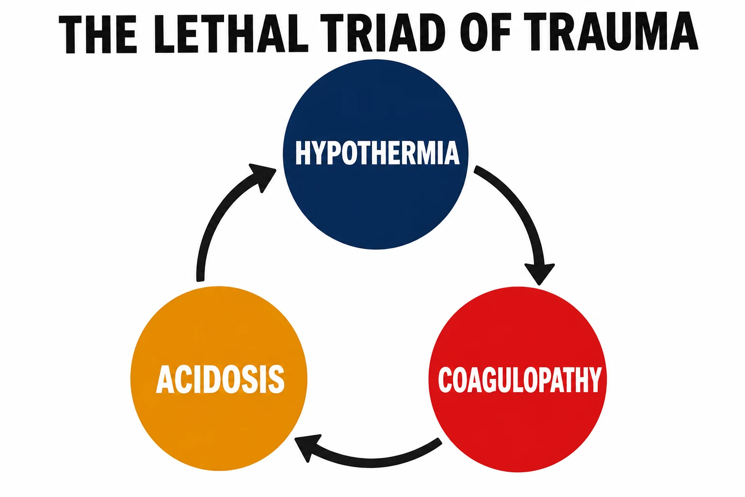
The lethal triad
The lethal triad — hypothermia, acidosis and coagulopathy — is the self-perpetuating cycle that kills the massively bleeding patient, and its prevention is the rationale for every element of damage-control resuscitation.[3] The hypothermia impairs the coagulation cascade and the platelet function (the enzymes of coagulation are temperature-dependent). The acidosis (from the poor perfusion and the shock) further inhibits the coagulation enzymes and depresses the cardiac function. The coagulopathy — from the consumption, the dilution by crystalloid, and the dysfunction of the cold, acidotic platelets and enzymes — worsens the bleeding, which worsens the shock, the acidosis and the hypothermia. The triad is prevented by warming the patient, the fluids and the blood; by giving blood products rather than crystalloid; by correcting the acidosis through the restoration of perfusion; and by giving the coagulation factors and the platelets proactively through the protocol.
Damage-control resuscitation
Damage-control resuscitation is the integrated approach to the massively bleeding patient, and it has four pillars.[3][1] First, early control of the bleeding — by direct pressure, a tourniquet, pelvic binding, surgery or interventional radiology — because no resuscitation succeeds against an uncontrolled source. Second, blood-product resuscitation in a balanced ratio (approaching 1-to-1-to-1 plasma-to-platelets-to-red-cells) rather than crystalloid, which dilutes the clotting factors and worsens the coagulopathy. Third, permissive hypotension — deliberately tolerating a lower than normal blood pressure (a systolic of 80 to 90 mmHg or a mean arterial pressure around 50 to 65 mmHg) until the bleeding is controlled, to avoid the disruption of early clots and the acceleration of haemorrhage; this is contraindicated in the traumatic brain injury, where the cerebral perfusion requires a higher pressure. Fourth, the early antifibrinolytic — tranexamic acid within three hours of injury. These four pillars, delivered through the MTP, are the evidence-based framework.
Activating and running the massive transfusion protocol
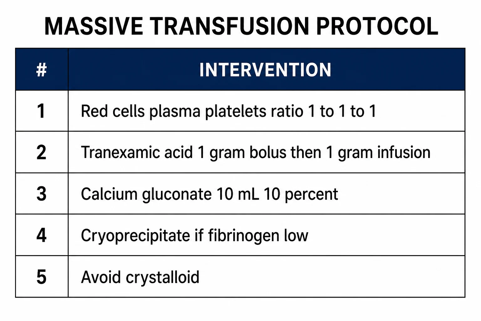
The massive transfusion protocol (MTP) is a pre-agreed, institutional pathway that delivers the blood products rapidly and in the right ratio, activated by a single phone call or button, and managed by a coordinated team with the blood bank. The MTP replaces the old ad-hoc requests for individual units with a protocolised delivery of "packs" or "boxes", each containing a set ratio of red cells, plasma and platelets, plus the tranexamic acid and the calcium, with the laboratory samples drawn at each pack for the coagulation, the full blood count, the ionised calcium and the viscoelastic testing. The team leader coordinates the resuscitation, the blood products, the source control and the monitoring; the blood bank prepares the products for the next pack before it is requested; and the communication is continuous. The MTP is deactivated when the bleeding is controlled and the haemostasis is achieved.[1]
The blood-product ratio: the evidence
The balanced ratio of blood products is the central operational decision of the MTP. The PROPPR trial compared a 1-to-1-to-1 ratio of plasma to platelets to red cells against a 1-to-1-to-2 ratio in patients with severe trauma and major bleeding, and found that the 1-to-1-to-1 ratio achieved earlier haemostasis and fewer deaths from exsanguination within the first 24 hours, though the 30-day mortality was not significantly different between the groups.[2] The practical message is that the balanced ratio (approaching 1-to-1-to-1) provides the coagulation factors and the platelets proactively, preventing the dilutional coagulopathy that arises when red cells alone are given, and it is the standard of the protocol.
Tranexamic acid
Tranexamic acid is the antifibrinolytic that reduces mortality from traumatic bleeding, and its early administration is a core element of the protocol. The CRASH-2 trial found that tranexamic acid, given as a 1 gram intravenous bolus followed by a 1 gram infusion over eight hours within three hours of injury, reduced all-cause mortality in trauma patients with significant haemorrhage, without an increase in vascular occlusive events.[1] Beyond three hours, the benefit is lost and there may be harm, so the timing is critical. The dose is given as early as possible, ideally at the scene or on arrival in the emergency department.
Calcium, fibrinogen and the laboratory
The rapid transfusion of stored blood carries a significant citrate load, and the citrate chelates the calcium, producing a hypocalcaemia that depresses the cardiac function and the coagulation. The ionised calcium is monitored and replaced — with 10 millilitres of 10 per cent calcium gluconate or 1 gram of calcium chloride — at regular intervals during the MTP. The fibrinogen is the first coagulation factor to fall to a critical level in massive haemorrhage, and the cryoprecipitate or the fibrinogen concentrate is given when the fibrinogen falls below 1.5 to 2.0 grams per litre, guided by the viscoelastic testing (ROTEM or TEG) where available, which provides a real-time assessment of the clot formation, the clot strength and the fibrinolysis. The standard laboratory tests (the INR, the APTT, the platelet count) are followed but they lag behind the real-time need; the viscoelastic test guides the targeted product administration. The pH and the temperature are maintained; the hypothermia and the acidosis are prevented.[3]
The anticoagulated patient
The patient on an anticoagulant who bleeds massively presents a specific challenge, and the reversal is integrated into the MTP. The warfarin is reversed with prothrombin complex concentrate (PCC) and intravenous vitamin K; the dabigatran with idarucizumab; the apixaban and rivaroxaban with andexanet alfa or, where unavailable, with PCC; and the antiplatelet agents are managed with platelet transfusion in the actively bleeding patient. The specific antidote is given early and in parallel with the MTP, guided by the coagulation testing and the clinical judgement. The aspirin and the clopidogrel are not reversed routinely in the non-bleeding patient, but are considered in the intracranial haemorrhage and the active bleeding. [1]
Transfusion complications
The rapid, large-volume transfusion carries a set of complications that the emergency physician must anticipate. The hypocalcaemia and the hypothermia (from the cold stored blood) have been discussed. The hyperkalaemia — from the potassium that leaks from the stored red cells — can cause arrhythmia, especially in the massive transfusion and the acidotic patient. The transfusion-associated circulatory overload (TACO) and the transfusion-related acute lung injury (TRALI) are recognised with the large-volume transfusion. The acute haemolytic reaction (from ABO incompatibility, which is prevented by the rigorous checking in the emergency of the massive transfusion) and the allergic and the febrile reactions are managed in the standard way. The dilutional thrombocytopenia and the dilutional coagulopathy are prevented by the balanced ratio. [1]
Specific scenarios
The traumatic haemorrhage is the paradigm: the MTP with the damage-control principles, the urgent source control (the operating theatre for the positive FAST and the unstable blunt trauma, the embolisation for the pelvic bleeding), and the ongoing assessment of the response. The upper gastrointestinal bleed — the variceal and the non-variceal — is managed with the specific interventions (the terlipressin, the endoscopy, the antibiotics) alongside the transfusion and the coagulation correction, and with the restrictive transfusion strategy (a haemoglobin threshold of around 70 to 80 grams per litre in the stable patient, except in the acute coronary syndrome). The obstetric postpartum haemorrhage follows the same MTP principles with the obstetric-specific interventions (the oxytocin, the misoprostol, the Bakri balloon, the surgical control). The surgical or the iatrogenic haemorrhage is managed with the MTP and the source control by the responsible team. [1]
Management — the drug doses and the blood-product ratios
The practical doses in the massive haemorrhage protocol: tranexamic acid 1 g IV bolus followed by 1 g IV infusion over 8 hours (within 3 hours of injury); calcium chloride 1 g IV (10 mL of 10 per cent) per pack or calcium gluconate 10 mL of 10 per cent IV for the citrate-induced hypocalcaemia; fresh frozen plasma 15 mL/kg (typically 4 units or approximately 1 L); cryoprecipitate 10 units (to maintain the fibrinogen above 1.5 g/L); prothrombin complex concentrate 25 to 50 IU/kg for the warfarin reversal; vitamin K 10 mg IV slow; idarucizumab 5 g IV (2 times 2.5 g vials) for the dabigatran reversal; and pooled platelets 1 adult dose (approximately 300 mL) to maintain the platelet count above 50 times 10 to the 9 per litre in the active bleed. [1]
Differential diagnosis — the cause of the massive haemorrhage
- Traumatic haemorrhage — the cavity bleeding (chest, abdomen, pelvis, long bones); the MTP with the damage-control principles.
- Upper GI bleed — the variceal, the peptic ulcer; the terlipressin 2 mg IV, the endoscopy, the restrictive transfusion strategy.
- Postpartum haemorrhage — the uterine atony, the retained products, the trauma; the oxytocin 10 IU IM, the misoprostol, the Bakri balloon.
- Ruptured AAA — the pulsatile abdominal mass, the sudden collapse; the urgent surgical or endovascular repair.
- Coagulopathy — the anticoagulated patient, the DIC; the targeted reversal (the PCC, the idarucizumab, the andexanet). [1]
Team coordination and regional practice
The MTP is a team operation that depends on the pre-agreed pathway, the blood bank liaison, the scribe and the time-stamped documentation, and the clear leadership. The communication between the emergency department, the blood bank, the theatre and the laboratory is continuous. The European guideline on the management of major bleeding after trauma provides the integrated evidence-based framework;[3] the ARC/NZRC guidelines and the Australian National Blood Authority patient blood management guidelines govern the Australasian practice.[1][1] The MTP is rehearsed, audited and refined.
Common pitfalls
The recurring errors are: resuscitating with crystalloid rather than blood products; delaying the tranexamic acid beyond three hours; targeting a normal blood pressure before the bleeding is controlled (worsening the haemorrhage); allowing the hypothermia and the acidosis to develop; not monitoring or replacing the ionised calcium; not using the viscoelastic testing to guide the targeted product administration; failing to reverse the anticoagulant early; and sending the unstable patient to computed tomography instead of the theatre. The patient whose bleeding is not controlled at the source will not survive, regardless of the MTP. [1]
Red flags
The following features identify massive haemorrhage that is uncontrolled or failing, in which the MTP is escalated and the source control is immediate: [1]
[1]Defining massive haemorrhage — the operational criteria
Massive haemorrhage has three operational definitions, any one of which triggers the protocol: the loss of more than one blood volume within twenty-four hours (approximately 70 mL/kg, or 5 litres in a 70 kg adult); the loss of more than half the blood volume within three hours; or a rate of blood loss above 150 mL per minute — equivalent to one unit of red cells every three to four minutes. A more practical and earlier trigger used at the bedside is the administration of four or more units of red cells within one hour with ongoing bleeding, or the anticipation of massive transfusion in the severely injured, shocked patient. The governing principle is that the MTP is activated on clinical judgement before the formal definition is met: if the clinician is considering activation, the protocol should already be running.[3][1]
[1]Activation criteria — the structured checklist
The MTP is activated by a single phone call or button that simultaneously mobilises the blood bank, the laboratory, the porters and the clinical team. The recognised activation criteria are:[1][1]
- Haemodynamic instability from bleeding — a systolic blood pressure under 90 mmHg (or a drop of more than 30 mmHg from baseline), a heart rate above 110, or a shock index (heart rate divided by systolic blood pressure) above 1.
- The transfusion burden — four or more units of red cells within one hour, or the anticipated need for ten or more units within twenty-four hours.
- The mechanism and the injury pattern — a positive FAST with shock, a major pelvic ring disruption, an unstable chest or abdominal trauma, a penetrating torso wound with compromise.
- The laboratory signature — a rising INR, a falling fibrinogen, a worsening acidosis, or a haemoglobin that is falling faster than the transfusion can replace it.
- The clinical judgement clause — any situation in which the clinician anticipates massive ongoing haemorrhage, regardless of the above. [1]
The structured running of the MTP
MTP activation, delivery and deactivation — the structured sequence
Step 1 — Activate (the single call)
A single phone call or button activates the protocol. The blood bank, the laboratory, the porters and the team mobilise simultaneously. The team leader is identified, the scribe is allocated, and the time-stamped record begins. Group O red cells and AB plasma (or low-titre group O whole blood) are issued immediately while the type-specific products are prepared.
Step 2 — Deliver the packs (1:1:1)
Pre-defined packs are delivered rapidly. A typical pack contains 4 units RBC + 4 units FFP + 1 adult dose of platelets (approaching 1:1:1), plus cryoprecipitate (10 units) and calcium. The products are warmed to ~37°C through a rapid infuser with an integrated warmer. Blood is drawn every 30-60 minutes for ROTEM/TEG, fibrinogen, platelet count, ionised calcium and haemoglobin.
Step 3 — Adjuncts with every pack
TXA 1 g IV bolus then 1 g over 8 hours (within 3 hours of injury); empiric calcium chloride 10 mmol (1 g) per ~4 units of blood products (the citrate chelates Ca²⁺); minimise crystalloid, and if unavoidable use a balanced solution (Hartmann or Plasma-Lyte), NEVER normal saline (the hyperchloraemic acidosis worsens coagulopathy). Maintain permissive hypotension (SBP 80-90) until the bleeding is controlled.
Step 4 — Transition to goal-directed therapy
As soon as the bleeding slows and the viscoelastic and the standard laboratory results return, switch from the fixed 1:1:1 ratio to the ROTEM/TEG-guided component therapy: a prolonged CT → FFP; a low α-angle/A10 → fibrinogen (cryoprecipitate or concentrate); a low MA/MCF → platelets; a raised ML/LY30 → TXA. This prevents the over-transfusion of plasma and platelets that drives the ARDS, the TACO and the multi-organ failure.
Step 5 — De-activate and screen
Stand the MTP down when the haemorrhage is controlled AND the haemodynamics are stable. After the de-activation, screen for the complications: the TRALI, the TACO, the hyperkalaemia (stored RBC potassium up to 70-80 mmol/L), the citrate-induced hypocalcaemia, the hypothermia, and recheck the coagulation, the electrolytes, the haemoglobin and the fibrinogen. Begin the VTE prophylaxis once it is safe.
The four pillars of damage-control resuscitation revisited
The damage-control philosophy rests on four pillars that run concurrently, not sequentially. The source control without the resuscitation fails; the resuscitation without the source control is futile. The PROMMTT study established the observational association between the early delivery of a higher plasma and platelet ratio and the improved survival, providing the rationale that the PROPPR trial then tested in a randomised design.[8]
Permissive hypotension
The physiology
- Target SBP 80-90 mmHg (MAP ~50-65) until the haemorrhage control
- Rationale: a higher pressure dislodges the early clots ("pop the clot"), dilutes the factors, accelerates the bleeding
- Give blood, NOT crystalloid, to hold this floor
- CONTRAINDICATED in the TBI (need SBP above 110, MAP above 80 for the CPP) and the spinal cord injury with the hypoperfusion
- Restore the normotension once the bleeding is controlled
Ratio-based transfusion
The blood
- Replace the shed blood with blood, not crystalloid
- RBC:FFP:platelets ~1:1:1 during the active uncontrolled phase (PROPPR)
- Give the empiric calcium, the TXA, and warm every product
- A bridge, not a destination — switch to the viscoelastic-guided therapy as soon as the bleeding slows
- The reflexive continuation of 1:1:1 causes the ARDS, the TACO and the multi-organ failure
Minimise crystalloid
The avoidance
- The crystalloid dilutes the clotting factors and the platelets and worsens the coagulopathy
- The normal saline causes the hyperchloraemic metabolic acidosis that worsens the lethal triad
- If the crystalloid is unavoidable, use a balanced solution (Hartmann or Plasma-Lyte)
- The goal is a blood-led, not a crystalloid-led, resuscitation
Source control
The anatomy
- No resuscitation succeeds against an uncontrolled source
- Direct pressure, tourniquet, pelvic binder, surgery, interventional radiology (embolisation)
- Damage-control surgery: the abbreviated laparotomy — pack, ligate, shunt, temporary closure
- The definitive repair is deferred to a planned re-operation once the lethal triad is corrected
Trauma-induced coagulopathy — the endogenous coagulopathy
Trauma-induced coagulopathy (TIC) is an endogenous hypocoagulable state present on arrival in approximately a quarter of severely injured patients — before any fluid or blood has been given. This is the conceptual keystone of the modern trauma resuscitation: the coagulopathy of the major bleeding is not purely iatrogenic (dilutional); it is generated by the shock and the tissue injury themselves, and it multiplies the mortality by three to four times.[3]
The unifying mechanism is the tissue hypoperfusion with the endothelial activation. The hypoperfusion and the tissue injury release the tissue factor and activate the endothelium; the thrombin-thrombomodulin complex activates the protein C (an endogenous anticoagulant that consumes the factors Va and VIIIa) and depletes the plasminogen activator inhibitor-1 (PAI-1), permitting the unopposed hyperfibrinolysis — the breakdown of the clots that do form. The endothelial glycocalyx shedding and the platelet dysfunction compound the effect. The result is a low-clot-strength, hyperfibrinolytic picture on the viscoelastic testing — a prolonged clotting time, a low clot firmness and a raised maximum lysis.[3]
The TIC is the target of the two most evidence-supported interventions in the trauma resuscitation: the early plasma (delivering the clotting factors concurrently with the red cells through the 1:1:1 ratio) and the tranexamic acid (counteracting the hyperfibrinolysis). It is also the rationale for minimising the crystalloid, which can only worsen a coagulopathy that is already endogenously present. [1]
Trauma-induced coagulopathy (TIC)
Endogenous, present on arrival
- Present BEFORE any fluid or blood given (~25 per cent of the severe trauma)
- Driven by the tissue hypoperfusion + the endothelial activation (NOT dilution)
- Mechanism: tissue factor + protein C activation → anticoagulation + hyperfibrinolysis
- Diagnosis: viscoelastic testing — prolonged CT, low clot strength, raised maximum lysis
- 3-4× mortality; the target of the TXA and the early plasma
Dilutional coagulopathy
Caused by our resuscitation
- Develops AFTER large volumes of crystalloid or stored RBC without plasma/platelets
- Driven by the dilution of factors and platelets + the citrate-induced hypocalcaemia
- Aggravated by the hypothermia and the acidosis (the lethal triad)
- Prevented by the DCR: minimise crystalloid, 1:1:1 ratios, give calcium, warm
Viscoelastic testing — the ROTEM/TEG decoder
Viscoelastic tests (ROTEM and TEG) measure the kinetics and the strength of the whole-blood clot formation and breakdown in real time (10-20 minutes), and they have superseded the reflexive use of the conventional ratios once the immediate bleeding is controlled. The conventional tests (INR, APTT, platelet count) report the plasma rather than the whole-blood function, take 30-60 minutes to return, and correlate poorly with the bleeding — they are followed, but they lag. The viscoelastic test guides the targeted, goal-directed administration of the specific deficient component.[1][1]
| TEG parameter | ROTEM equivalent | What it measures | Abnormal finding | Treatment |
|---|---|---|---|---|
| R (reaction time) | CT (clotting time) | Clotting factor initiation | Prolonged R/CT | FFP (or prothrombin complex concentrate) |
| K, α-angle | CFT, A10/A20, α-angle | Fibrin polymerisation (fibrinogen) | Low α / low A10 | Cryoprecipitate or fibrinogen concentrate |
| MA (maximum amplitude) | MCF (maximum clot firmness) | Platelet contribution to clot strength | Low MA / MCF | Platelets (1 adult dose) |
| LY30 | ML (maximum lysis) | Clot breakdown (fibrinolysis) | ML above 15 per cent | TXA 1 g IV |
TEG (thromboelastography)
Native technology
- Blood in a cup, a pin suspended — the clot moves the pin
- Native naming: R, K, α-angle, MA, LY30
- Widely used in the North American trauma centres
- The functional fibrinogen assay isolates the fibrinogen contribution
- The rapid TEG (rTEG) gives the results in ~15 minutes with the activators
ROTEM (rotational thromboelastometry)
European standard
- Blood in a cup, the pin rotates — the optical detection of the impedance
- Naming: CT, CFT, α-angle, A10/A20, MCF, ML
- Dominant in the European and the Australasian trauma practice
- EXTEM (tissue factor activation) and FIBTEM (cytochalasin D blocks the platelets → fibrinogen only) most used
- FIBTEM A10 is the rapid fibrinogen surrogate
The critical transition — and the most common exam trap — is that the ratio-based resuscitation is a bridge, not a destination. During the uncontrolled phase, give the 1:1:1 empirically; the moment the bleeding slows and the viscoelastic data return, switch to the goal-directed component therapy. The ITACTIC trial compared the viscoelastic-guided algorithm against the conventional laboratory-guided algorithm and found no significant difference in the mortality, but the viscoelastic approach reduced the over-transfusion of the plasma and is preferred where available; the consensus remains that the viscoelastic testing guides the targeted therapy, while the conventional tests are followed as a backstop.[7][3]
Calcium and citrate toxicity in depth
The rapid transfusion of the stored blood delivers a substantial citrate load. The citrate is the anticoagulant in the storage medium of the red cells and the plasma; it chelates the ionised calcium, producing a hypocalcaemia that is predictable, rapid and clinically significant in the massive transfusion. The ionised calcium is both a co-factor in the coagulation cascade (the conversion of the prothrombin to the thrombin and the activation of the factors IX and X and the platelets are calcium-dependent) and a determinant of the myocardial and the vascular smooth-muscle function — the hypocalcaemia therefore worsens the coagulopathy, depresses the cardiac contractility, blunts the response to the catecholamines, and produces a hypotension that is refractory to the vasopressors until the calcium is restored.[3][1]
The ionised calcium is monitored (the target is above 1.0 mmol/L) and replaced empirically during the MTP. The replacement is calcium chloride 10 mmol (1 g) or calcium gluconate 10 mL of 10 per cent per approximately every four units of the blood products. The calcium chloride provides more ionised calcium per mole and is preferred in the cardiac arrest and the massive transfusion, though it requires a central line (it is vesicant in a peripheral line); the calcium gluconate is safer peripherally but provides less ionised calcium per unit. The citrate is metabolised by the liver to the bicarbonate, which produces a late metabolic alkalosis — the early acidosis from the stored blood (pH approximately 6.6-7.0) gives way to a late alkalosis as the citrate is metabolised. [1]
[1]The landmark trials
CRASH-2 (Lancet 2010)
Multicentre, placebo-controlled randomised trial; 20,211 trauma patients with or at risk of major bleeding
Population: Adult trauma patients with significant haemorrhage, 274 hospitals across 40 countries
Key finding
TXA reduced the all-cause mortality (14.5 per cent vs 16.0 per cent, p=0.0035) and the bleeding death (4.9 per cent vs 5.7 per cent, p=0.0077), with no increase in the vascular occlusive events.
Practice change
Give TXA 1 g IV as early as possible in the trauma bleeding. The number needed to treat for death was around 120 — remarkable for a cheap, single, stable drug.
CRASH-2 time-window analysis (Lancet 2011)
Exploratory analysis of the CRASH-2 stratified by the time from injury to treatment
Population: 13,966 patients with the bleeding deaths analysed by the treatment timing
Key finding
The bleeding deaths were reduced within 1 hour (5.3 per cent vs 7.7 per cent) and within 1-3 hours. Given MORE than 3 hours after the injury, the TXA INCREASED the bleeding death (4.4 per cent vs 3.1 per cent).
Practice change
The 3-hour rule: the TXA must be given within 3 hours of the injury (ideally within 1 hour, pre-hospital if possible). Never give the TXA more than 3 hours after the injury.
MATTERs (Morrison, Archives of Surgery 2012)
Retrospective cohort study of the combat casualties in Afghanistan requiring the massive transfusion
Population: Military trauma patients, of whom a subgroup received the TXA
Key finding
The TXA reduced the mortality in the massive-transfusion group, at the cost of a higher rate of the thromboembolic events (the pulmonary embolism, the deep vein thrombosis).
Practice change
The military corroboration of the CRASH-2: the TXA confers a survival benefit in the most severely bleeding casualties, including those requiring the massive transfusion. The signal of the increased thromboembolism is acknowledged and managed with the early prophylaxis once the bleeding is controlled.
PROPPR (Holcomb, JAMA 2015)
Multicentre randomised trial; 680 severely injured adults predicted to need the massive transfusion
Population: Trauma patients at 12 Level I trauma centres in North America
Key finding
No significant difference in the 24-hour mortality (12.7 per cent vs 17.0 per cent, p=0.09) or the 30-day mortality. BUT 1:1:1 achieved the earlier haemostasis and fewer exsanguination deaths at 24 hours, with no increase in the ARDS or the multi-organ failure.
Practice change
1:1:1 is safe and reasonable empirically during the active massive bleeding — it improves the early haemostasis. There is no overall mortality advantage, so switch to the viscoelastic-guided therapy as soon as the bleeding slows.
WOMAN Trial (Shakur, Lancet 2017)
Multicentre, placebo-controlled randomised trial; 20,060 women with the postpartum haemorrhage
Population: Women with the clinical diagnosis of the postpartum haemorrhage after the vaginal or the caesarean delivery, across 21 countries
Key finding
The TXA reduced the death from bleeding (1.5 per cent vs 1.9 per cent; RR 0.81) with no increase in the thromboembolic events. The benefit was confined to the women treated within 3 hours.
Practice change
The CRASH-2 finding extended to the obstetric setting: the TXA 1 g IV within 3 hours of the onset of the postpartum haemorrhage reduces the death from bleeding, with no thromboembolic signal.
ITACTIC (Gonzalez, Annals of Surgery 2020)
Multicentre randomised trial; 395 trauma patients with the major bleeding
Population: Adult trauma patients predicted to need the massive transfusion
Key finding
No significant difference in the 24-hour mortality. The viscoelastic-guided group received less plasma and tended toward more fibrinogen and platelet use.
Practice change
The viscoelastic-guided algorithm is safe and reduces the over-transfusion of the plasma, but it did not improve the survival over the conventional algorithm in this trial. The consensus remains that the viscoelastic testing guides the targeted therapy where available.
Transition to definitive care — the structured phases
The damage-control philosophy is built on a deliberate transition between three phases: a temporising resuscitative phase, a restorative intensive-care phase, and a definitive reconstructive phase. The handover between them is governed by the physiology, not the clock.[3]
The three phases of damage control
Phase 0-1 — Index operation and damage-control resuscitation (minutes to under 2 hours)
Concurrent DCR and abbreviated surgery. Goals: permissive hypotension, 1:1:1 transfusion, TXA, calcium, warming. The operative goals are the haemostasis, the contamination control and the temporary closure. The patient is transferred to the ICU cold, acidotic and coagulopathic — the physiology is the priority.
Phase 2 — ICU resuscitation (the first 24-48 hours)
Correct the lethal triad: rewarm (forced-air warming, warmed fluids, warm ambient temperature); correct the acidosis (restore the perfusion — stop the bleed, give blood; avoid the bicarbonate except in extremis); correct the coagulopathy (viscoelastic-guided plasma, fibrinogen, platelets, TXA, calcium). Continue the lung-protective ventilation, treat the pain, begin the VTE prophylaxis once safe, and monitor the open abdomen for the abdominal compartment syndrome.
Phase 3 — The planned re-operation (24-48 hours)
Definitive reconstruction once the physiology is restored: remove the packs, perform the bowel anastomoses, repair the pancreas, the biliary tree and the bladder, and attempt the definitive fascial closure. If the abdomen is still hostile, re-pack and re-close temporarily, with a further planned re-operation. Each return to the theatre is an opportunity to close the abdomen before it becomes chronically open.
Phase 4 — Definitive care and closure
After the physiological recovery and the source control, pursue the early definitive fascial closure (ideally by days 5-7). If the primary closure is not achievable, manage the open abdomen with a planned ventral hernia and a split-thickness skin graft, with a delayed reconstruction. The transition is complete when the haemostasis is secure, the lethal triad is corrected and the definitive repairs are in place.
Postpartum haemorrhage — the obstetric MTP
The postpartum haemorrhage (PPH) follows the same damage-control principles as the trauma, with the obstetric-specific interventions layered on. The PPH is defined as a blood loss above 500 mL after a vaginal delivery or above 1000 mL after a caesarean, or any blood loss that causes the haemodynamic instability — the definition is clinical, not volumetric.[6]
The obstetric-specific escalation runs in parallel with the MTP: the uterotonics first (the oxytocin 10 IU IM or 5 IU slow IV, then the ergometrine, the carboprost 250 micrograms IM every 15 minutes to a maximum of 8 doses, and the misoprostol 800 micrograms PR), the mechanical measures (the bimanual uterine compression, the uterine balloon tamponade with the Bakri), and the surgical and the interventional measures (the B-Lynch or the brace suture, the ligation of the uterine or the internal iliac arteries, and the uterine artery embolisation), with the hysterectomy as the life-saving last resort. The tranexamic acid 1 g IV within 3 hours is given in parallel (the WOMAN trial).[6]
The E-MOTIVE trial established the value of the early detection and the bundled response: a drape-based blood-loss measurement with an early bundle (the uterotonics, the tranexamic acid, the IV access, the examination and the monitoring) reduced the severe PPH by 60 per cent. The implication for the emergency physician is the early recognition, the early tranexamic acid, the early activation of the obstetric and the haematology teams, and the simultaneous resuscitation with the blood products.[9]
Whole blood and the evolving resuscitation
The renaissance of whole blood (low-titre group O) is the contemporary frontier of the haemostatic resuscitation. The whole blood is the closest analogue to the 1:1:1 ratio in a single product — it contains the red cells, the plasma and the platelets in their physiological proportions, with functional platelets and clotting factors that the separated components lose in storage. The military and an increasing number of the civilian trauma centres use the low-titre group O whole blood as the primary resuscitation fluid in the active haemorrhage, with the 1:1:1 components as the standard civilian default where the whole blood is not available.[3]
Exam practice
SAQ — Massive haemorrhage in penetrating torso trauma
12 minutes · 12 marks
A 26-year-old man is brought to the emergency department 40 minutes after a single stab wound to the abdomen. He is drowsy (GCS 13), BP 74/46, HR 132, saturations 95 per cent on 15 L oxygen. The FAST is positive. He has received 2 units of O-negative red cells pre-hospital. Temperature 35.0°C, pH 7.16, lactate 7.2, INR 1.9, fibrinogen 1.2 g/L, ionised calcium 0.82 mmol/L. He is taken immediately to the theatre.
Clinical pearls — the high-yield points for the fellowship exam
References
- [1]CRASH-2 trial collaborators, Shakur H, Roberts I, et al. Effects of tranexamic acid on death, vascular occlusive events, and blood transfusion in trauma patients with significant haemorrhage (CRASH-2): a randomised, placebo-controlled trial Lancet, 2010.PMID 20554319
- [2]Holcomb JB, Tilley BC, Baraniuk S, et al. Transfusion of plasma, platelets, and red blood cells in a 1:1:1 vs a 1:1:2 ratio and mortality in patients with severe trauma: the PROPPR randomized clinical trial JAMA, 2015.PMID 25647203
- [3]Rossaint R, Bouillon B, Cerny V, et al. The European guideline on management of major bleeding and coagulopathy following trauma: sixth edition Crit Care, 2023.PMID 36859355
- [4]CRASH-2 collaborators. The importance of early treatment with tranexamic acid in bleeding trauma patients: an exploratory analysis of the CRASH-2 randomised controlled trial Lancet, 2011.PMID 21439633
- [5]Morrison JJ, Dubose JJ, Rasmussen TE, et al. Thin phytoplankton layers: characteristics, mechanisms, and consequences Ann Rev Mar Sci, 2012.PMID 22457973
- [6]WOMAN Trial Collaborators, Shakur-Still H, Roberts I, et al. Immune oncology in hepatocellular carcinoma-hype and hope Lancet, 2017.PMID 28434649
- [7]Gonzalez E, Moore EE, Moore HB, et al. A Ratiometric and Colorimetric Hemicyanine Fluorescent Probe for Detection of SO(2) Derivatives and Its Applications in Bioimaging Molecules, 2019.PMID 31694349
- [8]Holcomb JB, del Junco DJ, Fox EE, et al. Stepford doctors: an allegory Med Humanit, 2006.PMID 23674750
- [9]Galadanci N, Elhassan E, Mesleh M, et al. Predictive Value of the Neutrophil-Lymphocyte Ratio for Tumor Regression Grade and Prognosis of Local Advanced Rectal Cancer Patients Undergoing Neoadjuvant Chemoradiotherapy Technol Cancer Res Treat, 2023.PMID 37807729