EM · Peri-arrest arrhythmias
Peri-arrest arrhythmias
The peri-arrest arrhythmia algorithms: the stable-versus-unstable and broad-versus-narrow framework, the bradycardia and the tachycardia pathways, synchronised cardioversion versus defibrillation, the broad-complex VT and the torsades-with-magnesium, the narrow-complex SVT with adenosine, atrial fibrillation, the pre-excited AF trap, the peri-arrest drugs, and the electrolyte and reversible drivers.
On this page & tools
Your progress
Saved locally on this device.
Target exams
Red flags
The peri-arrest arrhythmias — the bradycardias and tachycardias that threaten to or do precipitate cardiac arrest — are managed by a set of algorithms built on two simple questions that the Fellowship candidate must apply reflexively. The first is whether the patient is unstable: a tachyarrhythmia with shock, syncope, myocardial ischaemia or heart failure is synchronised cardioverted immediately, whatever the rhythm. The second is the width and the regularity of the QRS complex, which directs the drug therapy of the stable patient. The algorithms are universal, they are applied with the defibrillator pads on and the team ready, and the cardinal errors — treating broad-complex VT with verapamil, or pre-excited atrial fibrillation with an AV-nodal blocker — are avoidable only by disciplined adherence to the framework. [1]
The stable-versus-unstable framework
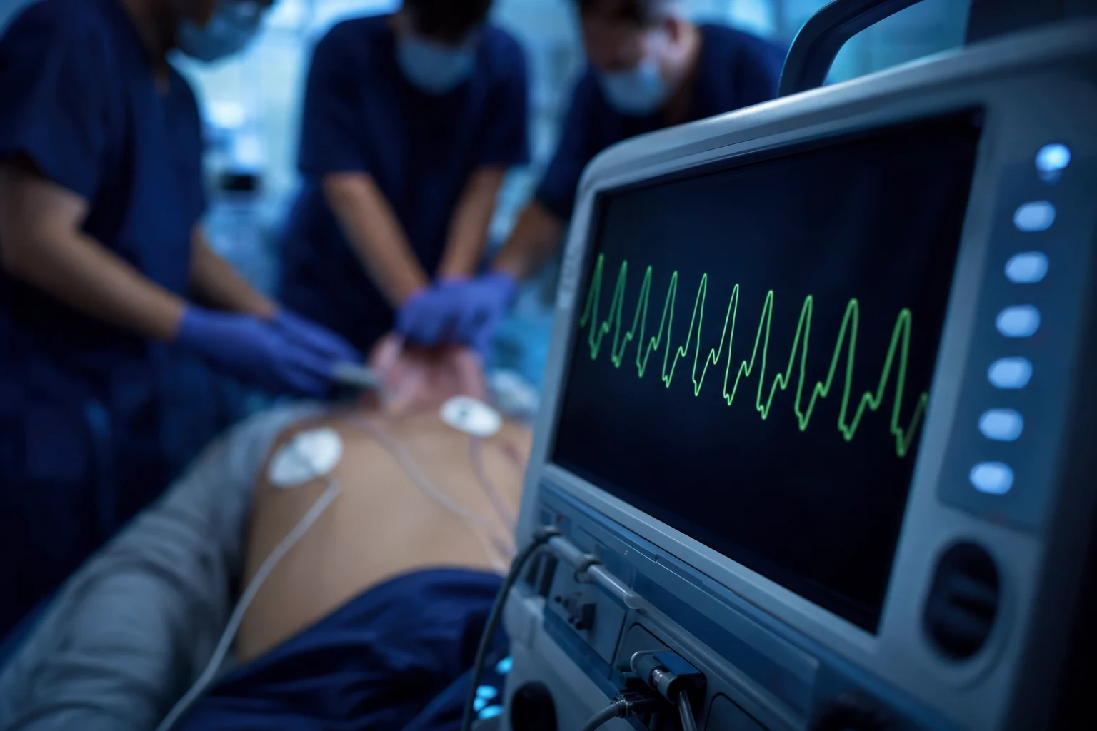
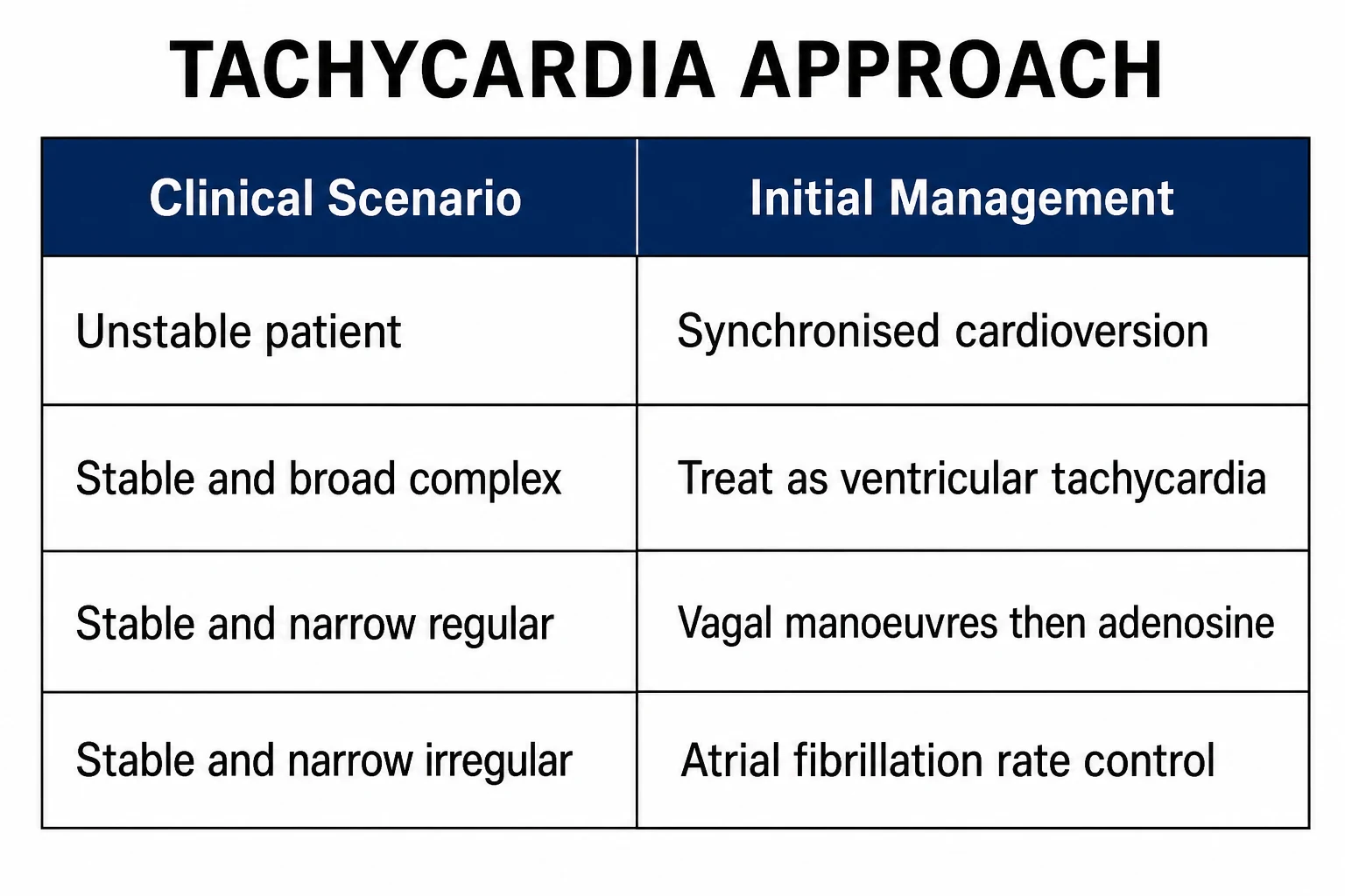
The first assessment of any peri-arrest arrhythmia is for the adverse features of instability — hypotension or shock, syncope or reduced conscious level, myocardial ischaemia (chest pain), and acute heart failure. The patient with any of these and a tachyarrhythmia is synchronised cardioverted without delay, irrespective of the rhythm, because the circulation cannot tolerate it. The stable patient — none of the adverse features — is managed down the drug algorithm, classified by the width and the regularity of the complex.[1][1]
Differential diagnosis
The peri-arrest arrhythmia is first framed by its morphology on the monitor, and within each frame lies a differential that the candidate must separate at the bedside before committing to a therapy: [1]
- A broad-complex tachycardia (QRS wider than 120 milliseconds) is ventricular tachycardia until proven otherwise in any patient with structural heart disease, but the differential also includes a supraventricular tachycardia conducted with fixed aberrancy (a pre-existing bundle branch block), a rate-related bundle branch block, pre-excited atrial fibrillation conducting down an accessory pathway (Wolff-Parkinson-White), ventricular flutter, and — importantly — artefact from tremor, lead movement or a failing electrode that can mimic a broad tachycardia and trigger inappropriate cardioversion. Where there is genuine diagnostic doubt and the patient is stable, an electrophysiology opinion or a 12-lead during the rhythm may resolve the origin; in the unstable patient the distinction is academic and the patient is cardioverted.
- A narrow-complex tachycardia is differentiated by regularity: a regular rhythm is most often atrioventricular nodal re-entry tachycardia (AVNRT) or atrioventricular re-entry tachycardia (AVRT) involving an accessory pathway, an atrial tachycardia, or atrial flutter with fixed atrioventricular block; an irregularly irregular rhythm is atrial fibrillation; an irregular rhythm with a sawtooth baseline is atrial flutter with variable block. A fast regular narrow complex at physiological rates should still prompt consideration of sinus tachycardia from a systemic driver — fever, sepsis, hypovolaemia, pain, pulmonary embolism or thyrotoxicosis — rather than a primary arrhythmia, because treating the rhythm while ignoring the driver harms the patient.
- A bradycardia requiring treatment is differentiated into the sinus-node causes (sick sinus syndrome, and drug-induced suppression from the beta-blockers, the calcium-channel blockers, digoxin or ivabradine), the atrioventricular causes (high-grade and complete heart block, often from inferior myocardial ischaemia, conduction-system fibrosis or drugs), and the systemic causes (hypothermia, hypothyroidism, raised intracranial pressure, hypoxia and the vagal excess of vasovagal syncope or inferior-wall stimulation). The site of the block is refined by the surface electrocardiogram, but in the peri-arrest setting the patient is paced first and the cause is sought in parallel.
- A polymorphic broad-complex tachycardia is differentiated by the QT interval: torsades de pointes with a long QT (the congenital long-QT syndromes, or drug-induced by the macrolides, the fluoroquinolones, the antipsychotics and the methadone) versus ischaemia-driven polymorphic VT with a normal QT, each pointing to a distinct corrective therapy — magnesium and the stopping of the offending drug for the former, and reperfusion for the latter. [1]
The unstable patient: synchronised cardioversion
Synchronised direct-current cardioversion delivers a shock timed to the R wave of the QRS, so that it does not fall on the T wave and precipitate ventricular fibrillation. The shock is delivered under sedation and analgesia, escalating the energy with each successive shock: for a regular broad-complex tachycardia and for a regular narrow-complex tachycardia, a biphasic energy of around 50 to 100 joules escalating; for atrial fibrillation, a higher initial energy of 120 to 150 joules escalating. The synchronisation must be confirmed on the monitor before each shock, and if the rhythm degenerates to VF the defibrillator is switched to the unsynchronised mode and the patient is defibrillated.[1]
Bradycardia
Bradycardia is treated when it is symptomatic — when it produces the adverse signs of hypotension, shock, syncope, ischaemia or heart failure. The first drug is atropine 500 micrograms intravenously, repeated every three to five minutes to a maximum of 3 milligrams. If the bradycardia is unresponsive to atropine, the escalation is transcutaneous pacing while an infusion of adrenaline (2 to 10 micrograms per minute) or dopamine is started, and a definitive transvenous pacing wire is placed. The causes — the negative chronotropic drugs (the beta-blockers, the calcium-channel blockers, digoxin), the high-grade atrioventricular blocks, the ischaemia (especially inferior myocardial infarction), and the vagal triggers (the excessive reflex in the vasovagal or the raised intracranial pressure) — are sought and treated in parallel. Atropine is ineffective in the high-grade block (the Mobitz II and the complete heart block), where pacing is the treatment. [1]
Broad-complex tachycardia: ventricular tachycardia
A broad-complex tachycardia (a QRS wider than 120 milliseconds) in a patient with structural heart disease is ventricular tachycardia until proven otherwise, and the commonest error is to mistake it for a supraventricular tachycardia with aberrancy and to treat it with verapamil — which precipitates collapse. The unstable patient is synchronised cardioverted. The stable patient with regular monomorphic VT is treated medically: amiodarone 300 milligrams intravenously over 20 to 60 minutes, followed by an infusion of 900 milligrams over 24 hours, is the first-line agent;[3][4] procainamide is an effective alternative, and some comparative evidence suggests it terminates stable VT as effectively as amiodarone;[5] lidocaine is the historical alternative. If the rhythm fails to terminate or recurs, the patient is cardioverted and referred for the definitive cardiology management. The stable broad-complex tachycardia of uncertain origin is treated as VT — this is the safe assumption in the emergency department.
Polymorphic VT and torsades de pointes
Polymorphic ventricular tachycardia — broad complexes that twist around the baseline — is treated by the correction of any ischaemia and electrolyte derangement and, where there is a long QT interval (torsades de pointes), by intravenous magnesium sulphate 2 grams over 10 minutes, followed by an infusion.[6] The potassium and the magnesium are corrected to the high-normal range, and any QT-prolonging drug is stopped. The patient with recurrent or refractory torsades may need overdrive pacing to shorten the QT. Ischaemia-driven polymorphic VT (without a long QT) is managed by the treatment of the ischaemia.
Narrow-complex tachycardia: SVT and adenosine
A regular narrow-complex tachycardia is a supraventricular tachycardia — the atrioventricular nodal re-entry tachycardia or the atrioventricular re-entry tachycardia (the re-entry involving an accessory pathway) — and the stable patient is treated with the vagal manoeuvres (the modified Valsalva, the carotid sinus massage in the patient without a bruit) followed by adenosine. Adenosine is given as a rapid intravenous bolus, 6 milligrams then 12 milligrams then 12 milligrams, through a large cannula in the antecubital fossa with a rapid saline flush, while the electrocardiogram is recorded continuously; it transiently blocks the AV node and terminates the re-entry circuit, and the meta-analytic evidence supports its efficacy and safety for this purpose.[2] The patient is warned of the flushing, the dyspnoea and the brief sense of doom, and the drug is used cautiously in the asthmatic (a relative contraindication) and avoided in the denervated heart (the transplant, where it causes prolonged asystole) and in decompensated heart failure.
Atrial fibrillation
Atrial fibrillation, the commonest sustained arrhythmia, presents in the emergency department as a rapid, irregularly irregular narrow-complex tachycardia. The haemodynamically unstable patient is synchronised cardioverted. The stable patient is managed by rate or rhythm control: for the recent-onset AF (within 48 hours, or fully anticoagulated beyond), a rhythm-control strategy with pharmacological or electrical cardioversion is reasonable; for the longer-standing AF, rate control with a beta-blocker or a rate-limiting calcium-channel blocker (diltiazem or verapamil) is the pragmatic approach, with assessment of the stroke risk and the anticoagulation by the CHA2DS2-VASc score and the onset-time-based anticoagulation decisions.[1][1] Digoxin is a second-line rate-control agent that is less effective in the high-adrenergic state.
The pre-excited atrial fibrillation (WPW) trap
The irregular broad-complex tachycardia is most often atrial fibrillation conducting down an accessory pathway (the Wolff-Parkinson-White syndrome), and it is the trap of the peri-arrest algorithms. The unstable patient is synchronised cardioverted. The stable patient is managed with an antiarrhythmic that blocks the accessory pathway (amiodarone, procainamide or flecainide) — and critically, the AV-nodal blockers (adenosine, verapamil, diltiazem and the beta-blockers) are avoided, because by blocking the AV node they preferentially conduct the atrial fibrillation down the fast accessory pathway, accelerating the ventricular rate and precipitating ventricular fibrillation. This single error — the AV-nodal blocker in the broad-irregular tachycardia — has killed patients, and the disciplined answer is to treat every irregular broad-complex tachycardia as pre-excited AF until proven otherwise. [1]
The peri-arrest drugs
The drugs of the peri-arrest algorithms are summarised here, and the candidate reproduces their doses.[1]
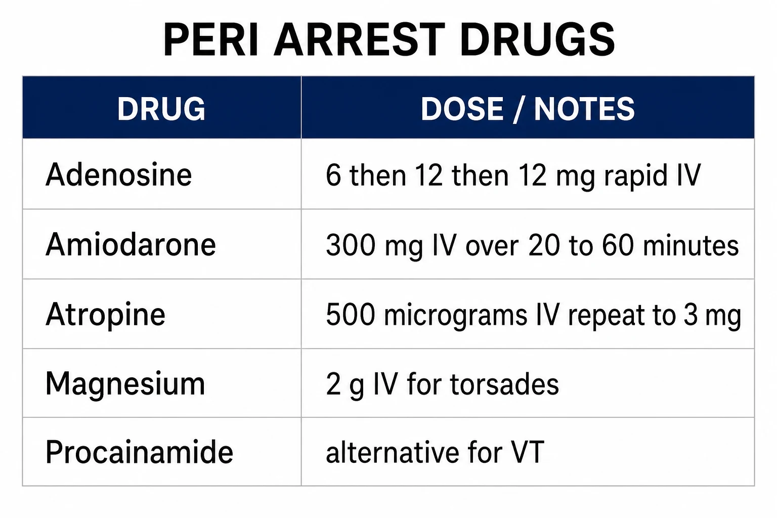
Adenosine terminates the regular narrow-complex SVT by the 6-12-12 milligram rapid-bolus method. Amiodarone terminates the stable broad-complex VT at 300 milligrams over 20 to 60 minutes, with procainamide and lidocaine as alternatives. Atropine treats the symptomatic bradycardia at 500 micrograms to a maximum of 3 milligrams. Magnesium 2 grams treats the torsades. The electrolytes — potassium, magnesium and calcium — are corrected to the high-normal range, and the QT-prolonging drugs are stopped. Each drug is given with the defibrillator pads on, the team ready, and the rhythm monitored. [1]
Electrolytes, reversible drivers, and the team
The arrhythmia is frequently driven by a reversible factor — ischaemia (the VT of the acute coronary syndrome, the AF of the atrial stretch), the electrolyte derangement (the hypokalaemia and the hypomagnesaemia of the torsades), the hypoxia, the sepsis, and the pro-arrhythmic drugs (the cocaine and the stimulants, the QT-prolonging agents, the digoxin toxicity). These are sought and corrected alongside the algorithm. The patient is monitored continuously, the defibrillator pads are attached, and a venous access and a 12-lead electrocardiogram are obtained. For any synchronised cardioversion, sedation and analgesia are given by a clinician competent in airway management, and the team is briefed. [1]
Special situations and regional practice
In the patient with the acute myocardial infarction, the VT or VF is managed by the standard algorithm with the addition of the urgent reperfusion. In the patient with the long-QT syndromes (the congenital and the drug-induced), the torsades is treated with magnesium and the removal of the offending drug. In the Brugada syndrome, the VT or VF is managed by the standard algorithm and the referral for the defibrillator. In pregnancy, the algorithms are unchanged, with the left lateral tilt and the awareness that adenosine and the DC cardioversion are safe. The guidelines are regional: the Resuscitation Council UK algorithms and the ARC/NZRC guidelines govern the peri-arrest practice, and the European Society of Cardiology guideline governs the atrial fibrillation.[1][1][1]
The peri-arrest algorithms — the rhythm-by-rhythm deep dive
The Fellowship candidate is examined on the application of each limb of the algorithm to a rhythm at the bedside, and the high-yield material is the dose, the energy and the sequence. The following sections take each of the common peri-arrest rhythms in turn — the unstable atrial fibrillation and flutter, the unstable broad-complex tachycardia, the pulseless ventricular tachycardia, the symptomatic bradycardia and the complete heart block — and give the exact algorithm that the examiner expects to be reproduced. The unifying rule is that the unstable tachyarrhythmia is cardioverted before the rhythm is diagnosed in detail, and the symptomatic bradycardia is paced when the atropine fails.[1][7]
[1]Synchronised cardioversion versus defibrillation
The distinction between the synchronised and the unsynchronised shock is the most examined technical point of the peri-arrest algorithms, and the cardinal error — delivering an unsynchronised shock that lands on the T wave and precipitates VF — is avoided by confirming the synchronisation marker on the R wave before each shock.[1]
Synchronised cardioversion
- A shock TIMED to the R wave, so it cannot fall on the T wave (the vulnerable repolarisation period) and precipitate VF
- Used for any UNSTABLE tachyarrhythmia with a discernible R wave — AF, atrial flutter, regular SVT, monomorphic VT, pre-excited AF
- Biphasic energy: 50–150 J escalating (120–200 J for AF, 50–100 J for the regular rhythms); start higher for AF
- Delivered under sedation and analgesia; confirm the SYNC marker on the monitor before each shock; re-synchronise after each escalation
Defibrillation (unsynchronised)
- A shock delivered IMMEDIATELY without synchronisation — the rhythm has no recognisable R wave to synchronise to
- Used for pulseless VT and ventricular fibrillation — the cardiac-arrest rhythms
- Biphasic energy: 150–200 J (manufacturer-specific); the shock is stacked into the CPR cycle and given as soon as the pads are on
- No sedation (the patient is in arrest); minimise the peri-shock pause — the chest compressions resume the instant the shock is delivered
Unstable atrial fibrillation and atrial flutter
The haemodynamically unstable patient with atrial fibrillation or atrial flutter — the hypotension, the shock, the syncope, the ischaemic chest pain or the acute pulmonary oedema — is synchronised cardioverted without delay. The higher initial energy for atrial fibrillation reflects the higher defibrillation threshold of the chaotic atrial substrate: a biphasic shock of 120 to 200 joules, escalating with each successive shock. For atrial flutter, the lower energy of 50 to 100 joules biphasic is usually sufficient because the re-entrant circuit is small and the threshold is low.[1][1]
Synchronised cardioversion for the unstable AF or flutter — the exact sequence
Attach the defibrillator pads to the anterolateral and the posterolateral positions (or the anteroposterior), switch the monitor on, and confirm the rhythm on the screen.
Assess the stability — the blood pressure, the perfusion, the conscious level, the chest pain, the pulmonary oedema. An adverse feature is the indication to cardiovert NOW, not after the diagnosis is refined.
Pre-oxygenase, assemble the airway team, and give the sedation and the analgesia — midazolam 1 to 2.5 mg IV titrated, fentanyl 50 to 100 micrograms IV, or a propofol bolus 0.5 to 1 mg/kg by a clinician competent in airway management and rescue.
Select the SYNC mode and confirm the synchronisation markers on the R wave of every QRS on the monitor. Select the biphasic energy: 120 to 200 J for AF, 50 to 100 J for flutter.
Charge, clear the patient, and deliver the shock. Reassess the rhythm. If the AF persists, re-synchronise, escalate the energy, and repeat — up to the maximum biphasic output.
If the rhythm degenerates to VF, switch OFF the SYNC mode and defibrillate immediately. If successful, reassess the blood pressure and the perfusion, obtain the 12-lead ECG, and admit for the cardiology assessment and the anticoagulation decision.
The unstable broad-complex tachycardia
The unstable broad-complex tachycardia — the hypotension, the shock, the syncope, the chest pain or the heart failure — is synchronised cardioverted, the same as any unstable tachyarrhythmia. If the patient is on the cusp (the borderline blood pressure, the patient who is deteriorating but not yet in shock), or if the cardioversion is being prepared, the stable-pathway drug therapy may be started in parallel: amiodarone 300 milligrams intravenously over 20 to 60 minutes (or over 10 minutes in the urgent case), followed by the 900 milligram infusion over 24 hours. If the rhythm is a regular monomorphic VT and the patient is stable, the amiodarone is the first-line agent, with procainamide and lidocaine as the alternatives.[3][4][7]
Unstable broad-complex tachycardia
- Adverse features present (shock, syncope, ischaemia, heart failure) → synchronised cardioversion NOW
- SYNC mode, biphasic 100 to 200 J escalating; re-synchronise between shocks
- If the patient is borderline or while the cardioversion is prepared, amiodarone 300 mg IV over 20 to 60 min may be started in parallel
- After termination, admit to the coronary care unit, search the driver (ischaemia, electrolytes, scar), refer for the electrophysiology
Stable regular monomorphic VT
- No adverse features → drug therapy; amiodarone 300 mg IV over 20 to 60 min then 900 mg over 24 h is first-line
- Procainamide 10 to 15 mg/kg IV at 20 to 30 mg/min is an effective alternative (and may terminate as well as amiodarone)
- Lidocaine 50 to 100 mg IV over 1 to 2 min is the historical alternative; avoid if there is concern for an ischaemic scar VT where amiodarone is preferred
- If the rhythm fails to terminate or recurs, synchronised cardioversion and the cardiology referral
VT of uncertain origin vs SVT with aberrancy
- In the ED, treat the stable broad-complex tachycardia of uncertain origin AS VT — this is the safe default
- The fusion beats, the capture beats, the concordance across the precordium and the AV dissociation favour VT
- A history of structural heart disease (the prior infarct, the cardiomyopathy) makes VT the overwhelming probability
- Never give verapamil to a broad-complex tachycardia — it can collapse a VT into asystole or VF
Pulseless ventricular tachycardia
Pulseless VT is a cardiac-arrest rhythm, and it is managed within the advanced life support algorithm, not the peri-arrest algorithm. The pulseless patient is defibrillated (an UNSYNCHRONISED shock, because there is no perfusing rhythm and no time to synchronise), at a biphasic energy of 150 to 200 joules (or the manufacturer's recommended energy). The shock is delivered within the CPR cycle, the chest compressions resume immediately, and after the third shock, amiodarone 300 milligrams intravenously is given, followed by a further 150 milligrams after a fifth shock if required. The reversible causes — the four Hs and the four Ts — are sought and treated in parallel, and the urgent reperfusion is arranged if the ischaemia is the driver.[7][8]
Pulseless VT within the ALS algorithm — the shockable-rhythm cycle
Confirm the cardiac arrest, call the arrest team, start the chest compressions at 100 to 120 per minute and a depth of 5 to 6 cm, and attach the defibrillator pads.
Confirm the rhythm is a shockable rhythm — pulseless VT or VF — on the monitor. Do NOT waste time analysing the morphology; a broad-complex tachycardia without a pulse is treated as pulseless VT.
Charge the defibrillator to 150 to 200 J biphasic (or the device default), clear the patient, and deliver the UNSYNCHRONISED shock. Minimise the peri-shock pause — the compressions resume the instant the shock is delivered.
Resume the CPR immediately for 2 minutes (one cycle), then reassess the rhythm. If the shockable rhythm persists, deliver the second shock, then resume the CPR.
After the THIRD shock, give amiodarone 300 mg IV bolus and start the adrenaline 1 mg IV every 3 to 5 minutes (every other cycle). Consider a further amiodarone 150 mg after the fifth shock.
Treat the reversible causes (the Hs and Ts) throughout, and arrange the urgent coronary reperfusion if the ischaemia is the driver. If the rhythm converts to a perfusing rhythm, reassess the pulses and the blood pressure and proceed to the post-cardiac-arrest care.
The bradycardia algorithm
The symptomatic bradycardia — the bradycardia with the adverse features of hypotension, shock, syncope, ischaemia or heart failure — is treated by the escalating bradycardia algorithm. The first drug is atropine 500 micrograms (0.5 mg) intravenously, repeated every 3 to 5 minutes to a maximum of 3 milligrams (6 micrograms per kilogram). If the bradycardia is unresponsive to the atropine, the escalation is the transcutaneous pacing, with an adrenaline infusion (2 to 10 micrograms per minute) or an isoprenaline infusion as the pharmacological bridge, and the definitive transvenous pacing wire. The atropine is ineffective in the high-grade atrioventricular block (the Mobitz II and the complete heart block) — these are paced directly.[1][10]
The symptomatic bradycardia algorithm — the escalation
Assess the bradycardia for the adverse features — the hypotension, the shock, the syncope or the reduced conscious level, the myocardial ischaemia (the chest pain), the acute heart failure. The asymptomatic bradycardia is monitored, not treated.
Give the atropine 500 micrograms IV (0.5 mg), repeated every 3 to 5 minutes to a maximum of 3 mg. The inadequate response (the heart rate still low with the symptoms) triggers the pacing.
Start the transcutaneous pacing — apply the pacing pads, set the rate to 60 to 80 per minute, and increase the mA until the electrical capture (a broad QRS following each pacing spike) and then the mechanical capture (a pulse) are achieved. Use the analgesia and the sedation — the transcutaneous pacing is painful.
In parallel, start the pharmacological chronotrope — an adrenaline infusion 2 to 10 micrograms per minute titrated to the response, or an isoprenaline or a dopamine infusion — as the bridge to the definitive pacing.
Place the transvenous pacing wire — the definitive treatment, ideally under the fluoroscopic or the echocardiographic guidance, via the right internal jugular or the left subclavian vein. Set the rate and the threshold, and confirm the capture.
Search and treat the cause — the negative-chronotropic drugs (beta-blockers, calcium-channel blockers, digoxin), the ischaemia (especially the inferior MI), the electrolytes, the hypoxia, the hypothermia, the raised intracranial pressure — and refer for the cardiology assessment and the permanent pacemaker.
Transcutaneous pacing
- The BRIDGE — applied within minutes, the pads on the chest, the capture by the mA titration
- Painful — requires the analgesia and the sedation; the skeletal-muscle contraction makes the capture assessment difficult
- Unreliable capture in the obese, the emphysematous and the patient with the large pericardial effusion
- Use while the transvenous wire is prepared; never the definitive treatment
Transvenous pacing
- The DEFINITIVE treatment — a pacing wire floated via the right internal jugular or the left subclavian into the right ventricular apex
- Set the rate to 60 to 80 per minute; determine the threshold (the minimum mA for the capture, typically under 1 mA) and set the output at 2 to 3 times the threshold
- Confirmed by the electrical capture (a QRS after each spike) and the mechanical capture (a pulse matching the paced rate)
- Risk: the ventricular perforation and the tamponade, the infection, the pneumothorax — the temporary wire is replaced by the permanent pacemaker within days
Episcopic / pharmacological chronotrope
- Adrenaline infusion 2 to 10 micrograms per minute, or isoprenaline, or dopamine — the bridge when the pacing is delayed
- Useful for the drug-induced bradycardia (the beta-blocker and the calcium-channel-blocker overdose) where the pacing may be needed but the chronotrope buys the time
- Causes the tachycardia, the hypertension, the ischaemia and the arrhythmia — titrate carefully and discontinue once the pacing is established
- Glucagon 5 to 10 mg IV is the specific antidote for the beta-blocker overdose; high-dose insulin for the calcium-channel-blocker overdose
Complete heart block and the high-grade AV block
The complete (third-degree) heart block — the atria and the ventricles beating independently, with the atrial rate faster than the escape ventricular rate — presents with the syncope, the dizziness, the exertional dyspnoea or the heart failure, or as an incidental finding on the ECG. The escape rhythm determines the haemodynamic stability: a junctional escape (a narrow-complex rhythm at 40 to 60 per minute) is usually stable enough to tolerate the transfer to the definitive pacing, but a ventricular escape (a broad-complex rhythm under 40 per minute) is unstable and is paced immediately. The Mobitz II (the intermittent non-conducted P waves without the preceding PR prolongation) and the high-grade AV block (two or more consecutive non-conducted P waves) carry a high risk of progression to the complete block and the asystole, and they are paced prophylactically.[1][10]
Complete heart block in the ED — the immediate management
Recognise the complete heart block on the ECG — the independent atrial and the ventricular activity, the atrial rate faster than the ventricular escape rate, the constant PR intervals with no relationship between the P and the QRS.
Assess the stability — the blood pressure, the perfusion, the conscious level, the ischaemia, the heart failure. The unstable complete heart block (the broad-complex escape under 40, the hypotension, the syncope) is paced immediately.
Give the atropine 500 micrograms IV — it is usually INEFFECTIVE in the complete heart block (the block is below the AV node), but it is given as the first-line drug while the pacing is prepared. Do not delay the pacing for the repeated atropine.
Start the transcutaneous pacing immediately — set the rate to 60 to 80 per minute, titrate the mA to the electrical and the mechanical capture, and give the analgesia and the sedation. Confirm the capture by the palpable pulse matching the paced rate (the monitor spikes alone are insufficient).
Place the transvenous pacing wire — the definitive temporary treatment, via the right internal jugular or the left subclavian vein, ideally under the fluoroscopic or the echocardiographic guidance, into the right ventricular apex.
Search the cause — the ischaemia (the inferior MI and the right ventricular infarction are the classic causes of the transient high-grade block), the conduction-system fibrosis (the Lev and the Lenègre disease), the drugs, the electrolytes, the infiltrative disease (the sarcoidosis, the amyloidosis), the infection (the Lyme disease, the Chagas disease) — and refer for the permanent pacemaker and the cardiology assessment.
The tachycardia drugs at a glance
The peri-arrest drug doses are reproduced here as the single reference the candidate memorises.[1][7]
Adenosine (regular narrow SVT)
- 6 mg then 12 mg then 12 mg IV rapid bolus via a large cannula in the antecubital fossa, followed by a rapid saline flush, with a continuous ECG
- Transiently blocks the AV node and terminates the AVNRT and the AVRT; warn the patient of the flushing, the dyspnoea and the brief sense of doom
- AVOID in the asthma (relative), the denervated heart (the transplant — prolonged asystole), the pre-excited AF, and the high-grade block without a pacemaker
- A 12 mg diagnostic bolus may reveal the underlying AF or flutter by the transient AV nodal block
Amiodarone (stable VT; post-shock VF/pulseless VT)
- Stable VT: 300 mg IV over 20 to 60 min, then 900 mg over 24 h. Cardiac arrest: 300 mg IV bolus after the third shock, then 150 mg after the fifth shock
- Blocks the potassium, sodium and calcium channels and the beta-receptors; broad-spectrum antiarrhythmic
- Side effects: the hypotension (give slowly), the bradycardia, the QT prolongation, the torsades (rare), the pulmonary fibrosis, the thyroid dysfunction, the hepatitis
- Avoid in the pregnancy (iodine load) and the pre-existing thyroid disease; the central line is preferred for the long-term infusion
Procainamide (stable VT alternative)
- 10 to 15 mg/kg IV at a rate of 20 to 30 mg/min, to a maximum of 17 mg/kg; followed by an infusion of 1 to 4 mg/min
- Class Ia; the PROCAT or the randomised evidence suggests it terminates the stable VT as effectively as the amiodarone
- Side effects: the hypotension (the commonest, give slowly and stop if the systolic falls), the QRS and the QT widening, the torsades, the prolonged half-life
- A 12-lead ECG during the infusion monitors the QRS widening (a 50 per cent widening is the stopping point)
Magnesium (torsades de pointes)
- 2 g IV (8 mmol) over 10 minutes, followed by an infusion of 0.5 to 1 g per hour; correct the potassium and the magnesium to the high-normal
- The first-line therapy for the torsades; works even when the magnesium level is normal (the membrane-stabilising effect)
- Side effects: the flushing, the hypotension, the areflexia, the respiratory depression at the high dose (the magnesium antagonises the calcium)
- Reversal: the calcium gluconate 10 mL of 10 per cent IV for the symptomatic hypermagnesaemia
Atropine (symptomatic bradycardia)
- 500 micrograms (0.5 mg) IV, repeated every 3 to 5 minutes to a maximum of 3 mg (6 micrograms per kg)
- Blocks the muscarinic receptor, removes the vagal tone, accelerates the sinus node and the AV-nodal conduction
- Ineffective in the Mobitz II and the complete heart block (the block is below the AV node) — do not delay the pacing
- A dose under 500 micrograms paradoxically slows the heart; the doses above 3 mg add no benefit
The evidence — the trials that shape the practice
ARREST — Kudenchuk, NEJM 1999 — amiodarone for shock-resistant VF
Design
Double-blind, randomised, placebo-controlled trial; 504 adults with out-of-hospital cardiac arrest and shock-resistant VF or pulseless VT
Intervention
Amiodarone 300 mg IV vs placebo, given after the paramedic-administered shocks failed to terminate the VF/VT
Primary result
Survival to hospital admission: amiodarone 44 per cent vs placebo 34 per cent (p=0.03); the benefit was preserved after the adjustment for the arrest variables
Bottom line
Amiodarone was the first antiarrhythmic shown to improve a clinically meaningful outcome in the shock-resistant VF, and it became the recommended agent after the third shock in the ALS algorithm. The benefit was to hospital admission, not to hospital discharge — the search for the perfect antiarrhythmic continues.
ALIVE — Dorian, NEJM 2002 — amiodarone vs lidocaine for shock-resistant VF
Design
Randomised, double-blind trial; 347 adults with out-of-hospital cardiac arrest and shock-resistant VF
Intervention
Amiodarone 5 mg/kg IV vs lidocaine 1.5 mg/kg IV, after the failed shocks
Primary result
Survival to hospital admission: amiodarone 22.8 per cent vs lidocaine 12.0 per cent (p=0.009)
Bottom line
Amiodarone was superior to lidocaine for the shock-resistant VF, confirming the ARREST finding and displacing lidocaine as the first-line antiarrhythmic in the cardiac-arrest algorithm. Lidocaine remains a reasonable alternative where amiodarone is unavailable.
Soar et al — ERC Guidelines 2021: adult advanced life support
Type
Authoritative consensus guideline — the European Resuscitation Council, the 2021 update
Scope
The shockable and the non-shockable rhythms, the airway management, the drugs (adrenaline, amiodarone, the reversible causes), the post-cardiac-arrest care, and the peri-arrest arrhythmias
Key recommendations
Pulseless VT and VF: defibrillate 150 to 200 J biphasic, amiodarone 300 mg after the third shock. The peri-arrest unstable tachycardia: synchronised cardioversion. The symptomatic bradycardia: atropine 500 micrograms to 3 mg, then the transcutaneous and the transvenous pacing
Bottom line
The European algorithm that governs the peri-arrest and the arrest practice in the ANZ, the UK and the European Fellowship exams, alongside the ARC/NZRC and the AHA equivalents.
Kelson & deSouza — procainamide vs amiodarone for the stable VT
Type
Comparative review and meta-analysis of the randomised and the observational evidence
Comparison
Procainamide vs amiodarone for the termination of the stable monomorphic ventricular tachycardia
Key finding
Both agents terminate the stable VT effectively; the procainamide may have a modest efficacy advantage and a faster onset, but the amiodarone is the better-studied and the more familiar agent in the ED
Bottom line
Procainamide is an evidence-supported alternative to amiodarone for the stable VT, and the candidate should know both. The procainamide requires the slow infusion and the QRS-width monitoring; the amiodarone requires the slow infusion and the blood-pressure monitoring.
Additional red flags
[1]Clinical pearls — the high-yield distillation
[1] [1] [1]Common pitfalls
The recurring errors are: treating broad-complex VT as SVT with aberrancy and giving verapamil, which collapses the patient; giving an AV-nodal blocker to the irregular broad-complex pre-excited AF, which precipitates VF; failing to synchronise the cardioversion shock (delivering it on the T wave and causing VF); giving atropine to the high-grade block without preparing to pace; using adenosine without warning the patient or without a continuous ECG; cardioverting without sedation and airway readiness; and treating the rhythm without correcting the electrolyte, the ischaemic or the toxic driver. [1]
SAQ — Symptomatic bradycardia and the high-grade block
10 minutes · 10 marks
A 74-year-old man presents with the syncope and the dizziness. The heart rate is 32 per minute, the blood pressure is 78 over 50, the electrocardiogram shows a complete heart block with a broad-complex ventricular escape at 38 per minute, and he is clammy and confused.
SAQ — Broad-complex tachycardia and the pre-excited atrial fibrillation trap
10 minutes · 10 marks
A 58-year-old man with the known ischaemic cardiomyopathy presents with the palpitations and the presyncope. The monitor shows a regular broad-complex tachycardia at 180 per minute, the blood pressure is 100 over 70, and the chest is clear.
Red flags
The following features identify the peri-arrest arrhythmia at risk of arrest or the treatment error, in which the disciplined algorithm is applied: [1]
[1]References
- [1]Van Gelder IC, Rienstra M, Bunting KV, et al. 2024 ESC Guidelines for the management of atrial fibrillation developed in collaboration with the European Association for Cardio-Thoracic Surgery (EACTS) Eur Heart J, 2024.PMID 39210723
- [2]Feng X, Liu J. Efficacy and safety of adenosine for supraventricular tachycardia: A meta-analysis utilizing BioMedGPT-LM-7B BMC Cardiovasc Disord, 2025.PMID 40055614
- [3]Foerster CR, Andrew E, Smith K, et al. Amiodarone for sustained stable ventricular tachycardia in the prehospital setting Emerg Med Australas, 2018.PMID 30084131
- [4]Long B, Koyfman A. Best Clinical Practice: Emergency Medicine Management of Stable Monomorphic Ventricular Tachycardia J Emerg Med, 2017.PMID 27751700
- [5]Kelson K, deSouza I. Procainamide Versus Amiodarone for Stable Ventricular Tachycardia Acad Emerg Med, 2019.PMID 31002448
- [6]Banai S, Tzivoni D. Drug therapy for torsade de pointes J Cardiovasc Electrophysiol, 1993.PMID 8269292
- [7]Soar J, Böttiger BW, Carli P, et al. Hollow microneedles: A perspective in biomedical applications Int J Pharm, 2021.PMID 33676993
- [8]Kudenchuk PJ, Cobb LA, Copass MK, et al. To tell the truth: a cancer diagnosis in other cultures is often a family affair J Natl Cancer Inst, 1999.PMID 10564674
- [9]Dorian P, Cass D, Schwartz B, et al. [Two cases of alcoholics associated with rhabdomyolysis and acute renal failure] Nihon Arukoru Yakubutsu Igakkai Zasshi, 2002.PMID 12462066
- [10]Link MS, Berkow LC, Kudenchuk PJ, et al. Part 7: Adult Advanced Cardiovascular Life Support: 2015 American Heart Association Guidelines Update for Cardiopulmonary Resuscitation and Emergency Cardiovascular Care Circulation, 2015.PMID 26472995