EM · Septic shock in the ED
Septic shock in the emergency department
Septic shock as infection complicated by a dysregulated host response and circulatory failure: the Sepsis-3 definitions and qSOFA, the pathophysiology, the time-critical principle, the Surviving Sepsis Hour-1 bundle, antibiotics and fluids and vasopressors, the MAP target, adjunctive corticosteroids and source control, the evolution from early goal-directed therapy, the endpoints of resuscitation, and the pitfalls.
On this page & tools
Your progress
Saved locally on this device.
Target exams
Red flags
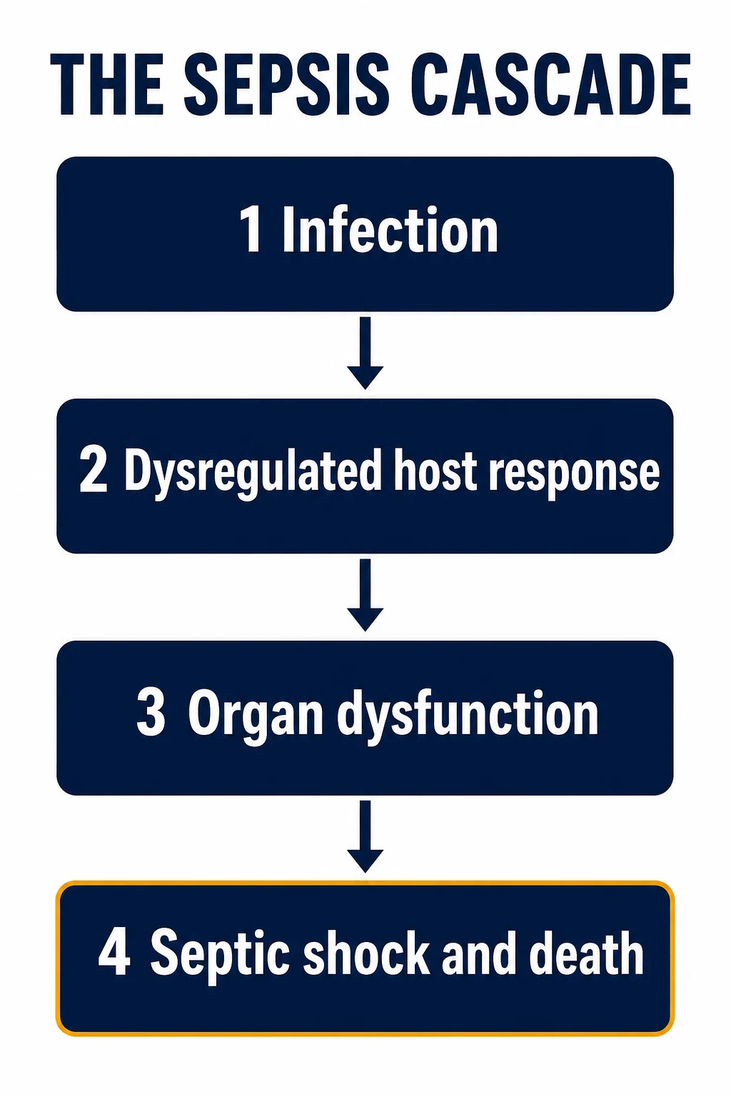
Septic shock is the most severe form of a syndrome that begins with infection and ends, if unrecognised, in multi-organ failure and death. It is among the commonest causes of shock seen in the emergency department, it carries a substantial mortality, and it is one of the few emergencies in which minutes genuinely matter: the speed with which the resuscitation is begun and the antibiotics delivered changes the outcome. The Fellowship-level understanding of septic shock rests on the modern definitions, the pathophysiology of the dysregulated response, and the Surviving Sepsis bundle that translates the evidence into a sequence of time-stamped actions performed in the first hour. [1]

Definitions
Sepsis is defined as a life-threatening organ dysfunction caused by a dysregulated host response to infection, with organ dysfunction operationalised as an acute change in the Sequential Organ Failure Assessment (SOFA) score of two points or more.[2] Septic shock is the subset of sepsis with circulatory and cellular abnormalities substantial enough to markedly increase mortality, defined clinically as sepsis with persisting hypotension requiring vasopressors to maintain a mean arterial pressure of 65 mmHg or more, and a serum lactate above 2 mmol per litre, despite adequate volume resuscitation.[3] This is not a semantic refinement: the patient who meets this definition has a markedly higher mortality than the patient with sepsis alone, and the definition directs the intensity of the resuscitation. The quick SOFA (qSOFA) — an altered mentation, a systolic blood pressure of 100 mmHg or less, and a respiratory rate of 22 per minute or more — is a bedside prompt to consider possible sepsis in a patient not already identified as such, not a diagnostic criterion.
Recognition in the emergency department
Recognition is the gate to the bundle, and the emergency department runs three complementary screens in parallel: a track-and-trigger score that flags the deteriorating patient from the observations, a sepsis-specific prompt that asks whether this deterioration is infection, and a lactate that measures the cellular cost. Each is imperfect alone; together they decide who is moved to the resuscitation bay and who is started on the clock. [1]
Track-and-trigger scores — MEWS, NEWS, NEWS2. A modified early warning score aggregates the routine observations — the respiratory rate, the oxygen saturation, the temperature, the systolic blood pressure, the heart rate and the conscious level — into a single number that rises with physiological derangement. The original MEWS validation showed that a score of five or more carried a markedly higher risk of death, intensive-care and high-dependency admission, and the modern Royal College of Physicians NEWS2 extends the principle with a separate scoring for the patient on supplemental oxygen.[12] The score's job is not to diagnose sepsis but to force a clinician to the bedside of the patient whose physiology is slipping — and the respiratory rate is the single most powerful individual component and the one most often unrecorded.
qSOFA — the sepsis prompt. The quick SOFA (two or three of an altered mentation, a systolic blood pressure of 100 mmHg or less, and a respiratory rate of 22 per minute or more) was proposed in Sepsis-3 as a bedside prompt for possible sepsis outside the intensive-care unit.[2] Its strength is simplicity and the fact that it is free at the bedside; its weakness is a modest sensitivity, so a low qSOFA does not exclude sepsis and must never be used to withhold the bundle in a patient who looks septic. NEWS2 outperforms qSOFA as a deterioration trigger, and the pragmatic emergency practice is to let NEWS2 trigger the assessment and qSOFA frame the sepsis question once the clinician arrives.
Lactate — the cellular cost. The venous or arterial lactate is the third screen and the one that quantifies the metabolic insult. A lactate above 2 mmol per litre is abnormal; a lactate of 4 or above marks severe disease and triggers the fluid arm of the Hour-1 bundle; a lactate that fails to clear with resuscitation is the cardinal sign of refractory shock. The pitfall is the well-compensated patient: a normal blood pressure and a reassuring conscious level with a lactate of 6 is occult shock until proven otherwise, and the lactate is what unmasks it. [1]
NEWS2
- A composite track-and-trigger score built from the observations: respiratory rate, SpO₂, air-or-oxygen, temperature, systolic blood pressure, heart rate and the AVPU conscious level
- The validated deterioration trigger; a high score mandates a clinician at the bedside and a structured search for a cause, sepsis high among them
- Superior sensitivity to qSOFA for predicting deterioration; it does not itself diagnose infection
- The respiratory rate is the most heavily weighted and the most often missing observation
qSOFA
- A three-variable bedside prompt: altered mentation, systolic blood pressure 100 mmHg or less, respiratory rate 22 per minute or more; two or more is positive
- A Sepsis-3 prompt to consider sepsis in a patient not already flagged; simple and free
- Modest sensitivity — a low score does not exclude sepsis and must not be used to withhold the bundle
- Use NEWS2 to trigger, qSOFA to frame the sepsis question
Lactate
- The cellular-cost screen: above 2 is abnormal, 4 or above is severe, and a failure to clear marks refractory shock
- Quantifies the metabolic insult the observations may hide; a lactate of 6 with a normal blood pressure is occult shock
- Obtain venous or arterial at the first blood draw; re-measure within two to four hours to chart clearance
- A normal early lactate does not exclude sepsis — trend it
Pathophysiology
Sepsis is a dysregulated host response, not simply an excessive one, and a Fellowship candidate should be able to defend the mechanism from first principles. Microbial products — the pathogen-associated molecular patterns, or PAMPs, such as endotoxin and lipoteichoic acid — and endogenous danger signals released from injured tissue — the damage-associated molecular patterns, or DAMPs, such as high-mobility-group-box-1 and mitochondrial DNA — bind the pattern-recognition receptors (the Toll-like receptors and others) on innate immune cells. The resulting activation releases a cytokine cascade (tumour necrosis factor, the interleukins, and the interferons) that is, in health, an appropriate and contained response. In sepsis the response becomes disordered: it is simultaneously excessive and deficient, because an early pro-inflammatory surge is accompanied and followed by a counter-regulatory anti-inflammatory response that produces immunoparalysis and leaves the patient vulnerable to further infection. [1]
Three downstream consequences drive the organ failure. First, the endothelium is activated and the glycocalyx is shed, so the capillary becomes leaky, plasma extravasates, and oedema forms in the tissues and the lungs. Second, the coagulation and inflammation systems are linked — sepsis is a prothrombotic state — and microvascular thrombosis starves the tissues, the extreme being the disseminated intravascular coagulation of meningococcaemia. Third, the microcirculation is maldistributed, with heterogeneous perfusion and shunting that starves the cells despite an adequate, even supra-normal, global oxygen delivery. Finally, the mitochondrion itself is injured — so-called cytopathic dysoxia — so that even restored oxygen delivery cannot be used, and the cell shifts to anaerobic metabolism. This is why the lactate can remain elevated in the face of a normal or high flow, and why a high lactate is a marker of cellular and microvascular failure as much as of inadequate global resuscitation. The goal of therapy is to interrupt this cascade early, before the organ failure becomes self-sustaining. [1]
Differential diagnosis — the other shock patterns
- Hypovolaemic shock — the haemorrhage, the dehydration; cold, clammy, the flat IVC; responds to the fluid.
- Cardiogenic shock — the MI, the myocarditis; cold, clammy, the pulmonary oedema, the raised JVP; does not respond to the fluid.
- Obstructive shock — the tamponade, the tension pneumothorax, the massive PE; cold, the raised JVP (except the tension PTX); the immediate cause treatment.
- Anaphylactic shock — the urticaria, the angioedema, the trigger; warm initially; the IM adrenaline 500 mcg.
- Adrenal crisis — the Addisonian crisis; the hyponatraemia, the hyperkalaemia, the hypoglycaemia; the hydrocortisone 200 mg IV. [1]
The time-critical principle
Time is the modifiable determinant of outcome. In a large cohort of patients studied under mandated emergency sepsis care, each additional hour to completion of the sepsis bundle — above all the administration of antibiotics — was associated with an increase in mortality, with the effect apparent from the first hour.[4] The practical consequence is that the recognition of possible sepsis triggers a simultaneous, time-stamped resuscitation bundle rather than a sequential workup. The clock starts at triage; the antibiotics, the fluids and the vasopressors are not deferred for imaging or a bed, and the lactate and the cultures are obtained in parallel.
The Surviving Sepsis Hour-1 bundle
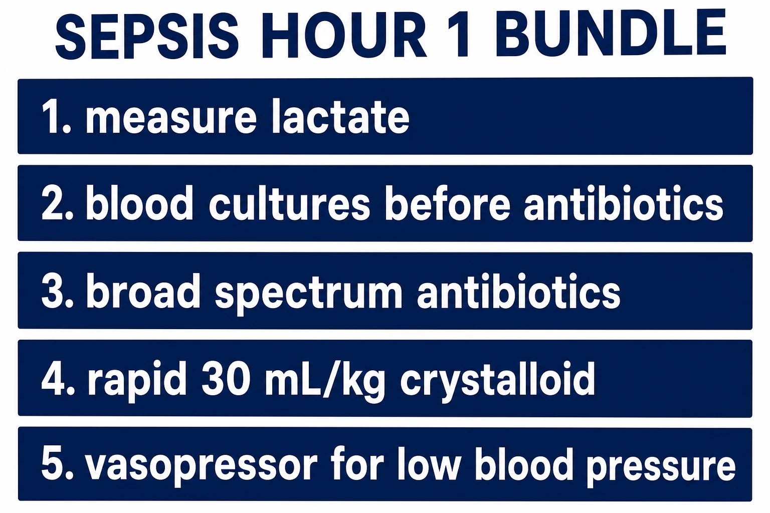
The Surviving Sepsis Campaign distils the evidence into a bundle to be delivered in the first hour.[1] Measure the lactate; obtain blood cultures before administering antibiotics where this does not delay them; administer broad-spectrum antibiotics; begin rapid crystalloid resuscitation at 30 millilitres per kilogram for hypotension or a lactate of 4 mmol per litre or more; and apply vasopressors for hypotension that is refractory to the initial fluid, targeting a mean arterial pressure of 65 mmHg or more. Each element is time-stamped and tracked, and the bundle is delivered as a whole rather than as a menu of options.
Antibiotics
The antibiotics are the single most time-sensitive element. They are broad-spectrum, intravenous, and delivered within the first hour, covering the likely source and the likely organisms — one or two agents active against the suspected pathogens, tailored to the local resistance patterns and de-escalated when cultures return. Typical empiric regimens cover Gram-negative and Gram-positive organisms (for example, a broad-spectrum beta-lactam with cover for resistant organisms, with or without an agent for source-specific cover), and the choice is refined by the source (a biliary source adds Gram-negative and anaerobic cover, a line infection adds Gram-positive cover, a necrotising soft-tissue infection adds broad cover). Cultures are drawn first where feasible because they guide de-escalation, but they must never delay the antibiotic beyond the hour. Typical adult empiric regimens include piperacillin-tazobactam 4.5 g IV every 6 to 8 hours, or ceftriaxone 2 g IV daily, with vancomycin 1.5 g IV every 12 hours for the suspected MRSA or the line infection, and meropenem 1 g IV every 8 hours for the resistant organism or the neutropenic patient. The metronidazole 500 mg IV every 8 hours adds the anaerobic cover when the biliary or the intra-abdominal source is suspected. [1]
Fluid resuscitation
The initial fluid is a rapid crystalloid bolus of 30 millilitres per kilogram for hypotension or a markedly raised lactate, given as balanced crystalloid rather than saline: in a large trial, balanced crystalloids led to fewer composite outcomes of death, new dialysis or persistent renal dysfunction than saline.[9] The fluid is given with reassessment, because the patient who does not respond to the first bolus — whose perfusion, blood pressure and lactate do not improve — needs a vasopressor, not a repetition of the same bolus, and excessive crystalloid in the non-responsive causes pulmonary and tissue oedema. Dynamic assessment of fluid responsiveness (the passive leg-raise, the pulse-pressure variation) refines the further fluid beyond the initial bolus. The practical doses: the balanced crystalloid (the Plasmalyte or the Hartmann's) at 30 mL/kg, given as 500 mL aliquots over 15 to 30 minutes; the noradrenaline 0.05 to 0.5 mcg/kg/min for the MAP below 65 mmHg after the initial fluid; the hydrocortisone 200 mg IV for the refractory shock; and the do-not-exceed 5 L of crystalloid without the critical-care reassessment.
Vasopressors and the MAP target
Noradrenaline is the first-line vasopressor for septic shock, restoring vascular tone and the mean arterial pressure. Vasopressin is a catecholamine-sparing adjunct, and adrenaline is an alternative or second-line agent. The target is a mean arterial pressure of 65 mmHg, and a higher target is not better: the SEPSISPAM trial found no survival benefit from targeting a mean of 80 versus 65 mmHg, and the higher target caused more arrhythmia, although a higher target may be reasonable in the chronically hypertensive patient. Vasopressors are started for fluid-refractory hypotension and titrated to the MAP and the perfusion; they are best given centrally, but in extremis a peripheral infusion is begun and converted to central early.[6]
Adjuncts: corticosteroids and source control
Adjunctive corticosteroids have a defined but limited role. The ADRENAL trial found that hydrocortisone did not reduce 90-day mortality in septic shock overall, though it hastened shock reversal and allowed earlier vasopressor cessation, and the Surviving Sepsis Campaign suggests a low-dose hydrocortisone infusion for septic shock that is refractory to adequate fluid and vasopressor therapy.[8][1] Source control is not an adjunct but a core element: an undrained focus — a collections, an infected line, an obstructed and infected biliary tree, necrotic tissue — will defeat any amount of resuscitation, and it is sought and addressed early by drainage, debridement or device removal. Transfusion thresholds, lung-protective ventilation for the associated respiratory failure, and glucose control are managed according to the standard critical-care targets.
The evolution of resuscitation targets
The modern approach to resuscitation has been shaped by a sequence of landmark trials, and a Fellowship candidate is expected to know them and what changed. Early goal-directed therapy, described by Rivers and colleagues in 2001, resuscitated the septic patient in the first six hours to a set of physiological endpoints measured through a central venous line and an oximetric catheter — a central venous oxygen saturation of 70 per cent or more, achieved by fluid, transfusion to a haematocrit, and dobutamine — and it reported a striking reduction in mortality that transformed practice.[5] The principle it established — that the early, aggressive, protocolised resuscitation of the septic patient to physiological endpoints changes outcome — survived everything that followed.
What changed was the need for the invasive protocol. Three large, multicentre, randomised trials — ProCESS in the United States, ARISE in Australasia, and ProMISe in the United Kingdom — compared formal protocolised EGDT (with the central venous oxygen saturation target, the early transfusion and the dobutamine) against usual care that already included early antibiotics, an initial fluid bolus and vasopressors, in an era in which practice had moved on.[7] All three found that the formal EGDT protocol did not improve outcome over contemporary usual care, and the invasive central venous oxygen-saturation target, the protocolised transfusion and the routine dobutamine were abandoned without harm. The contemporary synthesis, codified by the Surviving Sepsis Campaign, is that the benefit lies in the early recognition, the first-hour antibiotics, the initial balanced crystalloid bolus, the vasopressor to a mean arterial pressure target, and the serial lactate measurement — a less invasive, less protocolised resuscitation to the same physiological goals. The lesson the candidate takes is not that early resuscitation does not matter, but that the package that matters has simplified over time.
Monitoring, reassessment and endpoints
The septic patient is reassessed continuously against the targets: a mean arterial pressure of 65 mmHg or more, a falling lactate (a clearance of around 10 per cent per hour, or normalisation), a urine output of 0.5 millilitres per kilogram per hour or more, and an improving conscious level and peripheral perfusion. A static or rising lactate, persisting vasopressor requirements, or worsening organ dysfunction mark refractory shock and the need for critical-care admission, invasive monitoring and, in the deteriorating patient, intubation and mechanical ventilation. The response to the bundle is tracked and documented, and the patient who is not improving is escalated. [1]
Special populations
In children, septic shock is recognised from the tachycardia, the altered perfusion and the altered mental state, with age-specific physiological thresholds, and the resuscitation uses weight-based fluid boluses (10 to 20 millilitres per kilogram) with a low threshold for inotropes and intubation. In pregnancy, sepsis is a leading cause of maternal death, the physiology is altered, and the resuscitation is the same with a left lateral tilt. The immunocompromised and neutropenic patient may present with an occult, rapidly progressive infection and a blunted inflammatory response, with empirical broad-spectrum antibiotic cover that includes the resistant organism. The elderly patient presents atypically — with confusion or a fall — and has reduced reserve. [1]
Common pitfalls
The recurring errors are: delaying antibiotics for cultures, imaging or a bed; giving fluid by rote without assessing the response, and over-resuscitating the non-responsive patient; targeting a single blood pressure rather than the lactate and the perfusion; failing to achieve source control; forgetting the corticosteroid in refractory shock; and treating the bundle as a checklist completed rather than a resuscitation whose effect is tracked. The patient who improves initially and then deteriorates has either an undrained source, a resistant organism, or a complication, and the resuscitation is reassessed from the start. [1]
SAQ — Septic shock and the Surviving Sepsis Hour-1 bundle
10 minutes · 10 marks
A 68-year-old man with type 2 diabetes is brought from a nursing home with a 24-hour history of confusion and reduced urine output. He is warm and vasodilated, the respiratory rate is 30 per minute, the heart rate is 124, the blood pressure is 78 over 44, and the point-of-care lactate is 5.6 mmol per litre. He has received 500 mL of Hartmann solution in the ambulance.
SAQ — Refractory septic shock, the peripheral vasopressor and the dangerous intubation
10 minutes · 10 marks
A 52-year-old woman with a urinary tract infection is in the resuscitation bay with the septic shock. After 30 mL per kilogram of balanced crystalloid her mean arterial pressure is 52 mmHg, the lactate has risen from 4.1 to 6.8 mmol per litre, and she is becoming drowsy with a respiratory rate that has fallen from 32 to 12. The central line is not yet sited.
Red flags
The following features identify septic shock that is established or failing, in which escalation and critical-care involvement are immediate: [1]
[1]The ED sepsis pathway — from the door to the disposition
The emergency department management of septic shock is a single, parallel process that runs from the triage trigger to the disposition decision, with the bundle delivered in the first hour and the trajectory assessed continuously thereafter. The pathway below frames the operational steps; the detailed elements are expanded in the sections that follow. [1]
From triage to disposition
Recognise — the trigger
A high NEWS2, a positive qSOFA, a suspicion of infection, or simply a patient who looks unwell triggers the sepsis assessment. The clock starts at the moment of recognition, and that single clock governs everything that follows.
Resuscitate — the first hour
Move to the resuscitation bay; establish two large-bore cannulae; send the lactate, two sets of blood cultures, the full blood picture and the venous gas; give the antibiotic and the fluid; start the vasopressor for refractory hypotension. The bundle is delivered in parallel and time-stamped.
Reassess — the trajectory
After the first hour, reassess the mean arterial pressure, the lactate, the urine output, the peripheral perfusion and the conscious level. The trajectory — improving, static, or worsening — drives every subsequent decision, from further fluid to vasopressor titration to disposition.
Control the source
Seek and address the focus in parallel — the imaging, the drainage, the debridement, the infected line removal — because an undrained source defeats the resuscitation, and the patient who fails to improve is reassumed to have an uncontrolled source until proven otherwise.
Decide the disposition
Allocate to intensive care, the high-dependency unit or the ward on the basis of the trajectory and the level of organ support needed; document the trend, the plan and the explicit escalation criteria at handover.
The ED one-hour bundle in practice
The Hour-1 bundle is delivered as a parallel process, not a serial checklist, and the 2018 Surviving Sepsis Campaign recast the old three- and six-hour bundles into a single bundle to be started within the first hour from the moment of triage recognition.[1] The practical point is that the clock starts at recognition, that each element is time-stamped, and that nothing — imaging, a computed-tomography scan, a bed, a central line, a culture result — is allowed to delay the antibiotic or the fluid.
The Surviving Sepsis Hour-1 bundle
Measure the lactate
Draw a venous or arterial lactate at the first blood draw; a lactate of 4 or above, or persistent hypotension, triggers the full resuscitation arm. Re-measure within two to four hours to chart clearance and guide the ongoing resuscitation.
Obtain blood cultures before antibiotics
Two sets from separate sites, before the antibiotic dose, but only where this does not delay it. Cultures guide the later de-escalation; they must never push the antibiotic beyond the hour.
Administer broad-spectrum antibiotics
Within the first hour, intravenously, covering the likely source and the local resistance pattern — for example piperacillin-tazobactam or ceftriaxone, with vancomycin for the suspected MRSA or line infection and meropenem for the resistant organism or the neutropenic patient.
Begin rapid crystalloid at 30 mL/kg
Balanced crystalloid (Hartmann’s solution or Plasmalyte), 30 mL per kilogram for hypotension or a lactate of 4 or more, given in 500 mL aliquots over 15 to 30 minutes with a reassessment after each, not as an unmonitored pump.
Apply vasopressors for refractory hypotension
Noradrenaline for a mean arterial pressure below 65 mmHg that has not responded to the initial fluid, titrated to a MAP of 65; begin peripherally in extremis and convert to a central line early, in parallel with the resuscitation.
Lactate clearance versus central venous oxygen saturation
The endpoints of resuscitation have evolved from the invasive to the practical, and a Fellowship candidate must be able to compare the two that defined the modern era. Central venous oxygen saturation (ScvO₂), measured from a central line in the superior vena cava, quantifies the balance between global oxygen delivery and consumption: a low ScvO₂ implies that the tissues are extracting more oxygen because delivery is inadequate, and the original early goal-directed therapy protocol used it to drive fluid, transfusion and dobutamine. Lactate clearance, by contrast, is non-invasive and proximal to the cellular failure itself: a fall of about 10 per cent per hour, or normalisation within a few hours, marks a responding circulation; a static or rising lactate marks refractory shock. [1]
The decisive evidence is pragmatic. A randomised multicentre trial of early lactate-guided resuscitation in critically ill patients with a raised lactate found a reduction in hospital mortality when therapy was titrated to a falling lactate.[14] The three modern goal-directed-therapy trials (ProCESS, ARISE, ProMISe) then showed that protocolised ScvO₂-guided resuscitation added nothing over usual care that already included early antibiotics, an initial fluid bolus, vasopressors and serial lactate measurement.[7][10][11] The contemporary synthesis is that lactate clearance — measured, trended and acted upon — is the operational endpoint in the emergency department, and that the ScvO₂ catheter is reserved for the resuscitated patient in whom the macro-circulation is restored but the lactate remains high and the cause is unclear (a suspected persistent extraction deficit, mitochondrial dysfunction, or a mixed shock state).
Lactate clearance
- Non-invasive; a venous or arterial sample trended over hours
- Proximal to cellular failure: a clearance of roughly 10 per cent per hour, or normalisation, marks a responding circulation
- The contemporary operational endpoint in the ED; lactate-guided resuscitation improved outcomes in a randomised multicentre trial
- Falsely reassured by beta-blockade, liver failure and mitochondrial poisons — interpret with the perfusion, not in isolation
ScvO₂
- Invasive; requires a central line sampled from the superior vena cava, ideally with an oximetric catheter
- Quantifies global delivery versus consumption; a value below 70 per cent implies inadequate oxygen delivery
- The endpoint of the original early goal-directed therapy; the three modern trials showed it adds nothing over contemporary usual care
- Reserved for the resuscitated patient whose lactate stays high and whose shock type is unclear
Capillary refill time
- A bedside perfusion endpoint championed by the ANDROMEDA-SHOCK trial; a target of three seconds or less is reasonable
- Cheap, repeatable, and tracks the microcirculation that the lactate and the ScvO₂ only approximate
- Operator-dependent and confounded by a cold environment, but a useful adjunct at the bedside
- Not a replacement for the lactate — a complementary perfusion sign to be read alongside it
When to intubate the septic patient
Intubation in septic shock is a high-risk event: the patient who is hypoxic, acidotic and vasoplegic tolerates induction and the transition to positive-pressure ventilation badly, and the post-intubation cardiac arrest is a recognised and often lethal complication. The decision is therefore made deliberately and the resuscitation is optimised first — fluids and vasopressors running, the best oxygenation achievable, and a vasopressor bolus ready — but it is not deferred once a genuine indication is present. The four indications are: [1]
- Airway loss or a falling conscious level. A Glasgow Coma Scale of 8 or less, or a patient who cannot protect the airway, is intubated — septic encephalopathy is the commonest reason. The threshold is lower still when the conscious level is falling rapidly.
- Refractory hypoxaemia or failing respiratory effort. The tiring patient with a rising carbon dioxide, the acute respiratory distress syndrome picture with refractory hypoxaemia, or the respiratory distress that is exhausting the patient all mandate intubation. A rising respiratory rate that then falls is pre-arrest fatigue.
- Severe metabolic acidosis. A pH below about 7.15 to 7.20 with distressed breathing is intubated so that the ventilator can take over the work of breathing and the minute volume can be increased to blow off the carbon dioxide that is compensating for the metabolic acid.
- Haemodynamic collapse. The patient in extremis who cannot be resuscitated awake may need intubation as part of the spiral, accepting that induction may transiently worsen the pressure and demanding full resuscitation readiness. [1]
The induction itself is chosen to preserve the blood pressure — a reduced dose of ketamine or etomidate, with the vasopressor already running and a bolus drawn up — and post-intubation ventilation is lung-protective from the first breath (a tidal volume of 6 mL per kilogram of predicted body weight, plateau pressure below 30 cm of water), because the septic lung is primed for injury and ventilator-associated harm compounds the organ failure. [1]
[1] [1]Vasopressor initiation in the emergency department
Noradrenaline (norepinephrine) is the first-line vasopressor in septic shock: a predominantly alpha-adrenergic agonist that restores the vascular tone the septic vasoplegia has lost, with a modest beta-1 effect that supports rather than overdrives the heart, and a survival benefit over dopamine in the largest head-to-head comparison. The dose is titrated from about 0.05 to 0.5 (and up to 1) microgram per kilogram per minute to a mean arterial pressure of 65 mmHg or more, begun for hypotension that has not responded to the initial fluid, and continued until the vasoplegia resolves over hours to days. [1]
The practical question in the emergency department is access. The traditional teaching that vasopressors require a central line delayed their initiation and cost patients, and the evidence has moved: noradrenaline can be safely run through a large-bore peripheral cannula in a proximal vein, with close observation of the site, a low threshold to escalate, and early conversion to a central line once the patient is stable enough.[13] The principle is that the failure to perfuse is the emergency, not the absence of a central line — start the noradrenaline peripherally, then obtain central access as a controlled parallel task.
Vasopressin is added as a catecholamine-sparing adjunct (a fixed dose of about 0.03 units per minute) when noradrenaline alone is insufficient, and adrenaline is the second-line agent for the refractory case or where an inotropic component is needed. Adrenaline raises the lactate through beta-2-driven glycolysis, a fact that complicates lactate-based monitoring and is a reason to prefer noradrenaline first. [1]
Noradrenaline — first line
- Predominant alpha-agonist with a modest beta-1 effect; restores vascular tone; 0.05 to 1 mcg/kg/min titrated to a MAP of 65
- Superior to dopamine for mortality and arrhythmia; the default first-line agent in septic shock
- Can be run peripherally in extremis and converted to a central line early; sclerosing if it extravasates
- Raises the lactate far less than adrenaline, so lactate-based monitoring remains interpretable
Vasopressin — adjunct
- A V1-receptor peptide; a fixed dose around 0.03 units per minute, not titrated
- Catecholamine-sparing: added when noradrenaline alone is insufficient, it reduces the noradrenaline dose required
- Less tachyarrhythmia than the catecholamines; can cause mesenteric and digital ischaemia at higher doses
- Does not raise the lactate; a clean adjunct in the noradrenaline-refractory patient
Adrenaline — second line
- An alpha and beta agonist; for the refractory case or where an inotropic component is needed
- Potent and fast-acting; the agent when noradrenaline plus vasopressin has failed
- Raises the lactate through beta-2-driven aerobic glycolysis, confounding the lactate-clearance monitoring
- More arrhythmia and tachycardia than noradrenaline; not the first-line choice
Disposition from the emergency department
The disposition decision follows the response to the first hour. The patient who has been resuscitated to a MAP of 65 or more, whose lactate is falling, who is passing urine and whose conscious level is improving may go to a ward or a high-dependency unit depending on the degree of residual derangement. The patient who remains hypotensive on vasopressors, whose lactate is static or rising, who is oliguric or whose conscious level is falling needs intensive care. The disposition is a clinical judgement made from the trajectory, not a single number, and it is made in parallel with the resuscitation rather than at the end of it. [1]
Intensive care unit
- Vasopressor-requiring shock not yet stabilised; a rising or static lactate after the fluid; two or more failing organs
- Airway compromise or the need for mechanical ventilation; severe acidosis; the deteriorating conscious level
- The patient who is not on the improving limb of the resuscitation trajectory after the first hour
- Needs invasive monitoring, titrated vasopressors and the capacity for organ support such as ventilation or renal replacement therapy
High-dependency unit
- Single-organ failure or the need for a vasopressor infusion at a stable, low dose; a falling but still elevated lactate
- Close observation for a patient at risk of deterioration; the elderly or comorbid patient with limited reserve
- The patient who has responded but is not yet safe on an unmonitored ward
- Provides continuous monitoring, a nurse-to-patient ratio of about one to two, and a short-notice escalation path to the ICU
Ward
- Resolution or near-resolution of the physiological derangement; a normalising lactate; no vasopressor requirement
- A stable mean arterial pressure off support, a protected airway, an acceptable conscious level and reliable urine output
- The patient on the clearly improving trajectory in whom the focus shifts to oral antibiotics, source control and mobilisation
- Needs a documented sepsis-reassessment plan, a NEWS2 observation schedule, and an explicit escalation criterion at handover
The landmark trials in one place
The modern management of septic shock rests on a handful of trials that a Fellowship candidate must be able to summarise in a sentence each. The defining arc runs from the Rivers early goal-directed therapy study that established early aggressive resuscitation, through the three trials (ProCESS, ARISE, ProMISe) that retired the invasive protocol, to the Surviving Sepsis Campaign that codified the simplified, evidence-based Hour-1 bundle.[5]
Rivers — early goal-directed therapy
New England Journal of Medicine, 2001
A single-centre randomised trial of protocolised early resuscitation to a central venous oxygen saturation target versus usual care, in 263 emergency-department patients with severe sepsis or septic shock.
Key finding
In-hospital mortality fell from 46.5 per cent to 30.5 per cent with the protocol.
Practice change
Established that early, aggressive, protocolised resuscitation of the septic patient to physiological endpoints changes the outcome.
ProCESS, ARISE, ProMISe — the three modern trials
New England Journal of Medicine, 2014 to 2015
Three multicentre randomised trials comparing formal protocolised early goal-directed therapy (the ScvO₂ target, protocolised transfusion and dobutamine) against contemporary usual care, in over 4,000 patients combined.
Key finding
No mortality difference between protocolised goal-directed therapy and usual care in any of the three trials.
Practice change
Retired the invasive protocol; the benefit lies in early antibiotics, an initial fluid bolus, vasopressors to a MAP target and serial lactate measurement, not the ScvO₂ catheter.
SEPSISPAM — the blood-pressure target
New England Journal of Medicine, 2014
A multicentre randomised trial of a high (80 to 85 mmHg) versus a low (65 to 70 mmHg) mean arterial pressure target in 776 patients with septic shock on vasopressors.
Key finding
No difference in mortality overall; the higher target caused more atrial fibrillation, though it appeared to help the small subgroup with chronic hypertension.
Practice change
A MAP target of 65 mmHg is standard; a higher target is reserved for the chronically hypertensive patient.
ADRENAL — adjunctive corticosteroids
New England Journal of Medicine, 2018
A multicentre randomised trial of hydrocortisone 200 mg per day versus placebo in 3,658 patients with septic shock on vasopressors.
Key finding
No difference in 90-day mortality, but faster shock reversal and earlier vasopressor cessation with hydrocortisone.
Practice change
Hydrocortisone is reserved for septic shock that is refractory to adequate fluid and vasopressor therapy, not used routinely.
SMART — balanced crystalloids versus saline
New England Journal of Medicine, 2018
A pragmatic cluster-randomised crossover trial of balanced crystalloids versus saline in 15,802 critically ill adults.
Key finding
Balanced crystalloids reduced the composite outcome of death, new dialysis or persistent renal dysfunction.
Practice change
Balanced crystalloid is preferred over saline for the initial sepsis fluid bolus.
High-yield clinical pearls
[1] [1] [1] [1] [1] [1] [1]Additional red flags
[1]References
- [1]Evans L, Rhodes A, Alhazzani W, et al. Surviving sepsis campaign: international guidelines for management of sepsis and septic shock 2021 Intensive Care Med, 2021.PMID 34599691
- [2]Singer M, Deutschman CS, Seymour CW, et al. The Third International Consensus Definitions for Sepsis and Septic Shock (Sepsis-3) JAMA, 2016.PMID 26903338
- [3]Shankar-Hari M, Phillips GS, Levy ML, et al. Developing a New Definition and Assessing New Clinical Criteria for Septic Shock: For the Third International Consensus Definitions for Sepsis and Septic Shock (Sepsis-3) JAMA, 2016.PMID 26903336
- [4]Seymour CW, Gesten F, Prescott HC, et al. Time to Treatment and Mortality during Mandated Emergency Care for Sepsis N Engl J Med, 2017.PMID 28528569
- [5]Rivers E, Nguyen B, Havstad S, et al. Early goal-directed therapy in the treatment of severe sepsis and septic shock N Engl J Med, 2001.PMID 11794169
- [6]Asfar P, Meziani F, Hamel JF, et al. High versus low blood-pressure target in patients with septic shock N Engl J Med, 2014.PMID 24635770
- [7]ProCESS Investigators, Yealy DM, Kellum JA, et al. A randomized trial of protocol-based care for early septic shock N Engl J Med, 2014.PMID 24635773
- [8]Venkatesh B, Finfer S, Cohen J, et al. Adjunctive Glucocorticoid Therapy in Patients with Septic Shock N Engl J Med, 2018.PMID 29347874
- [9]Semler MW, Self WH, Wanderer JP, et al. Balanced Crystalloids versus Saline in Critically Ill Adults N Engl J Med, 2018.PMID 29485925
- [10]ARISE Investigators; ANZICS Clinical Trials Group, Peake SL, et al. Goal-directed resuscitation for patients with early septic shock N Engl J Med, 2014.PMID 25272316
- [11]Mouncey PR, Osborn TM, Power GS, et al. Trial of early, goal-directed resuscitation for septic shock N Engl J Med, 2015.PMID 25776532
- [12]Subbe CP, Kruger M, Rutherford P, Gemmel L. Validation of a modified Early Warning Score in medical admissions QJM, 2001.PMID 11588210
- [13]Cardenas-Garcia JF, Schaul YO, Barahona D, et al. Safety of peripheral intravenous administration of vasoactive medication J Hosp Med, 2015.PMID 26014852
- [14]Jansen TC, van Bommel J, Schoonderbeek FJ, et al. Three-dimensional imaging of the macular retinal nerve fiber layer in glaucoma with spectral-domain optical coherence tomography Invest Ophthalmol Vis Sci, 2010.PMID 20463326