EM · Chest trauma
Chest trauma
The chest trauma from the five immediately life-threatening injuries identified and treated in the primary survey (the tension pneumothorax, the massive haemothorax, the open pneumothorax, the flail chest with the contusion, the cardiac tamponade) through the eight potentially life-threatening injuries of the secondary survey (the blunt aortic injury, the tracheobronchial and the oesophageal injury, the diaphragmatic rupture), the chest-tube thoracostomy and the management principles.
On this page & tools
Your progress
Saved locally on this device.
8 MCQs with explanations
Target exams
Red flags
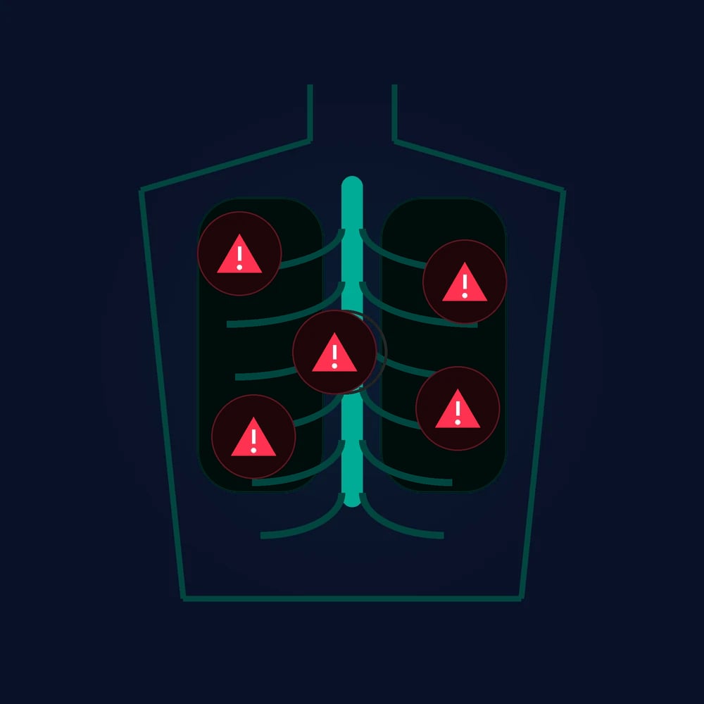
The chest trauma is the second leading cause of the trauma death, and it is the injury in which the emergency physician makes the most immediate interventions — the needle decompression, the chest drain, the pericardiocentesis and the resuscitative thoracotomy are all performed in the minutes of the primary survey. The chest is assessed in the breathing step (B) of the primary survey for the five immediately life-threatening conditions, each of which is identified by the clinical signs and treated at the bedside before any imaging.[1][1]
The five immediately life-threatening injuries
Tension pneumothorax. Air enters the pleural space through a one-way valve and cannot escape, raising the intrathoracic pressure, collapsing the lung, shifting the mediastinum to the opposite side, compressing the great veins, and producing the obstructive shock. The signs are the respiratory distress, the tracheal deviation (late), the unilateral absent breath sounds, the hyper-resonance, the distended neck veins and the hypotension. The treatment is the immediate needle decompression (the fourth or fifth intercostal space in the anterior axillary line, or the second intercostal space in the mid-clavicular line), followed by the definitive chest drain. The diagnosis is clinical and the treatment is immediate — waiting for the radiographic confirmation is fatal.[1]
Massive haemothorax. The accumulation of more than 1500 millilitres of blood in the pleural space, or the continued drainage of more than 200 millilitres per hour. The signs are the decreased breath sounds, the dullness to percussion, the hypotension and the hypoxia. The treatment is the large-bore chest drain (36 French or above), the simultaneous blood-product resuscitation, and the thoracotomy if the drainage exceeds the thresholds or the patient remains unstable. Crystalloid is minimised in favour of the blood products.[1][1]
Open pneumothorax (the sucking chest wound). A defect in the chest wall that communicates between the pleural space and the atmosphere. The treatment is the three-sided occlusive dressing (sealed on three sides to allow the air to escape and to prevent the tension), the chest drain placed remote from the wound, and the surgical closure of the defect.[1]
Flail chest with the pulmonary contusion. The fracture of three or more ribs in two or more places produces a segment of the chest wall that moves paradoxically (inward on inspiration, outward on expiration), impairing the ventilation, and the underlying pulmonary contusion (the alveolar haemorrhage and the oedema) impairs the gas exchange. The treatment is the aggressive analgesia (the regional block, the PCA, the epidural), the oxygen, and the ventilatory support (the non-invasive ventilation or the mechanical ventilation) for the respiratory failure. The fluid management is cautious because the contused lung is vulnerable to the fluid overload.[1][1]
Cardiac tamponade. The accumulation of blood in the pericardial space compresses the heart, producing the Beck triad (the hypotension, the distended neck veins, the muffled heart sounds) and the pulsus paradoxus. The treatment is the pericardiocentesis (a temporising measure) or the resuscitative thoracotomy (the definitive intervention in the arrest or the near-arrest after the penetrating torso trauma).[1]
The tube thoracostomy
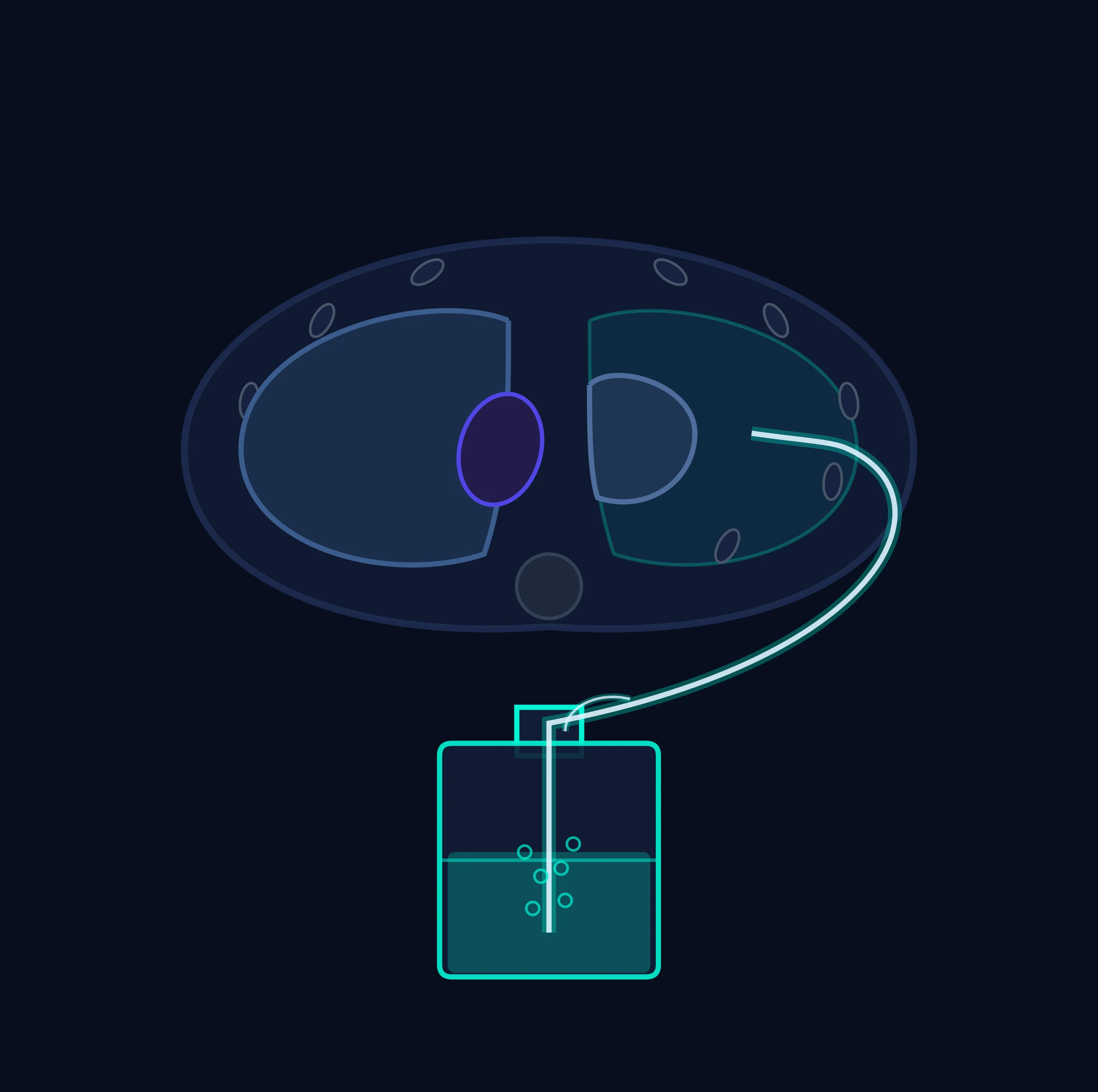
The tube thoracostomy (the chest drain) is the procedure that manages the pneumothorax, the haemothorax and the haemopneumothorax. The drain is placed in the fourth or fifth intercostal space in the anterior axillary line (the safe triangle — the border of the latissimus dorsi, the pectoralis major and the line of the nipple), using the blunt dissection (no trocar — the trocar causes the injury), the finger sweep to confirm the entry into the pleural space and to clear the clot, and the insertion of the large-bore tube (28 to 36 French) directed posteriorly and superiorly. The tube is connected to the underwater-seal drainage system, the chest radiograph confirms the position, and the output and the air leak are monitored. The complications are the infection, the bleeding (the intercostal artery), the tube malposition, and the subcutaneous emphysema.[1][1]
The eight potentially life-threatening injuries
The secondary survey identifies the injuries that are not immediately life-threatening but that have the significant morbidity if missed.[1] The blunt aortic injury — the traumatic deceleration tears the aorta at the isthmus (the ligamentum arteriosum), and the widened mediastinum on the chest radiograph is the sign that prompts the CT angiogram and the urgent endovascular or open repair. The tracheobronchial injury — the massive air leak, the haemoptysis and the subcutaneous emphysema. The oesophageal injury — rare, usually penetrating, with the pleural effusion and the surgical empyema risk. The diaphragmatic rupture — more common on the left (the liver protects the right), with the delayed presentation and the herniation of the abdominal contents. The myocardial contusion — the ECG changes, the troponin rise, the arrhythmia monitoring. The rib fractures — the pain control is the key, the epidural or the regional block, because the splinting and the atelectasis lead to the pneumonia.
Blunt versus penetrating chest trauma — the mechanism dictates the pattern
The mechanism of injury is the single most useful piece of information the pre-hospital team delivers, because it predicts the pattern. Blunt chest trauma — the motor-vehicle crash, the fall from height, the assault with a blunt object, the blast — transfers energy to the chest wall and the intrathoracic contents through compression, acceleration–deceleration and shear. The classic blunt patterns are the pulmonary contusion, the flail chest, the blunt aortic injury (at the ligamentum arteriosum), the blunt cardiac injury, the tracheobronchial rupture (within 2 cm of the carina), and the diaphragmatic rupture (more often left-sided). The rib fractures are the marker of force: the first and second rib, the scapula and the sternum are high-energy injuries that signal the mediastinal and the great-vessel injury.[1]
Penetrating chest trauma — the stab wound, the gunshot, the projectile — injures along the track of the weapon or missile. The injury is determined by the trajectory through the heart, the great vessels, the lung, the tracheobronchial tree, and the oesophagus. The penetrating wound below the nipple line anteriorly (the fourth intercostal space) or the tip of the scapula posteriorly may also breach the diaphragm and injure the intra-abdominal contents — the chest wound that crosses the diaphragm is an abdominal injury until proven otherwise, because the diaphragm rises to the fourth intercostal space on full expiration. Penetrating trauma is more likely to produce the cardiac tamponade, the great-vessel haemorrhage and the open pneumothorax; blunt trauma is more likely to produce the aortic injury, the flail chest and the pulmonary contusion.[1][1]
The weapon type and the velocity refine the penetrating pattern. The low-velocity stab produces the injury only along the blade; the low-velocity gunshot (handgun) deposits energy along a narrow track with cavitation that is temporary; the high-velocity rifle (over 600 metres per second) produces a permanent cavity and a blast effect that destroys tissue remote from the track. The exit wound is sought (and may be larger than the entry), and the patient is log-rolled early to find the posterior and the axillary wounds. [1]
| Feature | Blunt chest trauma | Penetrating chest trauma |
|---|---|---|
| Typical mechanism | Motor-vehicle crash, fall, assault, blast | Stab, gunshot, projectile, shrapnel |
| Energy transfer | Compression, deceleration, shear, blast | Along the track of the weapon/missile |
| Common life-threats | Flail chest, pulmonary contusion, blunt aortic injury, blunt cardiac injury | Cardiac tamponade, great-vessel haemorrhage, open pneumothorax |
| Aortic injury site | Isthmus (ligamentum arteriosum) — deceleration | Along the track — any site |
| Diaphragm | Rupture (usually left), delayed herniation | Laceration, more often left |
| Imaging priority | Whole-body trauma CT with angiogram | CT for the stable; RT/thoracotomy for the unstable/arrest |
| Resuscitative thoracotomy value | Very limited (rare survivors) | Potentially life-saving (tamponade, cross-clamp) |
The deadly dozen — the six primary and the six secondary chest injuries
The Fellowship candidate must be able to recite, in order, the six immediately life-threatening injuries of the primary survey (the breathing step B) and the six potentially life-threatening injuries of the secondary survey. The primary six kill in minutes and are treated at the bedside; the secondary six kill in hours to days and are identified on the head-to-toe examination and the imaging. The mnemonic of the primary survey is the ATLS framework, and the deadly dozen is the structural backbone of the chest trauma viva.[1][1]
The primary survey — the six immediately life-threatening injuries
The six injuries that must be identified and treated during the breathing assessment, before any imaging is obtained, are the airway obstruction (the upper-airway injury from the facial fracture, the haematoma, the burns, the laryngeal fracture), the tension pneumothorax, the massive haemothorax, the open pneumothorax (the sucking chest wound), the flail chest with the pulmonary contusion, and the cardiac tamponade. The airway obstruction is shared with the A step; the remaining five are the chest injuries that this topic addresses. Each is a clinical diagnosis, each has a defined bedside intervention, and each is fatal if missed or deferred.[1]
| The five chest life-threats (primary survey, B) | Bedside sign | Immediate intervention |
|---|---|---|
| Tension pneumothorax | Respiratory distress + unilateral hyper-resonance, reduced air entry, tracheal deviation (late) | Needle decompression 5th ICS mid-axillary → chest drain |
| Massive haemothorax | Dullness + reduced air entry + shock (>1500 mL initial or >200 mL/h) | Large-bore (32–36 Fr) chest drain + blood-product resuscitation |
| Open pneumothorax (sucking wound) | Audible air movement through a chest-wall defect | Three-sided occlusive dressing → chest drain at a separate site |
| Flail chest / pulmonary contusion | Paradoxical segment + hypoxia worsening over 24–48 h | Analgesia (regional/epidural) + NIV/IPPV ± restrictive fluid strategy |
| Cardiac tamponade | Beck triad (hypotension, JVD, muffled sounds), pulsus paradoxus | Echo → pericardiocentesis (bridge) / resuscitative thoracotomy |
The secondary survey — the six potentially life-threatening injuries
The six injuries identified on the secondary survey, each with significant morbidity if missed, are the simple pneumothorax, the haemothorax, the pulmonary contusion (without the flail segment), the blunt cardiac injury, the traumatic aortic disruption (the blunt aortic injury), and the diaphragmatic rupture. The tracheobronchial tree injury, the oesophageal injury and the sternum / first-rib fracture are often added to this list as the additional occult injuries. The principle: the secondary survey is the head-to-toe examination performed after the primary survey is complete and the patient is stable, and the missed secondary injury is the classic cause of the late deterioration.[1]
| The six secondary chest injuries | How it presents | Management |
|---|---|---|
| Simple pneumothorax | Decreased air entry, chest X-ray or CT finding | Chest drain before positive-pressure ventilation or transfer |
| Haemothorax (sub-massive) | Dullness, reduced air entry, blood on imaging | Large-bore chest drain; monitor output |
| Pulmonary contusion (isolated) | Hypoxia developing over 24–48 h, CT infiltrate | Restrictive fluids, analgesia, NIV/IPPV |
| Blunt cardiac injury | New ECG changes (arrhythmia, ST change), troponin rise | ECG + troponin screening, monitor the arrhythmia |
| Traumatic aortic disruption | Widened mediastinum on CXR, mechanism | CT angiogram → urgent endovascular (TEVAR) or open repair |
| Diaphragmatic rupture | Initially occult; delayed visceral herniation | CT, surgical repair (laparotomy/thoracotomy) |
Exam-exhaustive deep dive — the management of each life-threat in detail
The Fellowship candidate is examined on the decision-making — the indication, the technique, the dose, the timing, and the trap of each intervention. The sections below set out each of the deadly dozen at the depth the examiner expects. [1]
Tension pneumothorax — the clinical diagnosis and the decompression
Air enters the pleural space through a one-way valve (a lung laceration or a chest-wall defect that closes on inspiration) and cannot escape, raising the intrathoracic pressure above the venous pressure. The lung collapses, the mediastinum shifts to the opposite side, the great veins (the vena cava and the azygos) kink and compress, the venous return falls, the cardiac output falls, and the obstructive shock develops. The hypoxia from the shunt and the falling cardiac output produce the cardiovascular collapse.[1]
Needle decompression and the chest drain for tension pneumothorax — the modern sites
Recognise the tension
Severe respiratory distress, hypoxia, shock, reduced air entry and hyper-resonance on one side. Tracheal deviation and distended neck veins are late and often absent in the shocked patient. Treat on the clinical picture, not on the imaging.
Immediate needle decompression
The ATLS 10th-edition preferred site is the 5th intercostal space, anterior to the mid-axillary line, just anterior to the latissimus dorsi (the same site as the chest drain). This avoids the heart, the great vessels, and the thicker chest-wall muscle of the 2nd-ICS site. Use a large-bore cannula (14 or 16 gauge, at least 5 cm long — the standard cannula is often too short for the muscular or the obese trauma patient). A rush of air confirms the diagnosis and relieves the tension. The 2nd intercostal space mid-clavicular line remains an accepted alternative.
Definitive chest drain
A needle decompression is a bridge, not a treatment — the definitive management is the intercostal chest drain (size 28–32 Fr in the adult). Insert at the 5th intercostal space, anterior axillary line, blunt dissection over the upper border of the rib (NEVER the lower border — the neurovascular bundle runs in the subcostal groove), connected to an underwater seal. Do not clamp a bubbling drain.
The finger thoracostomy alternative
In the intubated, ventilated trauma patient in arrest or extremis, the finger thoracostomy (a rapid blunt incision into the pleural space, a finger sweeps to release the tension and the clotted haemothorax) is faster and more reliable than the needle, and is the technique of choice in the anaesthetised patient. The needle is reserved for the non-intubated patient in whom a chest drain cannot be immediately placed.
Massive haemothorax — the volume, the drain and the threshold for thoracotomy
The massive haemothorax is the accumulation of more than 1500 millilitres of blood in the pleural space, or the continued drainage of more than 200 millilitres per hour for 2 to 4 hours. It is both a lung problem (the collapsed lung produces the shunt and the hypoxia) and a volume problem (the blood in the chest is the blood that should be in the circulation). The signs are the decreased breath sounds, the dullness to percussion, the hypotension and the hypoxia. The treatment is the simultaneous chest drain and the resuscitation.[1][1]
The massive haemothorax — the drain, the resuscitation, and the thoracotomy threshold
Large-bore chest drain first
Insert a large-bore (32–36 Fr) chest drain at the 5th ICS mid-axillary line, blunt dissection, directed posteriorly and superiorly. The drain both evacuates the blood (re-expanding the lung, improving the gas exchange and the tamponade of the bleeding source) and quantifies the bleeding (the output drives the thoracotomy decision). Two drains may be needed for the massive haemopneumothorax (one apical for the air, one basal for the blood).
Blood-product resuscitation in parallel
The blood in the chest is the blood that should be in the circulation — the patient is in haemorrhagic shock. Activate the massive haemorrhage protocol, give the blood products in a balanced 1:1:1 ratio, tranexamic acid within 3 hours, and permissive hypotension to a systolic of 80–90 mmHg (unless there is a traumatic brain injury, in which case resuscitate to 110). Minimise the crystalloid — it worsens the lethal triad and dilutes the clotting.<Cite id="2" /><Cite id="4" />
The thoracotomy threshold
The indications for the urgent thoracotomy are: the initial drainage of more than 1500 mL of blood on the insertion of the drain; the continued drainage of more than 200 mL per hour for 2 to 4 hours; or the patient who remains in shock despite the drainage and the resuscitation. The thoracotomy controls the bleeding source (the lung laceration, the intercostal artery, the great vessel, the hilar vessel) and evacuates the clot.<Cite id="1" />
The retained haemothorax — the late complication
A haemothorax that is not fully drained (the clotted haemothorax) becomes the retained haemothorax, the substrate for the empyema and the trapped lung. The retained haemothorax is drained by the video-assisted thoracoscopic surgery (VATS) within 7 to 10 days; the delay beyond that converts the clot into the fibrous peel that requires the decortication.<Cite id="8" />
Open pneumothorax (the sucking chest wound) — the three-sided dressing
A defect in the chest wall that is at least two-thirds the cross-sectional area of the trachea allows the air to be preferentially drawn into the pleural space on inspiration (the path of least resistance), collapsing the lung. The defect may be large enough to prevent the spontaneous re-expansion of the lung even with the chest drain, and the open wound is the conduit for the tension if it is sealed incorrectly. [1]
Flail chest and the pulmonary contusion — the analgesia and the ventilation
The flail segment is the fracture of three or more ribs in two or more places, producing a free-floating segment of the chest wall that moves paradoxically (inward on inspiration, outward on expiration). The mechanical impairment is real but rarely the cause of the respiratory failure — the underlying pulmonary contusion is. The contusion is the alveolar haemorrhage and the capillary leak that worsens over 24 to 48 hours, producing the shunt, the hypoxaemia, and the falling compliance. The contused lung is the archetype of the lung that is vulnerable to the fluid overload and the high tidal-volume ventilation.[1][1]
The flail chest / pulmonary contusion — the management in steps
Aggressive analgesia
The regional block (serratus anterior plane, erector spinae plane) is transformative — it blocks the somatic pain of the rib fractures without the sedation of the opioid, allowing the patient to breathe deeply and cough. The epidural is the gold standard for the bilateral or the severe flail chest. The opioid-sparing multimodal regimen (paracetamol, NSAID if no contraindication, the regional block ± PCA) is the baseline; the opioid alone sedates, splints, and worsens the atelectasis.
High-flow oxygen and the NIV
The continuous positive airway pressure (CPAP) and the non-invasive ventilation (NIV) splint the flail segment, reduce the work of breathing, and improve the oxygenation — the NIV reduces the intubation rate in the selected flail-chest patient with the respiratory distress but the preserved mental state. The high-flow nasal cannula is the alternative for the less severe case.
The restrictive fluid strategy
The contused lung is vulnerable to the fluid overload — the capillary leak turns the extra crystalloid into the alveolar oedema. Use the minimum fluid to maintain the perfusion (the permissive hypotension, the early blood products, the vasopressor for the mean arterial pressure), and monitor the fluid balance and the lactate. The classical error is the over-resuscitation of the flail chest with the crystalloid that worsens the contusion.
The mechanical ventilation — when and how
Intubate for the respiratory failure (the refractory hypoxaemia, the rising respiratory rate, the exhaustion, the falling GCS), and ventilate with the lung-protective strategy (the low tidal volume of 6 mL/kg of the predicted body weight, the plateau pressure below 30 cm of water, the positive end-expiratory pressure titrated to the oxygenation). The high tidal volume re-injures the contused lung and converts the contusion into the ARDS.<Cite id="1" />
The surgical rib fixation
The operative fixation of the flail segment (the rib plating) is the selective intervention for the severe flail chest with the respiratory failure unresponsive to the conservative measures, the marked chest-wall deformity, or the failing non-invasive ventilation. The evidence supports a shorter ventilation, a lower pneumonia rate, and a faster return to work in the selected population.
Cardiac tamponade — the Beck triad, the echo and the relief
The accumulation of the blood in the relatively nondistensible pericardial space raises the intrapericardial pressure above the filling pressure of the heart, compressing the chambers (the right atrium and the right ventricle first, because their walls are thinner and their pressures lower), reducing the venous return, the stroke volume, and the cardiac output. The Beck triad (the hypotension from the falling output, the distended neck veins from the raised venous pressure, the muffled heart sounds from the fluid around the heart) and the pulsus paradoxus (the exaggerated fall in the systolic pressure on inspiration, greater than 10 mmHg) are the clinical signs.[1]
The resuscitative thoracotomy — the indications, the technique and the survivors
The resuscitative (emergency department) thoracotomy is the left anterolateral thoracotomy performed in the emergency department on the patient in (or the imminent) traumatic arrest, with the goals of the relief of the tamponade, the cross-clamping of the aorta (to redistribute the limited cardiac output to the heart and the brain and to control the subdiaphragmatic bleeding), the control of the intrathoracic haemorrhage, the open cardiac massage, and the direct repair of the cardiac laceration. It is the most aggressive intervention in the trauma resuscitation, and the indications are tightly defined because the survival and the neurological outcome are the function of the mechanism, the site and the timing.[1][7]
The indications for the resuscitative thoracotomy — the mechanism and the arrest
Penetrating trauma with arrest in the ED (best survival)
The highest-yield indication: the penetrating chest trauma with the cardiac arrest in the emergency department, or with the witnessed loss of the vital signs on the arrival or in transit. The survival is 20 to 35 percent in this group, because the reversible cause (the tamponade, the great-vessel bleed) is in the opened chest and the time to the relief is short. Perform the left anterolateral thoracotomy, relieve the tamponade, control the cardiac laceration, cross-clamp the aorta, and the open cardiac massage.
Penetrating trauma with arrest and short transport time
The penetrating chest trauma with the loss of the vital signs within 15 minutes of the arrival (the pre-hospital arrest with the short transport) is also the indication — the survival is lower (around 10 to 15 percent) but the benefit is real. The key variable is the time from the loss of the output to the relief: the survival is greatest when this is under 10 to 15 minutes.
Blunt trauma with a witnessed arrest (very selective)
The blunt trauma with the witnessed arrest in the emergency department, or with the rapid physiological deterioration and the loss of the vital signs on the arrival, is a much more selective indication. The survival is low (under 2 percent in most series), and the neurological outcome is often poor. The resuscitative thoracotomy is justified only if there is the witnessed or the rapid loss of the signs and the reversible intrathoracic cause (the tamponade from the blunt cardiac injury, the major haemothorax). The blunt trauma with the unwitnessed arrest or the prolonged pre-hospital CPR is NOT the indication — the survival is essentially nil and the futility and the staff-safety (the sharp injury, the blood exposure) considerations apply.<Cite id="7" />
The contraindications — the futility
The resuscitative thoracotomy is not performed for the blunt trauma with the unwitnessed arrest or the prolonged (over 15 minutes) pre-hospital CPR without the signs of life, because the survival is essentially nil and the procedure exposes the team to the sharp and the blood-borne risk without the benefit. The absence of the signs of life at the scene (the fixed dilated pupils, the absent pupillary response, the rigor, the dependent lividity) is the absolute contraindication.
The technique of the left anterolateral resuscitative thoracotomy
Incision
The left anterolateral thoracotomy in the 5th intercostal space (the inframammary fold), from the sternal border to the mid-axillary line, curving the incision to follow the rib. The incision is made rapidly through the skin, the subcutaneous tissue, and the intercostal muscles with the scalpel and the heavy scissors. The intercostal muscles are divided, the pleura is opened, and a retractor (the Finochietto) is placed. The incision can be extended across the sternum into the right chest (the clamshell) for the access to the right heart, the right lung, and the great vessels.
Relieve the tamponade
Open the pericardium longitudinally, anterior to the phrenic nerve (which runs along the lateral pericardium), and evacuate the clot and the blood. The relief of the tamponade often restores the cardiac output immediately.
Control the cardiac laceration
Control the bleeding cardiac laceration with the finger pressure, the skin staples, or the pledgeted suture (the 2-0 or 3-0 non-absorbable suture on the Teflon pledgets for the thin-walled ventricle). Avoid the coronary arteries. The atrial laceration is controlled with the vascular clamp.
Cross-clamp the aorta
Retract the left lung anteriorly, identify the descending thoracic aorta in the paravertebral gutter, bluntly dissect it from the oesophagus (the NG tube in the oesophagus is the landmark), and apply the aortic cross-clamp. The cross-clamp redistributes the limited cardiac output to the heart and the brain, controls the subdiaphragmatic bleeding, and improves the coronary and the cerebral perfusion. The cross-clamp time is documented — the limit is around 30 minutes before the irreversible ischaemia of the spinal cord and the viscera.
Open cardiac massage and the repair
Perform the bimanual open cardiac massage (the two-handed compression from the apex to the base), the internal defibrillation (the paddles directly on the heart), and the intracardiac adrenaline. Transfer to the operating theatre for the definitive repair once the output is restored.<Cite id="1" />
The six secondary chest injuries in detail
Simple pneumothorax and the occult (occult traumatic pneumothorax)
The simple pneumothorax is the air in the pleural space without the tension. The traumatic simple pneumothorax is identified on the chest X-ray or, more often, on the CT (the occult pneumothorax — visible on the CT but not on the supine chest X-ray). The management depends on the clinical context: the patient who is to be ventilated (the positive-pressure ventilation converts the simple pneumothorax into the tension), or who is to undergo the air transport (the gas expansion), or who is symptomatic (the respiratory distress), receives the chest drain. The asymptomatic occult pneumothorax in the stable, non-ventilated patient is managed conservatively with the close observation and the serial imaging — the supine chest X-ray is unreliable for the pneumothorax size, and the clinical monitoring for the evolving tension is the key.[1]
Blunt cardiac injury — the spectrum and the screening
The blunt cardiac injury (formerly the myocardial contusion) is the spectrum of the cardiac damage from the mild troponin rise to the wall-motion abnormality, the arrhythmia, the valvular disruption, the ventricular rupture, and the coronary artery injury. The clinically significant blunt cardiac injury presents with the new arrhythmia (the sinus tachycardia, the atrial arrhythmia, the premature ventricular complexes, the heart block, the ventricular tachycardia or the fibrillation), the cardiogenic shock, or the unexplained hypotension. The EAST practice management guideline provides the evidence-based screening: the ECG and the troponin at the arrival; if both are normal, the clinically significant blunt cardiac injury is excluded and no further cardiac monitoring is required. If either is abnormal, the patient is admitted for the continuous ECG monitoring for 24 to 48 hours (the period of the highest arrhythmia risk).[5]
Traumatic aortic disruption — the widened mediastinum and the CT angiogram
The blunt aortic injury is the traumatic tear of the aortic wall, most often at the isthmus (the junction of the arch and the descending aorta, at the ligamentum arteriosum), the site of the maximal shear from the deceleration (the ligamentum arteriosum tethers the aorta while the arch and the heart move forward with the deceleration). The mechanism is the high-energy deceleration: the motor-vehicle crash (especially the ejected, the side-impact, the fatality in the same vehicle), the fall from height (over 10 metres), and the crush. The tear may be partial (the intimal flap, the intramural haematoma) or full-thickness (the pseudoaneurysm, the rupture). The full-thickness rupture is the cause of the immediate death at the scene in the majority; the survivors are those with the contained rupture (the pseudoaneurysm) who reach the hospital.[1]
Diaphragmatic rupture — the occult injury and the delayed hernia
The diaphragmatic rupture is the tear of the diaphragm, more common on the left (the liver protects the right and dissipates the force) and more often from the blunt (the high-energy) mechanism. The tear is often missed on the initial imaging — the chest X-ray may be normal or show only the non-specific elevated hemidiaphragm or the effusion, and the CT may be equivocal in the acute phase. The diagnosis is the high index of the suspicion (the high-energy mechanism, the penetrating thoracoabdominal wound) plus the coronal and the sagittal CT reconstructions, and the occasional need for the MRI or the diagnostic laparoscopy/thoracoscopy. The danger of the missed injury is the delayed herniation of the abdominal viscera (the stomach, the colon, the spleen) into the chest, with the strangulation and the ischaemia — the classic presentation is the chest pain and the bowel sounds in the chest, weeks to years after the original injury.[1]
The tracheobronchial injury and the oesophageal injury
The tracheobronchial injury (the rupture of the trachea or the main bronchus, usually within 2 cm of the carina) presents with the massive air leak (the continuous bubbling through the chest drain), the haemoptysis, the subcutaneous emphysema, and the pneumomediastinum. The persistent air leak despite the well-placed chest drain, or the failure of the lung to re-expand, is the indication for the bronchoscopy (the diagnostic test) and the surgical repair. The oesophageal injury is rare, usually from the penetrating mechanism, and presents with the pleural effusion, the subcutaneous emphysema, the mediastinitis, and the surgical emphysema. The diagnostic tests are the contrast swallow (the water-soluble first, then the barium if the water-soluble is negative) and the endoscopy. The oesophageal injury is the surgical emergency — the untreated injury progresses to the mediastinitis and the sepsis with a high mortality.[1]
The damage-control resuscitation and the lethal triad in the chest trauma
The chest trauma patient with the major bleeding is resuscitated with the damage-control principles: the balanced blood-product ratio (the packed red cells, the plasma, and the platelets in a 1:1:1 ratio), the tranexamic acid within 3 hours, the permissive hypotension (to a systolic of 80 to 90 mmHg, unless the traumatic brain injury is present), the calcium replacement (the citrate in the stored blood binds the calcium), and the active warming to prevent the lethal triad. The lethal triad (the hypothermia, the acidosis, and the coagulopathy) is the proximate cause of the irreversibility — each component worsens the others, and the cycle ends in the death. The European guideline (Rossaint 2023) provides the evidence-based framework for the damage-control resuscitation that is integrated into the chest trauma management.[1][2][4]
CRASH-2 (Shakur, Lancet 2010) — tranexamic acid in the trauma haemorrhage
Design
Multicentre, randomised, placebo-controlled — 20,211 trauma patients with the significant bleeding across 274 hospitals in 40 countries
Intervention
Tranexamic acid 1 g IV loading over 10 min then 1 g over 8 h vs placebo, within 8 h of injury
Primary result
All-cause mortality reduced (14.5% vs 16.0%, RR 0.91, p=0.0035); bleeding death reduced (4.9% vs 5.7%); no increase in the vascular occlusive events or the thrombosis
Timing subanalysis
The benefit greatest if given within 1 h (the bleeding death halved); the benefit reduced at 1 to 3 h; the possible harm if given after 3 h
Bottom line
TXA is a cheap, safe, mortality-reducing drug in the trauma haemorrhage — give within 3 h, ideally pre-hospital. The greater-than-3-h harm signal is the exam favourite.
CRASH-2 timing analysis (Roberts, Lancet 2011) — the early TXA
Design
Exploratory analysis of the CRASH-2 data — the timing of the TXA and the mortality
Key finding
The benefit is greatest within the first hour (the bleeding death reduced by about a third); the benefit reduced at 1 to 3 h; the bleeding death increased when the TXA was given after 3 h
Bottom line
The TXA in the trauma haemorrhage is a time-critical drug — the earlier the better, and the harm signal after 3 h is the absolute cut-off. The pre-hospital and the immediate emergency-department administration is the standard.
PROPPR (Holcomb, NEJM 2015) — the 1:1:1 vs 1:1:2 ratio
Design
Multicentre randomised — 680 patients with the severe trauma and the major bleeding across 12 US level-1 trauma centres
Intervention
Plasma : platelets : red cells in a 1:1:1 ratio vs 1:1:2 ratio
Primary outcome
24-h and 30-d mortality: no significant difference (24-h 12.7% vs 17.0%, p=0.07; 30-d similar)
Key secondary
Fewer deaths from exsanguination at 24 h in the 1:1:1 group (9.2% vs 14.6%, p=0.03); no increase in the complications
Bottom line
The balanced 1:1:1 ratio is the standard for the massive transfusion — it does not improve the overall mortality but reduces the death from bleeding, without the added harm. The harm from the crystalloid-only resuscitation (the lethal triad) is the greater enemy.
Seamon EDT (J Trauma 2009) — the emergency department thoracotomy for the penetrating cardiac injury
Design
Multicentre retrospective — 1073 emergency department thoracotomies across multiple centres, the penetrating and the blunt trauma
Key finding
The survival after the penetrating cardiac injury with the arrest in the emergency department was the highest; the blunt trauma arrest survival was under 2 percent. The mechanism, the site (the cardiac), and the timing (the witnessed or the rapid loss of the signs) determined the survival.
Bottom line
The resuscitative thoracotomy is the life-saving intervention for the penetrating cardiac injury with the arrest in the emergency department, and the futile one for the blunt trauma with the unwitnessed arrest. The mechanism and the timing are the selection criteria.
Special populations in the chest trauma
The paediatric chest trauma has the greater compliance of the rib cage (the ribs bend rather than fracture), so the significant intrathoracic injury (the pulmonary contusion, the aortic injury, the cardiac injury) may occur without the rib fracture — the child with the chest trauma and the hypoxia has the contusion or the aortic injury even with the normal chest X-ray. The elderly chest trauma has the reduced reserve and the comorbidity (the ischaemic heart disease, the chronic lung disease) and the anticoagulation (the warfarin, the direct oral anticoagulants) — the rib fractures in the elderly are the significant injury with the pneumonia risk, and the early aggressive analgesia and the pulmonary toilet (and the early reversal of the anticoagulation) are the priorities. The pregnant chest trauma is managed with the manual left uterine displacement (the aortocaval compression), the permissive hypotension modified for the fetal perfusion, and the early consideration of the perimortem caesarean if the maternal arrest occurs.[1]
Common pitfalls
The recurring errors are: waiting for the radiograph to confirm the tension pneumothorax; under-sizing the chest drain for the massive haemothorax; using the trocar for the tube insertion (it causes the injury); not recognising the blunt aortic injury on the widened mediastinum; under-treating the pain of the rib fractures and the flail chest; over-resuscitating the contused lung with the crystalloid; and missing the diaphragmatic rupture on the initial imaging. [1]
SAQ — The tension pneumothorax and the massive haemothorax
10 minutes · 10 marks
A 28-year-old man is brought to the resuscitation bay after a stab wound to the right side of the chest. He is in severe respiratory distress, the heart rate is 130, the blood pressure is 76 over 50, the oxygen saturation is 84 per cent, the right side of the chest is silent with hyper-resonance to percussion, and the neck veins are distended. As you prepare to intervene, the registrar reports 1,400 mL of blood pouring from the right chest tube.
SAQ — Penetrating central chest trauma and the resuscitative thoracotomy
10 minutes · 10 marks
A 26-year-old man is brought to the emergency department with a single stab wound to the central chest, within the box bounded by the clavicles, the midclavicular lines and the costal margins. He was talking at the scene but lost the vital signs on arrival. The FAST shows the pericardial fluid with the right ventricular collapse.
Red flags
The following features identify the chest injury at immediate risk, in which the bedside intervention is performed in the primary survey: [1]
[1]References
- [1]Rossaint R, Bouillon B, Cerny V, et al. The European guideline on management of major bleeding and coagulopathy following trauma: sixth edition Crit Care, 2023.PMID 36859355
- [2]CRASH-2 trial collaborators, Shakur H, Roberts I, et al. Effects of tranexamic acid on death, vascular occlusive events, and blood transfusion in trauma patients with significant haemorrhage (CRASH-2): a randomised, placebo-controlled trial Lancet, 2010.PMID 20554319
- [3]CRASH-2 collaborators, Roberts I, Shakur H, et al. The importance of early treatment with tranexamic acid in bleeding trauma patients: an exploratory analysis of the CRASH-2 randomised controlled trial Lancet, 2011.PMID 21439633
- [4]Holcomb JB, Tilley BC, Baraniuk S, et al. Transfusion of plasma, platelets, and red blood cells in a 1:1:1 vs a 1:1:2 ratio and mortality in patients with severe trauma: the PROPPR randomized clinical trial JAMA, 2015.PMID 25647203
- [5]Clancy K, Velopulos C, Bilaniuk JW, et al. Screening for blunt cardiac injury: an Eastern Association for the Surgery of Trauma practice management guideline J Trauma Acute Care Surg, 2012.PMID 23114485
- [6]Demetriades D, Velmahos GC, Scalea TM, et al. Operative repair or endovascular stent graft in blunt traumatic thoracic aortic injuries: results of an American Association for the Surgery of Trauma Multicenter Study J Trauma, 2008.PMID 18332794
- [7]Seamon MJ, Smiddy D, pathology D, et al. Emergency department thoracotomy for penetrating injuries of the heart and great vessels: an appraisal of 283 consecutive cases from two urban trauma centers J Trauma, 2009.PMID 20009674
- [8]DuBose J, Inaba K, Demetriades D, et al. Management of post-traumatic retained hemothorax: a prospective, observational, multicenter AAST study J Trauma Acute Care Surg, 2012.PMID 22310111
- [9]Asensio JA, Soto SN, Forno W, et al. Penetrating cardiac injuries: a prospective study of variables predicting outcomes J Am Coll Surg, 1998.PMID 9449597