EM · Damage control resuscitation
Damage control resuscitation
Damage control resuscitation: the integrated strategy of haemorrhage control, blood-first resuscitation in the 1-to-1-to-1 ratio, permissive hypotension, the tranexamic acid within 3 hours, the prevention of the lethal triad of hypothermia, acidosis and coagulopathy, the massive transfusion protocol, the viscoelastic TEG and ROTEM guidance, the whole blood, the calcium replacement, and the transition to the definitive care.
On this page & tools
Your progress
Saved locally on this device.
8 MCQs with explanations
Target exams
Red flags
Damage control resuscitation is the integrated strategy for managing the exsanguinating trauma patient, developed from the US military experience in Iraq and Afghanistan, which transformed the survival of the bleeding soldier. It replaced the old approach (the aggressive crystalloid, the normal blood pressure, the definitive surgery at the first operation) with a strategy built on four pillars: the early haemorrhage control, the blood-first resuscitation, the permissive hypotension, and the early antifibrinolytic. The aim is to prevent the lethal triad of hypothermia, acidosis and coagulopathy that makes the bleeding irreversible.[3][1]
The shift from the "salt-water" era to the "blood-first" era is the single most important conceptual change in trauma resuscitation of the last thirty years. The old ATLS doctrine of "two large-bore cannulae and two litres of warmed normal saline" was a direct cause of the dilutional coagulopathy, the hyperchloroemic acidosis, and the failure of clot. The modern doctrine — minimise the crystalloid, give the blood products in a balanced ratio, keep the patient cool-but-not-cold and bleeding-but-not-normotensive, and administer the tranexamic acid before the clot lyses — was forged by the wartime data and the civilian trauma centres, and is now codified in the European, the ACS, and the military guidelines.[3][4]
Trauma-induced coagulopathy — the pathophysiology
The coagulopathy of the trauma patient is not a simple dilution from the fluid. It is a specific endogenous syndrome, the trauma-induced coagulopathy (TIC), that begins within minutes of the injury and is present in about a quarter of the severely injured patients on arrival. Three interlocking mechanisms drive the TIC: the endothelial activation and the glycocalyx shedding from the shock (the syndecan-1 release, the heparan sulphate activity), the protein C activation from the thrombin-thrombomodulin complex (the consumption of factors V and VIII, the auto-heparinisation), and the hyperfibrinolysis from the tissue factor release and the plasmin generation. The TIC is detected at the bedside by a raised prothrombin time, a low fibrinogen, and — most reliably — by the viscoelastic tests showing a prolonged clot-formation time and a fibrinolysis pattern.[3][8]
[1] [1]The four pillars
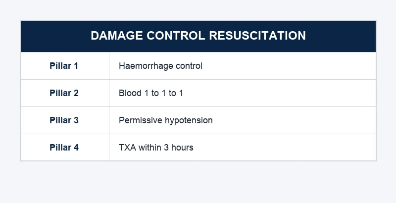
Pillar 1 — Haemorrhage control. The bleeding is stopped by the direct pressure, the tourniquet, the pelvic binder, the wound packing, the surgery, or the interventional radiology. No resuscitation succeeds against the uncontrolled source. The pre-hospital tourniquet for the exsanguinating limb injury is one of the most evidence-supported interventions in the trauma.[1]
Pillar 2 — Blood-first resuscitation (the 1-to-1-to-1 ratio). The resuscitation uses the blood products in a balanced ratio (the plasma, the platelets and the red cells approaching 1-to-1-to-1) rather than the crystalloid, which dilutes the clotting factors and worsens the coagulopathy. The PROPPR trial showed the 1-to-1-to-1 ratio achieved the earlier haemostasis and the fewer deaths from the exsanguination at 24 hours, though the 30-day mortality was not significantly different. The massive transfusion protocol delivers the products rapidly and in the right ratio. The whole blood (the fresh, the warm, the whole blood from the walking donor or the pre-hospital stock) is the modern evolution of this pillar.[2][3]
Pillar 3 — Permissive hypotension. The blood pressure is deliberately kept below the normal (a systolic of 80 to 90 mmHg, or a mean arterial pressure of 50 to 65 mmHg) until the bleeding is controlled, because the normal blood pressure disrupts the early clots, accelerates the haemorrhage, and dilutes the clotting factors. The permissive hypotension is contraindicated in the traumatic brain injury (where the cerebral perfusion requires a systolic of 110 mmHg or above).[3][1]
Pillar 4 — Tranexamic acid within 3 hours. The CRASH-2 trial showed that the tranexamic acid, given as 1 gram intravenous bolus followed by 1 gram infusion over 8 hours within 3 hours of injury, reduced the mortality from the traumatic bleeding. Beyond 3 hours, the benefit is lost and there may be harm. The TXA is given as early as possible — at the scene or on the arrival.[1]
Crystalloid era (pre-2005)
- Two large-bore cannulae and two litres of warmed saline
- Aggressive fluid to a normal blood pressure
- Definitive surgical repair at the first operation
- Crystalloid-driven dilutional coagulopathy
- Mortality from the lethal triad was the norm
DCR era (modern)
- Minimise crystalloid; blood products first
- Permissive hypotension to systolic 80 to 90
- TXA within 3 hours of injury
- Damage control surgery with the planned re-operation
- Viscoelastic-guided, calcium-replaced, warmed resuscitation
Permissive hypotension — the deeper physiology
Permissive hypotension is not "no resuscitation" — it is the deliberate titration of the mean arterial pressure to the lowest level that preserves the end-organ perfusion while the bleeding is uncontrolled. The mechanistic rationale is twofold. First, the higher blood pressure mechanically disrupts the soft early clot that forms at the bleeding site — the "pop-the-clot" phenomenon — and reopens the bleeding vessel. Second, the higher pressure drives more blood out of the injured vessel, dilutes the clotting factors, and accelerates the consumption. The animal and the human data converge on the systolic of 80 to 90 mmHg (or a MAP of 50 to 65 mmHg) as the safe lower limit in the conscious, non-head-injured patient.[3][10]
The clinical endpoint of the permissive hypotension is the level of consciousness, not a number. The patient who is talking, obeys commands, has a normal mentation, and produces the urine has an adequate perfusion; the precise systolic — 82 or 88 — is irrelevant. The patient who becomes confused, agitated, or loses consciousness is being underperfused, and small aliquots of blood product (not crystalloid) are given to restore the mentation. The bolus is 250 mL of the O-negative red cells or the balanced crystalloid, titrated to the mentation, never to a number.[3][1]
[1] [1] [1]Haemostatic resuscitation and the 1-to-1-to-1 ratio
The 1-to-1-to-1 ratio means that for every unit of red blood cells, the patient receives one unit of plasma and one apheresis (or six pooled) unit of platelets. The ratio is a unit-based approximation of the whole blood, which contains the red cells, the plasma and the platelets in roughly equal proportions. The mechanistic rationale is that the resuscitation should mirror the composition of the blood being lost — give back what is being shed. The crystalloid, by contrast, restores the volume but dilutes the clotting factors and the platelets, producing the iatrogenic dilutional coagulopathy.[2][6]
The PROPPR trial randomised 680 patients with the severe trauma and the major transfusion to the 1-to-1-to-1 versus the 1-to-1-to-2 ratio. The 1-to-1-to-1 group achieved the earlier haemostasis and the fewer deaths from the exsanguination at 24 hours, but the 30-day mortality — the primary outcome — was not significantly different (12.7 per cent versus 17.3 per cent, a difference that fell just short of the statistical significance). The PROMMTT observational study, run in parallel, found that the patients who received the higher plasma-to-red-cell ratio earlier had a lower mortality in the first six hours, when most of the haemorrhagic deaths occur. The synthesis: the 1-to-1-to-1 ratio is the target, the early plasma is the mediator of the benefit, and the protocolised massive transfusion is the delivery vehicle.[2][6]
[1] [1] [1]Tranexamic acid — the three-hour cliff
The tranexamic acid is the lysine analogue that reversibly blocks the lysine-binding site of the plasminogen, preventing its conversion to the plasmin and the subsequent lysis of the fibrin clot. The mechanistic rationale in the trauma is the early hyperfibrinolysis — the plasmin-driven clot lysis that is one of the three drivers of the trauma-induced coagulopathy. The TXA halts the hyperfibrinolysis and preserves the early clot at the bleeding site.[1][4]
The CRASH-2 trial (Shakur et al., 2010) randomised over 20,000 trauma patients with the significant bleeding to the TXA (1 g loading dose over 10 minutes, then 1 g over 8 hours) versus the placebo, within 8 hours of injury. The TXA reduced the all-cause mortality from 13.5 per cent to 14.5 per cent (relative risk 0.85, number-needed-to-treat around 120) with no increase in the vascular occlusive events. The pre-specified subgroup analysis by the time-to-treatment showed the striking effect of the timing: the TXA given within 1 hour reduced the mortality by a third; given between 1 and 3 hours reduced it by a fifth; given beyond 3 hours increased the mortality. This is the "three-hour cliff" — the single most important TXA fact for the Fellowship candidate.[1]
The MATTERs study (Morrison et al., 2012) examined the TXA in the military trauma — the battlefield casualties in Afghanistan with the severe bleeding and the high transfusion requirement. The TXA reduced the mortality from 17.4 per cent to 13.5 per cent in the matched cohort — a larger absolute benefit than the CRASH-2, attributable to the sicker military population and the early administration. Notably, the MATTERs also reported a higher rate of the thromboembolic events (the DVT, the PE, the MI) in the TXA group, a finding that has driven the guideline emphasis on the early but not the late administration.[4]
The WOMAN trial (Shakur et al., 2017) extended the TXA evidence to the post-partum haemorrhage and confirmed the same three-hour cliff: the TXA reduced the death from the bleeding in the PPH, and the subgroup analysis showed the benefit only when the drug was given within 3 hours of the onset. The WOMAN trial is the obstetric parallel to the CRASH-2 and reinforces the universal principle that the antifibrinolytic works in the early phase of the haemorrhage, when the hyperfibrinolysis is the mechanism, and is harmful in the late phase, when the established thrombosis is the competing risk.[5]
TXA within 1 hour
- CRASH-2: mortality reduced by around a third
- MATTERs: largest absolute benefit
- The hyperfibrinolysis is the active mechanism
- Give on the scene or on the arrival
TXA 1 to 3 hours
- CRASH-2: mortality reduced by around a fifth
- Benefit attenuating but still present
- The CRASH-2 protocol allowed up to 8 hours but the data support 3
- Reasonable if the patient arrives late
TXA beyond 3 hours
- CRASH-2: mortality INCREASED
- The established thrombosis dominates the risk
- Avoid if the time of injury is over 3 hours ago
- Confirm the time of injury before the dose
CRASH-2 — tranexamic acid in trauma (Lancet 2010)
Lancet
PMID 20554319
Key finding
A multicentre randomised placebo-controlled trial of 20,211 trauma patients with significant bleeding, treated within 8 hours of injury with TXA (1 g IV over 10 min, then 1 g over 8 h) or placebo. All-cause mortality was reduced from 13.5% to 14.5% (RR 0.85), with no increase in vascular occlusive events. A pre-specified time-to-treatment subgroup showed benefit within 3 hours and harm beyond 3 hours.
Practice change
The foundation of the modern TXA-in-trauma doctrine: give early (ideally pre-hospital), and do not give beyond 3 hours of injury.
MATTERs — military TXA in combat trauma (Arch Surg 2012)
Archives of Surgery
PMID 22704304
Key finding
A retrospective cohort of 896 combat casualties with severe bleeding and massive transfusion in Afghanistan, comparing the TXA (1 g IV bolus then 1 g over 8 h) to no TXA. The unadjusted mortality was 17.4% with TXA versus 23.9% without; in the propensity-matched cohort the mortality fell from 14.4% to 11.6%. Thromboembolic events were more frequent with TXA.
Practice change
In the sicker, military population with the early administration, the TXA benefit is larger than in the civilian CRASH-2; the trade-off is the thrombosis risk, which underlines the importance of the timing.
WOMAN — TXA in post-partum haemorrhage (Lancet 2017)
Lancet
PMID 28449780
Key finding
A randomised placebo-controlled trial of 20,060 women with post-partum haemorrhage, treated with TXA 1 g IV (repeated once if bleeding continued after 30 min) or placebo. Death from bleeding was reduced from 1.9% to 1.5% (RR 0.81), with no increase in thromboembolic events or maternal complications. The subgroup analysis showed the benefit confined to the administration within 3 hours.
Practice change
The obstetric confirmation of the CRASH-2 three-hour cliff: the antifibrinolytic works in the early phase of the haemorrhage and is ineffective or harmful in the late phase.
PROPPR — plasma, platelets and red cells in a 1:1:1 vs 1:1:2 ratio (JAMA 2015)
JAMA
PMID 25647203
Key finding
A multicentre randomised trial of 680 patients with severe trauma and massive transfusion (within 1 hour of admission, expected to need more), comparing a 1:1:1 versus a 1:1:2 ratio of plasma:platelets:red cells. The 1:1:1 group achieved earlier haemostasis and fewer deaths from exsanguination at 24 hours; the 30-day mortality (primary outcome) was not significantly different.
Practice change
The 1:1:1 ratio is adopted as the target for the massive transfusion protocol — it changes the mode of the early death even if the overall survival is not changed.
PROMMTT — observational timing of plasma (J Trauma 2013)
Journal of Trauma and Acute Care Surgery
PMID 23532247
Key finding
A prospective observational study of 1,245 trauma patients receiving the massive transfusion at ten US level-1 trauma centres, examining the relationship between the time-varying plasma-to-red-cell ratio and the mortality. The patients who received the higher plasma ratio earlier had a lower in-hospital mortality, particularly in the first 6 hours when most of the haemorrhagic deaths occur.
Practice change
The observational evidence that the early plasma delivery — not just the cumulative ratio — is the mediator of the survival benefit, and that the MTP must deliver the plasma in parallel with the red cells, not after.
The lethal triad and its prevention
The lethal triad of hypothermia, acidosis and coagulopathy is the self-perpetuating cycle that makes the bleeding irreversible. The hypothermia impairs the coagulation enzymes and the platelet function (the coagulation is a temperature-dependent cascade). The acidosis (from the poor perfusion) further inhibits the enzymes and the cardiac function. The coagulopathy (from the consumption, the dilution, and the dysfunction of the cold, acidotic platelets and enzymes) worsens the bleeding, which worsens the shock, the acidosis and the hypothermia.[3]
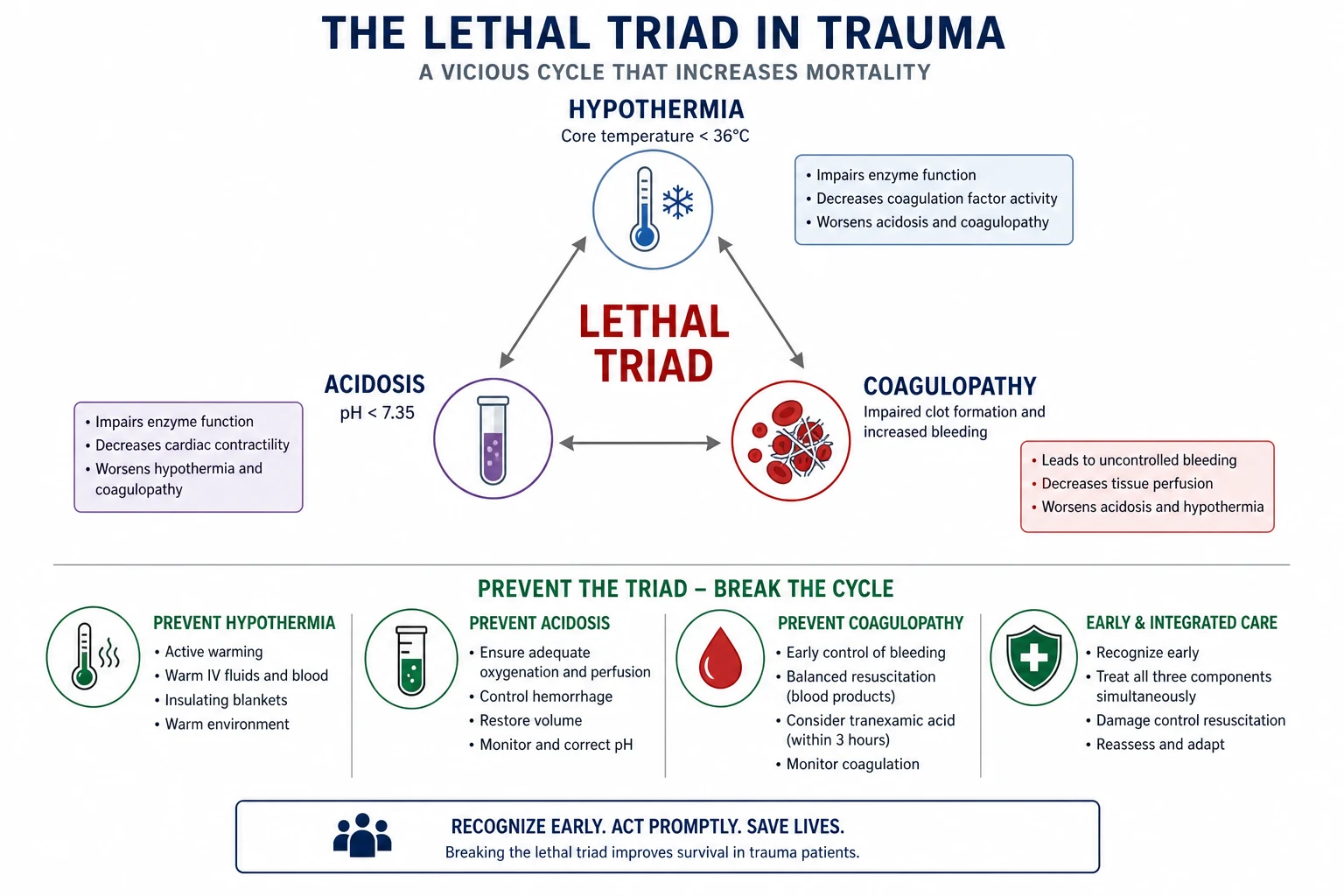
The prevention starts from the first minute: the patient is warmed (the blankets, the warmed fluids, the high ambient temperature, the body wrap), the blood products are given instead of the crystalloid (which does not cause the dilutional coagulopathy or the hyperchloroemic acidosis), and the acidosis is corrected through the restoration of the perfusion (the blood, the vasopressor, the surgical control).[3][1]
[1] [1] [1]Damage control surgery
The damage control surgery is the surgical counterpart of the damage control resuscitation: the abbreviated operation that controls the bleeding and the contamination without the definitive repair. The bleeding is controlled by the packing, the ligation, or the shunt; the contamination is controlled by the resection without the anastomosis, the stapling, or the stoma; and the abdomen is left open (the temporary vacuum closure) for the planned second-look after the lethal triad is corrected in the intensive care. The definitive repairs are deferred to the re-operation, when the physiology is restored.[3][1]
The three classical stages of the damage control surgery are: the abbreviated initial operation (the haemorrhage and the contamination control, the temporary abdominal closure, target operative time under 60 minutes), the physiological resuscitation in the ICU (the rewarming to 36°C, the correction of the coagulopathy to a normal INR and a fibrinogen above 2 g/L, the clearance of the lactate to under 2.5 mmol/L, the restoration of the perfusion), and the planned re-operation at 24 to 48 hours for the definitive repair (the anastomosis, the stoma, the closure of the abdomen, the evaluation of the viability).[3]
[1] [1]Viscoelastic testing — TEG and ROTEM
The viscoelastic haemostatic assays (the thromboelastography, TEG; the rotational thromboelastometry, ROTEM) are the point-of-care tests that provide a whole-blood, dynamic, real-time picture of the clot formation and the clot lysis. Unlike the conventional coagulation tests (the INR, the APTT, the fibrinogen) — which are plasma-based, slow (30 to 60 minutes), and informative only of the clot initiation — the viscoelastic assays use the whole blood, return the actionable result in 10 to 15 minutes, and characterise the entire clot lifecycle: the clot initiation (the reaction time, R or CT), the clot propagation (the kinetics, K or CFT, the alpha angle), the clot strength (the maximum amplitude, MA, or the MCF, reflecting the platelet number and function and the fibrinogen), and the clot lysis (the LY30 or the ML, the marker of the hyperfibrinolysis).[8]
The viscoelastic-guided resuscitation replaces the empirical 1-to-1-to-1 with the goal-directed product replacement: a prolonged R or CT (the clotting factors deficient) → the plasma; a low alpha angle or a prolonged K (the fibrinogen deficient, the early clot strength) → the cryoprecipitate or the fibrinogen concentrate; a low MA or MCF (the platelet deficient) → the platelets; an elevated LY30 or ML (the hyperfibrinolysis) → the tranexamic acid. The Gonzalez randomised trial (2016) compared the viscoelastic to the conventional coagulation-test-guided resuscitation in the trauma and found the viscoelastic strategy reduced the mortality (a relative reduction of around 30 per cent) and the plasma and platelet transfusion, with a higher fibrinogen concentrate use. The European and the American guidelines now recommend the early viscoelastic testing in the major haemorrhage.[3][8]
Conventional tests (INR, APTT, fibrinogen)
- Plasma-based, not whole-blood
- Turnaround 30 to 60 minutes
- Inform on the clot initiation only
- Do not detect the hyperfibrinolysis
- Do not guide the fibrinogen replacement well
- The standard since the 1970s
Viscoelastic (TEG, ROTEM)
- Whole-blood, point-of-care
- Actionable result in 10 to 15 minutes
- Characterise the clot initiation, propagation, strength, lysis
- Detect the hyperfibrinolysis (the LY30 elevated)
- Guide the product replacement (factors, fibrinogen, platelets, TXA)
- Reduced mortality in the Gonzalez RCT
The massive transfusion protocol
The massive transfusion is defined as the replacement of one blood volume (around 10 units of red cells) within 24 hours, or half a blood volume (around 5 units) within 3 hours, or the ongoing transfusion at a rate of more than 150 mL per minute. The massive transfusion protocol (MTP) is the institutional pathway that delivers the blood products in the 1-to-1-to-1 ratio, in the pre-assembled packs, with the rapid infuser, the point-of-care testing and the laboratory support, in response to a single activation call. The MTP is the operational vehicle for the haemostatic resuscitation.[3][1]
The activation of the MTP is a clinical decision — it does not wait for the laboratory confirmation of the coagulopathy or the haemoglobin (the haemoglobin falls late, only after the dilution has reached 30 per cent of the blood volume). The triggers for the activation are the clinical shock with the suspected active bleeding, the positive focused assessment with sonography for trauma (FAST), the haemodynamic instability after the crystalloid, the penetrating mechanism to the torso, and the major pelvic or the junctional injury. The early over-activation is preferable to the late under-activation.[3]
The massive transfusion protocol — the first hour
Recognise and activate
Sample and crossmatch
TXA within 3 hours of injury
Rapid infuser, warmed blood
Calcium replacement
Viscoelastic-guided product replacement
Source control
Transition from permissive hypotension to restoration
Stand down the MTP
Calcium and citrate toxicity in the massive transfusion
The stored red cells and the plasma are preserved with the sodium citrate, the anticoagulant that chelates the calcium and prevents the clotting in the bag. When the citrated blood is transfused rapidly (more than one unit every 5 minutes, or the rapid infuser at the full rate), the citrate overwhelms the hepatic metabolism and the calcium chelation produces the acute hypocalcaemia — an ionised calcium that falls within minutes. The hypocalcaemia impairs the myocardial contractility (the calcium is the trigger of the excitation-contraction coupling), the vascular tone (the calcium is the smooth-muscle contractile agent), and the coagulation (the calcium is factor IV of the cascade).[3][7]
The clinical picture of the citrate-induced hypocalcaemia is the worsening shock despite the adequate transfusion — the falling blood pressure, the narrow pulse pressure, the prolonged QT interval, the poor response to the vasopressor, and the decreased cardiac output on the bedside ultrasound. The ionised calcium (not the corrected total calcium) is the actionable test, and the bedside point-of-care ionised calcium is part of the MTP monitoring. The treatment is the calcium chloride 1 g (10 mL of 10 per cent) intravenously after every 4 units of the red cells or plasma, and as clinically indicated for the falling ionised calcium.[7]
[1] [1] [1]Whole blood, lyophilised plasma and the alternative fluids
The modern trauma resuscitation is moving beyond the reconstituted 1-to-1-to-1 toward the fluids that more closely approximate the lost blood. The low-titre group-O whole blood (LTOWB) — the fresh whole blood from the donors with a low anti-A and anti-B titre, stored cold for up to 14 days — delivers the red cells, the plasma and the platelets in a single bag, at the physiological ratio, with a single donor exposure and a lower citrate load. The LTOWB is now the pre-hospital fluid of choice in the US military and several civilian systems, and reduces the donor exposure, the storage lesion and the anticoagulant burden.[9]
The lyophilised (freeze-dried) plasma is the plasma that has been freeze-dried and can be stored at the room temperature for years, reconstituted with the sterile water in 3 to 6 minutes. The French military has used the lyophilised plasma in the combat setting for two decades, and the civilian systems are adopting it for the pre-hospital and the rural-retrieval use, where the frozen plasma is logistically impossible. The lyophilised plasma contains the full complement of the clotting factors and the natural anticoagulants, and is the functional equivalent of the fresh-frozen plasma.[3]
[1]Special circumstances
The concurrent traumatic brain injury
The traumatic brain injury is the absolute contraindication to the permissive hypotension. The injured brain loses the autoregulation, the cerebral blood flow becomes the pressure-passive, and the single episode of the systolic blood pressure below 90 mmHg doubles the mortality. The European guideline and the Brain Trauma Foundation target a MAP of 80 mmHg or above in the TBI, with the systolic of 110 mmHg or above as the operational target. The concurrent bleeding and the TBI are managed with the higher blood-pressure target, the early vasopressor (the noradrenaline), and the prompt source control to minimise the duration of the shock.[3][1]
The pregnant trauma patient
The pregnant trauma patient has the expanded blood volume (a 40 per cent increase by the third trimester) and the relative haemodilution, so the signs of the shock appear later than in the non-pregnant patient — the tachycardia and the hypotension are the late signs. The permissive hypotension is modified in the pregnancy (the lower MAP compromises the placental perfusion), and the target is a systolic of 100 mmHg or above. The supine hypotensive syndrome is avoided by the left lateral tilt of 15 to 30 degrees. The TXA is safe in the pregnancy and the post-partum haemorrhage (the WOMAN trial) and is given for the traumatic bleeding within the 3-hour window.[5]
The anticoagulated trauma patient
The anticoagulated patient (the warfarin, the direct oral anticoagulants, the antiplatelet agents) presents a specific challenge: the pre-existing coagulopathy compounds the trauma-induced coagulopathy. The warfarin is reversed with the prothrombin complex concentrate (PCC, 25 to 50 units per kg) and the intravenous vitamin K 5 to 10 mg; the dabigatran with the idarucizumab 5 g (two 2.5 g vials); the apixaban and the rivaroxaban with the andexanet alfa or the PCC. The viscoelastic test guides the reversal; the conventional INR is unreliable in the direct-oral-anticoagulant patient. The early involvement of the haematology and the toxicology is the standard.[3]
The paediatric trauma patient
The paediatric trauma patient has the greater physiological reserve and the delayed signs of the shock (the child compensates with the tachycardia and the vasoconstriction until the late decompensation). The permissive hypotension is applied with the age-adjusted target (a systolic of 70 plus twice the age in years). The TXA dose is 15 mg/kg (max 1 g) over 10 minutes, then 2 mg/kg per hour for 8 hours. The blood volume is calculated at 80 mL/kg, and the MTP is delivered in the weight-based ratio. The intraosseous access is the default in the shocked child with the difficult peripheral access. [1]
The first 60 minutes — the integrated timeline
The first 60 minutes of the exsanguinating trauma patient
0 to 5 minutes — the primary survey and the simultaneous resuscitation
The airway with the cervical spine control, the breathing (the oxygen, the tension pneumothorax excluded), the circulation (the two large-bore cannulae, the FAST, the pelvic binder for the suspected pelvic fracture, the tourniquet for the limb bleed). The team leader calls the MTP and the surgical/vascular team. The TXA is prescribed at the moment of the call.
5 to 15 minutes — the MTP first pack, the TXA, the warming
The first MTP pack (the O-negative red cells, the AB plasma, the platelets) is hung on the rapid infuser, warmed. The TXA 1 g IV over 10 minutes is started (within the 3-hour window from the injury). The patient is wrapped and warmed (the forced-air warmer, the warmed fluids, the high ambient temperature). The permissive hypotension is maintained (systolic 80 to 90, or the MAP 80 in the TBI).
15 to 30 minutes — the source control and the viscoelastic
The source control in parallel: the surgery for the torso bleed (the damage control laparotomy or the thoracotomy), the interventional radiology for the pelvic and the solid-organ bleeding, the wound packing for the junctional bleed. The first viscoelastic test (TEG or ROTEM) is sent and the result is used to direct the second MTP pack. The ionised calcium is checked and the calcium chloride is given for every 4 units of the red cells.
30 to 60 minutes — the transition and the disposition
As the bleeding is controlled, the transition from the permissive hypotension to the perfusion restoration: the MAP to 65 mmHg (or 80 in the TBI), the lactate clearance, the correction of the coagulopathy (the viscoelastic-guided fibrinogen, plasma, platelets), the normalisation of the ionised calcium and the temperature. The disposition: the theatre for the damage control surgery, the angiography suite for the embolisation, or the ICU for the ongoing resuscitation. The definitive imaging (the CT, the pan-scan) is deferred for the unstable patient — the theatre first, the imaging second.
Common pitfalls
The recurring errors are: resuscitating with crystalloid instead of blood products; delaying the TXA beyond 3 hours; targeting a normal blood pressure before the bleeding is controlled; not preventing the hypothermia; not applying the pre-hospital tourniquet; sending the unstable patient to the CT instead of the theatre; not replacing the calcium during the massive transfusion; not using the viscoelastic testing to guide the product replacement; giving the TXA beyond the 3-hour cliff; not transitioning from the permissive hypotension once the bleeding is controlled; and not activating the MTP early enough.[3][1][7]
[1] [1]Exam practice
SAQ — The four pillars of damage control resuscitation in exsanguinating polytrauma
10 minutes · 10 marks
A 34-year-old man is brought to the trauma bay 25 minutes after a high-speed motorcycle crash. He is pale, diaphoretic and agitated (GCS 14), BP 72/46, HR 132, RR 28, SpO2 94 per cent on 15 L oxygen via a non-rebreather mask. He has a markedly distended abdomen with free fluid on the focused assessment with sonography in trauma (FAST), an open femoral shaft fracture with active external bleeding, and clinical pelvic instability. The initial haemoglobin is 134 g/L, lactate 7.2 mmol/L, INR 1.9, fibrinogen 1.4 g/L, ionised calcium 0.88 mmol/L. The time of injury is confirmed at 25 minutes ago. He has no head injury.
SAQ — Permissive hypotension with TXA in penetrating torso trauma
10 minutes · 10 marks
A 28-year-old man is brought to the trauma bay 90 minutes after a single stab wound to the right upper quadrant. He is pale, cold and confused (GCS 14), BP 84/56, HR 124, RR 26, SpO2 96 per cent on 15 L oxygen. The FAST is positive in the right upper quadrant. Lactate 5.8 mmol/L, INR 1.6, ionised calcium 0.92 mmol/L, temperature 35.2 degrees C. The time of injury is confirmed at 90 minutes ago. He has no head injury and the surgical team is in attendance.
SAQ — Damage control surgery principles in exsanguinating polytrauma
10 minutes · 10 marks
A 42-year-old man is brought to the trauma bay 40 minutes after a high-speed motorcycle crash. He is in haemorrhagic shock (BP 70/48, HR 138, GCS 14), with a markedly distended abdomen positive on FAST, an open femoral shaft fracture, and a clinically unstable pelvis. The pelvic binder and the tourniquet are applied, the massive transfusion protocol is activated, the O-negative red cells are running, and he is taken directly to the theatre, NOT to the CT scanner. The laparotomy reveals a Grade IV liver laceration, a destructive splenic injury, and a non-expanding pelvic haematoma. The core temperature is 34.1 degrees C, the pH 7.18, the INR 2.1, the lactate 8.2 mmol/L.
SAQ — Damage control resuscitation with tranexamic acid: mechanism, evidence, and the three-hour cliff
10 minutes · 10 marks
A 30-year-old soldier is evacuated from the battlefield after an improvised explosive device detonation, with bilateral below-knee amputations and a clinically unstable pelvis. He arrives at the role-2 facility 165 minutes after the injury. He is in haemorrhagic shock (BP 78/52, HR 132, GCS 14), with bilateral above-knee tourniquets and a pelvic binder in situ. The lactate is 6.4 mmol/L, the ionised calcium 0.90 mmol/L, the temperature 35.0 degrees C, and the TEG shows an elevated LY30 of 8 per cent. The massive transfusion protocol is running. The medical officer asks whether to give the tranexamic acid given that the casualty is almost at the three-hour mark.
Red flags
[1]References
- [1]CRASH-2 trial collaborators, Shakur H, Roberts I, et al. Effects of tranexamic acid on death, vascular occlusive events, and blood transfusion in trauma patients with significant haemorrhage (CRASH-2): a randomised, placebo-controlled trial Lancet, 2010.PMID 20554319
- [2]Holcomb JB, Tilley BC, Baraniuk S, et al. Transfusion of plasma, platelets, and red blood cells in a 1:1:1 vs a 1:1:2 ratio and mortality in patients with severe trauma: the PROPPR randomized clinical trial JAMA, 2015.PMID 25647203
- [3]Rossaint R, Bouillon B, Cerny V, et al. The European guideline on management of major bleeding and coagulopathy following trauma: sixth edition Crit Care, 2023.PMID 36859355
- [4]Morrison JJ, Dubose JJ, Rasmussen TE, Midwinter MJ. Can anyone spare a little indigo carmine? The drug shortage crisis Can J Urol, 2012.PMID 22704304
- [5]WOMAN Trial Collaborators, Shakur H, Roberts I, et al. Transcatheter Mitral Valve Replacement: Insights From Early Clinical Experience and Future Challenges J Am Coll Cardiol, 2017.PMID 28449780
- [6]Holcomb JB, Fox EE, Wade CE, PROMMTT Study Group. Macrolides and bronchiectasis: clinical benefit with a resistance price JAMA, 2013.PMID 23532247
- [7]Napolitano LM, Cohen MJ, Cotton BA, Schreiber MA, Moore EE. Racial-ethnic disparities in management and outcomes among children with type 1 diabetes Pediatrics, 2015.PMID 25687140
- [8]Gonzalez E, Moore EE, Moore HB, et al. Nonaqueous electrocatalytic water oxidation by a surface-bound Ru(bda)(L)₂ complex Dalton Trans, 2016.PMID 26974188
- [9]Cotton BA, Podbielski J, Camp E, et al. Synthesis of trifluoromethyl ketones via tandem Claisen condensation and retro-Claisen C-C bond-cleavage reaction J Org Chem, 2013.PMID 23517488
- [10]Rowell SE, Meier EN, McKnight B, et al. Pathological and clinical features of enteric adenocarcinoma of the thymus. A pooled analysis of cases from a reference center and systematic review of the literature Cancer Treat Rev, 2021.PMID 33296826