EM · Limb & extremity trauma
Limb and extremity trauma
The limb trauma from the open fracture and the Gustilo classification, the vascular injury and the hard signs, the compartment syndrome, the neurovascular examination, the dislocation reduction, the open-fracture management with the antibiotics and the washout, and the crush injury with the rhabdomyolysis.
On this page & tools
Your progress
Saved locally on this device.
Target exams
Red flags
The limb and the extremity trauma encompasses the fractures, the dislocations, the vascular and the nerve injuries, the compartment syndrome and the crush injury, and it is the most common form of the trauma that the emergency physician manages. Though rarely immediately life-threatening, the limb injury is the common source of the long-term disability and the limb loss if it is not managed promptly and correctly. The Fellowship candidate must know the Gustilo classification of the open fracture, the hard and the soft signs of the vascular injury, the clinical diagnosis of the compartment syndrome, and the principles of the open-fracture management.[2][3]
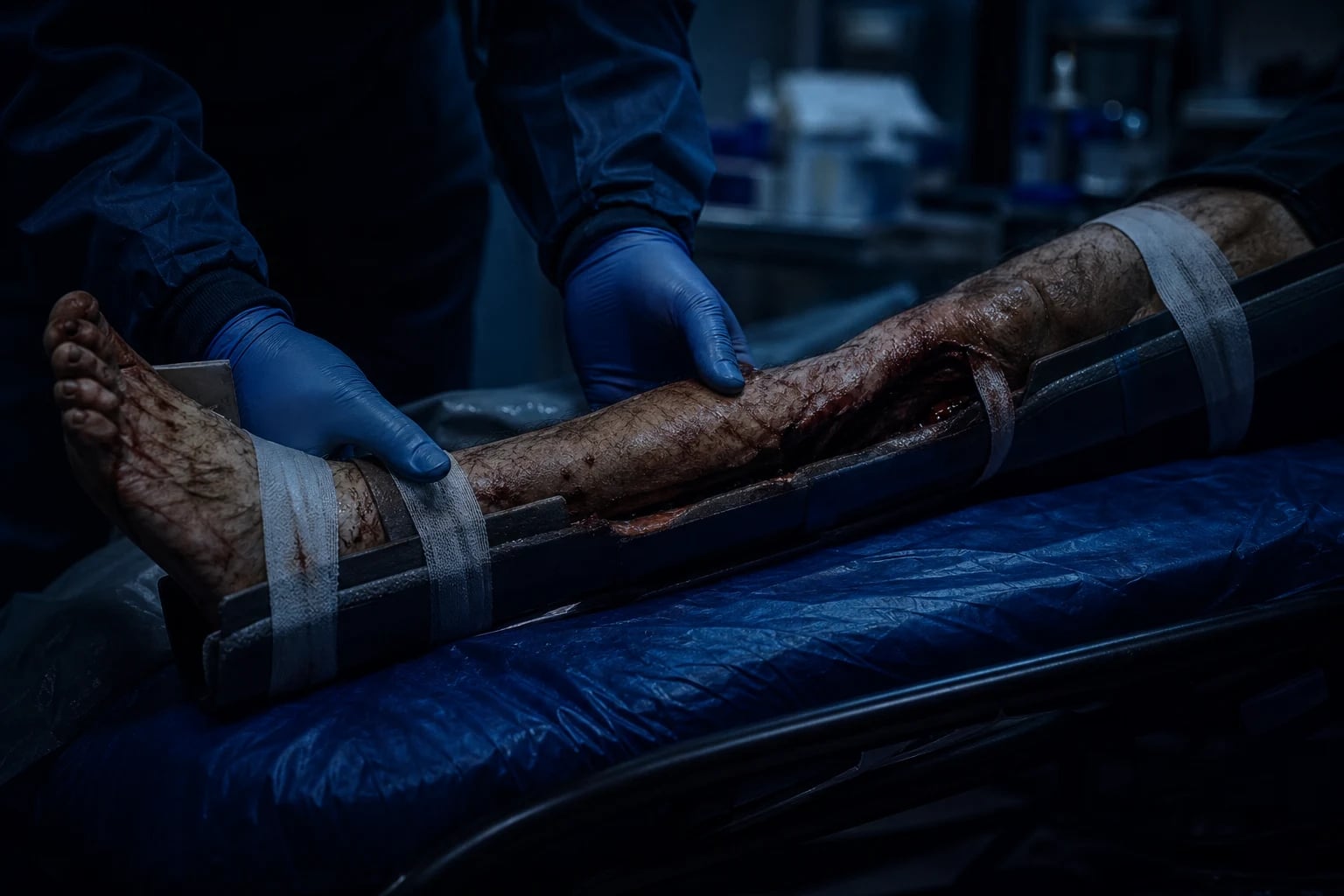
The open fracture: the Gustilo classification
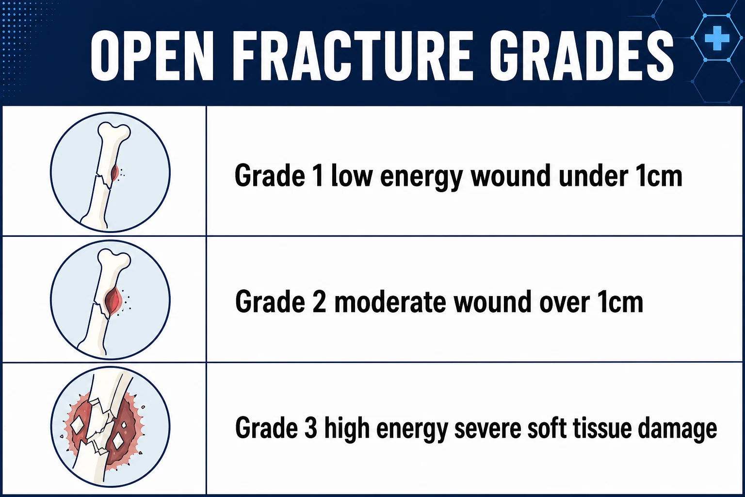
The open fracture — the fracture with a wound communicating between the bone and the external environment — is classified by the Gustilo-Anderson system, which predicts the infection risk and guides the management.[2] Grade I: the wound under 1 centimetre, the low-energy, the clean puncture. Grade II: the wound over 1 centimetre, the moderate soft-tissue damage, no flap needed. Grade III: the severe soft-tissue damage, the high-energy, further subdivided: Grade IIIA the adequate soft-tissue coverage, Grade IIIB the flap needed for the coverage, Grade IIIC the vascular injury requiring the repair. The Grade III has the high rate of the infection and the amputation, and the Grade IIIC has the highest amputation rate of all.
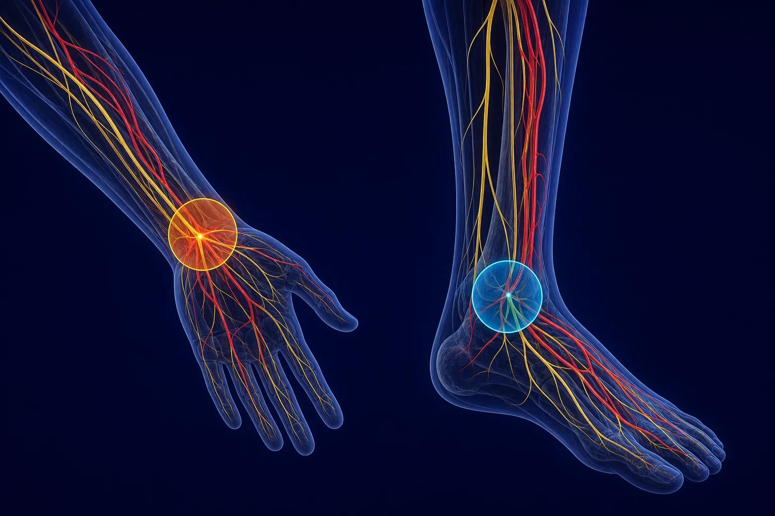
The vascular injury
The vascular injury is identified by the hard signs (the pulsatile bleeding, the expanding or the pulsatile haematoma, the bruit or the thrill, the absent pulse, the signs of the acute ischaemia — the pallor, the pulselessness, the paraesthesia, the paralysis, the poikilothermia, the pain) and the soft signs (the reduced but present pulse, the small non-expanding haematoma, the nerve deficit, the proximity of the injury to a major vessel). The hard signs mandate the immediate surgical or endovascular intervention — the on-table angiography and the repair. The soft signs warrant the CT angiogram for the definitive assessment. The knee dislocation has a particularly high rate of the popliteal artery injury (up to a third), and the CT angiogram is indicated even if the distal pulse is initially present, because the intimal tear may thrombose over the hours.[2][3]
The compartment syndrome
The compartment syndrome is the diagnosis that cannot be missed — it is the most time-critical of the limb injuries, because the delay in the fasciotomy beyond 6 to 8 hours produces the irreversible necrosis and the limb loss or the death. The diagnosis is clinical: the pain out of proportion to the injury, the pain on the passive stretch of the affected compartment's muscles, and the tense, swollen compartment. The late signs (the pulselessness, the paraesthesia, the paralysis) are the signs of the irreversible damage and must not be awaited. The fasciotomy — the incision of the fascia to release the pressure — is the treatment, performed without the delay for the pressure measurement if the clinical diagnosis is clear. The causes include the fracture (the tibial shaft), the crush, the reperfusion after the vascular repair, the burns, the prolonged immobilisation, and the bleeding into a compartment (the anticoagulated patient).[2][3]
The neurovascular examination
The neurovascular examination of the injured limb is performed and documented at the presentation and after every intervention (the splinting, the reduction, the surgery). The pulses (the radial, the ulnar, the femoral, the popliteal, the dorsalis pedis, the posterior tibial), the capillary refill, the sensation (the dermatomes and the individual nerves), the motor function, and the compartment softness are assessed. The documentation is essential for the medico-legal and the clinical follow-up.[2]
The open-fracture management
The open fracture is managed by the washout within 6 hours of the injury, the empirical intravenous antibiotics (the first-generation cephalosporin cefazolin 1 to 2 g — the cefazolin 25 to 50 mg/kg in the child — for the Grade I and II; for the Grade III the addition of the gentamicin 5 to 7 mg/kg and, for the farm or the water contamination, the high-dose benzylpenicillin; for the patient with the severe penicillin allergy the vancomycin 15 mg/kg or the clindamycin 600 to 900 mg replaces the cefazolin), the tetanus prophylaxis (the tetanus immunoglobulin 250 IU intramuscularly and the toxoid booster for the incomplete immunisation), the splinting, the wound coverage with the saline-soaked gauze, and the orthopaedic referral for the definitive surgical management. The wound is not closed primarily in the emergency department.[2][3]
The dislocations
The dislocation is reduced promptly after the neurovascular documentation, using the sedation and the analgesia (the procedural sedation) and the appropriate technique. The shoulder is reduced by the Stimson, the Kocher or the scapular manipulation. The hip is reduced urgently (the avascular necrosis risk within 6 hours) by the Allis or the Stimson. The knee is reduced immediately and splinted — the popliteal artery is assessed by the CT angiogram. The patella and the finger dislocations are reduced and splinted. The post-reduction neurovascular examination and the radiograph are documented.[2]
The crush injury and the rhabdomyolysis
The crush injury (the prolonged compression of the limb, as in the entrapment or the collapse) releases the myoglobin and the potassium from the damaged muscle, producing the rhabdomyolysis — the myoglobinuric acute kidney injury, the hyperkalaemia and the metabolic acidosis. The CK is markedly elevated (above 5000 units per litre), the urine is dark (the tea-coloured) with the dipstick positive for the blood but the microscopy negative for the red cells (the myoglobin). The management is the aggressive fluid resuscitation (the saline at a high rate to maintain the urine output above 1 to 2 millilitres per kilogram per hour), the bicarbonate (to alkalinise the urine and to prevent the myoglobin precipitation in the tubules), and the treatment of the hyperkalaemia. The fasciotomy is performed if the compartment syndrome coexists. The fat embolism syndrome (the triad of the respiratory failure, the neurological dysfunction and the petechial rash, 24 to 72 hours after the long-bone fracture) is managed with the supportive care and the oxygenation.[1][3]
Common pitfalls
The recurring errors are: not performing and documenting the neurovascular examination before and after the intervention; missing the compartment syndrome (the pain attributed to the fracture rather than the compartment); not imaging the popliteal artery after the knee dislocation; closing the open wound primarily; delaying the washout of the open fracture beyond 6 hours; under-dosing the antibiotics for the Grade III; and not recognising the rhabdomyolysis in the crush injury.[4]
Differential diagnosis
- The open fracture versus the closed fracture: the open fracture has the wound communicating with the bone and the external environment and mandates the empirical antibiotics, the tetanus prophylaxis and the washout within 6 hours, whereas the closed fracture does not breach the skin and is managed by the reduction and the splinting alone.
- The vascular injury versus the compartment syndrome: both may produce the pulselessness and the paraesthesia, but the vascular injury shows the hard signs (the pulsatile bleeding, the expanding haematoma, the absent pulse) and the acute ischaemia from the moment of the injury, while the compartment syndrome develops over hours with the pain out of proportion and the pain on the passive stretch before the late pulselessness.
- The fat embolism versus the venous thromboembolism: both cause the respiratory distress after the limb trauma, but the fat embolism arises within 24 to 72 hours of the long-bone or the pelvic fracture with the triad of the respiratory failure, the cerebral dysfunction and the petechial rash, while the venous thromboembolism develops later from the immobilisation and lacks the petechial rash.
- The crush injury with the rhabdomyolysis versus the isolated fracture haematoma: the crush injury releases the myoglobin and the potassium with the markedly elevated CK and the dark urine (the dipstick-positive, microscopy-negative blood), while the simple fracture haematoma does not produce the systemic hyperkalaemia, the acidosis or the myoglobinuric renal failure.
- The nerve injury (the neuropraxia, the axonotmesis) versus the cord injury: a focal limb nerve deficit points to the peripheral nerve, but a bilateral or a sensory-level deficit, the urinary retention or the priapism indicates the spinal cord injury and demands the different, time-critical management.[5]
Exam practice
SAQ — The Gustilo Grade IIIC open tibial fracture with the vascular injury
10 minutes · 10 marks
A 24-year-old motorcyclist is brought to the trauma bay 1 hour after a collision with a car at 70 km/h. He has an open fracture of the right mid-tibia and fibula with a 12-cm wound, the bone visible, and the gross contamination with the road debris. The right foot is pale, cold and pulseless — the dorsalis pedis and the posterior tibial pulses are absent, the capillary refill is greater than 5 seconds, and the patient reports the paraesthesia in the foot. The systolic BP is 110, HR 110, and the rest of the primary survey is unremarkable.
SAQ — The knee dislocation with the popliteal artery injury
10 minutes · 10 marks
A 35-year-old footballer presents to the emergency department 90 minutes after a tackle in which his right knee was hyperextended and dislocated. The dislocation was reduced on the field by the team physiotherapist. On arrival the knee is swollen and deformed, the patient is in severe pain, the dorsalis pedis pulse is present but weaker than the left, the capillary refill is 3 seconds, and the ankle-brachial index is 0.8. The patient reports numbness in the first web space of the foot.
Red flags
[8]The vascular injury in detail — the hard signs, the soft signs, the decision to operate
The limb vascular injury is the injury where the emergency physician's decision-making has the most direct bearing on the limb survival. The blood supply to the limb is a single arterial axis with the limited collateral flow at the joints (the shoulder, the elbow, the knee), and the complete interruption produces the irreversible ischaemia of the muscle within 4 to 6 hours and the nerve within a similar window. The diagnostic question is never "is the vessel injured?" but "is the limb ischaemic?" — and the answer determines whether the patient goes to the operating theatre from the resus bay or to the CT scanner for the angiogram. The hard signs answer the question by the bedside; the soft signs answer it by the imaging.[2][3]
The hard signs of the vascular injury are the five findings that, alone or in combination, prove the arterial disruption and mandate the immediate surgical exploration without the further imaging: the pulsatile bleeding from the wound, the expanding or the pulsatile haematoma, the bruit or the thrill over the injury, the absent distal pulse, and the signs of the acute limb ischaemia (the six Ps — the pain, the pallor, the pulselessness, the paraesthesia, the paralysis, the poikilothermia). The presence of any one hard sign carries a positive predictive value for the major vascular injury above 90 per cent, and the on-table angiography — not the CT angiogram — is the next step. The emergency physician's role is to recognise the hard sign, apply the direct pressure (never a blind clamp), resuscitate, and mobilise the vascular surgeon; the angiogram in the radiology suite wastes the ischaemic window and is contraindicated.[2]
The soft signs of the vascular injury are the findings that suggest but do not prove the arterial injury: the reduced but present distal pulse, the small stable non-expanding haematoma, the peripheral nerve deficit (which may reflect the proximity of the vessel or the direct nerve injury), the history of the arterial bleeding at the scene that has now stopped, and the proximity of the wound or the fracture to a major vessel (the axillary, the brachial, the superficial femoral, the popliteal). The soft signs warrant the CT angiography for the definitive assessment, because the injury may be the intimal flap, the pseudoaneurysm or the arteriovenous fistula that is not yet ischaemic but threatens to thrombose or rupture over the hours.[3]
Hard signs → immediate surgery
- The pulsatile bleeding from the wound; the expanding or pulsatile haematoma
- The bruit or the thrill over the injury (the arteriovenous fistula)
- The absent distal pulse; the signs of the acute ischaemia (the six Ps)
- Positive predictive value above 90 per cent — the theatre, not the scanner
- The on-table angiography and the repair; the direct pressure, never the blind clamp
Soft signs → CT angiography
- The reduced but present distal pulse; the small stable non-expanding haematoma
- The peripheral nerve deficit; the history of the bleeding at the scene that has stopped
- The proximity of the wound or the fracture to the named vessel
- The intimal flap, the pseudoaneurysm, the arteriovenous fistula — not yet ischaemic
- The CT angiogram is the definitive assessment; the serial observation if negative
The vascular injury — the decision to operate, to image, or to observe
Step 1 — Identify the hard signs at the bedside
Inspect the wound for the pulsatile or the bright-red bleeding and the expanding haematoma. Auscultate and palpate over the injury for the bruit and the thrill. Palpate the distal pulses (the radial, the ulnar, the dorsalis pedis, the posterior tibial) and compare with the contralateral limb. Assess the six Ps of the ischaemia. Any hard sign sends the patient to the operating theatre.
Step 2 — The hard signs: the theatre and the on-table angiography
Apply the direct manual pressure (the air not the clamp), resuscitate with the blood products, give the antibiotics if the wound is open, and alert the vascular surgeon. The on-table angiography confirms the level and the extent of the injury, and the repair follows — the primary anastomosis, the interposition vein graft (the reversed saphenous from the contralateral limb), or the temporary shunt to restore the flow before the definitive reconstruction. The fasciotomy is performed prophylactically after the prolonged ischaemia to prevent the reperfusion compartment syndrome.
Step 3 — The soft signs: the CT angiography
The haemodynamically stable patient with the soft signs undergoes the CT angiography of the affected limb, which identifies the intimal flap, the thrombosis, the pseudoaneurysm, the arteriovenous fistula and the active contrast extravasation. The positive CTA sends the patient to the vascular surgeon; the negative CTA with the stable pulse is followed by the serial observation and the repeat examination at 4 to 6 hours, because the intimal tear may declare itself late.
Step 4 — The special case of the knee dislocation
The knee dislocation (even the spontaneous reduction) carries the popliteal artery injury rate of up to a third, because the artery is tethered at the adductor hiatus and the popliteal fossa and is stretched across the dislocated tibia. The hard signs send the patient to the theatre; in their absence, the CT angiogram or the admission with the serial ABI (the ankle-brachial index) monitoring is mandatory, because the intimal tear may thrombose over the hours and convert the salvageable limb to the amputation.<Cite id="2"/><Cite id="3"/>
The peripheral nerve injury — the examination and the classification
The peripheral nerve injury accompanies the limb trauma in up to a fifth of the high-energy injuries, and it is the most commonly missed of the limb injuries because the fracture dominates the attention. The nerve injury may be the direct laceration (the sharp glass, the knife), the crush (the blunt force), the stretch (the traction across the joint), or the compression (the haematoma, the swelling, the compartment syndrome). The diagnosis is made by the focused examination of the individual nerves of the affected limb, performed and documented at the presentation and repeated after every intervention — because the nerve deficit that develops after the splinting or the reduction is the iatrogenic injury and the medico-legal hazard.[2]
The Seddon classification describes the three grades of the nerve injury by the structural disruption and the prognosis. The neuropraxia is the concussion of the nerve — the conduction block without the structural damage, the recovery within weeks to months, the prognosis excellent. The axonotmesis is the disruption of the axon with the intact endoneurium — the Wallerian degeneration distal to the injury, the regeneration at a millimetre a day (roughly an inch a month), the recovery over the months to a year, the prognosis good but slow. The neurotmesis is the complete transection of the nerve and its sheath — the no spontaneous recovery, the surgical repair or the grafting required, the prognosis guarded. The Sunderland classification refines this into the five degrees by the disruption of the individual layers, but the Seddon trio is the examination answer.[2]
The examination of the individual nerves of the upper and the lower limb is the core skill. The median nerve is tested by the opposition of the thumb (the abductor pollicis brevis) and the sensation over the thenar eminence; its injury at the wrist causes the loss of the thumb opposition and the numbness of the radial three and a half digits. The ulnar nerve is tested by the abduction of the fingers (the first dorsal interosseous, the "crossed fingers" test) and the sensation over the little finger; its injury at the elbow causes the claw hand (the hyperextension of the fourth and fifth metacarpophalangeal joints) and the numbness of the ulnar one and a half digits. The radial nerve is tested by the wrist and the finger extension and the sensation over the first dorsal webspace; its injury in the spiral groove of the humerus causes the wrist drop and the numbness of the dorsum of the hand. The axillary nerve (the surgical neck of the humerus, the anterior dislocation) is tested by the deltoid abduction and the regimental badge patch of the numbness over the lateral shoulder. The peroneal (the common fibular) nerve (the fibular neck, the knee dislocation) is tested by the dorsiflexion of the ankle and the toes and the sensation over the dorsum of the foot; its injury causes the foot drop. The tibial nerve (the popliteal fossa) is tested by the plantarflexion and the sensation over the sole; its injury is rare in isolation. The femoral nerve is tested by the knee extension (the quadriceps) and the anterior thigh sensation.[2]
Median nerve
- The wrist laceration (the "suicide attempt"); the supracondylar humerus in the child
- The loss of the thumb opposition (the abductor pollicis brevis); the thenar numbness
- The "pointing index" — the inability to flex the index finger
- The sensory loss over the radial three and a half digits; the poor precision grip
Ulnar nerve
- The elbow injury (the medial epicondyle); the wrist laceration at the Guyon canal
- The claw hand (the fourth and fifth MCP hyperextension); the "crossed fingers" test positive
- The first dorsal interosseous wasting; the sensory loss over the ulnar one and a half digits
- The loss of the power grip and the fine manipulation; the highest-yield nerve at the viva
Radial nerve
- The spiral groove of the humerus (the "Saturday night palsy"); the holstein-lewis fracture
- The wrist drop; the loss of the finger and the thumb extension
- The sensory loss over the first dorsal webspace (the snuffbox) — small and easily missed
- The most common of the upper-limb nerve palsies; often recovers (the neuropraxia)
Peroneal (fibular) nerve
- The fibular neck fracture; the knee dislocation; the prolonged squatting or the cast
- The foot drop — the loss of the dorsiflexion and the eversion
- The sensory loss over the dorsum of the foot and the lateral lower leg
- The commonest lower-limb nerve palsy; the high fall-risk; the splint to hold the foot neutral
Tibial nerve
- The popliteal fossa injury; the displaced posterior tibia or the calcaneus
- The loss of the plantarflexion (the calf); the loss of the toe flexion
- The sensory loss over the sole; the clawing of the toes
- The rare injury in isolation; the tarsal tunnel at the medial malleolus
Axillary nerve
- The anterior shoulder dislocation; the surgical neck of the humerus
- The loss of the deltoid abduction beyond the first 15 degrees
- The "regimental badge" — the numbness over the lateral deltoid
- The most common nerve injured by the shoulder dislocation; the reduction is followed by the re-examination
The open fracture in detail — the Gustilo classification, the antibiotics, the debridement
The open fracture is the orthopaedic emergency that the emergency physician initiates, because the delay in the antibiotics and the debridement converts the salvageable limb into the infected, the amputated or the dead limb. The Gustilo-Anderson classification, derived from the prospective analysis of over a thousand open fractures, grades the injury by the wound size, the soft-tissue damage and the vascular involvement, and it dictates the antibiotic regimen, the urgency of the debridement and the prognosis.[5][4]
Grade I: the puncture wound under 1 centimetre, the low-energy mechanism, the clean wound from the inside-out (the bone spike pierced the skin) or the low-velocity gunshot. Grade II: the wound over 1 centimetre, the moderate soft-tissue damage, the no flap, the no major vascular injury. Grade III: the severe soft-tissue damage, the high-energy mechanism, the heavy contamination — subdivided into Grade IIIA (the adequate soft-tissue coverage of the bone, the primary closure possible), Grade IIIB (the flap or the muscle cover needed for the soft-tissue defect, the exposed bone) and Grade IIIC (the vascular injury requiring the repair, irrespective of the soft-tissue degree). The Grade III carries the infection rate of 10 to 50 per cent and the amputation rate that climbs from the IIIA to the IIIC, with the IIIC having the amputation rate of 40 to 60 per cent — the highest of all.[4][5]
Grade I
- The wound under 1 cm; the low-energy; the clean puncture (often inside-out)
- The bone pierces the skin transiently; the minimal contamination
- Cefazolin alone; the washout and the debridement within 24 h
- Infection rate under 2 per cent; the excellent prognosis
Grade II
- The wound over 1 cm; the moderate soft-tissue damage; the no flap needed
- The moderate energy; the contamination present but limited
- Cefazolin alone; the washout within 24 h; the delayed or the primary closure
- Infection rate 2 to 7 per cent; the good prognosis
Grade IIIA
- The wound over 10 cm; the high-energy; the heavy contamination
- The adequate soft-tissue coverage of the bone; the primary closure possible
- Cefazolin plus gentamicin (and the penicillin for the farm or the water)
- Infection rate 10 to 25 per cent; the flap or the graft not required
Grade IIIB
- The extensive soft-tissue loss; the exposed bone; the flap or the muscle cover required
- The heavy contamination; the high-energy crush or the shearing
- The triple antibiotic; the serial debridement; the delayed flap cover
- Infection rate up to 50 per cent; the amputation rate 10 to 25 per cent
Grade IIIC
- The vascular injury requiring the repair — the hard signs, the ischaemia
- The arterial disruption at any level of the soft-tissue damage
- The vascular repair or the shunt first, then the skeletal fixation, then the cover
- The amputation rate of 40 to 60 per cent — the highest of all the grades.
The open fracture — the emergency-department pathway in the first hour
1 — Cover, photograph and culture
The wound is covered with the sterile saline-soaked gauze and the bandage; a photograph is taken (to avoid the repeated uncovering and the contamination); the wound is NOT explored, debrided or closed in the emergency department. The contamination with the obvious debris is gently removed if accessible; the embedded object is left in situ for the theatre.
2 — The antibiotics within one hour
The intravenous antibiotics are given as soon as the open fracture is recognised, ideally within one hour of the arrival — the earlier the better, because the infection rate climbs with each hour of the delay. The Grade I and II receive the cefazolin 1 to 2 g (or 25 to 50 mg/kg in the child). The Grade III receives the cefazolin plus the gentamicin 5 to 7 mg/kg, and the farm, the soil, the fresh water or the marine contamination adds the high-dose benzylpenicillin (the clostridial cover). The severe penicillin allergy substitutes the vancomycin 15 mg/kg or the clindamycin 600 to 900 mg for the cefazolin.
3 — The tetanus prophylaxis
The tetanus status is assessed and the booster (the toxoid 0.5 mL intramuscularly) is given if the last dose was over 5 years ago. The contaminated, the necrotic or the over-24-hour-old wound in the incompletely immunised patient receives the tetanus immunoglobulin 250 IU intramuscularly at a separate site. The tetanus is the universally fatal preventable disease — the status is never assumed, always confirmed.
4 — The splint and the analgesia
The limb is splinted in the position of the function (the above-knee or the below-knee backslab, the plaster or the padded rigid splint) to reduce the pain, the bleeding and the further soft-tissue damage. The femur fracture is splinted in the traction splint (the Sager or the Kendrick). The analgesia is the intravenous opioid titrated (the morphine 0.1 mg/kg, the fentanyl 1 to 2 mcg/kg) with the antiemetic.
5 — The debridement and the fixation in the theatre
The patient goes to the operating theatre for the formal debridement, the lavage and the skeletal stabilisation — ideally within 24 hours of the injury (the historical "6-hour rule" has been revised by the modern evidence, but the earlier remains the better). The external fixation is the standard for the Grade III and the contaminated fracture (the damage-control orthopaedics); the definitive internal fixation is delayed until the soft-tissue envelope has settled. The wound is left open and the delayed primary closure or the flap follows.<Cite id="2"/><Cite id="3"/><Cite id="4"/>
FLOW (Bhandari, NEJM 2015) — the irrigation of the open fracture wound
The amputation — the indications and the preservation of the part
The traumatic amputation is the complete severance of the limb or the digit, and the emergency physician's role is the preservation of the life (the haemorrhage control), the preservation of the stump (the soft-tissue handling) and the preservation of the amputated part (the cooling) for the potential reimplantation. The decision to reimplant is the surgical decision, made on the basis of the part, the level, the mechanism, the patient and the time, but the emergency physician determines whether the reimplantation is even an option by the care of the amputated part in the first minutes.[3]
The absolute indications for the amputation (the completion of the amputation that has functionally occurred) are: the life over the limb (the uncontrolled haemorrhage, the crush with the prolonged ischaemia, the sepsis from the necrotic limb in the unstable patient), the irreversible ischaemia (the mangled limb with the warm ischaemia over 6 hours and the non-viable muscle), and the Gustilo IIIC with the unrepairable vascular injury. The relative indications are: the Gustilo IIIB/IIIC with the MESS over 7, the severe nerve injury (the complete transection of the major nerve with the anticipated useless limb), the delayed presentation (the warm ischaemia of 6 to 8 hours), and the patient factors (the age, the comorbidity, the occupation, the preference). The decision is the multidisciplinary one — the orthopaedic, the plastic, the vascular and the rehabilitation — and it is made in the light of the fact that the modern prosthesis often outperforms the poorly salvaged limb.[2]
Absolute indications
- The life over the limb — the uncontrolled haemorrhage or the septic necrotic limb
- The irreversibly ischaemic limb (the warm ischaemia over 6 h, the non-viable muscle)
- The Gustilo IIIC with the unrepairable or the multiply-segmented vessel
- The multi-level crush with the open fracture, the nerve and the vessel all transected
Relative indications
- The Gustilo IIIB/IIIC with the MESS of 7 or above
- The anticipated useless limb (the complete nerve transection, the bone loss)
- The warm ischaemia of 6 to 8 hours (the grey zone)
- The patient factors — the age, the comorbidity, the occupation, the preference
- The decision is the multidisciplinary, never the single-surgeon act
The management of the amputated part — the saline, the gauze, the bag, the ice
1 — Control the stump bleeding
The stump bleeding is controlled by the direct manual pressure with the bulky gauze and the elevation, or the pneumatic tourniquet proximal to the amputation if the pressure fails. The blind clamping and the ligation of the nerves and the vessels in the field are avoided (they destroy the tissue needed for the reimplantation). The patient is resuscitated with the blood products; the massive transfusion protocol is activated for the proximal amputation.
2 — Elevate and wrap the stump
The stump is elevated to reduce the swelling and the bleeding, and wrapped in the sterile saline-soaked gauze and the bulky bandage. The stump is NOT cleaned, debrided or explored in the emergency department — the theatre is the place for the stump management. The photograph is taken for the documentation.
3 — Recover and prepare the amputated part
The amputated part is recovered from the scene if possible (the digit, the hand, the forearm). It is gently rinsed with the sterile saline to remove the gross contamination, wrapped in the saline-soaked gauze, placed in the sealed plastic bag, and the bag is placed on the ice (the ice-water slurry at 4 degrees Celsius). The part is NEVER placed directly on the ice (the frostbite damages the tissue), and NEVER immersed in the water or the formalin.
4 — The time, the level and the mechanism are documented
The time of the amputation, the level (the digit, the hand, the forearm, the above-knee), the mechanism (the guillotine — the clean cut, the best for the reimplantation; the crush; the avulsion — the worst), and the patient factors (the age, the occupation, the handedness, the comorbidity) are documented. The reimplantation centre is contacted within the first hour, because the warm ischaemia time for the digit is 6 hours and the cold ischaemia time is 12 hours; for the major limb the window is shorter.
5 — The transfer and the reimplantation
The patient and the part are transferred to the reimplantation centre (the plastic and the orthopaedic microsurgery service). The thumb, the multiple digits, the hand and the forearm in the adult are the strong indications for the reimplantation; the single digit (other than the thumb), the crush and the avulsion amputations, and the heavily contaminated limbs are the relative contraindications. The decision is the surgical one; the emergency physician preserves the option.<Cite id="3"/>
The crush injury in detail — the fluid before the extraction
The crush injury — the prolonged compression of the limb by the heavy object, as in the earthquake, the building collapse, the trench collapse or the entrapment under the machinery — produces the crush syndrome, the systemic illness first described by Bywaters and Beall in the London Blitz of 1941. The damaged muscle releases the myoglobin, the potassium, the phosphate and the lactic acid into the circulation, producing the hyperkalaemia (the cardiac arrest), the myoglobinuric acute kidney injury (the pigmented casts in the tubules), the metabolic acidosis and the hypovolaemic shock (the fluid sequestered in the injured muscle). The mortality of the untreated crush syndrome is high; the mortality of the promptly treated crush syndrome is low, and the difference is the fluid.[7][8]
The cardinal principle of the crush syndrome management is the fluid loading before the extraction. The moment the extrication begins and the compression is released, the sequestered potassium and the myoglobin flood into the circulation, and the patient who was stable under the rubble arrests within minutes of the release. The intravenous access is established and the isotonic saline is infused at a high rate (the 1 litre per hour in the adult, the 15 mL/kg/h in the child) BEFORE the limb is freed, and the infusion continues through the extraction and into the emergency department. The urine output is targeted at 1 to 2 mL/kg/h, the urine is alkalinised with the bicarbonate (the 50 mmol per litre of the fluid) to prevent the myoglobin precipitation, and the potassium is lowered with the insulin-dextrose, the calcium chloride (the membrane stabilisation against the hyperkalaemic cardiotoxicity), and the salbutamol.[7]
The crush syndrome — the management from the scene to the intensive care
1 — The fluid before the release
Establish the intravenous access and begin the isotonic saline at 1 L/h (the adult) BEFORE the limb is freed. The compression acts as a tourniquet; its release dumps the potassium and the myoglobin into the circulation. The fluid loading dilutes the load and supports the renal perfusion. The patient who is pulled out without the prior fluid is the patient who arrests in the ambulance.
2 — The hyperkalaemia is anticipated and treated
The potassium released from the damaged muscle may exceed 7 mmol/L and produce the widened QRS, the sine wave and the cardiac arrest within minutes of the release. The ECG is monitored continuously. The calcium chloride 10 mL of the 10% (the membrane stabilisation), the insulin-dextrose (the 10 units of the soluble insulin in 50 mL of the 50% dextrose), the salbutamol 10 to 20 mg nebulised, and the bicarbonate (the shift and the alkalinisation) are given for the hyperkalaemia. The potassium exchange resin and the dialysis are the slower adjuncts.
3 — The alkalinisation of the urine
The myoglobin precipitates in the acidic urine and the distal tubule, forming the pigmented casts that obstruct and the nephron and the acute kidney injury. The sodium bicarbonate (the 50 mmol per litre of the maintenance fluid, targeting the urine pH above 6.5) keeps the myoglobin in the solution and the urine flowing. The bicarbonate is continued until the CK falls and the urine clears; the acetazolamide is added if the systemic alkalosis develops without the urinary alkalinisation.
4 — The CK, the urine and the renal monitoring
The creatine kinase peaks at 24 to 48 hours and may exceed 50,000 units per litre in the severe crush. The urine is dark (the "tea" or the "cola") and the dipstick is positive for the blood with the microscopy negative for the red cells (the myoglobin pseudoperoxidase). The fluid is titrated to the urine output of 1 to 2 mL/kg/h; the mannitol (the 1 to 2 g/kg) is the adjunct for the osmotic diuresis but is avoided in the oliguric patient (it worsens the volume overload). The renal team is alerted for the possible continuous dialysis.
5 — The fasciotomy for the compartment syndrome
The crushed limb develops the compartment syndrome as the swelling fills the closed osseofascial compartments, and the fasciotomy is performed if the compartment is tight, the pain is on the passive stretch, or the pressures exceed 30 mmHg. The fasciotomy releases the pressure, decompresses the muscle and prevents the further necrosis — but it also releases more myoglobin, so the fluid and the alkalinisation are intensified after the fasciotomy. The limb is observed for the progressive necrosis and the amputation is the last resort.<Cite id="7"/><Cite id="8"/>
Bywaters & Beall (1941, reprinted JASN 1998) — the original description of the crush syndrome
Better (Nephron 1990) — the crush syndrome revisited (1940-1990)
The disposition
The limb-trauma patient is discharged, admitted, or transferred to the tertiary service depending on the injury. The discharge is for the simple closed fracture (the undisplaced, the stable), the reduced dislocation with the documented neurovascular intactness, and the soft-tissue injury, with the splint, the analgesia, the follow-up and the safety-net advice. The admission is for the open fracture (the antibiotics, the theatre), the vascular injury (the repair or the observation), the compartment syndrome (the fasciotomy), the multiple injuries and the analgesia requirement beyond the oral. The transfer to the tertiary limb service is for the reimplantation (the digit, the hand), the complex soft-tissue cover (the flap), and the limb salvage in the mangled extremity. The crush syndrome and the major vascular injury go to the intensive care. The venous thromboembolism prophylaxis (the low-molecular-weight heparin) is started within 24 to 48 hours of the major limb trauma, balancing the bleeding and the clot risk.[1]
Common pitfalls (the expanded list)
The recurring errors in the limb trauma are added to: not loading the crush patient with the fluid before the extrication, with the consequent hyperkalaemic arrest on the release; placing the amputated part directly on the ice or in the water, destroying the tissue for the reimplantation; under-dosing the Grade III antibiotic regimen (omitting the gentamicin or the penicillin); delaying the antibiotic beyond the first hour for the radiograph or the consultation; not imaging the popliteal artery after the knee dislocation even when the pulse returns; not examining and documenting the individual peripheral nerves before and after the reduction; closing the open wound or exploring it in the emergency department; using the high-pressure irrigation on the open wound (driving the bacteria into the tissue); and not anticipating the hyperkalaemia and the myoglobinuria in the first hours of the crush.[6]
Red flags (the expanded list)
[5]The examiner's mental map
The Fellowship candidate is expected to walk the examiner through the limb trauma as a sequence of the limb-threatening and the limb-saving decisions. The map below is the structure the examiner listens for — the decisions that frame the answer.[1]
The six decisions of the limb trauma — the structure the examiner expects
Decision 1 — Is the limb ischaemic? (the hard signs)
The pulsatile bleeding, the expanding haematoma, the bruit, the absent pulse, the six Ps. Any hard sign sends the patient to the theatre for the on-table angiography and the repair — no CT. The direct pressure, the resuscitation, the vascular surgeon. The limb has 4 to 6 hours before the irreversible ischaemia.
Decision 2 — Is the compartment threatened? (the pain)
The pain out of proportion, the pain on the passive stretch, the tense compartment — the clinical diagnosis, the fasciotomy without the delay for the pressure measurement. The late signs (the pulselessness, the paralysis) are the irreversible. The causes: the tibial fracture, the crush, the reperfusion, the burn, the anticoagulant.
Decision 3 — Is the fracture open? (the Gustilo grade)
The wound communicating with the bone — the Grade I, II, IIIA/B/C. The antibiotic within the hour (the cefazolin; the gentamicin and the penicillin for the Grade III and the contamination), the tetanus prophylaxis, the saline-soaked gauze, the photograph, the theatre for the debridement within 24 hours, the external fixation for the Grade III.
Decision 4 — Is the nerve injured? (the individual nerves)
The median, the ulnar, the radial, the axillary in the arm; the peroneal, the tibial, the femoral, the sciatic in the leg. The motor and the sensory of each, examined and documented before and after the reduction. The Seddon classification — the neuropraxia, the axonotmesis, the neurotmesis — and the prognosis of each.
Decision 5 — Is the amputation or the reimplantation needed? (the part)
The absolute indications (the life over the limb, the irreversible ischaemia, the IIIC unrepairable); the relative (the MESS, the nerve, the time, the patient). The management of the part — the saline, the gauze, the sealed bag, the ice slurry at 4 degrees. The thumb and the multiple digits are reimplanted; the decision is the microsurgeon's.
Decision 6 — Is the crush syndrome developing? (the fluid and the potassium)
The fluid before the release; the hyperkalaemia anticipated (the calcium, the insulin-dextrose, the salbutamol); the alkalinisation of the urine (the bicarbonate); the CK and the urine monitored; the fasciotomy for the compartment. The crush is the systemic illness — the kidney, the heart and the limb are all at risk.<Cite id="1"/><Cite id="2"/><Cite id="3"/><Cite id="7"/>
References
- [1]Rossaint R, Bouillon B, Cerny V, et al. The European guideline on management of major bleeding and coagulopathy following trauma (fifth edition). Critical Care, 2023.PMID 36859355
- [2]Galvagno SM Jr, Nahmias JT, Young DA Advanced Trauma Life Support(®) Update 2019: Management and Applications for Adults and Special Populations. Anesthesiology clinics, 2019.PMID 30711226
- [3]Halvorson JJ, Anz A, Langfitt M, et al. Vascular injury associated with extremity trauma: initial diagnosis and management. The Journal of the American Academy of Orthopaedic Surgeons, 2011.PMID 21807917
- [4]Gustilo RB, Mendoza RM, Williams DN. Problems in the management of type III (severe) open fractures: a new classification of type III open fractures. Journal of Trauma, 1984.PMID 6471139
- [5]Gustilo RB, Anderson JT. Prevention of infection in the treatment of one thousand and twenty-five open fractures of long bones: retrospective and prospective analyses. Journal of Bone and Joint Surgery (Am), 1976.PMID 773941
- [6]FLOW Investigators, Bhandari M, Jeray KJ, et al. A trial of wound irrigation in the initial management of open fracture wounds. New England Journal of Medicine, 2015.PMID 26448371
- [7]Better OS. The crush syndrome revisited (1940-1990). Nephron, 1990.PMID 2194135
- [8]Bywaters EGL, Beall D. Crush injuries with impairment of renal function. 1941. Journal of the American Society of Nephrology, 1998.PMID 9527411