EM · Major trauma resuscitation
Major trauma resuscitation
The integrated resuscitation of the multiply-injured patient: the trimodal distribution of trauma death, the simultaneous primary survey with damage-control resuscitation, the blood-product-first strategy with the 1-to-1-to-1 ratio and tranexamic acid, the permissive hypotension, the resuscitative thoracotomy and REBOA for the arresting patient, and the damage-control surgery and the whole-body CT for the stable.
On this page & tools
Your progress
Saved locally on this device.
Target exams
Red flags
The major trauma patient — the multiply-injured, the physiologically deranged, the time-critical — is the archetype of the emergency that cannot be managed sequentially. The primary survey, the resuscitation, the source control and the definitive imaging all proceed in parallel, from the moment of the arrival, because the bleeding patient who is sent for the CT while the blood products are ordered is a patient who bleeds to death in the scanner. The major trauma resuscitation is the integration of the primary survey (the C-A-B-C-D-E), the damage-control resuscitation (the blood products, the tranexamic acid, the permissive hypotension), the source control (the surgery, the interventional radiology, the REBOA), and the definitive imaging (the whole-body CT for the stable), all led by the trauma team leader and delivered by the coordinated trauma team.[3][1]

The trimodal distribution of trauma death
Trauma kills in three peaks, and the resuscitation is built around preventing the second and the third.[1] The first peak is the immediate death — the seconds to the minutes after the injury — from the catastrophic brain or the great-vessel injury, which is largely preventable only by the primary prevention (the seatbelt, the helmet, the speed limit). The second peak is the early death — the minutes to the several hours — from the treatable conditions: the tension pneumothorax, the cardiac tamponade, the exsanguinating haemorrhage, the expanding intracranial haematoma. This is the peak the emergency department targets, and it is the rationale for the golden hour, the trauma team, the primary survey and the damage-control resuscitation. The third peak is the late death — the days to the weeks — from the multiple organ failure and the sepsis, prevented by the high-quality critical care and the tertiary survey.

The parallel approach
The major trauma resuscitation is not a series of sequential steps; it is a set of parallel streams that run simultaneously from the arrival. The primary survey (the C-A-B-C-D-E) identifies and treats the immediately life-threatening conditions. The damage-control resuscitation delivers the blood products, the tranexamic acid and the permissive hypotension through the massive transfusion protocol. The source control is pursued in parallel — the operating theatre for the positive FAST with the ongoing bleeding, the interventional radiology for the pelvic embolisation, the neurosurgery for the expanding intracranial haematoma. The definitive imaging (the whole-body CT) is obtained for the stable patient or after the stabilisation. The team leader coordinates these streams and makes the decision points — the theatre or the CT, the transfer, the escalation, the termination.[3][1]
Damage-control resuscitation
The damage-control resuscitation is the evidence-based framework for the fluid and the blood-product management of the bleeding trauma patient, and it is detailed in the massive haemorrhage topic. Its four pillars are the early source control, the blood-product resuscitation in the balanced 1-to-1-to-1 ratio (the PROPPR trial showed it achieved earlier haemostasis and fewer deaths from exsanguination at 24 hours),[2] the tranexamic acid within three hours (the CRASH-2 trial showed it reduced mortality from traumatic bleeding within this window),[1] and the permissive hypotension (the lower blood pressure tolerated until the bleeding is controlled, except in the traumatic brain injury). The lethal triad of hypothermia, acidosis and coagulopathy is prevented by the warming, the blood products and the correction of the perfusion.[3]
The resuscitative thoracotomy
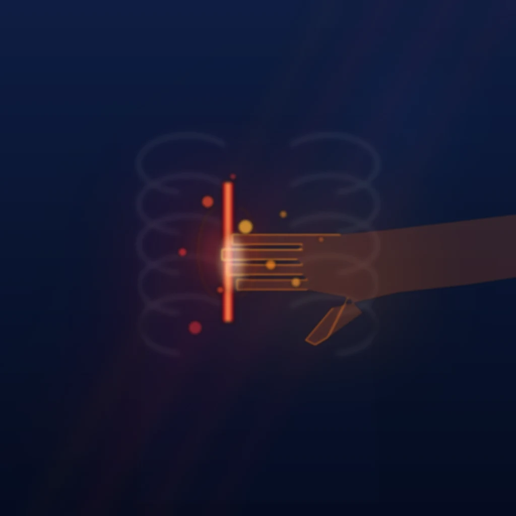
The resuscitative thoracotomy — the emergency department thoracotomy — is the definitive intervention for the trauma patient in arrest or with the imminent arrest after a penetrating torso injury. The left anterolateral thoracotomy (the incision below the nipple in the fifth intercostal space, extending from the sternal border to the mid-axillary line) gives access to the pericardium (to relieve the tamponade), the thoracic aorta (to cross-clamp and to redirect the blood to the brain and the heart), the lung (to control the bleeding), and the heart (to repair the laceration or to perform the open cardiac massage). The outcomes are best after the penetrating cardiac injury with the recent loss of the output, and poor after the blunt trauma with the prolonged downtime — the indication is narrow, and the decision is made by the team leader at the point of the arrest.[1]
REBOA and the modern adjuncts
The Resuscitative Endovascular Balloon Occlusion of the Aorta (the REBOA) is the modern adjunct to the resuscitative thoracotomy for the patient with the non-compressible torso haemorrhage. A balloon is inserted through the femoral artery (percutaneously or by the cut-down) and inflated in the aorta at Zone 1 (the descending thoracic aorta, for the intra-abdominal bleeding) or Zone 3 (the infrarenal aorta, for the pelvic bleeding), occluding the distal flow and raising the proximal blood pressure while the definitive source control is achieved. It is less invasive than the thoracotomy and the cross-clamping, but it has its own complications (the ischaemia of the distal tissues, the vascular injury), and its role is evolving in the trauma systems that have the capability.[3]
Damage-control surgery and the whole-body CT
The damage-control surgery — the abbreviated operation that controls the bleeding and the contamination without attempting the definitive repair — is the surgical counterpart of the damage-control resuscitation. The bleeding is controlled (the packing, the ligation, the shunt), the contamination is controlled (the resection without the anastomosis, the stapling, the stomas), and the abdomen is left open (the temporary closure) for the planned second-look after the physiology is restored in the intensive care. The whole-body CT (the pan-scan, from the head to the pelvis with the arterial and the venous phases) is the definitive imaging for the stable patient or the stabilised patient, and it identifies the injuries that direct the definitive management. The unstable patient who cannot go to the CT goes to the operating theatre for the damage-control surgery, with the imaging deferred to the post-operative period.[1][1]
Trauma team activation criteria
The trauma team activation is the trigger that converts a single-clinician assessment into the parallel, multi-personnel resuscitation. The governing principle is one of deliberate over-triage: it is far better to call the full team for a patient who turns out to be minimally injured than to under-call and discover the deteriorating patient alone in the resus bay. The activation criteria are tiered into the physiological, the anatomical and the mechanistic, and most pre-hospital and emergency systems combine the three so that any single positive criterion triggers the team.[1][1]
The physiological criteria are the most reliable because they reflect derangement already present, not the mechanism that might have produced it. A systolic blood pressure below 90 mmHg, a Glasgow Coma Score below 13, and a respiratory rate below 10 or above 29 are the classic triggers; together they identify the patient who is shocked, brain-injured, or failing to ventilate. The anatomical criteria flag injuries that are inherently life- or limb-threatening regardless of the current numbers: the flail chest, the two or more long-bone fractures, the unstable pelvic fracture, the amputation proximal to the wrist or ankle, the penetrating injury to the head, neck or torso, the burns of greater than twenty per cent of the body surface area, and the suspected paralysis or spinal-cord injury. The mechanistic criteria are the least specific but capture the high-energy transfer that may have produced an occult injury: a fall from greater than 6 metres (or twice the patient height), a high-speed motor-vehicle crash (especially with ejection, rollover, or a death in the same vehicle), a pedestrian or cyclist struck by a vehicle, a motorcycle crash above 30 km/h, and a crush, blast, or extrication time over 20 minutes. [1]
[1]Physiological
- The most reliable — derangement already present
- SBP below 90 mmHg, GCS below 13, RR below 10 or above 29
- Any single criterion triggers the full trauma team
- Reflects shock, brain injury, or ventilatory failure
Anatomical
- Inherently life- or limb-threatening regardless of numbers
- Flail chest, two or more long bones, pelvic fracture, amputation proximal to wrist or ankle
- Penetrating injury to head, neck or torso; burns greater than 20% body surface area
- Paralysis; suspected spinal-cord injury; crushed or degloved limb
Mechanism
- High-energy transfer — may have produced an occult injury
- Fall greater than 6 m or twice patient height; high-speed MVC, ejection, rollover, death in same vehicle
- Pedestrian or cyclist struck; motorcycle crash above 30 km/h
- Crush, blast, or extrication over 20 min — least specific, designed for sensitivity
The trauma team and the roles
The trauma team is the human engine of the parallel approach. The defining feature is the team leader — the single clinician who stands back from the bedside, does not touch the patient, and orchestrates the resuscitation by calling the structured assessments, allocating the tasks, hearing the findings aloud, and making the disposition decisions. The team leader is the only person who makes the global decisions; everyone else executes within their allocated role. The remaining roles are distributed around the patient: the airway doctor at the head, the breathing and circulation doctors at the chest and the side, the procedure doctor running the lines and the FAST, the scribe documenting the timeline, the radiology liaison coordinating the films and the CT, and the nursing team handling the monitoring, the drugs, the warming and the family.[1]
The trauma team — who stands where, and who does what
Team leader
Stands at the foot of the bed, hands off the patient, eyes on the monitor and the team. Calls the structured assessments, allocates the tasks, hears the findings aloud, and makes the disposition (theatre, CT, angiography). The only person who makes the global decisions.
Airway doctor
Head of the bed. Cervical-spine control with manual in-line stabilisation, airway assessment, suction, the adjuncts, and the rapid-sequence induction and intubation. The surgical airway (cricothyroidotomy) if the airway is lost.
Breathing doctor
Right chest. Assesses the chest, applies the oxygen, performs the needle decompression and the chest drain for the tension and the haemothorax, and auscultates after the intubation to confirm the tube.
Circulation doctor
Left side. Two large-bore cannulae, the blood samples (group and crossmatch, coagulation, venous gas), the massive haemorrhage activation, the FAST scan, the pelvic binder, and the haemorrhage control of the limbs.
Procedure doctor
The arterial line and the central line if needed, the urinary catheter (after excluding the urethral injury), the gastric tube, and the assisting of every other procedure as it arises.
Scribe
Documents the timeline — the vital signs every five minutes, the interventions and their times, the drugs and the doses, the examination findings, the imaging results, and the GCS trend. The scribe is the medicolegal and the clinical memory of the resuscitation.
Radiology and nursing
Radiology coordinates the portable chest and pelvis films and the FAST, and the CT booking. Nursing applies the monitoring, prepares and times the drugs, warms the patient, handles the specimens, and liaises with the family.
The primary survey — the C-A-B-C-D-E in depth
The primary survey is the structured, simultaneous identification and treatment of the immediately life-threatening conditions. The modern sequence — C-A-B-C-D-E — reorders the traditional A-B-C by placing the catastrophic haemorrhage (the first C) ahead of the airway, on the military evidence that exsanguination from a compressible limb can kill faster than airway obstruction. The catastrophic external haemorrhage is controlled by direct pressure, then the tourniquet, then wound packing with the haemostatic dressing, all within the first 60 seconds of the patient arriving.[3][1]
A — Airway with cervical-spine protection
The airway is assessed for patency and protected, while the cervical spine is immobilised from the moment of the accident. The signs of airway compromise — the gurgling, the snoring, the agonal breathing, the cyanosis, the obvious facial or neck injury — are sought. The jaw thrust (not the head tilt) opens the airway without moving the neck; suction clears the blood and vomit; the oropharyngeal or nasopharyngeal airway maintains patency; and the definitive airway is the endotracheal tube, placed by the rapid-sequence induction with the manual in-line stabilisation (MILS) of the neck. The indications for the definitive airway in trauma are the inability to maintain or protect the airway, the anticipated deterioration (the facial burns, the neck haematoma), the ventilation or oxygenation failure, and the GCS of 8 or below. [1]
[1] [1]B — Breathing
The breathing is assessed for the rate, the effort, the symmetry of chest movement, and the oxygen saturation. The five immediately life-threatening thoracic conditions are sought: the tension pneumothorax, the massive haemothorax, the open (sucking) pneumothorax, the flail chest with pulmonary contusion, and the cardiac tamponade. High-flow oxygen is applied; the tension pneumothorax is decompressed immediately (the 5th intercostal space, anterior axillary line, then the definitive chest drain); the massive haemothorax is drained (and triggers the thoracotomy threshold if more than 1500 mL is drained on insertion or more than 200 mL per hour continues); and the flail chest is managed with the regional analgesia and the ventilatory support as the underlying contusion dictates. [1]
[1]C — Circulation with haemorrhage control
The circulation is assessed for the pulse rate and character, the capillary refill, the skin colour and temperature, and the blood pressure — recognising that the blood pressure is a late sign, because the young trauma patient maintains a normal pressure until thirty per cent of the blood volume is lost. The five sources of bleeding are the external surface (controlled in the catastrophic-haemorrhage step), the chest (the haemothorax), the abdomen (the FAST scan, the distension), the pelvis (the binder), and the long bones (the splint and the traction). Two large-bore cannulae are placed in the antecubital fossae, blood is drawn for the group and crossmatch, the coagulation and the venous gas, and the massive haemorrhage protocol is activated. Permissive hypotension — a systolic pressure of 80 to 90 mmHg in the bleeding patient without a head injury — is the target until the bleeding is controlled. [1]
[1]D — Disability
The disability is the rapid neurological assessment: the GCS (with the eye, verbal and motor components recorded separately, not just the sum), the pupil size and reactivity, and the blood glucose (the hypoglycaemia is a reversible cause of coma). The GCS of 8 or below mandates the definitive airway; the falling GCS signals the rising intracranial pressure; and the unilateral fixed dilated pupil signals the uncal herniation against the tentorium. The AVPU (Alert, Voice, Pain, Unresponsive) is the rapid alternative for the pre-hospital or the deteriorating patient. [1]
[1]E — Exposure and environmental control
The patient is fully exposed for the complete examination (the log-roll for the back), and then immediately covered and warmed — the prevention of hypothermia is the prevention of one arm of the lethal triad. The wet clothes are removed, the blanket and the warmed fluids are applied, and the core temperature is monitored. The log-roll examines the occiput, the spine, the buttocks, the perineum (for the urethral meatal blood and the perineal bruising), and the rectum (for the tone, the blood, and the prostate position). [1]
C — Catastrophic haemorrhage
- Direct pressure, tourniquet, haemostatic packing within 60 seconds
- A bleeding limb kills faster than an obstructed airway
- The reordering from A-B-C to C-A-B-C-D-E
- Military tourniquet data drove the change
A — Airway + C-spine
- Jaw thrust, suction, OPA/NPA, ETT with MILS
- GCS 8 or below; anticipated deterioration; ventilation failure
- NPA contraindicated in the basal-skull fracture
- Surgical airway if the airway is lost
B — Breathing
- Five life-threats: tension PTX, massive haemothorax, open PTX, flail chest/contusion, tamponade
- Tension is clinical — decompress without the chest X-ray
- 5th intercostal space, anterior axillary line
- Drain over 1500 mL or 200 mL/h — thoracotomy threshold
C — Circulation
- Five sources: external, chest, abdomen, pelvis, long bones
- Two large-bore cannulae; activate MHP; permissive hypotension SBP 80 to 90
- FAST positive and unstable — straight to laparotomy
- Blood pressure is a late sign; thirty per cent volume lost first
D — Disability
- GCS components recorded separately; pupils; blood glucose
- GCS 8 or below — definitive airway; falling GCS — rising ICP
- Unilateral fixed dilated pupil — uncal herniation
- Glucose — the reversible cause of the coma
E — Exposure
- Fully expose, examine, log-roll, then cover and warm
- Prevent hypothermia (core above 35 degrees) — one arm of the lethal triad
- Examine occiput, spine, buttocks, perineum, rectal tone and prostate
- Warmed fluids, blanket, remove wet clothes
Damage-control resuscitation in depth
The four pillars of the damage-control resuscitation are the early surgical or interventional source control, the balanced haemostatic resuscitation in a 1:1:1 ratio of plasma to platelets to red cells, the permissive hypotension until the haemorrhage is controlled, and the prevention and correction of the lethal triad of hypothermia, acidosis and coagulopathy. The two ancillary pharmacological pillars are the tranexamic acid within three hours and the minimisation of the crystalloid — the dilutional coagulopathy and the hyperchloraemic acidosis of the saline are iatrogenic contributors to the trauma coagulopathy, and they are avoided from the first minute.[3]
The permissive hypotension target is a systolic blood pressure of 80 to 90 mmHg (a mean arterial pressure around 65 mmHg) until the bleeding is controlled — enough to preserve the conscious level and the urine output, not enough to dislodge the clot. The two absolute contraindications are the traumatic brain injury, where a single episode of hypotension doubles the mortality and the cerebral perfusion pressure must be defended, and the patient with the compromising ischaemia. The 1:1:1 ratio — packed red cells, fresh-frozen plasma and platelets in equal units — reflects the PROPPR finding of fewer exsanguination deaths at 24 hours; the historical crystalloid-first approach is abandoned, and the Borgman military data showed the higher plasma-to-red-cell ratio independently reduced mortality in the massively transfused casualty.[2][4]
Activating and running the massive haemorrhage protocol
Trigger with one call
A single call to the blood bank activates the protocol. Triggers: haemorrhagic shock, known or anticipated massive bleeding, the SBP below 90 after the crystalloid bolus, or the positive FAST in the unstable patient. Do not wait for the haemoglobin to fall — it is the last parameter to move.
Blood products in a 1:1:1 ratio
Packed red cells, fresh-frozen plasma and platelets in a 1:1:1 ratio, delivered in defined packs at fixed intervals. Give O-negative blood immediately if exsanguinating, then switch to group-specific then crossmatched. PROPPR showed fewer exsanguination deaths at 24 hours with the balanced ratio.
Tranexamic acid within 3 hours
TXA 1 g IV over 10 min, then 1 g over 8 h. CRASH-2 showed reduced mortality; the benefit is greatest within the first hour and is lost, with a signal of harm, after 3 hours. Give it early — the pre-hospital administration is now standard.
Minimise crystalloid, prevent the lethal triad
Crystalloid dilutes the clotting factors, causes the hyperchloraemic acidosis, and worsens the coagulopathy. Warm the patient (core above 35 degrees), give calcium (the citrate chelation in the massive transfusion causes hypocalcaemia), and target the permissive hypotension until the haemorrhage is controlled.
Source control in parallel
The massive haemorrhage protocol buys time — it does not stop the bleeding. Run the operating theatre, the interventional radiology, or the REBOA in parallel; the definitive haemorrhage control is the endpoint, and it is pursued from the moment the protocol is activated.
CRASH-2 (Shakur, Lancet 2010) — tranexamic acid in traumatic haemorrhage
Design
Multicentre randomised placebo-controlled — 20,211 trauma patients with significant bleeding across 274 hospitals in 40 countries
Intervention
Tranexamic acid 1 g IV over 10 min then 1 g over 8 h vs placebo, within 8 h of injury
Primary result
All-cause mortality reduced (14.5% vs 16.0%, RR 0.91, p=0.0035); death from bleeding reduced (4.9% vs 5.7%); no increase in vascular occlusive events
Timing
Benefit greatest within 1 h of injury; reduced thereafter; signal of harm if given after 3 h
Bottom line
A cheap, safe, mortality-reducing drug in traumatic haemorrhage — give within 3 h, ideally pre-hospital. The post-3-h harm signal is the exam favourite.
PROPPR (Holcomb, JAMA 2015) — plasma:platelets:red cells 1:1:1 vs 1:1:2
Design
Multicentre randomised — 680 patients with severe trauma and major bleeding across 12 US level-1 trauma centres
Intervention
Plasma:platelets:red cells in a 1:1:1 ratio vs 1:1:2 ratio
Primary outcome
24-h and 30-d mortality — no significant difference overall (24-h 12.7% vs 17.0%, p=0.07)
Secondary
Earlier haemostasis and fewer deaths from exsanguination at 24 h with the 1:1:1 ratio
Bottom line
The balanced 1:1:1 ratio is the standard of the massive transfusion; it does not improve overall mortality but reduces death from bleeding without added harm. Crystalloid-only resuscitation (the lethal triad) is the greater enemy.
MATTERs (Morrison, Arch Surg 2012) — TXA in military combat trauma
Design
Retrospective cohort — 896 NATO soldiers with combat injury requiring transfusion at a Role 3 facility in Afghanistan
Intervention
TXA 1 g IV bolus then 1 g infusion vs no TXA
Result
TXA reduced unadjusted mortality (17.4% vs 23.9%) and the multivariate odds of death (OR 0.45); benefit greatest in the massive-transfusion subgroup
Harm
Increased venous thromboembolism, but the survival benefit outweighed it
Bottom line
The military data that corroborated CRASH-2 in the battlefield setting and embedded TXA into the pre-hospital and damage-control pathway.
CRASH-3 (Lancet 2019) — TXA in traumatic brain injury
Design
Multicentre randomised placebo-controlled — 12,737 patients with traumatic brain injury (intracranial bleeding on CT or GCS up to 12)
Intervention
TXA 1 g IV loading then 1 g over 8 h vs placebo, within 8 h of injury
Primary result
No overall reduction in head-injury death, but a reduction in mild-to-moderate TBI (RR 0.78) when given within 3 h
Harm
No increase in vascular occlusive events or seizures
Bottom line
TXA is safe and reduces death in mild-to-moderate TBI when given early — the strongest case for not withholding TXA in the head-injured bleeding patient.
The whole-body CT pan-scan
The whole-body CT — the pan-scan from the vertex to the symphysis pubis, with the non-contrast head and the arterial-phase trunk and the venous phase where indicated — is the definitive imaging of the stable or the stabilised trauma patient. The Huber-Wagner multicentre study demonstrated a survival benefit for the immediate whole-body CT during the trauma resuscitation, greatest in the severely injured (ISS above 25) subgroup, and it is the basis of the modern pan-scan. The pan-scan is for the patient who is stable or stabilised; the unstable patient goes to the operating theatre, never to the scanner, because the bleeding patient sent to the CT is the patient who bleeds to death in the gantry. The radiation dose and the rare contrast reaction are acceptable in the multiply-injured patient, and they are never a reason to defer the pan-scan in the indicated casualty.[6]
[1]Huber-Wagner (Lancet 2009) — whole-body CT in trauma resuscitation
Design
Retrospective multicentre registry — 4,621 polytrauma patients in the German Trauma Registry
Comparison
Whole-body CT during the primary resuscitation vs selective imaging
Result
Whole-body CT associated with improved survival (OR 0.73), greatest in the severe-injury (ISS above 25) subgroup
Bottom line
The defining evidence that the immediate whole-body CT during the trauma resuscitation saves lives in the severely injured patient — the basis of the pan-scan for the stable or stabilised casualty.
The resuscitative thoracotomy — selection and outcomes
The resuscitative thoracotomy is the definitive intervention for the trauma patient in arrest or with the imminent arrest after the penetrating torso injury, and its outcomes are determined entirely by the mechanism, the site and the time since the loss of output. The survival after a penetrating cardiac injury with the thoracotomy performed within minutes of the arrest may reach 30 per cent; the survival after the blunt trauma with a prolonged downtime is essentially nil, and the thoracotomy is not indicated. The accepted indications — codified in the EAST practice-management guideline — are the penetrating torso trauma with the witnessed loss of output (within 15 minutes), the persistent severe shock after the penetrating torso injury, and the blunt trauma with the witnessed loss of a vital sign in the emergency department. The resuscitative thoracotomy for the blunt trauma with the prolonged downtime is futile, exposes the team to the blood-borne infection, and consumes the resource that should be directed elsewhere.[8]
EAST / Seamon (J Trauma Acute Care Surg 2015) — evidence-based selection for ED thoracotomy
Document
Practice-management guideline from the Eastern Association for the Surgery of Trauma
Survival
Overall survival about 8 to 10 per cent; penetrating torso trauma about 14 per cent; blunt trauma about 2 per cent; majority of survivors neurologically intact
Selection
Survival concentrated in the penetrating torso trauma with the short downtime and the witnessed loss of output
Bottom line
The ED thoracotomy is justified for the penetrating torso trauma with the witnessed loss of output within 15 minutes; it is futile and not indicated after the blunt trauma with the prolonged downtime.
Clinical pearls for the examination
[1] [1] [1] [1] [1]Special populations and the trauma system
The paediatric major trauma follows the same framework with the weight-based drug doses and the awareness of the non-accidental injury. The pregnant major trauma is managed with the left lateral tilt, the early consideration of the perimortem caesarean, and the monitoring of the foetal health. The elderly major trauma has the comorbidity, the anticoagulation and the reduced reserve that convert the minor mechanism into the major injury, and the higher threshold for the imaging and the admission. The trauma system — the pre-hospital triage, the major trauma centre designation, the retrieval network, and the rehabilitation pathway — is the structural framework that delivers the right patient to the right place at the right time.[1][1]
Common pitfalls
The recurring errors are: treating the primary survey and the resuscitation as sequential rather than parallel; resuscitating with crystalloid rather than blood products; delaying the tranexamic acid; targeting a normal blood pressure before the bleeding is controlled; sending the unstable patient to the CT; performing the resuscitative thoracotomy for the blunt trauma with the prolonged downtime; and not preventing the lethal triad from the first minute. [1]
Exam practice
SAQ — The massive transfusion in the polytrauma patient with the haemorrhagic shock
10 minutes · 10 marks
A 32-year-old man is brought to the trauma bay 40 minutes after a high-speed motorcycle collision. He is GCS 8 (intubated at the scene by the retrieval team), BP 76/48, HR 138 in sinus tachycardia, with the absent breath sounds on the right (a chest drain has been inserted, draining 800 mL initially and 250 mL in the last hour). The FAST is positive in the Morrison pouch. The pelvis is unstable on the lateral compression. The haemoglobin is 72 g per litre, the INR is 1.7, the lactate is 6.5 mmol per litre, the temperature is 35.1 degrees, and the pH is 7.18.
SAQ — The resuscitative thoracotomy for the penetrating torso trauma in arrest
10 minutes · 10 marks
A 26-year-old man is brought to the trauma bay by the paramedics 8 minutes after a single stab wound to the left chest, anterior to the mid-axillary line at the level of the fourth intercostal space. He was talking at the scene but lost consciousness on the way. On arrival he is pulseless, with an organised rhythm on the monitor (the PEA at 40 per minute), the CPR has been in progress for 6 minutes, and the FAST shows a large pericardial effusion with the right-ventricular collapse. The bilateral breath sounds are present.
Red flags
The following features identify the major trauma resuscitation that is failing, in which the escalation is immediate: [1]
[1]References
- [1]CRASH-2 trial collaborators, Shakur H, Roberts I, et al. Effects of tranexamic acid on death, vascular occlusive events, and blood transfusion in trauma patients with significant haemorrhage (CRASH-2): a randomised, placebo-controlled trial Lancet, 2010.PMID 20554319
- [2]Holcomb JB, Tilley BC, Baraniuk S, et al. Transfusion of plasma, platelets, and red blood cells in a 1:1:1 vs a 1:1:2 ratio and mortality in patients with severe trauma: the PROPPR randomized clinical trial JAMA, 2015.PMID 25647203
- [3]Rossaint R, Bouillon B, Cerny V, et al. The European guideline on management of major bleeding and coagulopathy following trauma: sixth edition Crit Care, 2023.PMID 36859355
- [4]Borgman MA, Spinella PC, Perkins JG, et al. The ratio of blood products transfused affects mortality in patients receiving massive transfusions at a combat support hospital J Trauma, 2007.PMID 18090009
- [5]Morrison JJ, Dubose JJ, Rasmussen TE, et al. Military Application of Tranexamic Acid in Trauma Emergency Resuscitation (MATTERs) Study Arch Surg, 2012.PMID 22006852
- [6]Huber-Wagner S, Lefering R, Qvick LM, et al. Effect of whole-body CT during trauma resuscitation on survival: a retrospective, multicentre study Lancet, 2009.PMID 19321199
- [7]CRASH-3 trial collaborators. Effects of tranexamic acid on death, disability, vascular occlusive events and other morbidities in patients with acute traumatic brain injury (CRASH-3): a randomised, placebo-controlled trial Lancet, 2019.PMID 31623894
- [8]Seamon MJ, Haut ER, Van Arendonk K, et al. Life after near death: long-term outcomes of emergency department thoracotomy survivors J Trauma Acute Care Surg, 2013.PMID 23609284