EM · Pelvic trauma
Pelvic trauma
The pelvic ring fracture and its life-threatening haemorrhage: the Young and Burgess classification, the pelvic binder at the greater trochanters, the FAST and the CT angiogram, the angiographic embolisation, the external fixation and the pelvic packing, the REBOA, and the associated injuries of the bladder, the urethra and the rectum.
On this page & tools
Your progress
Saved locally on this device.
Target exams
Red flags
The pelvic fracture is one of the most dangerous injuries in the trauma, because the pelvis is a ring of bone surrounded by a rich venous plexus and the branches of the internal iliac artery, and the retroperitoneal space can hold 3 to 4 litres of blood before the tamponade. The unstable pelvic ring fracture — the open-book or the vertical shear — is the source of the massive haemorrhage that kills in the first hours, and its management is one of the few situations in which the emergency physician's intervention (the pelvic binder) directly saves the life. The pelvic fracture carries an overall mortality of 10 to 15 per cent, rising to 30 to 50 per cent in the open pelvic fracture and in the haemodynamically unstable patient, so the recognition of the unstable pattern and the immediate mechanical and the haematological resuscitation are the central competencies of the trauma team.[1][1]
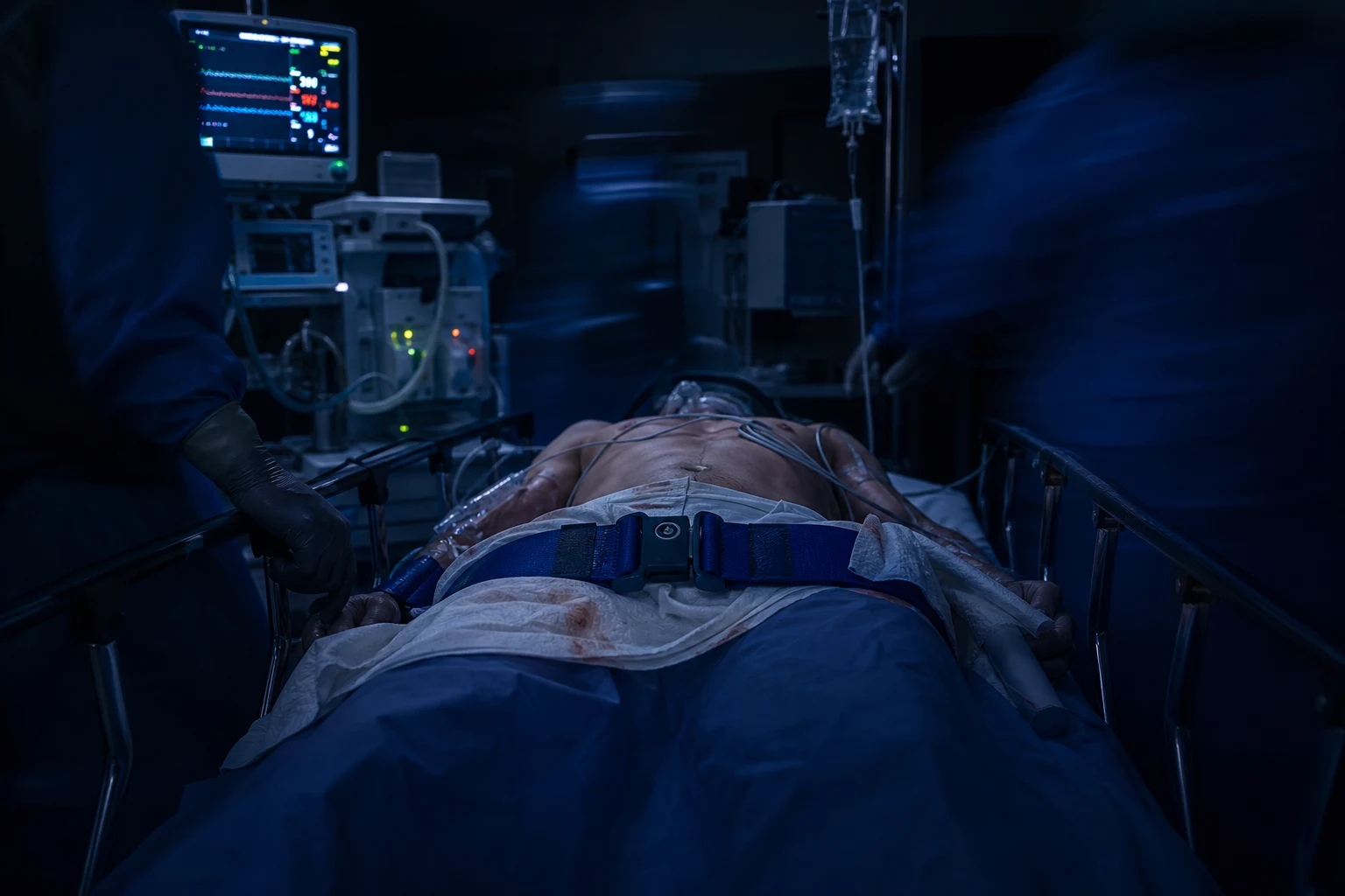
The mechanism and the classification
The pelvic ring fracture is produced by the lateral compression (the side-impact collision, the crush), the anteroposterior compression (the open-book, the frontal collision), or the vertical shear (the fall from a height, the dashboard impact). The Young and Burgess classification classifies the patterns by the force vector and predicts the bleeding and the mortality. The lateral compression (LC) is the most common pattern (around 60 per cent of the pelvic fractures) and is generally the most stable; it is caused by the side-impact and produces the internal rotation and the crescent fracture of the sacrum, but the higher grades (the LC2 and the LC3) with the iliac wing or the sacral crushing can still bleed. The anteroposterior compression (APC) — the open-book — widens the pelvic volume, opens the sacroiliac joints and the symphysis pubis, and carries the highest transfusion requirement and a mortality of 20 to 30 per cent. The vertical shear (VS) — the Malgaigne — shears the hemipelvis vertically through the sacroiliac joint and the rami and is unstable in all the planes, with a mortality around 25 per cent. The combined mechanism (CM) is the fourth pattern and is the most lethal because it mixes the vectors and disrupts the ring in every direction.[1]
The Tile classification (the equivalent of the AO/OTA) classifies the biomechanical stability for the surgeon: the Type A is the stable, the avulsion and the isolated ramus; the Type B is the rotationally unstable but the vertically stable (the open-book and the lateral compression); the Type C is the unstable in all the planes (the vertical shear and the combined) and requires the surgical fixation. The Tile type guides the orthopaedic fixation, and the Young-Burgess type guides the emergency resuscitation because it predicts the bleeding. The two classifications are complementary and both are asked in the Fellowship examination.[1]
Tile A (stable)
- The avulsion, the isolated ramus, the transverse sacral below S2
- Stable in all planes — no ring disruption
- No binder needed; the analgesia and the mobilisation
- AO/OTA 61-A
Tile B (rotationally unstable, vertically stable)
- The open-book (B1), the lateral compression (B2), the bilateral (B3)
- The posterior sacroiliac ligaments intact — the vertical stability preserved
- The binder and the external fixation reduce and stabilise
- AO/OTA 61-B; the highest-yield binder responders
Tile C (unstable in all planes)
- The vertical shear (C1), the bilateral (C2), the acetabular with the ring (C3)
- The complete disruption of the posterior and the sacroiliac complex
- Requires the surgical fixation; the highest mortality and transfusion
- AO/OTA 61-C; the angiographic and the packing candidates
LC (lateral compression)
- Around 60 per cent — the commonest pattern; the side-impact and the crush
- The internal rotation; the crescent sacral fracture; the rami overlap
- Generally stable (LC1); the LC2/LC3 bleed with the iliac wing or the sacral crush
- The binder less beneficial — may over-reduce
APC (open-book)
- The frontal collision, the crush from the front; the external rotation
- The symphysis and the sacroiliac widening; the increased pelvic volume
- The highest transfusion requirement; the mortality of 20 to 30 per cent
- The binder is the first-line and the most effective
VS (vertical shear)
- The fall from the height, the dashboard impact; the cranial displacement
- The hemipelvis sheared vertically through the SI joint and the rami
- Unstable in all planes (Tile C); the mortality around 25 per cent
- The binder plus the skeletal traction for the displaced hemipelvis
CM (combined mechanism)
- The mix of the vectors; the ring disrupted in every direction
- The most lethal pattern; the polytrauma and the head injury common
- Demands the immediate binder, the blood and the multidisciplinary escalation
- The highest mortality of all the Young-Burgess patterns
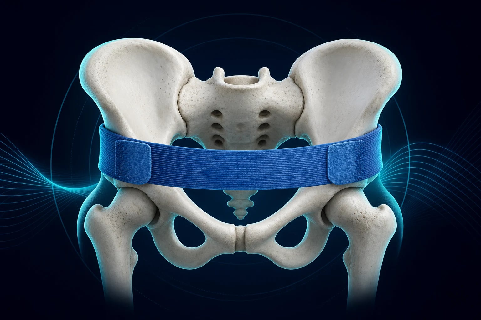
The pelvic binder
The pelvic binder is the circumferential sheet or the commercial binder (the T-POD, the SAM Sling) applied at the level of the greater trochanters (not the iliac crests), and it reduces the pelvic volume, reapproximates the fracture fragments and tamponades the venous bleeding. Applied correctly it can reduce the pelvic volume by up to a third. It is applied pre-hospital for the suspected unstable pelvic fracture and maintained in the emergency department; the binder is not removed until the pelvic stability is confirmed by the CT and the orthopaedic assessment, because the removal can release the tamponade and precipitate the rebleeding. The incorrect placement at the iliac crests fails to reduce the volume and may worsen the displacement; the binder that is too loose fails to tamponade; the binder left for more than 24 hours causes the pressure necrosis of the skin. The log-roll is avoided or minimised in the suspected pelvic fracture, because the rolling displaces the fragments and disrupts the clot. The binder is the first-line intervention for the APC and the vertical shear; in the pure lateral compression the binder is less beneficial and may theoretically over-reduce the internal rotation, but it is applied whenever the pattern is uncertain, because the cost of the missed unstable fracture is the death.[1][1]
Applying the pelvic binder — the technique and the traps
Select and position
Use the commercial binder (the T-POD, the SAM Sling) or the folded sheet. The patient is supine; the binder is slid under the patient on the flat slide (the log-roll is avoided). The correct level is the greater trochanters — the same level as the pubic symphysis — NOT the iliac crests, where the binder levers the pelvis open and worsens the displacement.
Apply and tighten
The binder is centred over the symphysis pubis and the sacrum, then tightened symmetrically. The commercial slings have a calibrated buckle that releases at the target tension (about 180 N); do not over-tighten past the buckle in the lateral compression, where the over-reduction is the risk. Confirm the position by palpating the greater trochanters under the binder.
Confirm the effect
The binder has worked if the leg-length discrepancy and the internal or external rotation improve, the pain eases, and the haemodynamics stabilise. The repeat springing of the pelvis (the spring test) is FORBIDDEN once the binder is on — the repeated displacement disrupts the clot and restarts the bleeding.
Decide when to remove
The binder stays on until the pelvic stability is confirmed by the CT and the orthopaedic review — typically 24 to 48 hours. Beyond 24 hours the pressure necrosis of the skin over the trochanters and the sacrum becomes the risk. The removal is a planned, multidisciplinary act in a controlled setting, never a reflex in the resus bay.<Cite id="1" /><Cite id="1" />
The FAST and the imaging
The FAST scan is often negative in the isolated pelvic fracture, because the bleeding is retroperitoneal, not intraperitoneal; a positive FAST in the pelvic fracture suggests the concomitant intraperitoneal injury (the splenic or the hepatic) and shifts the management toward the laparotomy. A negative FAST must never be used to exclude the pelvic bleeding. The plain pelvic radiograph in the trauma bay identifies the fracture pattern and the radiographic signs of the instability: the pubic symphysis diastasis greater than 2.5 cm, the sacroiliac joint widening greater than 1 cm, the avulsion of the ischial spine or the L5 transverse process (the sign of the sacroiliac ligament disruption), and the crescent fracture of the sacrum. The CT with the intravenous contrast (the CT angiogram) is the definitive imaging for the stable or the stabilised patient — it identifies the fracture pattern, the active contrast extravasation (the arterial bleeding, the "blush"), the volume of the pelvic haematoma, and the associated injuries of the bladder, the urethra and the sacrum. The CT cystography and the CT urethrogram are added when the bladder or the urethral injury is suspected.[1]
FAST scan
- Bedside, repeatable, performed in the trauma bay during the primary survey
- Detects the intraperitoneal fluid (the Morrison, the splenorenal, the pouch of Douglas, the pericardium)
- Negative in the isolated pelvic fracture — the blood is retroperitoneal
- Positive in the shocked patient — the indication for the laparotomy
Plain pelvic radiograph
- The single AP film in the trauma bay identifies the pattern and the instability
- The symphysis diastasis over 2.5 cm, the SI widening over 1 cm, the avulsion of the ischial spine or L5, the crescent fracture
- Does not quantify the bleeding; the LC1 can be subtle on the film
- The first imaging in the unstable patient who cannot go to the CT
CT angiogram
- The definitive imaging for the stable or the stabilised patient
- Defines the fracture, the haematoma volume, the contrast blush (the arterial bleeding)
- The CT cystogram and the urethrogram added when the bladder or the urethra is injured
- NOT for the patient in extremis — the CT kills the unstable patient who arrests in the scanner
The primary survey and the resuscitation
The pelvic fracture is managed within the ATLS primary survey. The airway with the cervical spine is secured; in the shocked, the agitated or the head-injured patient the early endotracheal intubation is performed with a haemodynamically stable induction such as the ketamine 1 to 2 mg/kg or the fentanyl 1 to 2 mcg/kg with the midazolam. The breathing is assessed for the tension pneumothorax, the haemothorax and the flail chest that coexist in the high-energy trauma. The circulation is the priority: two large-bore cannulae, the blood sent for the group and the crossmatch, the lactate and the base excess as the markers of the shock, and the early activation of the massive transfusion protocol with the balanced 1:1:1 ratio of the plasma to the platelets to the red cells, the tranexamic acid 1 g intravenous bolus over 10 minutes followed by the 1 g infusion over 8 hours within the first three hours, and the permissive hypotension to a systolic of 80 to 90 mmHg (or a MAP of 65) until the bleeding is controlled, avoided in the traumatic brain injury. The calcium gluconate 10 mL of the 10% solution is given after the massive transfusion to correct the citrate-induced hypocalcaemia, and the morphine 0.1 mg/kg for the analgesia. The disability and the exposure complete the survey, with the warmth and the pelvic binder in place.[2][1]
The management of the bleeding
The bleeding from the pelvic fracture is managed by a stepwise, multidisciplinary approach that pairs the emergency physician, the trauma surgeon, the orthopaedic surgeon and the interventional radiologist. First, the pelvic binder reduces the volume and tamponades the venous bleeding. Second, the massive transfusion protocol delivers the blood products in the 1:1:1 ratio, the tranexamic acid 1 g bolus then the 1 g infusion, the calcium gluconate 10 mL of the 10% solution, and the permissive hypotension at the systolic of 80 to 90 mmHg. Third, the angiographic embolisation is the definitive treatment for the arterial bleeding (the active extravasation, the "blush" on the CT angiogram), targeting the branches of the internal iliac artery; it is preferred when the patient stabilises after the binder. Fourth, the external fixation (the anterior pelvic frame) stabilises the rotationally unstable fracture and reduces the bleeding further. Fifth, the preperitoneal pelvic packing (the three packs on each side) is the salvage for the patient too unstable to reach the angiography suite, performed in the emergency department or the operating theatre, and it directly packs the retroperitoneum. Sixth, the REBOA (the resuscitative endovascular balloon occlusion of the aorta) in Zone 3 (the infrarenal aorta, below the renal arteries) is the modern adjunct that occludes the distal flow to the pelvis while the definitive haemostasis is achieved; it is the alternative to the resuscitative thoracotomy for the non-compressible truncal haemorrhage. The sequence is individualised: the patient who arrests or deteriorates despite the binder goes to the packing or the REBOA; the patient who stabilises goes to the angiography and the fixation.[1][2][1]
CRASH-2 (Shakur, Lancet 2010) — tranexamic acid in the trauma haemorrhage
Design
Multicentre, randomised, placebo-controlled — 20,211 trauma patients with the significant bleeding across 274 hospitals in 40 countries
Intervention
Tranexamic acid 1 g IV over 10 min then 1 g over 8 h vs placebo, within 8 h of injury
Primary result
All-cause mortality reduced (14.5% vs 16.0%, RR 0.91, p=0.0035); the bleeding death reduced (4.9% vs 5.7%); no increase in the vascular occlusive events
Timing
The benefit is greatest within the first hour; lost, with a signal of harm, after 3 h. Give early — the pre-hospital administration is now standard
Bottom line
A cheap, safe, mortality-reducing drug in the traumatic haemorrhage. The pelvic-fracture patient in the shock gets the TXA within 3 h, ideally pre-hospital.
PROPPR (Holcomb, JAMA 2015) — the 1:1:1 vs 1:1:2 ratio
Design
Multicentre randomised — 680 patients with the severe trauma and the major bleeding across 12 US level-1 trauma centres
Intervention
Plasma, platelets and red cells in a 1:1:1 ratio vs 1:1:2 ratio
Primary outcome
24-h and 30-d mortality: no significant difference (24-h 12.7% vs 17.0%, p=0.07)
Secondary
Fewer deaths from the exsanguination at 24 h with the 1:1:1 ratio; no increase in the complications
Bottom line
The balanced 1:1:1 ratio is the standard for the massive transfusion in the pelvic haemorrhage. The crystalloid-only resuscitation (the lethal triad) is the greater enemy.
MATTERs (Morrison, Arch Surg 2012) — TXA in the military combat trauma
Design
Retrospective cohort — 896 NATO soldiers with the combat injury requiring the transfusion at a Role 3 facility in Afghanistan
Intervention
Tranexamic acid 1 g IV bolus then 1 g infusion vs no TXA
Result
TXA reduced the unadjusted mortality (17.4% vs 23.9%) and the multivariate odds of death (OR 0.45); the benefit greatest in the massive-transfusion subgroup
Bottom line
The military data that corroborated CRASH-2 in the battlefield and embedded the TXA into the damage-control pathway of the high-energy pelvic trauma.
The bleeding algorithm — the stable vs the unstable vs the crashing patient
All patients — the binder, the blood, the TXA
Apply the pelvic binder at the greater trochanters on the suspicion of the unstable fracture. Activate the massive transfusion protocol, deliver the 1:1:1 ratio, the tranexamic acid within 3 hours, the calcium gluconate, and the permissive hypotension (unless the head injury). Take the trauma series — the chest and the pelvic radiograph, the FAST — and send the group, the crossmatch and the lactate.
The responder (the haemodynamics stabilise)
Proceed to the CT angiogram to define the fracture, the haematoma and the contrast blush. The blush sends the patient to the angiographic embolisation of the internal iliac branches. No blush but the unstable pattern sends the patient to the external fixation and the orthopaedic admission. The CT is the destination of the patient who stabilises.
The transient responder (the rise then the fall)
The patient who lifts with the blood then drifts down again is still bleeding. A positive FAST sends the patient to the laparotomy. A negative FAST with the pelvic pattern sends the patient to the preperitoneal pelvic packing (the three packs each side) and the external fixation, then the angiography if still bleeding. Avoid the CT in the transient responder — the scanner is where they arrest.
The non-responder and the crash
The patient who fails to lift with the blood and the binder, or who arrests, goes to the REBOA (Zone 3) and the preperitoneal packing in the operating theatre or the resus bay. The resuscitative thoracotomy with the aortic cross-clamp is the older alternative for the arrest. The angiographic embolisation is NOT for the patient in extremis — it is for the stabilised patient with the arterial blush.<Cite id="1" /><Cite id="2" />
Preperitoneal pelvic packing and external fixation — the salvage of the unstable
Preperitoneal pelvic packing (PPP)
A lower-midline laparotomy incision (or the Pfannenstiel) is made, the retroperitoneal space is opened and the three large laparotomy packs are placed on each side of the bladder, directly compressing the bleeding venous plexus and the bony surfaces. It is performed in the operating theatre or the resus bay for the patient too unstable for the angiography suite. The packs are removed at 24 to 48 hours; the re-bleed at the removal sends the patient to the angio.
Anterior pelvic external fixation
The external frame (the two pins in the iliac crests or the supra-acetabular pins, connected by the anterior bar) closes the open-book and stabilises the rotationally unstable fracture, reducing the volume further and tamponading the bleeding. Applied in the resus bay or the theatre, it is the complement to the binder for the Tile B and C.
Angiographic embolisation
The definitive treatment for the arterial bleed: the femoral access, the selective catheterisation of the internal iliac and its branches, the gelatin-sponge particles or the coils to occlude the bleeding vessel. Performed for the CT blush, or the continued bleed despite the binder and the packing. Bilateral internal iliac embolisation risks the gluteal or the bladder necrosis but is sometimes necessary.
The order — pack first, then angio, then fix
For the unstable patient the sequence is: binder, then packing plus the external fixation in the theatre, then the angiography if the bleeding persists. For the stabilised patient: binder, then the CT, then the angio for the blush, then the definitive fixation. The REBOA bridges the patient who cannot reach the theatre. The sequence is individualised — but the principle is constant: the venous bleed by the reduction, the arterial by the embolisation.<Cite id="1" /><Cite id="2" />
The associated injuries
The pelvic ring injury rarely occurs in isolation, and the search for the associated injuries is the second half of the assessment. The bladder rupture — the extraperitoneal from the bony fragment, or the intraperitoneal from the direct blow to the full bladder — presents with the gross haematuria or the inability to void and is assessed by the CT cystography; the extraperitoneal rupture is managed conservatively with the catheter, the intraperitoneal by the repair. The urethral injury — the blood at the meatus, the perineal or the scrotal haematoma, the high-riding or the non-palpable prostate on the rectal examination, and the difficulty voiding — is excluded by the retrograde urethrogram before the catheterisation; a urethral catheter is never forced through the suspected urethral injury, and the suprapubic catheter is placed instead. The rectal injury — the blood on the rectal examination, the boggy prostate and the loss of the sphincter tone — requires the diverting colostomy and the presacral drainage, because the faecal contamination of the pelvic haematoma is catastrophic. The vaginal laceration is sought by the speculum examination in the female, because the open fracture communicating with the vagina converts the fracture to the open pelvic fracture. The lumbosacral plexus injury and the sacral nerve root injury produce the lower-limb weakness, the saddle anaesthesia and the bladder and the bowel dysfunction, and are documented before the instrumentation. The open pelvic fracture — the communication with the perineum, the vagina or the rectum — carries a mortality of 30 to 50 per cent from the sepsis and the bleeding, and demands the immediate antibiotics with the gram-negative and the anaerobic cover (the piperacillin-tazobactam 4.5 g), the tetanus prophylaxis and the urgent debridement.[1]
[1]Bladder rupture
- The extraperitoneal (the bony fragment) is the commoner; the intraperitoneal (the direct blow to the full bladder) is the rarer
- The gross haematuria, the suprapubic pain, the inability to void
- Diagnosed by the CT cystography (the retrograde contrast)
- The extraperitoneal managed with the catheter; the intraperitoneal by the surgical repair
Urethral injury
- The posterior urethra (the membranous, above the urogenital diaphragm) with the pelvic fracture; the anterior with the straddle injury
- The blood at the meatus, the perineal and scrotal haematoma, the high-riding prostate, the difficulty voiding
- Diagnosed by the retrograde urethrogram BEFORE the catheter
- The partial tear with the gentle catheter or the suprapubic; the complete transection with the suprapubic and the delayed repair
Rectal injury
- The blood on the rectal examination; the boggy prostate; the lost tone
- The faecal contamination of the pelvic haematoma is catastrophic — the sepsis
- Mandates the diverting colostomy, the presacral drainage and the distal rectal washout
- The antibiotics with the gram-negative and the anaerobic cover (the piperacillin-tazobactam)
Open pelvic fracture
- The communication with the perineum, the vagina or the rectum
- The mortality of 30 to 50 per cent — the sepsis and the bleeding
- The immediate antibiotics, the tetanus, the debridement and the diversion
- The highest-risk pelvic injury — the multidisciplinary, the urgent, the intensive-care
Differential diagnosis
In the emergency department the pelvic trauma is approached from two angles: the differentiation of the fracture patterns, which predicts the bleeding and the stability, and the differentiation of the associated injuries that masquerade as, or accompany, the pelvic ring injury. [1]
- The stable lateral compression versus the unstable anteroposterior compression (the open-book) versus the vertical shear (the Malgaigne) versus the combined mechanism — the lateral compression presents with the internal rotation and the crescent sacral fracture and bleeds the least; the anteroposterior compression widens the symphysis and the sacroiliac joint and bleeds the most from the venous plexus and the internal iliac branches; the vertical shear displaces the hemipelvis cranially and is unstable in all the planes; the classification is made on the pelvic radiograph and the CT and it predicts the transfusion and the mortality.
- The bladder rupture versus the urethral injury — both present after the pelvic fracture with the haematuria and the inability to void, but the bladder rupture shows the gross haematuria and is diagnosed by the CT cystography, whereas the urethral injury shows the blood at the meatus, the perineal haematoma and the high-riding prostate and is diagnosed by the retrograde urethrogram; the urethral catheter is withheld until the urethra is cleared.
- The vaginal laceration versus the rectal injury versus the open pelvic fracture — the speculum examination finds the vaginal laceration that converts the fracture to the open, the rectal examination finds the blood that mandates the colostomy, and either finding elevates the mortality and demands the antibiotics and the surgical exploration; both are easily missed without the focused examination.
- The lumbosacral plexus and the sacral nerve root injury — the sacral plexus injury presents with the lower-limb weakness, the saddle anaesthesia and the bowel and the bladder dysfunction; it is differentiated from the simple fracture pain and the spinal cord injury by the focused neurological examination before the instrumentation, and it predicts the long-term disability.
- The non-pelvic sources of the shock — the tension pneumothorax, the haemothorax, the intra-abdominal bleeding (the splenic and the hepatic) and the femoral shaft fracture — must be sought and excluded, because the pelvic fracture patient is frequently the polytrauma patient, and the attribution of the shock to the pelvis alone misses the second life-threatening bleed; the FAST, the chest radiograph and the CT sort the sources. [1]
Common pitfalls
The recurring errors in the pelvic trauma are: applying the binder at the iliac crests rather than the greater trochanters, where it fails to reduce the volume; accepting the negative FAST as the exclusion of the pelvic bleeding, when the blood is the retroperitoneal; failing to activate the angiography and the massive transfusion early in the unstable pattern; forcing the urethral catheter through the suspected urethral injury instead of performing the retrograde urethrogram; not recognising the open pelvic fracture through the vagina or the rectum, which demands the antibiotics and the colostomy; under-resuscitating with the crystalloid instead of the blood products and the balanced ratio; and neglecting the sacral plexus and the nerve root examination before the instrumentation. [1]
The disposition
The haemodynamically unstable patient with the pelvic fracture is resuscitated in the trauma bay and transferred to the angiography suite, the operating theatre or the intensive care unit, depending on the bleeding source and the response to the binder. The stable patient with the CT-proven fracture is admitted for the analgesia, the fixation and the rehabilitation; the isolated stable pubic ramus fracture in the elderly may be managed conservatively with the early mobilisation and the venous thromboembolism prophylaxis. The open pelvic fracture, the bladder, the urethral and the rectal injuries need the multidisciplinary involvement of the orthopaedic, the urological and the general surgeons. The full recovery is measured in the months, and the long-term morbidity includes the chronic pain, the leg-length discrepancy, the neurological deficit and the sexual and the voiding dysfunction. [1]
Exam practice
SAQ — The open-book pelvic fracture with the haemorrhagic shock
10 minutes · 10 marks
A 28-year-old man is brought to the trauma bay 50 minutes after a head-on motor-vehicle collision at 80 km/h. He is GCS 15, BP 78/50, HR 132, RR 28, SpO2 96 per cent on 15 L oxygen. The pelvis is unstable and tender, with the widening of the symphysis on the palpation. There is blood at the urethral meatus. The FAST is negative in the Morrison pouch, the splenorenal and the suprapubic windows. The chest and the abdominal examinations are otherwise unremarkable. The haemoglobin is 68 g per litre and the lactate is 7.2 mmol per litre.
SAQ — The vertical shear pelvic fracture with the bladder injury
10 minutes · 10 marks
A 45-year-old window cleaner is brought to the trauma bay after a fall from the second-floor scaffolding (5 metres), landing on the feet then the buttocks. He is GCS 15, BP 102/68, HR 110, with the right-leg shortening and the right hemipelvis displaced cranially. The abdomen is tender in the suprapubic region and there is gross haematuria from the urethral meatus. The CT scan shows the vertical displacement of the right hemipelvis through the sacroiliac joint and the bladder contusion with the extravasation of the contrast.
Red flags
[1] [1]The examiner's mental map
The Fellowship candidate is expected to walk the examiner through the pelvic fracture as a time-critical, multidisciplinary problem with a clear sequence of decisions. The map below is the structure the examiner listens for — the five decisions that frame the answer. [1]
The five decisions of the pelvic fracture — the structure the examiner expects
Decision 1 — Is the pattern unstable? (the mechanism and the radiograph)
The high-energy mechanism (the motor-vehicle collision, the fall from the height, the crush), the perineal bruising, the blood at the meatus, the leg-length discrepancy. The pelvic radiograph: the symphysis over 2.5 cm, the SI over 1 cm, the vertical displacement. The Young-Burgess type predicts the bleeding; the Tile type predicts the fixation.
Decision 2 — Apply the binder and start the blood (the C of the primary survey)
The binder at the greater trochanters on the suspicion — do not wait for the radiograph if the mechanism is suggestive. The massive transfusion protocol, the 1:1:1 ratio, the TXA within 3 hours, the permissive hypotension (abandon if the head injury). Exclude the urethral injury before the catheter (the meatus, the prostate, the urethrogram).
Decision 3 — The FAST and the source of the shock (the abdomen or the pelvis?)
The FAST answers: is there the intraperitoneal bleed (the splenic, the hepatic)? A positive FAST in the shocked patient sends the patient to the laparotomy. A negative FAST does not exclude the pelvic bleed — it is expected. The chest radiograph and the long-bone exam exclude the other sources.
Decision 4 — The CT or the operating theatre (the responder vs the non-responder)
The responder and the stable go to the CT angiogram, the blush to the embolisation, the unstable pattern to the fixation. The transient responder goes to the preperitoneal packing and the external fixation. The non-responder goes to the REBOA, the packing and the resuscitative thoracotomy. The CT is NEVER the destination of the unstable patient.
Decision 5 — The associated injuries and the secondary survey
The bladder (the CT cystogram), the urethra (the retrograde urethrogram), the rectum (the colostomy if injured), the vagina (the open fracture), the sacral plexus (the examination before the instrumentation), the open fracture (the antibiotics, the tetanus, the debridement). The long leg of the pelvic fracture is the rehabilitation and the late morbidity — the chronic pain, the leg-length, the nerve deficit, the sexual and the voiding dysfunction.<Cite id="1" /><Cite id="2" /><Cite id="1" />
References
- [1]Cullinane DC, Schiller HJ, Zielinski MD, et al. Eastern Association for the Surgery of Trauma practice management guidelines for hemorrhage in pelvic fracture--update and systematic review J Trauma, 2011.PMID 22182895
- [2]Rossaint R, Bouillon B, Cerny V, et al. The European guideline on management of major bleeding and coagulopathy following trauma: sixth edition Crit Care, 2023.PMID 36859355
- [3]CRASH-2 trial collaborators, Shakur H, Roberts I, et al. Effects of tranexamic acid on death, vascular occlusive events, and blood transfusion in trauma patients with significant haemorrhage (CRASH-2): a randomised, placebo-controlled trial Lancet, 2010.PMID 20554319
- [4]Holcomb JB, Tilley BC, Baraniuk S, et al. Transfusion of plasma, platelets, and red blood cells in a 1:1:1 vs a 1:1:2 ratio and mortality in patients with severe trauma: the PROPPR randomized clinical trial JAMA, 2015.PMID 25647203
- [5]Morrison JJ, Dubose JJ, Rasmussen TE, Midwinter MJ. Military Application of Tranexamic Acid in Trauma Emergency Resuscitation (MATTERs) Study Arch Surg, 2012.PMID 22006852