EM · Primary survey
Primary survey
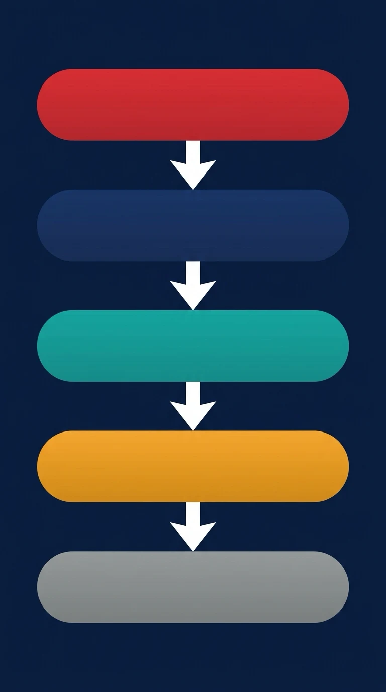
The ATLS primary survey — the structured, prioritised, 60-second assessment of the trauma patient using the C-A-B-C-D-E sequence: catastrophic haemorrhage control, airway with cervical-spine protection, the five immediately life-threatening chest injuries, the circulation with haemorrhage control, the disability, and the exposure, with the adjuncts, the transition to the secondary survey, and the trauma team coordination.
On this page & tools
Your progress
Saved locally on this device.
Target exams
Red flags
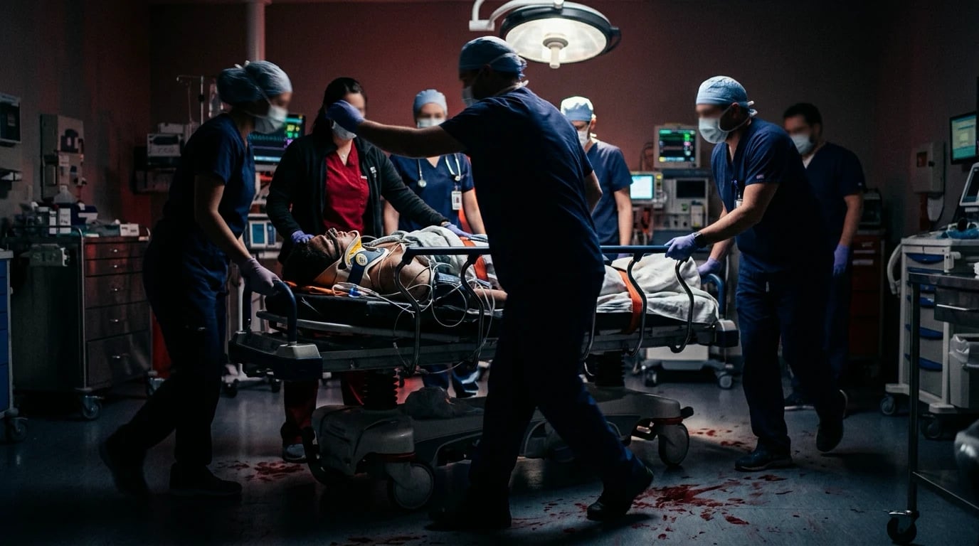
The primary survey is the structured, prioritised, 60-second assessment of the trauma patient that identifies and treats the immediately life-threatening conditions in the order that they kill. It is the Advanced Trauma Life Support framework, and it is built on a single principle: treat the greatest threat to life first, resuscitate as you go, and do not allow the history or the diagnostic uncertainty to delay the life-saving intervention. The sequence, evolved from the original A-B-C-D-E to the modern C-A-B-C-D-E (catastrophic haemorrhage, airway, breathing, circulation, disability, exposure), reflects the military and the civilian evidence that exsanguination from a limb can kill faster than a compromised airway. The Fellowship candidate must apply this survey reflexively, efficiently, and with the trauma team around them.[1][1]
C — Catastrophic haemorrhage control
The modern primary survey begins with the catastrophic haemorrhage step, a recognition born of the military experience that a bleeding limb can exsanguinate in the time it takes to secure the airway. The step is rapid: look for the source of the catastrophic external bleeding — the limb, the junctional zone, the scalp, the torso — and control it. The tourniquet, applied high and tight on the thigh or the upper arm, is the definitive intervention for the exsanguinating limb, and the evidence from the civilian and the military data is that the pre-hospital tourniquet saves lives.[2] The pelvic binder is applied for the suspected unstable pelvic fracture. The wound packing (with the gauze or the haemostatic agent) controls the junctional bleeding. The direct pressure controls the scalp and the smaller limb bleeding. The internal bleeding (the chest, the abdomen, the retroperitoneum) is addressed in the circulation step and the definitive imaging or the surgery.
A — Airway with cervical-spine protection
The airway is assessed for patency, for protection, and for the need for the definitive airway, all with the cervical spine protected. The trauma patient is assumed to have a cervical-spine injury until it is cleared, so the manual in-line stabilisation is maintained throughout the airway manoeuvres (the head tilt is replaced by the jaw thrust), and the patient is left in the hard collar and the head blocks. The airway is cleared (the suction, the finger sweep of the obstructing clot or the vomit), the basic adjuncts are placed (the oropharyngeal airway, the nasopharyngeal airway — the latter contraindicated in the suspected basal-skull fracture), and the oxygen is given. If the airway cannot be maintained or protected, or if the patient is comatose (a GCS of 8 or below), a definitive airway by rapid sequence intubation is placed, with the cervical spine maintained in-line and the difficult-airway equipment and the surgical airway ready.[1]
B — Breathing: the five immediately life-threatening chest injuries
The breathing assessment identifies and treats the five conditions that will kill the trauma patient in the minutes after the airway is secured.[1][1]
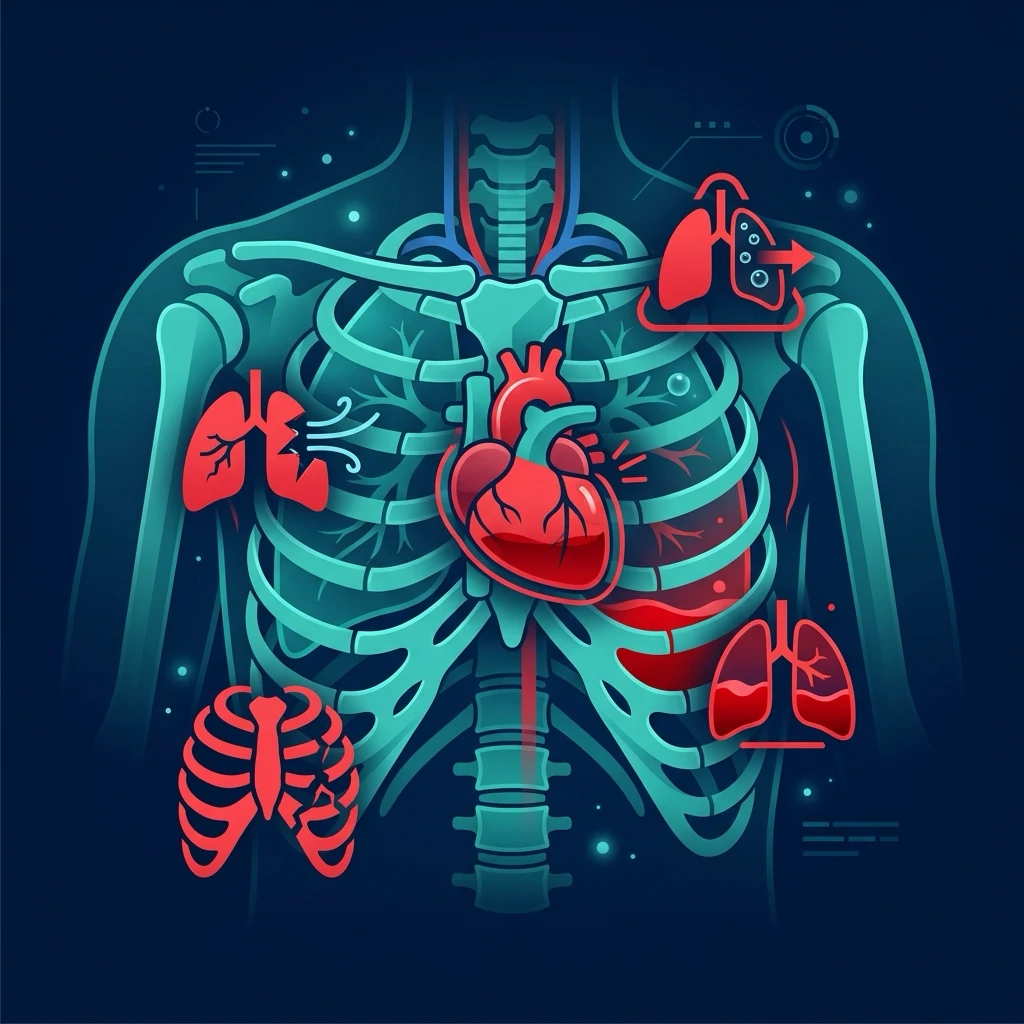
The tension pneumothorax is decompressed with the needle thoracocentesis and the chest drain. The massive haemothorax (more than 1500 mL or more than 200 mL per hour) is drained with the large-bore chest tube and resuscitated with the blood. The open pneumothorax (the sucking chest wound) is sealed with the three-sided dressing and the chest drain. The flail chest with the pulmonary contusion is managed with the analgesia, the oxygen, and the ventilatory support as needed. The cardiac tamponade is drained by the pericardiocentesis or the resuscitative thoracotomy. The respiratory rate, the oxygen saturation, the chest movement, the auscultation, the percussion and the tracheal position are assessed, and the life-threatening condition is treated at the point of its identification.[1]
C — Circulation with haemorrhage control
The circulation is assessed for the perfusion (the heart rate, the blood pressure, the capillary refill, the skin colour, the conscious level) and the source of the bleeding is identified — the external bleeding (controlled in the C step), the chest (the haemothorax, already identified), the abdomen (the FAST scan, the distension, the mechanism), the pelvis (the unstable fracture, the binder applied), and the long bones (the femoral fracture, the splint and the traction). The two large-bore intravenous cannulae are placed, the bloods are drawn including the crossmatch, the coagulation and the lactate, and the resuscitation is begun with the blood products in the balanced ratio (not the crystalloid) if the bleeding is significant, with the tranexamic acid and the permissive hypotension.[1][1] The shock is classified by the estimated blood loss (class I to IV), and the class III and IV shock is the indication for the massive transfusion protocol. The focused assessment with sonography in trauma (the FAST) is the key adjunct for the identification of the intra-abdominal bleeding in the unstable patient, and the pelvic and the chest radiographs are the standard adjuncts.
D — Disability
The disability is assessed with the Glasgow Coma Score, the pupil examination, the bedside glucose, and the lateralising or the spinal-cord signs. The GCS of 8 or below is the indication for the definitive airway, and a falling GCS during the resuscitation signals the intracranial haematoma or the rising intracranial pressure. The pupils are examined for the size, the symmetry and the reactivity (the unilateral fixed dilated pupil suggests the herniation). The glucose is checked for the hypoglycaemia. The alcohol and the drugs are considered as contributors, but the assumption in the trauma patient is that the altered consciousness is from the injury until proven otherwise.[1]
E — Exposure and environmental control
The patient is fully exposed (the clothes cut away, the full body examined) to identify the wounds, the entry and exit sites, the rashes and the drug patches, but the hypothermia is prevented by the warm blankets, the warmed fluids, and the high ambient temperature, because the lethal triad of hypothermia, acidosis and coagulopathy is the proximate cause of the irreversibility in the trauma patient. The log roll is performed to examine the back, the buttocks, the perineum and the spine.[1]
Adjuncts to the primary survey
The adjuncts run alongside the primary survey: the electrocardiographic monitoring, the pulse oximetry, the non-invasive blood pressure, the arterial blood gas, the urinary catheter (after the exclusion of the urethral injury), the gastric tube, the chest and the pelvic radiographs, and the FAST. The computed tomography is the definitive imaging for the stable patient (the whole-body trauma CT), but it follows, never precedes, the resuscitation of the unstable patient.[1][1]
The transition to the secondary survey
Once the primary survey is complete and the life-threatening conditions are treated and the patient is stable, the secondary survey is performed: the head-to-toe examination, the AMPLE history (the Allergies, the Medications, the Past history, the Last meal, the Events), the full neurological examination, the re-examination of the vital signs, and the complete imaging. The patient who remains unstable does not proceed to the secondary survey until they are stabilised or taken to the operating theatre, because the secondary survey is interrupted by the deterioration and the missed injuries of the unstable patient are the classic cause of the preventable death.[1]
The trauma team and the lethal triad
The primary survey is delivered by the trauma team with the allocated roles, the team leader, and the scribe, and the lethal triad of hypothermia, acidosis and coagulopathy is actively prevented throughout. The European guideline provides the evidence-based framework for the damage-control resuscitation that is integrated into the primary survey.[1][1][1]
Special populations
The paediatric trauma primary survey follows the same C-A-B-C-D-E, with the weight-based drug doses, the age-appropriate physiological ranges, and the awareness that the child maintains the blood pressure until late. The pregnant trauma patient is managed with the manual left uterine displacement, the early consideration of the perimortem caesarean, and the recognition that the mother's signs are attenuated by the physiological reserve of the pregnancy. The elderly trauma patient has the comorbidity, the anticoagulation and the reduced reserve that convert a minor mechanism into a major injury. [1]
Common pitfalls
The recurring errors are: not controlling the catastrophic haemorrhage before the airway; not protecting the cervical spine during the airway manoeuvres; missing the tension pneumothorax or the tamponade during the breathing assessment; resuscitating with crystalloid rather than blood; not applying the pelvic binder for the suspected pelvic fracture; sending the unstable patient to the computed tomography; not preventing the hypothermia; and proceeding to the secondary survey before the primary survey is complete and the patient is stable. [1]
Exam-exhaustive deep dive — C-A-B-C-D-E in detail
The Fellowship candidate is examined on the decision-making, not the slogan. Each letter of the survey is a discrete assessment with a defined set of findings, a defined threshold to act, and a defined intervention that must be performed before moving on. The sections below lay out each step at the depth the examiner expects: the technique, the indication, the dose, the timing, and the trap. [1]
A — Airway with cervical-spine protection (in depth)
The airway assessment answers three questions in order: Is it patent? Is it protected? Does this patient need a definitive airway? Every manoeuvre is performed with the cervical spine held in manual in-line stabilisation (MILS) — the hard collar is opened or removed for the airway manoeuvre, but an assistant holds the head in the neutral position throughout. The head is never tilted and the chin lift is avoided; the jaw thrust is the only airway-opening manoeuvre used in the suspected C-spine injury.[1]
[1]Basic adjuncts. Once the airway is cleared with suction (blood, vomit, broken teeth, foreign body), the basic adjuncts maintain patency. The oropharyngeal (Guedel) airway is sized from the angle of the mouth to the ear lobe (or the corner of the mouth to the angle of the jaw) and inserted upside-down then rotated 180 degrees in the adult; it is tolerated only in the unconscious patient (intact gag reflex = contraindication). The nasopharyngeal airway (NPA) is sized by length to the nares and lubricated; it is tolerated at lighter levels of consciousness and is the adjunct of choice for the semi-conscious trauma patient — but it has one absolute contraindication the examiner rewards. [1]
The definitive-airway decision in trauma — who is intubated, and how
Indication 1 — cannot maintain or protect the airway
Ongoing obstruction, pooling secretions, absent gag, or a GCS of 8 or below (the classic threshold). The patient who cannot protect the airway aspirates and dies; the definitive airway is an endotracheal tube.
Indication 2 — anticipated deterioration
The inhalation injury with facial burns (oedema will close the airway at 24-48 h), the neck haematoma, the sustained seizure, the severe traumatic brain injury. Intubate early before the airway is lost.
Indication 3 — ventilation or oxygenation failure
Refractory hypoxaemia, a failing respiratory effort, or the need for transport where the airway cannot be reassessed.
The technique — rapid sequence intubation with MILS
Pre-oxygenase with 100% oxygen (no bagging — the full stomach). Give a induction agent (ketamine 1-2 mg/kg or fentanyl/etomidate) and a fast-onset paralysing agent (suxamethonium 1.5 mg/kg or rocuronium 1 mg/kg). Apply cricoid pressure (debated, often released on difficulty). The assistant holds manual in-line stabilisation; the collar is opened. The most experienced operator performs the laryngoscopy; a video laryngoscope improves the view with MILS.
The failed airway — the surgical airway
If the endotracheal tube cannot be passed after the optimised attempt(s), do NOT persist — declare the failed airway and perform a surgical cricothyroidotomy. This is a cannae-intubate-cannot-oxygenate emergency. The scalpel-bougie technique: a transverse incision through the cricothyroid membrane, a bougie passed into the trachea, and a size 6 cuffed tube railroaded over the bougie.<Cite id="1" />
| Airway adjunct | Sizing | Consciousness required | C-spine safe | Key contraindication |
|---|---|---|---|---|
| Jaw thrust | Technique only | Any | Yes (no neck movement) | None — first manoeuvre |
| Oropharyngeal (Guedel) | Mouth angle to ear lobe | Unconscious (no gag) | Yes | Intact gag reflex — vomiting/vasovagal |
| Nasopharyngeal (NPA) | Length to nares (7–9 cm) | Semi-conscious tolerated | Yes | Suspected basal-skull / midface fracture |
| Supraglottic (i-gel/LMA) | Weight-based | Unconscious | Yes (rescue device) | Not a definitive airway; risk of aspiration |
| Endotracheal tube (RSI) | Cuffed, size-dependent | Unconscious | Yes with MILS | Cannot-intubate → surgical airway |
| Surgical cricothyroidotomy | Scalpel-bougie, size 6 tube | — | Yes (C-spine neutral) | Children under ~10 (needle cric preferred) |
B — Breathing: inspect, palpate, percuss, auscultate, and the five life-threatening chest injuries
The breathing assessment has two halves: the four-part examination of the chest (inspect, palpate, percuss, auscultate) and the active hunt for the five immediately life-threatening chest injuries — the conditions that kill the trauma patient in the minutes after the airway is secured and that are diagnosed clinically at the bedside, not by the chest X-ray.[1][1]
Inspect — respiratory rate, depth and pattern of breathing, chest-wall movement (asymmetry, paradoxical movement of a flail segment), the open wound, the tracheal position (deviation away from a tension, toward an collapse), the distended neck veins (tamponade, tension), the use of accessory muscles, and the colour (cyanosis, pallor). Palpate — chest-wall tenderness, crepitus (surgical emphysema), the flail segment, the subcutaneous air, the tracheal position, and the paradoxical movement. Percuss — hyper-resonance (pneumothorax), dullness (haemothorax). Auscultate — the absence of breath sounds (haemothorax, pneumothorax), the reduced air entry, and the added sounds. The chest X-ray and the extended FAST confirm but never replace the clinical assessment. [1]
Needle decompression and the chest drain for tension pneumothorax — the modern sites
Recognise the tension
Severe respiratory distress, hypoxia, shock, reduced air entry and hyper-resonance on one side. Tracheal deviation and distended neck veins are late and often absent in the shocked patient.
Immediate needle decompression
The ATLS 10th-edition preferred site is the 5th intercostal space, anterior to the mid-axillary line, just anterior to the latissimus dorsi (the same site as the chest drain) — this avoids the heart, the great vessels, and the thicker chest-wall muscle of the 2nd intercostal space. Use a large-bore cannula (14 or 16 gauge, at least 5 cm — the standard cannula is often too short for the muscular trauma patient). A rush of air confirms the diagnosis and relieves the tension.
Definitive chest drain
A needle decompression is a bridge, not a treatment — the definitive management is the intercostal chest drain (size 28–32 Fr in the adult). Insert at the 5th intercostal space, anterior axillary line, blunt dissection over the rib (NEVER under — the neurovascular bundle), connected to an underwater seal. Do not clamp a bubbling drain.<Cite id="1" />
The finger thoracostomy alternative
In the intubated, ventilated trauma patient in arrest or extremis, the finger thoracostomy (a rapid blunt incision into the pleural space, a finger sweeps to release the tension and the clotted haemothorax) is faster and more reliable than the needle, and is the technique of choice in the anaesthetised patient. The needle is for the non-intubated patient in whom a chest drain cannot be immediately placed.
The remaining four life-threatening injuries, each identified and treated in the breathing assessment: [1]
- Massive haemothorax — over 1500 mL of blood on the initial chest-tube drainage, or over 200 mL per hour for 2 to 4 hours. The chest is dull to percussion with reduced air entry; the patient is shocked. The treatment is the simultaneous chest drain (large-bore) and the resuscitation with blood products (the haemothorax is a volume problem as much as a lung problem). Do not autoclave or reinfuse.
- Open pneumothorax (sucking chest wound) — a chest-wall defect that is at least two-thirds the cross-sectional area of the trachea, through which air is drawn in on inspiration. Seal with a three-sided occlusive dressing (a square dressing taped on three sides, creating a flutter-valve that lets air escape but not enter), place an intercostal chest drain away from the wound, and plan the definitive surgical closure. A fully occlusive dressing converts the open pneumothorax into a tension pneumothorax — the three-sided dressing is the exam answer.
- Flail chest — three or more ribs fractured in two or more places (a segment free-floating), producing paradoxical inward movement on inspiration. The danger is the underlying pulmonary contusion (the capillary leak and the shunt) more than the mechanical segment. Management is the adequate analgesia (the regional block — the serratus anterior or the erector spinae plane block — is transformative), the high-flow oxygen, and the ventilatory support (non-invasive or invasive) for the failing respiratory effort.
- Cardiac tamponade — the Beck triad (hypotension, distended neck veins, muffled heart sounds) in the context of penetrating central chest trauma, with a pulsus paradoxus. The bedside FAST shows the pericardial effusion. The management is the pericardiocentesis (a temporising measure) or, in the arrest, the resuscitative thoracotomy with the relief of the tamponade and the repair of the cardiac laceration.[1]
| The five life-threatening chest injuries | Key clinical sign | Immediate treatment |
|---|---|---|
| Tension pneumothorax | Respiratory distress + unilateral hyper-resonance and reduced air entry | Needle decompression → chest drain |
| Massive haemothorax | Dullness + reduced air entry + shock (>1500 mL or >200 mL/h) | Large-bore chest drain + blood resuscitation |
| Open pneumothorax (sucking wound) | Visible chest-wall defect with audible air movement | Three-sided dressing → chest drain (separate site) |
| Flail chest / pulmonary contusion | Paradoxical segment + hypoxia worsening over 24–48 h | Analgesia (regional block) ± NIV/invasive ventilation |
| Cardiac tamponade | Beck triad (hypotension, JVD, muffled sounds), penetrating central chest | Pericardiocentesis / resuscitative thoracotomy |
C — Circulation with haemorrhage control (in depth)
The circulation assessment has two threads: the perfusion assessment (the heart rate, the blood pressure, the capillary refill, the skin colour and temperature, the conscious level) and the source-and-control assessment (the external bleeding, the chest, the abdomen, the pelvis, the long bones). The resuscitation runs in parallel — the access is secured, the bloods are drawn, and the fluids or the blood products are given — while the source is identified and controlled.[1][1]
Source identification. The five compartments that bleed in the trauma patient are: the external surface (controlled in the C step — direct pressure, tourniquet, pelvic binder, wound packing), the chest (the haemothorax — drained in the B step), the abdomen (the FAST scan, the distension, the mechanism — a seat-belt bruise, the handlebar injury), the pelvis (the unstable fracture — the binder applied), and the long bones (the femoral fracture — the splint and the traction reduce the volume of the pelvic-femoral compartment). The retroperitoneum and the great vessels are the hidden compartments; the mechanism (the high-energy deceleration, the fall from height) raises the index of suspicion. [1]
The massive haemorrhage protocol (MHP) — the balanced resuscitation
Activate with one call
The single call to the blood bank activates the protocol. The clinical trigger is the haemorrhagic shock, the major bleeding, the anticipated massive transfusion. Do not wait for the haemoglobin to fall — it is the last parameter to move.
Blood products in a 1:1:1 ratio
Packed red cells, fresh-frozen plasma, and platelets in a 1:1:1 ratio — the PROPPR trial showed no overall mortality difference between 1:1:1 and 1:1:2 but fewer deaths from exsanguination at 24 hours with the balanced ratio, and a trend to earlier death-avoidance. Give O-negative blood immediately if exsanguinating, then switch to group-specific then crossmatched.<Cite id="3" />
Tranexamic acid within 3 hours
TXA 1 gram intravenous bolus over 10 minutes, then 1 gram over 8 hours. The CRASH-2 trial showed a reduction in all-cause mortality; the timing analysis showed the benefit is greatest within the first hour and is lost (and may be harmful) after 3 hours. Give it early — the pre-hospital administration is now standard.<Cite id="4" /><Cite id="5" />
Calcium and the viscoelastic guidance
The citrate in the stored blood binds calcium — give calcium chloride 10% 10 mL (central line) or gluconate (peripheral) for every 4 units of blood. Use the ROTEM or TEG to guide the ratio in real time: a low fibrinogen (the FIBTEM A5 under 10) — give cryoprecipitate (fibrinogen target over 1.5 g/L); a low platelet count — give platelets (target over 50); a prolonged clotting — give FFP (INR target under 1.5).<Cite id="1" />
Damage-control surgery and angioembolisation
The resuscitation is a bridge to the definitive source control: the damage-control laparotomy (pack, control the contamination, restore the perfusion, close the abdomen) for the intra-abdominal bleed, and the angioembolisation for the pelvic and the solid-organ arterial bleeding. The patient who is not bleeding externally and is in shock has a bleeding compartment — find it and control it.
CRASH-2 (Shakur, Lancet 2010) — tranexamic acid in trauma haemorrhage
Design
Multicentre, randomised, placebo-controlled — 20,211 trauma patients with significant bleeding across 274 hospitals in 40 countries
Intervention
Tranexamic acid 1 g IV loading over 10 min then 1 g over 8 h vs placebo, within 8 h of injury
Primary result
All-cause mortality reduced (14.5% vs 16.0%, RR 0.91, p=0.0035); bleeding death reduced (4.9% vs 5.7%); NO increase in vascular occlusive events or thrombosis
Timing subanalysis
Benefit greatest if given within 1 h (bleeding death halved); benefit reduced at 1-3 h; possible harm if given after 3 h
Bottom line
TXA is a cheap, safe, mortality-reducing drug in trauma haemorrhage — give it within 3 h, ideally pre-hospital. The >3-h harm signal is the exam favourite.
PROPPR (Holcomb, NEJM 2015) — the 1:1:1 vs 1:1:2 ratio
Design
Multicentre randomised — 680 patients with severe trauma and major bleeding across 12 US level-1 trauma centres
Intervention
Plasma:platelets:red cells in a 1:1:1 ratio vs 1:1:2 ratio
Primary outcome
24-h and 30-d mortality: no significant difference (24-h 12.7% vs 17.0%, p=0.07; 30-d similar)
Key secondary
Fewer deaths from exsanguination at 24 h in the 1:1:1 group (9.2% vs 14.6%, p=0.03); no increase in complications
Bottom line
The balanced 1:1:1 ratio is the standard for the massive transfusion — it does not improve overall mortality but reduces death from bleeding, without adding harm. The harm from the crystalloid-only resuscitation (the lethal triad) is the greater enemy.
D — Disability (in depth)
The disability assessment is the rapid neurological evaluation that drives the airway decision and identifies the raised intracranial pressure. The four elements are the Glasgow Coma Score, the pupil examination, the bedside glucose, and the lateralising or the spinal-cord signs.[1]
E — Exposure and environmental control (in depth)
The patient is fully exposed (the clothes cut from the collar to the feet, the full body examined) to identify the wounds, the entry and exit sites, the seat-belt bruising, the rashes and the drug patches (the glyceryl-trinitrate patch, the fentanyl patch), the deformities, and the perineal and the back findings. But the hypothermia is prevented — the lethal triad of hypothermia, acidosis and coagulopathy is the proximate cause of the irreversibility, and the hypothermia worsens the coagulopathy (the clotting enzymes are temperature-dependent) and the acidosis.[1]
Catastrophic haemorrhage — the C step in detail
The C step is the hunt for the catastrophic external bleed and its immediate control, performed in under a minute. The order of the interventions is the direct pressure, then the tourniquet, then the wound packing, then the pelvic binder. The military and the civilian evidence converge: the pre-hospital tourniquet for the exsanguinating limb saves lives, and the earlier the application the greater the benefit.[2][6]
MATTERS / the tourniquet registry (Kragh, J Trauma 2014) — tourniquets save lives
Design
Retrospective analysis of the US military Combat Ready Counter-AFV Program and the MATTERS registry — the largest tourniquet dataset
Key finding
Pre-hospital tourniquet application in major limb trauma was associated with a survival benefit; the benefit was greatest when the tourniquet was applied before the onset of shock. No limbs were lost to the tourniquet alone.
Bottom line
The pre-hospital tourniquet is the standard of care for the catastrophic limb haemorrhage. The lesson translates to the civilian practice: apply early, apply high and tight, and document the time.
MATTERs study (Morrison, Arch Surg 2012) — TXA in military trauma
Design
Retrospective cohort — 896 military casualties with combat injury requiring transfusion, Afghanistan
Intervention
TXA vs no TXA in the bleeding military trauma patient
Key finding
TXA was associated with reduced mortality (17.4% vs 23.9%) and, in the subgroup requiring massive transfusion, a survival benefit was large (14.4% vs 55%). The benefit was independent of the transfusion.
Bottom line
Corroborated the CRASH-2 civilian data in the military setting and established TXA as a near-universal drug in the trauma resuscitation. Give early.
Adjuncts — the monitoring and the tubes
The primary-survey adjuncts — the timing and the order
Monitoring (immediate)
ECG, pulse oximetry, non-invasive blood pressure on the arrival, and the capnography as soon as the airway is secured (the end-tidal CO2 confirms the tube position and the adequacy of the ventilation). The arterial blood gas at 15 to 30 minutes (the lactate and the base excess are the markers of the adequacy of the resuscitation — a rising lactate is the subclinical shock).
Tubes (after the exclusion of the contraindications)
The urinary catheter is placed after the exclusion of the urethral injury (the blood at the meatus, the perineal bruising, the high-riding prostate, the scrotal haematoma — any of these = the urethrogram before the catheter, and the suprapubic if the urethrogram is positive). The gastric tube decompresses the stomach and reduces the aspiration risk; the orogastric route (not the nasogastric) is preferred in the suspected basal-skull fracture.
Radiographs (the trauma series)
The chest and the pelvic radiograph in the resus bay — the chest for the haemothorax, the pneumothorax, the widened mediastinum (the aortic injury), and the pelvic for the fractures that drive the binder and the angiography. The lateral cervical film is now largely replaced by the CT.
FAST and the CT
The FAST at the bedside for the intra-abdominal and the pericardial fluid. The whole-body trauma CT (the head-to-pelvis CT angiogram) is the definitive imaging for the stable patient — it follows, never precedes, the resuscitation of the unstable patient. The unstable patient goes to the operating theatre, not the CT scanner.<Cite id="1" />
Exam practice
SAQ — The C-A-B-C-D-E primary survey in the polytrauma patient
10 minutes · 10 marks
A 40-year-old unrestrained driver is brought to the trauma bay 35 minutes after a high-speed motor-vehicle collision. He was trapped for 15 minutes and extricated by the fire service. On arrival he is making gurgling sounds, cyanosed, with the flail segment of the right chest wall and the paradoxical movement, BP 80/55, HR 130, GCS 12, and there is active bleeding from an open right femur fracture with the visible bone. The right pupil is fixed and dilated. The paramedics report a brief loss of consciousness at the scene.
SAQ — The failed airway and the cannot-intubate-cannot-oxygenate in the trauma patient
10 minutes · 10 marks
A 55-year-old man is brought to the trauma bay after a fall from a horse, with the facial fractures, the swollen tongue and the blood in the oropharynx. The team attempts the rapid sequence intubation — the first attempt with the video laryngoscope fails (the Cormack-Lehane grade 4 view, the blood and the swelling), the second attempt with the bougie fails, and now the saturation has dropped to 72 per cent despite the bag-valve-mask ventilation, and the airway is increasingly difficult to maintain.
Red flags
The following features identify the trauma patient at immediate risk, in whom the primary survey is the priority and the definitive imaging follows: [1]
[1]References
- [1]Rossaint R, Bouillon B, Cerny V, et al. The European guideline on management of major bleeding and coagulopathy following trauma: sixth edition Crit Care, 2023.PMID 36859355
- [2]Scerbo MH, Mumm JP, Lundy JB, et al. The trauma center is too late: Major limb trauma without a pre-hospital tourniquet has increased death from hemorrhagic shock J Trauma Acute Care Surg, 2017.PMID 29190257
- [3]Holcomb JB, Tilley BC, Baraniuk S, et al. Modulation of ochratoxin A induced DNA-damage in urothelial cell cultures Mycotoxin Res, 2005.PMID 23605211
- [4]CRASH-2 trial collaborators, Shakur H, Roberts I, et al. Endoscopic assisted laparoscopic removal of a gastric foreign body Surgeon, 2010.PMID 20569944
- [5]CRASH-2 collaborators, Roberts I, Shakur H, et al. The effects of antidepressants on human brain as detected by imaging studies. Focus on major depression Prog Neuropsychopharmacol Biol Psychiatry, 2011.PMID 21138750
- [6]Kragh JF Jr, Walters TJ, Baer DG, et al. Prenatal nicotinic exposure suppresses fetal adrenal steroidogenesis via steroidogenic factor 1 (SF-1) deacetylation Toxicol Appl Pharmacol, 2014.PMID 24709674