EM · Trauma in pregnancy
Trauma in pregnancy
The trauma in pregnancy: the physiological changes that alter the response to the injury, the primary survey modifications (the left lateral tilt and the foetal assessment), the placental abruption and the uterine rupture, the perimortem caesarean at 4 minutes, the foeto-maternal haemorrhage and the Anti-D, the radiation exposure and the safe imaging, and the seatbelt advice.
On this page & tools
Your progress
Saved locally on this device.
Target exams
Red flags
The trauma in pregnancy is the management of two patients simultaneously — the mother and the foetus — and the Fellowship candidate must know the physiological changes of the pregnancy that alter the response to the injury, the modifications to the primary survey, the perimortem caesarean, the foetal complications (the abruption and the uterine rupture), and the foeto-maternal haemorrhage with the Anti-D prophylaxis. The principles of the trauma resuscitation are unchanged; the modifications are the adaptations that protect the foetus and address the altered physiology.[1]

The physiological changes of pregnancy
The pregnancy alters the physiology in ways that directly affect the trauma management.[1] The blood volume increases by 40 to 50 per cent (so the pregnant patient loses more blood before showing the signs of the shock — the normal maternal blood pressure does not exclude the 1500 millilitres of the blood loss). The heart rate rises by 15 to 20 beats per minute. The blood pressure falls by 5 to 15 mmHg in the second trimester (the progesterone-mediated vasodilation). The clotting factors increase (the pregnancy is a hypercoagulable state — the factor VII, the factor VIII, the fibrinogen, the von Willebrand factor — the VTE risk is 4 to 5 times the non-pregnant). The functional residual capacity falls and the oxygen consumption rises (so the pregnant patient desaturates rapidly on the apnoea). The enlarged uterus (above 20 weeks) compresses the inferior vena cava and the aorta in the supine position (the aortocaval compression), reducing the venous return and the cardiac output by up to 30 per cent.
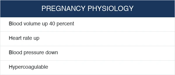
The cardiovascular system — the expanded but diluted volume
The cardiovascular adaptation of the pregnancy is the expanded blood volume with the relatively reduced red-cell mass — the dilutional anaemia of the pregnancy (the haemoglobin falls by around 20 grams per litre, to a physiologically normal of 105 to 110 by the third trimester). The plasma volume rises more than the red-cell mass (the plasma rises by 50 per cent, the red cells by 20 to 30 per cent), so the pregnant patient is both better protected against the acute haemorrhage (the larger volume to lose) and worse at carrying oxygen (the lower haemoglobin, the lower reserve before the tissue hypoxia). The cardiac output rises by 30 to 50 per cent by the late second trimester (the stroke volume and the heart rate both rise), and the systemic vascular resistance falls (the progesterone-mediated vasodilation, the low-resistance uteroplacental circulation).[1]
The net effect on the trauma management is the deceptively well-looking patient: the pregnant woman compensates through the tachycardia, the vasoconstriction and the expanded volume until 30 to 35 per cent of the circulating volume is lost, and the hypotension is the late, pre-terminal sign. The shock index (the heart rate divided by the systolic blood pressure) is more sensitive than either value alone, and a shock index above 0.9 to 1.0 in the pregnant trauma patient (where the baseline is 0.7 to 0.8) is the early warning of the occult blood loss. [1]
1st trimester
- Blood volume rising; modest dilutional anaemia begins
- HR +5 to 10 bpm; BP near normal
- Uterus a pelvic organ — protected by the bony pelvis
- Aortocaval compression NOT yet significant
- Penetrating trauma injures the maternal viscera as in the non-pregnant
2nd trimester
- Blood volume near peak; dilutional anaemia established (Hb ~105 to 110)
- HR +15 to 20 bpm; SBP 5 to 15 mmHg below baseline
- Uterus rises out of the pelvis — exposed to the direct blow
- Aortocaval compression begins at 20 weeks (uterus at umbilicus)
- Fetal viability from 23 to 24 weeks — perimortem caesarean considered
3rd trimester
- Blood volume at peak (40 to 50% above non-pregnant); maximum dilution
- HR +15 to 20 bpm; SBP 10 to 15 mmHg low, rises slightly toward term
- Uterus fills the abdomen — large target, displaces viscera
- Full aortocaval compression in supine — the supine hypotensive syndrome
- PMCS for any arrest; the fetus at the gestational age of the survival
The respiratory system — the reduced reserve and the rapid desaturation
The functional residual capacity falls by 20 per cent by the third trimester (the gravid uterus elevates the diaphragm), the oxygen consumption rises by 20 to 30 per cent (the metabolic demand of the fetus and the maternal organs), and the closing volume encroaches on the functional residual capacity (the small airways close during the tidal breathing). The net effect is the rapid, precipitous desaturation on the apnoea — the pregnant trauma patient desaturates within 60 to 90 seconds of the apnoea (versus 5 to 8 minutes in the non-pregnant), and the pre-oxygenation (the three minutes of the high-flow oxygen, or the eight vital-capacity breaths of the 100 per cent oxygen) is the non-negotiable before the rapid sequence induction. The minute ventilation rises (the progesterone-driven hyperventilation), the PaCO2 falls to 30 to 32 mmHg (the respiratory alkalosis of the pregnancy — the renal compensation brings the bicarbonate to 18 to 21), and the PaCO2 of 35 in the pregnant patient is the rising CO2, the fatigue, and the impending respiratory failure.[1]
The difficult airway of the pregnancy adds to the risk: the laryngeal and the pharyngeal oedema, the enlarged and friable mucosa (the easy bleeding on the laryngoscopy), the enlarged breasts (the difficult laryngoscopy and the difficult bag-mask), the reduced FRC (the reduced apnoeic tolerance), and the increased aspiration risk (the relaxed lower oesophageal sphincter from the progesterone, the delayed gastric emptying, the raised intra-abdominal pressure). The pregnant airway is the difficult airway, the aspiration prophylaxis (the ranitidine and the sodium citrate before the anaesthesia) and the rapid sequence induction are the standard, and the senior airway operator is summoned early. [1]
[1] [1]The gastrointestinal system and the aspiration risk
The progesterone relaxes the lower oesophageal sphincter (the reflux is the near-universal complaint of the pregnancy), the gravid uterus displaces and compresses the stomach (the raised intra-gastric pressure), and the gastric emptying is delayed (the opioid analgesia and the pain of the trauma worsen it). The combination makes the pregnant trauma patient the high aspiration risk — the "full stomach" applies even in the absence of the recent food intake. The rapid sequence induction with the pre-oxygenation, the cricoid pressure (the debated but the standard in many units), and the cuffed endotracheal tube are the airway protection, and the aspiration prophylaxis (the sodium citrate 30 mL orally, the ranitidine 50 mg IV, the metoclopramide 10 mg IV) is given before the anaesthesia where the time allows. [1]
[1]The haematological system — the hypercoagulable state
The pregnancy is the hypercoagulable state: the factor VII, the factor VIII, the factor X, the fibrinogen and the von Willebrand factor all rise, the protein S falls, and the acquired activated protein C resistance develops. The fibrinogen at the term is 4 to 6 grams per litre (versus 2 to 4 in the non-pregnant), and the normal fibrinogen in the bleeding pregnant patient is the abnormal finding — a fibrinogen below 4 g/L in the bleeding third-trimester patient is the marker of the consumptive coagulopathy (the abruption, the amniotic fluid embolism, the massive haemorrhage). The VTE risk is 4 to 5 times the non-pregnant and rises further in the immobility, the surgery and the trauma — the early mechanical and the pharmacological thromboprophylaxis (once the bleeding is controlled) is the non-negotiable.[3]
[1]The primary survey modifications
The primary survey follows the C-A-B-C-D-E sequence with the pregnancy-specific modifications. The catastrophic haemorrhage step is unchanged. The airway is managed normally, with the awareness that the pregnant patient desaturates rapidly and the intubation is more difficult (the laryngeal oedema, the enlarged breasts, the reduced FRC). The breathing is managed with the higher oxygen flow and the earlier ventilatory support. The circulation is the key modification: the patient is placed in the left lateral tilt of 15 to 30 degrees or the manual left uterine displacement (pushing the uterus to the left) from the first minute, to relieve the aortocaval compression and restore the venous return. The fluids and the blood products are given with the awareness that the expanded blood volume masks the loss. The disability is assessed normally. The exposure includes the vaginal examination (the bleeding, the rupture of the membranes, the cervical dilation) and the abdominal examination (the uterine tenderness, the contractions, the foetal heart rate after 20 weeks).[1][1]
The aortocaval compression and the left lateral tilt
The gravid uterus, from 20 weeks gestation, compresses the inferior vena cava and the aorta against the lumbar spine when the patient is supine — the supine hypotensive syndrome of the pregnancy. The venous return to the heart falls, the cardiac output falls by 10 to 30 per cent, the uteroplacental perfusion falls (the fetal distress), and the lower-body venous pressure rises (the engorgement of the epidural veins, the leg veins). The aortic compression, less discussed but equally important, reduces the perfusion of the lower limbs and worsens the distal ischaemia. The compression is relieved by the left lateral tilt of 15 to 30 degrees (the wedge or the tilted spine board) or the manual left uterine displacement (the operator pushes the uterus to the left with both hands), which shifts the gravid uterus off the great vessels and restores the venous return.[1]
The manual left uterine displacement is the preferred technique in the trauma and the cardiac arrest: it is the hands-only manoeuvre that does not interfere with the CPR (the chest compressions are delivered on the flat, firm surface), it does not move the spine (the cervical-spine protection is maintained), and it is applied from the first minute of the resuscitation. The lateral tilt is the alternative for the supine patient on the trolley (the wedge under the right buttock), but the tilt impairs the CPR effectiveness (the patient rolls during the compressions) and the manual displacement is the better technique in the arrest. [1]
[1] [1]The modified primary survey in the pregnant trauma patient
C — Catastrophic haemorrhage
Identify and control the external catastrophic bleeding with the direct pressure, the tourniquet for the limbs, and the pelvic binder for the pelvic fracture. Two large-bore (14 to 16 G) intravenous cannulae, bloods sent including the blood group and save, the Kleihauer-Betke, the beta-hCG and the coagulation. Activate the massive transfusion protocol early if the shock is present.
A — Airway with the rapid sequence induction
Assess and secure the airway. The pregnant patient is the difficult airway and the rapid desaturator. Pre-oxygenate for three minutes (or eight vital-capacity breaths), prepare the videolaryngoscope, the bougie, the supraglottic airway and the senior operator. Perform the rapid sequence induction with the cricoid pressure and the cuffed tube. The aspiration prophylaxis (sodium citrate, ranitidine) where the time allows.
B — Breathing with the higher oxygen flow
Apply the high-flow oxygen (15 L/min via the non-rebreather) to all the pregnant trauma patients until the hypoxia and the hypovolaemia are excluded — the fetus benefits from the maternal hyperoxia. Exclude and decompress the tension pneumothorax. Ventilate to the pregnant baseline PaCO2 of 30 to 32 mmHg (avoid the permissive hypercapnia in the raised intracranial pressure). Assess the respiratory rate against the pregnant baseline of 18 to 24.
C — Circulation with the tilt and the access
Apply the left lateral tilt or the manual left uterine displacement from the first minute (from 20 weeks). Two large-bore cannulae, the warmed balanced crystalloid and the early blood products. The permissive hypotension is modified (the SBP target above 90 mmHg to protect the uteroplacental perfusion). The tranexamic acid within three hours if the bleeding is significant. The vasopressors are the adjunct, not the substitute for the volume.
D — Disability with the AVPU and the glucose
Assess the GCS, the pupils, the blood glucose and the temperature. The altered consciousness may reflect the hypoxia, the hypovolaemia, the eclampsia, the substance use or the head injury. The glucose is checked — the hypoglycaemia is the rapidly reversible cause. The pregnant patient with the reduced GCS is intubated for the protection and the imaging.
E — Exposure and the foetal assessment
Fully expose and examine, including the abdominal and the vaginal examination (the speculum for the bleeding, the leaking liquor, the cervical dilation). Apply the continuous CTG monitoring from the arrival. Keep the patient warm (the hypothermia worsens the coagulopathy). Document the fundal height, the presence of the contractions, the foetal movements and the foetal heart rate. The obstetric review from the first minute.
The perimortem caesarean
The perimortem caesarean (the resuscitative hysterotomy) is performed at 4 minutes of the cardiac arrest in the pregnant patient with the gestation over 20 weeks (the uterus at the level of the umbilicus). The procedure is performed in the resus bay, through a midline vertical incision from the umbilicus to the symphysis pubis, without the anaesthesia (the patient is in the arrest), and the baby is delivered, the cord is clamped, and the placenta is removed. The rationale is the maternal, not just the foetal, survival: the emptying of the uterus relieves the aortocaval compression, restores the venous return and the cardiac output, and improves the effectiveness of the CPR — the maternal survival improves significantly after the perimortem caesarean. The baby survival is best when the delivery occurs within 5 minutes of the arrest.[1]
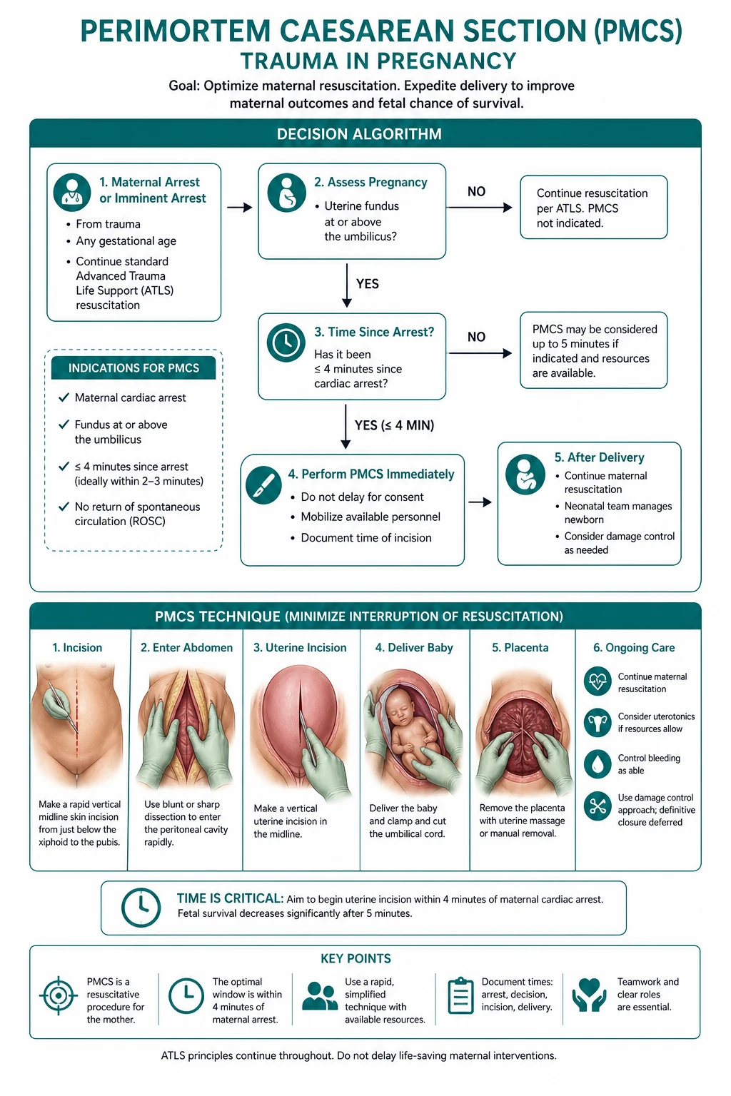
The timing — the four-minute rule
The American Heart Association's 2015 scientific statement on the cardiac arrest in pregnancy set the operational framework: the chest compressions begin immediately, the resuscitative hysterotomy is begun at 4 minutes of the arrest (the start of the incision, not the delivery), and the delivery is completed by 5 minutes, in the patient with the gestation over 20 weeks (the uterus at or above the umbilicus).[6] The rationale for the 4-minute start is the realistic observation that the preparation (the scalpel, the incision, the delivery, the cord clamp) consumes 1 minute, so the start at 4 minutes achieves the delivery at 5 minutes. The 5-minute delivery is the threshold of the improved neurological outcome for the fetus, and the early emptying of the uterus is the maternal benefit — the cardiac output after the relief of the aortocaval compression rises by 10 to 30 per cent, and the maternal ROSC is more likely after the perimortem caesarean.[6][7]
The historical 5-minute rule (the Katz framework from 1986, refined by the modern guidance) is the operational target, and the reality of the maternal cardiac arrest in the literature (the Get with the Guidelines cohort of over 500 arrests) is that the perimortem caesarean is performed in fewer than a third of the eligible cases, and the median time to the delivery is well beyond 5 minutes.[7] The team that prepares early — the call for the PMCS at the start of the arrest, the obstetric and the neonatal teams summoned, the equipment prepared — is the team that meets the 4-minute target. The delay is the most common failure.
Under 20 weeks
- Uterus below the umbilicus — no significant aortocaval compression
- PMCS NOT indicated — the uterus is too small to benefit the maternal haemodynamics
- The CPR and the standard resuscitation without the PMCS
- The fetal survival at this gestation is negligible — focus on the mother
20 to 23 weeks
- Uterus at the umbilicus — aortocaval compression significant
- PMCS for the MATERNAL benefit — the empty uterus restores the venous return
- Fetal survival unlikely but the procedure is justified by the maternal indication
- The team proceeds for the maternal reason, the fetal survival is the bonus
24 weeks and over
- PMCS for BOTH the maternal and the fetal benefit
- Fetal survival at 24+ weeks is the gestational age of the viability
- Begin at 4 minutes, deliver by 5 minutes — the team prepares from the start of the arrest
- Neonatal team present for the resuscitation of the newborn
The resuscitative hysterotomy — the procedure
Decide and announce
At the start of the arrest, the team leader announces the PMCS plan: gestation 20+ weeks, begin at 4 minutes, deliver by 5 minutes. The obstetric and the neonatal teams are summoned. The clock is started at the first compression. The decision is not delayed for the ultrasound — the fundal height at the umbilicus is the sufficient landmark.
Prepare the equipment
The number 10 or 22 scalpel, the blunt dissection scissors, the retractors, the cord clamp, the bowel clamps, the suction, the swabs. The equipment is gathered by the nurse while the compressions continue. The anaesthetic is not required (the patient is in the arrest) but the procedural analgesia is given if the ROSC occurs during the procedure.
Incise at 4 minutes
The midline vertical incision from the umbilicus to the symphysis pubis, through the skin, the subcutaneous fat, the rectus sheath and the peritoneum. The bladder is retracted inferiorly (the Foley catheter decompresses it). The vertical incision is faster than the Pfannenstiel and gives the better access, and it is the standard in the resuscitative setting.
Open the uterus and deliver
A vertical incision through the uterine wall, avoiding the placenta if it is anterior and visible. The baby is delivered, the cord is clamped and cut, and the baby is handed to the neonatal team. The placenta is delivered or manually removed, and the uterus is emptied — the relief of the aortocaval compression is the maternal goal.
Continue the resuscitation
The compressions continue throughout and after the delivery. The uterine atony is anticipated (the oxytocin 10 IU IM, the bimanual uterine massage, the uterotonic infusion). The bleeding from the uterine incision is controlled with the running suture or the uterine packing. The ROSC is the goal, and the post-arrest care (the targeted temperature, the perfusion, the ICU) follows the standard.
The foetal assessment and the CTG monitoring
The foetal assessment is the parallel to the maternal resuscitation — the continuous cardiotocography (the CTG) is applied from the arrival in every pregnant trauma patient over 20 weeks, and the monitoring is maintained for a minimum of 4 hours after the injury (the longer for the significant mechanism or the concerning features).[1] The CTG is the most sensitive indicator of the foetal distress, and the foetal heart-rate abnormalities often precede the maternal haemodynamic decompensation — the foetus is the canary in the maternal haemodynamic coal mine. The abnormal CTG triggers the obstetric review, the ultrasound for the foetal viability and the biophysical profile, and the consideration of the emergency delivery.
The indications for the minimum 4 to 6 hours of the continuous CTG monitoring after the trauma are: any abdominal pain or the uterine tenderness, the vaginal bleeding, the uterine contractions, the significant mechanism (the high-speed collision, the fall from the height, the pedestrian struck), the ejection from the vehicle, the maternal tachycardia or hypotension, and the maternal Rh-negative status with the suspected foeto-maternal haemorrhage. The patient with the concerning CTG (the recurrent decelerations, the reduced variability, the foetal tachycardia or bradycardia) is monitored longer and referred for the obstetric assessment. [1]
The foetal assessment after the maternal trauma
Apply the CTG from the arrival
All the pregnant trauma patients at 20+ weeks have the continuous CTG applied in the resus bay, alongside the maternal monitoring. The CTG is run for a minimum of 20 minutes; the foetal heart rate, the variability and the presence or absence of the decelerations are documented. The maternal vital signs are correlated with the CTG — the foetal distress may be the first sign of the maternal hypovolaemia.
Observe for the minimum 4 to 6 hours
The patient with the significant mechanism or any concerning feature is observed for a minimum of 4 to 6 hours of the continuous CTG. The discharged patient must have a normal CTG, no abdominal pain, no vaginal bleeding, no uterine contractions and no concerning mechanism. The patient with the persistent abnormalities is admitted for the longer monitoring.
Assess the Kleihauer-Betke in all the Rh-negative patients
The Kleihauer-Betke test (or the flow cytometry) is sent in every Rh-negative pregnant trauma patient to quantify the foeto-maternal haemorrhage. The Anti-D is given within 72 hours, and an additional dose is given if the Kleihauer-Betke shows a large bleed (over 4 mL of the foetal cells).
Perform the obstetric ultrasound
The bedside ultrasound (the FAST extended to the pelvis) assesses the foetal cardiac activity, the foetal movement, the amniotic fluid volume and the placental location. The formal obstetric ultrasound is performed for the detailed biophysical profile if the CTG is concerning or the monitoring is prolonged.
Escalate the abnormal CTG
The recurrent decelerations, the reduced variability, the bradycardia (below 110) or the tachycardia (above 160) escalate the urgency. The obstetric team is called, the maternal resuscitation is optimised (the oxygen, the volume, the left lateral tilt), and the decision for the emergency caesarean is made if the foetal distress persists despite the optimised maternal state.
The placental abruption
The placental abruption is the most common significant complication of the trauma in pregnancy (the shearing of the placenta from the uterine wall by the deceleration or the direct blow).[1][2] It presents with the abdominal pain, the uterine tenderness, the uterine contractions, the vaginal bleeding (though the bleeding may be concealed behind the placenta), and the foetal distress (the abnormal foetal heart rate pattern, the foetal death). The abruption can present up to 24 hours after the minor trauma, so the patient with the significant mechanism is observed with the continuous foetal monitoring for 4 to 24 hours. The management is the obstetric referral, the foetal monitoring, the resuscitation, and the delivery if the abruption is significant.
The Couvelaire uterus and the consumptive coagulopathy
The concealed abruption is the dangerous variant: the blood is trapped behind the placenta, the vaginal bleeding is minimal or absent despite the massive retroplacental clot, and the diagnosis is missed if the team waits for the external bleeding. The concealed abruption drives the consumptive coagulopathy (the disseminated intravascular coagulation from the release of the thromboplastin and the consumption of the fibrinogen and the platelets), and the Couvelaire uterus — the blood extravasates into the myometrium and the broad ligament, producing the purple, tense, non-contractile uterus that fails to respond to the oxytocin after the delivery. The fibrinogen below 4 g/L in the bleeding third-trimester patient is the marker of the abruption-driven coagulopathy, and the cryoprecipitate and the fresh-frozen plasma are the targeted replacement. [1]
[1] [1]The uterine rupture
The uterine rupture is the catastrophic complication of the major trauma (the high-speed collision, the direct blow to the abdomen), more common in the previously-scarred uterus (the previous caesarean). It presents with the severe abdominal pain, the loss of the foetal station (the foetal parts felt through the abdominal wall), the shock, and the foetal death. The management is the emergency laparotomy.[1][1]
The foeto-maternal haemorrhage and the Anti-D
The trauma can cause the foetal blood to enter the maternal circulation (the foeto-maternal haemorrhage), which sensitises the Rh-negative mother and produces the haemolytic disease of the newborn in the future pregnancies. The Kleihauer-Betke test (or the flow cytometry) quantifies the foetal cells in the maternal blood. The Anti-D immunoglobulin is given to the Rh-negative mother within 72 hours of the trauma to prevent the sensitisation, at the standard dose (500 IU for the first trimester, 1250 IU for the second or third), with an additional dose if the Kleihauer-Betke shows a large bleed.[1]
The Kleihauer-Betke test and the dose calculation
The Kleihauer-Betke acid-elution test relies on the resistance of the foetal haemoglobin (the HbF) to the acid elution — the maternal adult haemoglobin (the HbA) is eluted from the red cells by the acid, leaving only the foetal cells to stain, and the ratio of the foetal to the adult cells on the blood film estimates the volume of the foeto-maternal haemorrhage. The flow cytometry (the anti-HbF or the anti-D antibody) is the more accurate modern alternative and is increasingly preferred, especially in the larger bleeds. The test is sent in every Rh-negative pregnant trauma patient, and the result guides the additional Anti-D beyond the standard dose. [1]
The additional Anti-D is calculated at 25 IU (or 125 IU in some guidelines) per millilitre of the foetal whole blood identified on the Kleihauer-Betke. The standard prophylactic dose (500 IU in the first trimester, 1500 IU in the second and third) covers up to 4 mL of the foetal cells, and the additional dose is given for the larger bleeds. The test is repeated at 48 hours if the initial Kleihauer-Betke shows a large bleed, because the foeto-maternal haemorrhage can continue. The Anti-D is given within 72 hours of the trauma, and it is given even after a stillbirth or a miscarriage, because the sensitisation risk persists. [1]
[1] [1]The mechanisms of injury in pregnancy
The trauma in pregnancy reflects the activities and the physiology of the pregnant woman: the motor-vehicle collision (the commonest), the fall (the second commonest, the altered balance and the shifted centre of gravity), the penetrating injury (the stab, the gunshot), the assault and the domestic violence (the under-recognised and the universally screened), and the rare but the distinctive mechanisms (the thermal injury, the electrical injury, the near-drowning). The mechanism-specific injury patterns and the modifications of the management follow from the mechanism. [1]
The motor-vehicle collision and the seatbelt pattern
The motor-vehicle collision is the leading mechanism of the trauma in pregnancy (the deceleration, the direct impact, the ejection). The proper seatbelt use — the three-point restraint with the lap belt below the uterus, across the upper thighs and the pubic symphysis, and the shoulder belt between the breasts and over the mid-clavicle — is the most important modifiable factor in the foetal survival.[8][9] The seatbelt sign (the bruise across the abdomen where the lap belt was incorrectly placed over the uterus) is the marker of the high-energy transfer to the uterus and the foetus, and it is the indication for the prolonged foetal monitoring and the search for the abruption, the uterine rupture and the direct foetal injury. The airbag deployment adds to the chest and the abdominal impact but does not significantly worsen the foetal outcome when the seatbelt is worn correctly.[8]
[1] [1]The fall — the altered balance and the shifted centre of gravity
The pregnant woman is at the increased risk of the fall: the shifting of the centre of the gravity (the gravid uterus), the joint laxity (the relaxin, the progesterone), the postural instability, the syncope (the supine hypotensive syndrome, the orthostatic hypotension), and the altered gait. The fall from the standing height is the common mechanism, and the injury is usually minor (the soft-tissue, the sprain), but the significant mechanism (the fall down the stairs, the fall from the height, the syncopal fall onto the abdomen) is the indication for the full trauma assessment and the foetal monitoring. The syncopal fall is investigated for the cause (the arrhythmia, the seizure, the ectopic, the pulmonary embolism) — the pregnancy is the pro-thrombotic, the pro-syncopal, and the pro-seizure state. [1]
The penetrating injury and the protecting uterus
The penetrating injury to the pregnant abdomen has a distinctive pattern after the 12 weeks gestation: the gravid uterus, filling the pelvis and rising into the abdomen, becomes the large, vascular, muscular organ that interposes between the penetrating object and the maternal viscera (the bowel, the bladder, the great vessels). The uterus absorbs the energy of the penetrating object, and the maternal visceral injury is less common and less severe than in the non-pregnant patient — but the uterine injury (the foetal injury, the amniotic fluid leak, the abruption, the uterine vascular injury) is the consequence. The lower trajectory of the penetrating object (the pelvic entry, the buttock, the thigh) bypasses the uterus and injures the viscera directly, and the upper trajectory (the chest, the upper abdomen) is managed as in the non-pregnant patient. The management is the surgical exploration for the significant visceral injury, the foetal monitoring, and the delivery if the uterus or the foetus is compromised. [1]
[1]The domestic violence — the universal screening
The intimate partner violence affects roughly one in six pregnant women (the higher in the younger, the unsupported, the substance-using), and the pregnancy is the time of the increased risk (the controlling partner, the financial dependence, the stress of the new child, the disputed paternity). The trauma from the domestic violence is the under-reported, the under-recognised and the recurring mechanism — the repeated presentations, the inconsistent history, the delay in the presentation, the injury inconsistent with the stated mechanism. The universal screening of every pregnant woman for the intimate partner violence (the structured questions, the private setting, the compassionate and the non-judgmental approach) is the standard of the antenatal and the emergency care, and the positive screen triggers the safety assessment, the social-work referral, the documentation, and the safeguarding plan.[8]
[1]Motor-vehicle
- The commonest mechanism; the deceleration and the direct impact
- Seatbelt sign — prolonged monitoring, search for the abruption
- Correct restraint: lap belt below the uterus, shoulder between the breasts
- Ejection and the high-speed collision — the full trauma protocol
Fall
- The altered balance, the shifted centre of the gravity, the joint laxity
- The syncopal fall — investigate the cause (the arrhythmia, the ectopic, the PE)
- The significant fall (the stairs, the height) — the full assessment and the monitoring
- The standing fall — usually minor, but the foetal monitoring for the abdominal impact
Penetrating
- The uterus protects the viscera after 12 weeks (the gravid uterus interposes)
- The foetal mortality high (40 to 60%) from the uterine injury
- The lower trajectory bypasses the uterus — manage as the non-pregnant
- The surgical exploration for the significant visceral injury
Domestic violence
- One in six pregnant women; the under-reported and the recurring
- Universal screening at every encounter — HITS, HARK, WAST in the private setting
- Inconsistent history, the delay, the injury inconsistent with the mechanism
- Safety assessment, the social work, the documentation, the safeguarding plan
The imaging and the radiation
The computed tomography is the definitive imaging for the major trauma, and the radiation exposure to the foetus is minimised by the shielding (the lead apron over the uterus where possible), the limited scans (the head, the chest, the abdomen), and the clinical justification (the maternal life comes first — a single CT of the abdomen and the pelvis exposes the foetus to about 25 millisieverts, well below the teratogenic threshold of 100 millisieverts). The FAST scan is the preferred initial bedside modality (no radiation, quick). The MRI is safe in pregnancy but impractical in the acute trauma.[1]
The radiation exposure — the thresholds and the principles
The foetal radiation exposure follows the principle of the ALARA (as low as reasonably achievable) and the clinical justification (the maternal life and the limb come first). The teratogenic threshold is widely cited at 100 millisieverts (the deterministic effects — the growth retardation, the microcephaly, the intellectual disability — peak at 8 to 15 weeks gestation), and the carcinogenic risk (the stochastic effect — the small increase in the childhood-cancer risk) is the small, linear, non-threshold effect. The common exposures: the chest radiograph 0.001 mSv, the abdominal radiograph 1.4 mSv, the pelvic radiograph 0.4 mSv, the CT head 0 mSv (shielded), the CT chest 0.3 mSv, the CT abdomen and pelvis 25 mSv, the CT pulmonary angiogram 0.1 to 0.5 mSv, the ventilation-perfusion scan 0.1 to 0.5 mSv. A single trauma series that includes the CT of the head, the chest, the abdomen and the pelvis keeps the foetal exposure well below the teratogenic threshold, and the maternal clinical indication is never deferred for the foetal radiation concern.[1]
The contrast safety: the iodinated contrast (the CT) crosses the placenta but the modern non-ionic low-osmolar agents are safe in the pregnancy; the gadolinium (the MRI) crosses the placenta, is excreted in the amniotic fluid, and is avoided in the pregnancy unless the essential. The iodinated contrast is given for the indicated CT (the trauma, the pulmonary embolism workup) without the deferral, and the post-contrast hydration is the only precaution. [1]
[1]The FAST scan — the bedside assessment
The focused assessment with sonography in trauma (the FAST) is the preferred initial bedside imaging modality in the pregnant trauma patient — it is the radiation-free, the rapid, the repeatable and the bedside assessment of the free fluid in the peritoneum (the Morrison's pouch, the splenorenal recess, the pelvis) and the pericardium. The extended FAST (the E-FAST) adds the lung and the pleural views for the pneumothorax and the haemothorax. The pregnant uterus does not significantly interfere with the FAST, and the scan is performed at the arrival as the part of the primary survey. The FAST is the negative in the retroperitoneal bleed and the hollow-viscus injury, so the negative FAST in the haemodynamically unstable patient is the indication for the CT or the laparotomy, and the negative FAST in the stable patient is the reassurance but not the exclusion of the significant intra-abdominal injury. [1]
[1]The resuscitation, the massive transfusion and the tranexamic acid
The massive transfusion in the pregnant trauma follows the principles of the damage-control resuscitation — the balanced ratio of the packed red cells to the plasma to the platelets (the 1-to-1-to-1 or the 2-to-1-to-1, scaled to the weight), the early tranexamic acid, the warming, the calcium replacement, and the minimisation of the crystalloid.[3] The fibrinogen is the particular concern in the obstetric haemorrhage (the high baseline, the rapid consumption in the abruption and the amniotic fluid embolism), and the cryoprecipitate (two pools, ten units) or the fibrinogen concentrate is the targeted replacement for the fibrinogen below 4 g/L. The O-negative blood is the universal donor for the pregnant patient (the Rh-D negative to protect the current and the future pregnancies), used until the crossmatched blood is ready.[3]
The tranexamic acid is given early (within three hours of the injury) at the dose of 1 gram intravenously over ten minutes, followed by 1 gram over eight hours, in the pregnant trauma patient with the significant bleeding or the risk of the bleeding.[4] The CRASH-2 trial established the mortality benefit of the tranexamic acid in the trauma (the non-pregnant), and the WOMAN trial extended the evidence to the obstetric haemorrhage — the early tranexamic acid reduced the death from the bleeding in the post-partum haemorrhage without the increase in the thromboembolic events.[5] The tranexamic acid crosses the placenta but the safety in the pregnancy (the absence of the teratogenicity) is established, and the benefit outweighs the theoretical risk in the significant haemorrhage.
CRASH-2 (Shakur, Lancet 2010) — tranexamic acid in trauma
Design
Multicentre randomised placebo-controlled — 20,211 trauma patients with significant bleeding, across 274 hospitals in 40 countries
Intervention
Tranexamic acid 1 g IV over 10 min, then 1 g over 8 h, within 8 h of the injury; started within 3 h in the protocol analysis
Primary result
All-cause mortality at 28 days reduced — 14.5% TXA vs 16.0% placebo (RR 0.91, p=0.0035); death due to bleeding 4.9% vs 5.7% (RR 0.85)
Benefit greatest
When given within 3 h — death due to bleeding reduced by a third (RR 0.72 within 1 h, RR 0.79 within 1 to 3 h). After 3 h the benefit was lost and mortality trended higher.
Bottom line
The foundational evidence for the early tranexamic acid in the trauma haemorrhage — extrapolated to the pregnant trauma patient with the significant bleeding, the 1-g loading dose within the 3-hour window is the standard.
WOMAN (Lancet 2017) — tranexamic acid in post-partum haemorrhage
Design
International randomised placebo-controlled — 20,060 women with post-partum haemorrhage after vaginal or caesarean delivery, across 193 hospitals in 21 countries
Intervention
Tranexamic acid 1 g IV over 10 min, within 3 h of delivery; a second 1-g dose given if the bleeding continued after 30 min
Primary result
Death due to bleeding reduced — 1.5% TXA vs 1.9% placebo (RR 0.81, p=0.045); no increase in thromboembolic events (PE, DVT, MI, stroke)
Laparotomy avoided
The hysterectomy for bleeding was not reduced overall, but the death from the bleeding was — the benefit confined to the early (within 3 h) administration.
Bottom line
The evidence that the tranexamic acid is safe and effective in the obstetric haemorrhage — the foundation for the early TXA in the pregnant trauma patient with the significant bleeding, alongside the obstetric source control.
The venous thromboembolism — the early prophylaxis
The pregnancy is the hypercoagulable state and the trauma adds the immobility, the surgery, the endothelial injury and the indwelling lines — the VTE risk in the pregnant trauma patient is among the highest in the medicine (50 to 100 times the age-matched non-pregnant). The mechanical prophylaxis (the intermittent pneumatic compression) is applied from the arrival, the pharmacological prophylaxis (the low-molecular-weight heparin, the enoxaparin 40 mg subcutaneously daily) is started as soon as the bleeding is controlled (typically 12 to 24 hours after the injury or the surgery), and the early mobilisation is the adjunct. The pregnant trauma patient is among the highest-risk patients in the hospital for the VTE, and the prophylaxis is the non-negotiable — the omission is the recurring and the litigious error. [1]
Common pitfalls
The recurring errors in the pregnant trauma patient are: not applying the left lateral tilt or the manual uterine displacement (the aortocaval compression worsens every parameter); accepting the normal blood pressure as excluding the shock (the expanded volume masks the loss, the hypotension is late and pre-terminal); failing to pre-oxygenate before the rapid sequence induction (the reduced FRC and the increased oxygen consumption cause the precipitous desaturation); not applying the continuous CTG monitoring from the arrival (the foetal distress is the early warning of the maternal hypovolaemia); discharging the patient before the four-hour CTG (the abruption presents up to 24 hours later); delaying the perimortem caesarean beyond the 4-minute mark (the preparation must precede the start, the delivery is by 5 minutes); forgetting the Anti-D in the Rh-negative patient (the sensitisation harms the future pregnancies); omitting the Kleihauer-Betke (the standard dose may not cover the large bleed); under-appreciating the domestic violence (the universal screening, the repeated presentations, the inconsistent history); deferring the life-saving maternal imaging for the foetal radiation concern (the maternal death is the greater foetal threat); omitting the VTE prophylaxis (the highest-risk patient in the hospital); and the team that focuses on the foetus before the mother (the maternal resuscitation is the foetal resuscitation). [1]
Exam practice
SAQ — The primary survey modifications in the pregnant trauma patient with the abruption
10 minutes · 10 marks
A 32-year-old woman at 32 weeks gestation (G2P1) is brought to the trauma bay 1 hour after a moderate-speed motor-vehicle collision. She was the restrained driver, the airbags deployed. She is GCS 15, BP 100/65, HR 108, RR 22, SpO2 97 per cent on room air. She reports the abdominal pain and the uterine contractions. There is a small amount of the vaginal bleeding. The foetal heart rate monitor shows the baseline of 160 with the late decelerations. The FAST is negative.
SAQ — The perimortem caesarean in the cardiac arrest in pregnancy
10 minutes · 10 marks
A 35-year-old woman at 36 weeks gestation collapses in the emergency department waiting room. She is pulseless and apnoeic. The CPR is started immediately. The rhythm on the monitor is ventricular fibrillation. The patient is on the floor, the resuscitation team is assembling, and the obstetric and the theatre teams have been called but are 10 minutes away.
Red flags
[1]References
- [1]El-Kady D, Gilbert WM, Anderson J, et al. Trauma during pregnancy: an analysis of maternal and fetal outcomes in a large population Am J Obstet Gynecol, 2004.PMID 15284764
- [2]El Kady D. Perinatal outcomes of traumatic injuries during pregnancy Clin Obstet Gynecol, 2007.PMID 17762411
- [3]Rossaint R, Bouillon B, Cerny V, et al. The European guideline on management of major bleeding and coagulopathy following trauma: sixth edition Crit Care, 2023.PMID 36859355
- [4]CRASH-2 trial collaborators, Shakur H, Roberts I, et al. Effects of tranexamic acid on death, vascular occlusive events, and blood transfusion in trauma patients with significant haemorrhage (CRASH-2): a randomised, placebo-controlled trial Lancet, 2010.PMID 20554319
- [5]WOMAN Trial Collaborators. Effect of early tranexamic acid administration on mortality, hysterectomy, and other morbidities in women with post-partum haemorrhage (WOMAN): an international, randomised, double-blind, placebo-controlled trial Lancet, 2017.PMID 28456509
- [6]Jeejeebhoy FM, Zelop CM, Lipman S, et al. Cardiac Arrest in Pregnancy: A Scientific Statement From the American Heart Association Circulation, 2015.PMID 26443610
- [7]Zelop CM, Einav S, Mhyre JM, et al. Characteristics and outcomes of maternal cardiac arrest: A descriptive analysis of Get with the guidelines data Resuscitation, 2018.PMID 30170022
- [8]Owattanapanich N, Moungmaithong K, Khunamornpong S, et al. Motor vehicle crashes in pregnancy: Maternal and fetal outcomes J Trauma Acute Care Surg, 2021.PMID 33496550
- [9]Klinich KD, Flannagan CA, Rupp JD, et al. Fetal outcome in motor-vehicle crashes: effects of crash characteristics and maternal restraint Am J Obstet Gynecol, 2008.PMID 18395036