EM · Traumatic brain injury
Traumatic brain injury
The traumatic brain injury from the primary impact through the preventable secondary injury, the GCS severity classification, the emergency management that protects the brain from hypoxia, hypotension and raised intracranial pressure, the Canadian CT Head Rule, the osmolar therapy (hypertonic saline and mannitol), the ICP staircase, the corticosteroid evidence (harmful), the decompressive craniectomy evidence (DECRA and RESCUEicp), the BEST-TRIP, POLAR and prehospital hypertonic saline trials, the specific injuries (extradural, subdural, diffuse axonal) and the neurosurgical referral.
On this page & tools
Your progress
Saved locally on this device.
Target exams
Red flags
The traumatic brain injury is the leading cause of the preventable death after trauma, and it is the injury in which the emergency physician makes the greatest difference, because the primary brain injury — the mechanical damage at the moment of the impact — is irreversible, while the secondary brain injury, the cascade that follows over the hours, is preventable. The emergency management of the TBI is built on a single principle: protect the injured brain from the second insult. The hypoxia, the hypotension, the raised intracranial pressure, the seizure, the fever and the hyperglycaemia each compound the primary injury, and each is preventable or treatable in the emergency department. The Fellowship candidate must know the pathophysiology, the Glasgow Coma Scale and its severity classification, the Canadian CT Head Rule that governs the imaging decision, the Brain Trauma Foundation guidelines, the evidence that transformed the practice (the corticosteroids that harm, the prophylactic hypothermia that does not help, the decompressive craniectomy that helps the selected patient), and the specific injuries that need the urgent neurosurgical intervention.[1][1]

Primary versus secondary brain injury — the central concept
If a Fellowship candidate retains a single idea about the TBI, it is this: the primary injury is done, but the secondary injury is still preventable, and the whole of the emergency management is the prevention of the secondary injury. This is the most important concept in the TBI, and it is the framework against which every intervention is judged — does this action prevent a second insult, or does it cause one? [1]
Primary versus secondary brain injury — the framework of every TBI decision
| Feature | Primary brain injury | Secondary brain injury |
|---|---|---|
| Timing | At the moment of the impact (milliseconds) | Minutes to days after the impact |
| Mechanism | Mechanical — the contusion, the laceration, the haematoma, the diffuse axonal shearing, the skull fracture | The ischaemia, the oedema, the inflammation, the excitotoxicity and the biochemical cascade that compounds the primary damage |
| Reversibility | Irreversible — the damage is done; the tissue is dead or sheared | Preventable and treatable — this is the target of all emergency care |
| The drivers | The energy transfer (the fall, the motor-vehicle collision, the assault, the blast) | Hypotension, hypoxia, raised ICP, hypercapnia, hyperthermia, hyperglycaemia, seizure, anaemia |
| What the emergency physician does | Nothing can undo the primary injury | Prevents every one of the secondary insults — this is the whole job |
| The evidence | The mechanical damage is fixed at the scene | A single SBP <90 mmHg doubles the mortality; a single SpO2 <90% doubles the mortality |
The secondary brain injury is driven by the systemic and the intracranial insults that reduce the cerebral oxygen delivery or raise the intracranial pressure. The landmark data are from the Traumatic Coma Data Bank and the secondary-injury analyses: the hypotension and the hypoxia are not merely associated with a worse outcome — they cause it, and their prevention is the single greatest opportunity to change the outcome.[5][6][7]
The preventable secondary insults — the target, the threshold, the consequence
| Secondary insult | The threshold that harms | The mechanism of the harm | The emergency intervention |
|---|---|---|---|
| Hypotension | A single SBP <90 mmHg (target SBP >110) | The cerebral perfusion pressure falls (CPP = MAP − ICP); the injured brain loses autoregulation and is perfusion-pressure-dependent | Isotonic or hypertonic crystalloid; blood products for haemorrhage; noradrenaline to keep SBP >110 |
| Hypoxia | A single SpO2 <90% (target SpO2 >94%) | The direct neuronal hypoxic injury; the secondary rise in ICP from the cerebral vasodilation | Oxygen; the definitive airway if the GCS ≤8; avoid the hyperventilation |
| Raised ICP | ICP >22 mmHg (treat >22) | The perfusion falls and the herniation threatens | Head up 30°, sedation, osmolar therapy (3% saline or mannitol), surgery |
| Hypocapnia | PaCO2 <35 mmHg from the hyperventilation | The cerebral vasoconstriction → the ischaemia | Ventilate to normocapnia (PaCO2 35–45); reserve the hyperventilation for the herniation |
| Hypercapnia | PaCO2 >45 mmHg | The cerebral vasodilation → the raised cerebral blood volume → the raised ICP | Adjust the minute volume to normocapnia |
| Hyperthermia | Temperature >37.5 °C | Each 1 °C raises the cerebral metabolic rate ~10% → the raised flow and the raised ICP | Paracetamol, the cooling, treat the infection |
| Hyperglycaemia | Glucose >10 mmol/L | The cellular acidosis, the free-radical injury, the infection risk | Insulin to keep glucose 6–10 mmol/L; avoid the hypoglycaemia |
| Seizure | Any clinical or electrographic seizure | The metabolic surge, the raised ICP, the secondary neuronal injury | Prophylaxis (levetiracetam or phenytoin) in the high-risk patient for 7 days |
| Anaemia | Hb <70–80 g/L in the severe TBI | The reduced oxygen carriage to the injured brain | Transfuse to keep Hb >70–80 g/L (an individualised threshold) |
The secondary brain injury
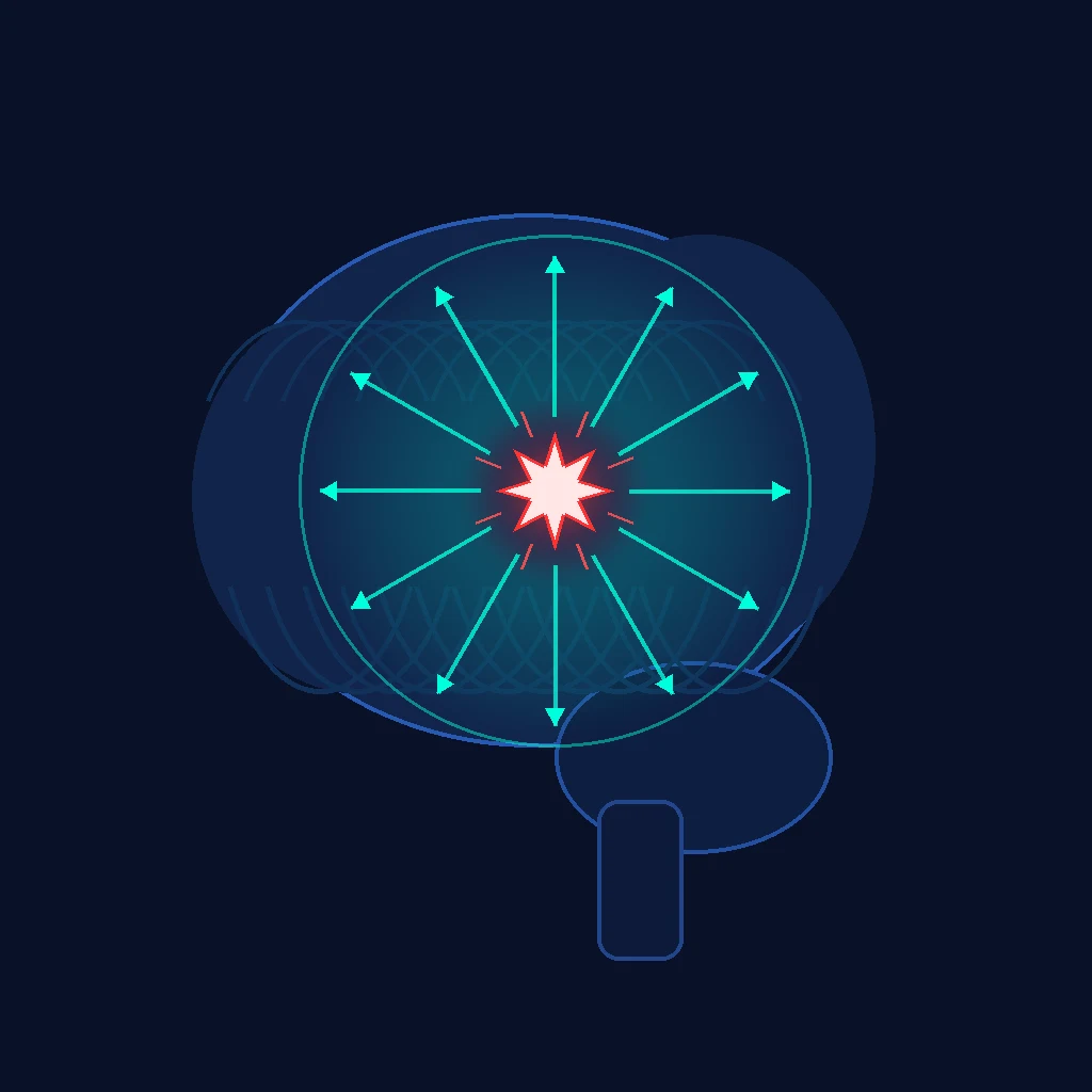
The primary injury — the contusion, the diffuse axonal injury, the haematoma — is done at the moment of the impact and is irreversible. The secondary injury, which develops over the minutes to the days, is the cascade of the ischaemia, the oedema, the inflammation and the biochemical injury that compounds the primary damage, and it is driven by the preventable insults: the hypoxia (a saturation below 90 per cent doubles the mortality), the hypotension (a single systolic below 90 mmHg doubles the mortality), the raised intracranial pressure (from the haematoma, the oedema or the hypercapnia), the seizure, the fever and the hyperglycaemia.[1][5][7] The whole of the emergency management of the TBI is the prevention of these insults — the airway and the oxygenation, the blood pressure, the intracranial pressure, and the metabolic control — and the candidate who internalises this principle has the framework for every decision.
The Glasgow Coma Scale and the severity classification
The Glasgow Coma Scale (GCS), introduced by Teasdale and Jennett in 1974, is the universal language of the traumatic brain injury, and the severity classification that flows from it governs the airway decision, the imaging decision and the disposition.[4]
The Glasgow Coma Scale — the three components and the scoring
| Component | The response | The score |
|---|---|---|
| Eye opening (E) | Spontaneous / to speech / to pain / none | 4 / 3 / 2 / 1 |
| Verbal response (V) | Oriented / confused / inappropriate words / incomprehensible sounds / none | 5 / 4 / 3 / 2 / 1 |
| Motor response (M) | Obeys commands / localises pain / withdrawes / flexion (decorticate) / extension (decerebrate) / none | 6 / 5 / 4 / 3 / 2 / 1 |
| Total | E + V + M | 3 to 15 |
The severity classification that follows from the total score is the single most examined fact in the TBI viva, and it carries the airway threshold with it: [1]
The TBI severity classification — the score, the airway, the disposition
| Severity | GCS | The implication | The airway |
|---|---|---|---|
| Mild | 13–15 | The vast majority; the Canadian CT Head Rule governs the imaging; discharge possible with the safety-net advice | Observes own airway |
| Moderate | 9–12 | The combative or the confused patient; the CT mandatory; the high risk of the deterioration — monitor closely | Prepare for the intubation; intubate if the GCS falls |
| Severe | 3–8 | The comatose patient; the ICP risk is high; the intensive care and the neurosurgery | Intubate — a GCS ≤8 is the indication for the definitive airway |
The GCS pitfalls are perennial exam points. The score is reported as the best motor response (not the worst); the localising or the withdrawing is tested with the central pain (the supraorbital pressure or the trapezius squeeze), and the patient who localises with one limb and withdraws with the other scores the better response. The verbal score is unobtainable in the intubated patient — record it as "Vt" or "1t" and never invent a number. The painful stimulus must be central, because a peripheral limb-withdrawal can be a spinal reflex in the brain-dead patient. The GCS trends — the serial measurements matter more than the single number, because a falling GCS (especially a falling motor score) is the herald of the expanding mass lesion or the rising ICP.[1][4]
The pupil examination and the motor asymmetry are the essential adjuncts to the GCS. The unilateral fixed dilated pupil with a falling GCS is the uncal herniation syndrome — the third nerve is compressed against the edge of the tentorium by the medial temporal lobe — and it is the signal to give the osmolar therapy immediately and call the neurosurgeon. A bilateral fixed dilated pupil in the hypothermic or the intoxicated patient is not necessarily terminal; resuscitate, correct the hypoxia and the hypotension, give the osmolar therapy, and reassess. [1]
Airway, breathing and the oxygenation
The airway is secured early in the patient with a GCS of 8 or below (the standard threshold for the definitive airway), using the rapid sequence intubation with the cervical spine in-line and the neuroprotective induction (propofol or thiopental, with attention to the blood pressure; ketamine is acceptable and maintains the haemodynamics). The oxygenation is maintained at a saturation of 94 per cent or above, and the ventilation is to a normocapnia (PaCO2 35 to 45 mmHg or 4.5 to 6.0 kPa). The hyperventilation, which lowers the intracranial pressure by the cerebral vasoconstriction, is reserved as a temporising measure for the patient with the acute neurological deterioration and the impending herniation (the fixed dilated pupil, the decerebrate posturing), while the definitive treatment (the osmolar therapy, the surgery) is prepared; the routine hyperventilation is harmful because it causes the cerebral ischaemia.[1][1]
The intubation of the severe TBI is the first opportunity to prevent (or to cause) the secondary injury. The induction must not drop the blood pressure, because the hypotension at the induction doubles the mortality; the ketamine is haemodynamically safe and is no longer considered contraindicated (the historical concern about the raised ICP was overstated and is outweighed by the preservation of the blood pressure). The rocuronium (1.2 mg/kg) or the suxamethonium (1.5 mg/kg) for the paralysis; the in-line manual stabilisation of the cervical spine; the pre-oxygenation to the maximal denitrogenation; and the preparation for the hypotension (the fluid, the vasopressor drawn up) are the elements of the neuroprotective RSI. [1]
Circulation and the blood pressure
The blood pressure is maintained at a systolic of 110 mmHg or above (or a mean arterial pressure of 80 or above) from the scene to the intensive care, because the single episode of hypotension after the severe TBI doubles the mortality. The fluid resuscitation uses the isotonic or the hypertonic crystalloid (not the hypotonic, which worsens the cerebral oedema), and the blood products are given for the associated haemorrhage. The vasopressor (noradrenaline) is used to maintain the blood pressure if the fluid is insufficient. The cerebral perfusion pressure — the mean arterial pressure minus the intracranial pressure — is the target that links the systemic and the cerebral management, and it is maintained at 60 to 70 mmHg.[1][6]
The dextrose-containing fluid (the 5% dextrose) is avoided, because the free water worsens the cerebral oedema and the hyperglycaemia. The hypotonic fluid (the 0.45% saline) is avoided for the same reason. The preferred fluids are the 0.9% saline, the balanced crystalloid (the Plasma-Lyte) and, for the osmolar effect, the hypertonic saline. The glucose is controlled to 6 to 10 mmol/L, because both the hyperglycaemia and the hypoglycaemia worsen the secondary injury. [1]
The CT head: the Canadian CT Head Rule
The imaging decision in the mild head injury (GCS 13–15) is governed by the validated decision rule — the Canadian CT Head Rule of Stiell and colleagues, derived and validated in over 3,000 patients and now the standard in the Australasian, the British and the North American practice.[13] The rule identifies the high-risk criteria (the CT is mandatory — the finding threatens the life and needs the neurosurgical intervention) and the medium-risk criteria (the CT is recommended — the finding needs the observation or the intervention).
The Canadian CT Head Rule — when to scan the adult with the minor head injury (GCS 13–15)
| Tier | The criterion | The action |
|---|---|---|
| High-risk (CT mandatory) | GCS <15 at 2 h after the injury | CT |
| Suspected open or depressed skull fracture | CT | |
| Any sign of basal skull fracture (the haemotympanum, the racoon eyes, the CSF leak, the Battle sign) | CT | |
| Vomiting ≥2 episodes | CT | |
| Age ≥65 years | CT | |
| Medium-risk (CT recommended) | Dangerous mechanism (the pedestrian struck, the fall >3 ft or 5 stairs, the ejection from the motor vehicle) | CT |
| Amnesia before the impact >30 min | CT | |
| (On the original rule) dangerous mechanism OR amnesia — observation or CT | CT or observe |
The rule applies to the adult with the minor head injury and the GCS of 13–15 who sustained the witnessed loss of consciousness, the amnesia or the disorientation. It does not apply to the patient on the anticoagulation without the full reassessment (many centres scan the anticoagulated patient after any head strike, because the intracranial bleeding risk is high and the rule was not derived in this group), the pregnant patient, the patient with the bleeding disorder, or the patient younger than 16. The cervical spine is imaged by the Canadian C-Spine Rule in parallel, because the head and the cervical injury coexist in up to 15% of the serious trauma.[13][1]
The CT findings that demand the urgent neurosurgical referral are: the extradural or the acute subdural haematoma with the midline shift; the depressed skull fracture greater than the thickness of the skull; the intracerebral haematoma with the mass effect; the diffuse axonal injury; and any haemorrhage with the effacement of the basal cisterns (the herniation imminent). [1]
The intracranial pressure and the osmolar therapy
The intracranial pressure is managed in a staircase, with each step added if the preceding step is insufficient.[1]
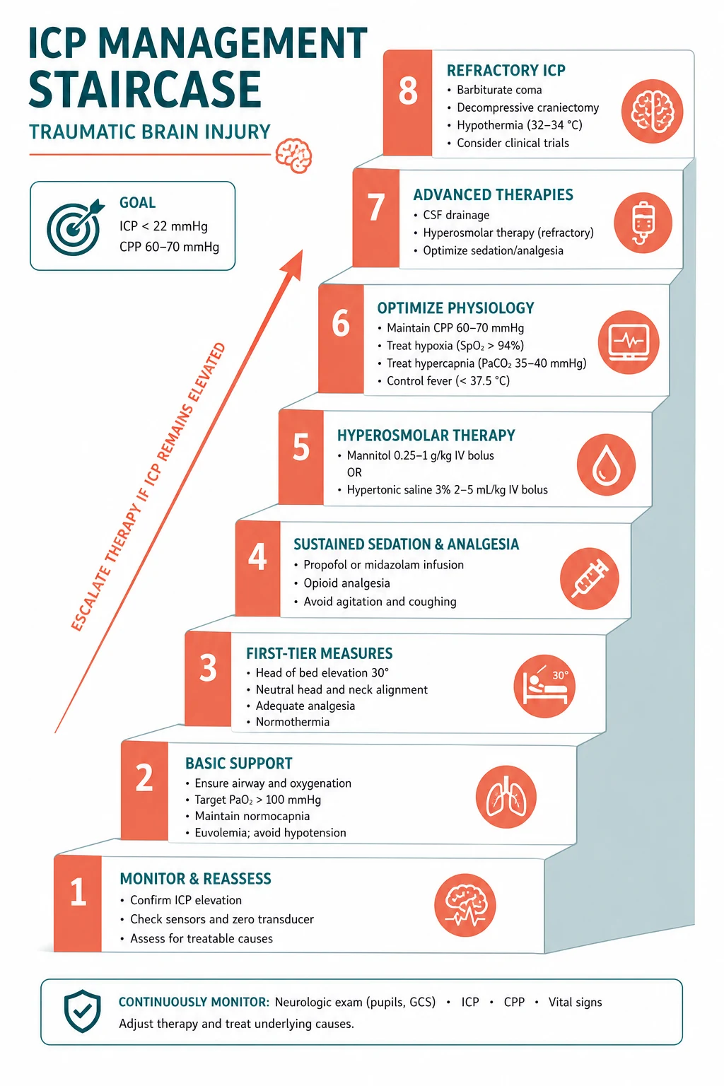
The head is elevated to 30 degrees (to promote the venous drainage). The sedation and the analgesia reduce the metabolic demand and the coughing. The cerebrospinal fluid is drained if an external ventricular drain is in place. The osmolar therapy — the mannitol (0.25 to 1 gram per kilogram intravenously, over 10 to 15 minutes) or the hypertonic saline (3 per cent, in boluses or the infusion) — draws the water from the brain into the intravascular space and lowers the intracranial pressure. The decompressive craniectomy is the surgical step, and the metabolic suppression (the barbiturate coma) is the last pharmacological step. The seizure prophylaxis (the levetiracetam or the phenytoin for the first seven days in the high-risk patient) and the temperature and the glucose control are maintained throughout. [1]
Mannitol versus hypertonic saline — the osmolar therapy compared
| Feature | Mannitol (20%) | Hypertonic saline (3% / 5% / 7.5%) |
|---|---|---|
| The dose | 0.25–1 g/kg IV over 10–15 min (a typical 250 mL of 20%) | 3% — 250 mL bolus, or 5% / 7.5% via the central line; many EDs give 3% 250 mL for the acute rise |
| The onset | Minutes | Minutes |
| The effect on the serum sodium | May fall (the free-water diuresis) | Rises — monitor the Na+ (aim <160) |
| The effect on the volume | Diuresis → the hypovolaemia and the hypotension | Volume expansion — may raise the blood pressure (the dual benefit in the hypotensive patient) |
| The osmolar gap | The diuresis; monitor the osmolality (stop if >320) | Less of an osmotic diuresis |
| The renal risk | The acute kidney injury if the patient is hypovolaemic | Lower, but the hypernatraemia and the hyperchloraemic acidosis |
| The evidence | The traditional first-line; the BTF guidelines as the option | The increasingly preferred agent in the ED (the volume expansion, the blood pressure support, the comparable ICP reduction) |
| The exam answer | Effective, but the diuresis may drop the blood pressure → the secondary injury | Effective, and the volume expansion preserves the blood pressure → the preferred agent in the ED where the hypotension is the threat |
The Brain Trauma Foundation treats the ICP threshold above 22 mmHg as the trigger for the treatment, because the mortality rises sharply above that level. The cerebral perfusion pressure (CPP = MAP − ICP) is maintained between 60 and 70 mmHg — the too-low CPP causes the ischaemia, and the too-high CPP (driven by the excessive vasopressors) causes the adult respiratory distress syndrome and the fluid overload. [1]
The ED neuroprotection — the structured first hour
The emergency management of the severe TBI is the disciplined application of the ABCDE with the neuroprotective targets at every step. The candidate who walks through this structure in the viva demonstrates the command of the secondary-injury prevention. [1]
The severe TBI in the ED — the structured neuroprotective first hour
A — Airway with the cervical spine control
GCS ≤8 → the RSI and the definitive airway. In-line manual stabilisation of the cervical spine (the cervical collar, the head blocks, the tape). The pre-oxygenation; the haemodynamically safe induction (the ketamine or the propofol with the vasopressor ready); the rocuronium or the suxamethonium. The target: a secure airway and no hypoxia and no hypotension.
B — Breathing and the ventilation
Oxygen to keep the SpO2 >94% (the target >90% as the absolute floor — a single SpO2 <90% doubles the mortality). Ventilate to the NORMOCAPNIA, PaCO2 35–45 mmHg. Do NOT hyperventilate routinely (the hypocapnia causes the cerebral ischaemia); reserve the hyperventilation (PaCO2 30–35) as a TEMPORISING measure only for the impending herniation while the osmolar therapy and the surgery are prepared.
C — Circulation — maintain the SBP >110
The single SBP <90 doubles the mortality — maintain the SBP >110 (or the MAP >80) from the scene. The isotonic or the hypertonic crystalloid (NOT the dextrose, NOT the hypotonic); the blood products for the haemorrhage; the noradrenaline if the fluid is insufficient. Maintain the CPP 60–70 mmHg. Check the glucose (keep 6–10) and the haemoglobin (keep >70–80 g/L).
D — Disability — the GCS, the pupils, the osmolar therapy
The GCS (E + V + M), the pupil size and the reactivity, the limb power. The falling GCS or the unilateral fixed dilated pupil → the herniation → give the osmolar therapy NOW (the 3% saline 250 mL or the mannitol 0.5 g/kg) and call the neurosurgeon. The seizure prophylaxis (the levetiracetam or the phenytoin) in the high-risk patient. Do NOT give the corticosteroids.
E — Exposure and the environment
The full secondary survey once the A–D are stable (but the CT may precede the full secondary survey in the unstable severe TBI). Maintain the NORMOTHERMIA — do not allow the hypothermia from the exposure, and do not induce the prophylactic hypothermia (the POLAR trial — no benefit). The glucose, the sodium, the temperature controlled throughout.
The CT head (non-contrast) — and the cervical spine
Once the A–C are stable, the urgent non-contrast CT head and the CT cervical spine. In the haemodynamically unstable patient, do not delay the life-saving surgery (the laparotomy, the decompression) for the CT — resuscitate, operate, then the CT. The CT findings that demand the urgent neurosurgical referral: the extradural or the acute subdural with the midline shift, the depressed fracture, the mass effect, the effaced basal cisterns.
The neurosurgical referral and the disposition
Refer early and refer with the structured handover (the mechanism, the GCS and the trend, the pupils, the CT, the other injuries, the coagulation, the time since the injury). The severe TBI → the ICU with the ICP monitoring (if the CT abnormal or the GCS ≤8 with the normal CT and two of: age >40, the posturing, the SBP <90). The extradural / the subdural with the shift → the urgent theatre.
The temporising management of the impending herniation
The patient who develops the unilateral fixed dilated pupil or the sudden fall in the GCS while awaiting the scan or the theatre has the impending herniation, and the temporising measures buy the minutes to the definitive treatment. This is the ONE setting in which the hyperventilation is justified. [1]
The impending herniation — the temporising sequence while the definitive treatment is prepared
1. Recognise the herniation syndrome
The falling GCS (especially the falling motor score), the unilateral fixed dilated pupil (the third-nerve compression), the decerebrate or the decorticate posturing, the Cushing triad (the hypertension, the bradycardia, the irregular respiration — the late sign of the markedly raised ICP). Time is brain — act now.
2. Give the osmolar therapy immediately
The 3% hypertonic saline 250 mL IV over 10 min, OR the mannitol 0.5–1 g/kg IV over 10–15 min. The osmolar agent draws the water out of the brain into the intravascular space and lowers the ICP within minutes. Check the sodium and the osmolality. If the patient is hypotensive, prefer the 3% saline (it expands the volume) over the mannitol (which causes the diuresis and may drop the blood pressure).
3. The hyperventilation — the ONE temporising indication
Ventilate to the PaCO2 30–35 mmHg (the mild hypocapnia) — the cerebral vasoconstriction lowers the intracranial blood volume and the ICP. This is the ONLY justified use of the hyperventilation, and it is TEMPORISING (minutes to an hour), because the sustained hypocapnia causes the cerebral ischaemia. Revert to the normocapnia as soon as the definitive treatment (the osmolar agent, the surgery) takes effect.
4. Elevate the head to 30°
The head-up 30° position promotes the venous drainage and the jugular emptying, which lowers the intracranial blood volume and the ICP. Ensure the neck is midline (the tape, the collar, the ties) — the kinked neck obstructs the venous return and raises the ICP.
5. The sedation and the paralysis
Deepen the sedation (the propofol, the midazolam) and paralyse (the rocuronium) to abolish the coughing, the straining and the metabolic demand. Ensure the blood pressure is maintained (the vasopressor) — the deeper sedation may drop the blood pressure and must not cause the secondary injury.
6. The urgent CT and the neurosurgery
The urgent non-contrast CT head (once the A–C are secure) and the neurosurgical referral for the definitive treatment (the evacuation of the mass lesion, the decompressive craniectomy in the refractory case). The temporising measures buy the time — they are not the definitive treatment.
Corticosteroids: the evidence
The corticosteroids, once given routinely for the presumed anti-oedema effect, are harmful in the traumatic brain injury. The CRASH trial, in over 10,000 adults with the clinically significant head injury, found that the intravenous corticosteroids increased the risk of the death within two weeks compared with the placebo, and the practice was abandoned. The candidate who gives the corticosteroids for the TBI is the candidate who has not read the evidence; the corticosteroids are reserved for the tumour-related oedema and the spinal cord injury, not for the traumatic brain injury.[1]
The decompressive craniectomy: DECRA and RESCUEicp
The decompressive craniectomy — the removal of a large skull bone flap to allow the swollen brain to expand outward — is one of the most debated interventions in the TBI, and the candidate must know the two landmark trials. The DECRA trial found that the early bifrontal decompressive craniectomy for the diffuse TBI with the raised ICP did not improve the functional outcome and was associated with a worse outcome in some measures, which argued against the routine early craniectomy.[2] The RESCUEicp trial found that the decompressive craniectomy as the last-tier intervention for the refractory intracranial hypertension reduced the mortality compared with the medical management, but at the cost of a higher rate of the vegetative and the lower-good-recovery states in the survivors.[3] The synthesis: the craniectomy is not routine or early, but it is considered for the refractory intracranial hypertension as the last resort, with the frank discussion of the outcomes with the family.
DECRA versus RESCUEicp — the two craniectomy trials compared
| Feature | DECRA (Cooper 2011) | RESCUEicp (Hutchinson 2016) |
|---|---|---|
| The population | The diffuse TBI with the raised ICP early (within 72 h) | The refractory intracranial hypertension (the ICP >25 sustained after the tiered medical therapy) |
| The intervention | The early bifrontal decompressive craniectomy | The decompressive craniectomy (last-tier) vs the barbiturate (the continued medical therapy) |
| The timing | Early (prophylactic / first-tier) | Late (salvage / last-tier) |
| The outcome | The craniectomy had a WORSE functional outcome at 6 months (more unfavourable outcomes) | The craniectomy REDUCED the mortality but at the cost of more vegetative and dependent survivors |
| The lesson | The routine early craniectomy for the diffuse TBI is not justified | The salvage craniectomy as the last resort reduces the death but trades it for the disability — the family counselling is essential |
The ICP monitoring — the BEST-TRIP trial
The intracranial pressure monitoring has been the cornerstone of the severe-TBI management on the rationale that the treatment of the raised ICP improves the outcome. The BEST-TRIP trial (Chesnut and colleagues, 2012) tested this directly in the Bolivian and Ecuadorian centres: the severe TBI randomised to the ICP-monitor-guided management versus the imaging-and-examination-guided management. The outcome was no different between the groups — and the result is not interpreted as the ICP monitoring is useless, but as the disciplined protocolised care (with or without the monitor) is what matters, and the monitor does not replace the careful clinical and the imaging assessment.[8] The Brain Trauma Foundation recommends the ICP monitoring in the severe TBI (the salvageable patient with the abnormal CT, or the normal CT with two of: age >40, the motor posturing, the SBP <90), with the treatment threshold above 22 mmHg.
The trials that did NOT work — hypothermia and the prehospital hypertonic saline
Two large randomised trials tested the interventions that were long hoped to be neuroprotective in the severe TBI, and both were negative. The Fellowship candidate must know them, because the examiner asks not only what to do but what has been tried and abandoned. [1]
POLAR (Cooper 2018) — prophylactic hypothermia in severe TBI
Design
Multicentre randomised controlled trial — Australia, New Zealand, France; 511 patients with the severe TBI (GCS 3–8)
Intervention
Early sustained prophylactic hypothermia (33–35 °C for >72 h) vs normothermia
Primary outcome
Favourable neurological outcome at 6 months — NO difference (lower in the hypothermia group, though not significantly)
Harms
More episodes of the hypothermia-related bradycardia; no benefit and a signal toward harm
Bottom line
Prophylactic hypothermia does NOT improve the outcome after the severe TBI. Do not cool the head-injured patient for the neuroprotection alone — control the fever, but maintain the normothermia, not the induced hypothermia
Cooper 2004 (JAMA) — prehospital hypertonic saline in severe TBI
Design
Randomised controlled trial — 229 patients with the hypotension and the severe TBI in the prehospital setting
Intervention
Prehospital 7.5% hypertonic saline vs Ringer's lactate resuscitation
Primary outcome
Neurological outcome at 6 months — NO difference between the groups
Bottom line
The prehospital hypertonic saline did not improve the neurological outcome despite the theoretical advantage (the volume expansion, the osmolar effect). It remains a reasonable resuscitation fluid (especially for the combined hypotension and the raised ICP), but it is not the neuroprotective magic bullet. Use it as the 3% saline in the ED for the documented or the suspected raised ICP, not as the routine prehospital resuscitation
The seizure prophylaxis — the Temkin trial
The post-traumatic seizure compounds the secondary injury (the metabolic surge, the raised ICP, the excitotoxicity). The early post-traumatic seizures (within seven days) are reduced by the prophylactic phenytoin — the landmark Temkin trial (1990) showed that the phenytoin halved the rate of the early seizures compared with the placebo. The modern practice uses the levetiracetam increasingly (the comparable efficacy, the easier administration, the fewer interactions), given for seven days to the high-risk patient (the intracerebral haematoma, the depressed fracture, the penetrating injury, the subdural or the extradural haematoma, the seizure at the scene). The prophylaxis does NOT prevent the late seizures (after seven days) and is not continued beyond the acute period unless the seizures recur.[11]
The prognostic models — the CRASH calculator
The outcome prediction after the TBI is requested by the families and the clinicians, and the validated model is the CRASH prognostic calculator (Perel and the CRASH collaborators, 2008), derived from over 10,000 patients and externally validated. The model uses the age, the GCS, the pupil reactivity, the CT findings, the presence of the major extracranial injury and the country (the high- vs the low-income) to estimate the 14-day mortality and the 6-month outcome. The model is the tool for the honest family counselling and the goals-of-care discussion, not for the early withdrawal of the care in the patient who may still recover.[12]
The specific injuries
The extradural (epidural) haematoma — the lentiform (biconvex) blood collection between the skull and the dura, classically from the middle meningeal artery tear after a temporal fracture — presents with the lucid interval (the initial concussion, the recovery, then the deterioration as the haematoma expands), and it needs the urgent surgical evacuation; the prognosis is excellent if evacuated before the herniation. The acute subdural haematoma — the crescent-shaped collection between the dura and the arachnoid, from the bridging-vein tear or the cortical laceration — carries a worse prognosis because it reflects the underlying brain injury, and it needs the evacuation if it is thick or causing the midline shift. The diffuse axonal injury — the shearing of the white-matter tracts from the rapid acceleration-deceleration — may have a normal CT initially (the MRI shows the micro-haemorrhages) and carries the worst prognosis; there is no specific surgical treatment. The traumatic subarachnoid haemorrhage and the intracerebral contusion are managed by the ICP control and the monitoring for the expansion.[1][1]
The specific traumatic brain injuries — the imaging, the mechanism, the management
| Injury | The CT appearance | The mechanism | The management |
|---|---|---|---|
| Extradural (epidural) | The lentiform / biconvex, does NOT cross the sutures | The middle meningeal artery (the temporal fracture) | The URGENT surgical evacuation — the prognosis excellent if before the herniation; the lucid interval is the classic but not the universal presentation |
| Acute subdural | The crescent, crosses the sutures, the midline shift | The bridging-vein tear or the cortical laceration | The evacuation if >10 mm thick or the midline shift >5 mm; the prognosis worse (the underlying brain injury) |
| Diffuse axonal injury | The CT may be NORMAL; the micro-haemorrhages on the MRI (the corpus callosum, the brainstem) | The rotational acceleration-deceleration shears the axons | No specific surgical treatment; the supportive ICP control; the prognosis often poor |
| Traumatic subarachnoid | The blood in the sulci / the basal cisterns | The tearing of the surface vessels | The ICP control; the seizure prophylaxis; the monitoring for the vasospasm (rarely clinically significant) |
| Intracerebral contusion | The superficial haemorrhage (the frontal and the temporal poles — the coup and the contrecoup) | The brain striking the rough floor of the frontal / the temporal fossa | The ICP control; the monitoring for the EXPANSION (the "blossoming contusion" over 24–48 h) — re-scan if the GCS falls |
| Depressed skull fracture | The bone fragment depressed greater than the skull thickness | The focal blow | The surgical elevation; the antibiotic prophylaxis; the tetanus; the seizure prophylaxis |
Common pitfalls
The recurring errors are: allowing the hypoxia or the hypotension (the greatest preventable causes of the secondary injury); giving the corticosteroids (harmful per the CRASH trial); the routine hyperventilation (causes the ischaemia); delaying the CT for the secondary survey before the primary survey is complete; not referring the mass lesion urgently; not giving the seizure prophylaxis to the high-risk patient; inducing the prophylactic hypothermia (no benefit, the POLAR trial); and not maintaining the normothermia and the normoglycaemia. [1]
The recurring TBI errors — the error, the harm, the correction
| The error | The harm it causes | The correction |
|---|---|---|
| Allowing the hypotension (SBP <90) | The mortality doubles — the cerebral ischaemia | Maintain the SBP >110 from the scene; the fluid, the blood, the noradrenaline |
| Allowing the hypoxia (SpO2 <90%) | The mortality doubles — the direct neuronal injury | The oxygen; the intubation if the GCS ≤8 |
| The routine hyperventilation | The cerebral ischaemia (the hypocapnic vasoconstriction) | Ventilate to the normocapnia; reserve the hyperventilation for the herniation |
| Giving the corticosteroids | The increased mortality (the CRASH trial) | Do NOT give the corticosteroids for the TBI (reserved for the tumour, the spinal cord) |
| The prophylactic hypothermia | No benefit, the bradycardia, the coagulopathy (the POLAR trial) | Maintain the normothermia; control the fever, do not cool |
| The hypotonic or the dextrose fluid | The cerebral oedema, the hyperglycaemia | The isotonic or the hypertonic crystalloid only |
| Delaying the CT for the full secondary survey | The delayed diagnosis of the expanding mass lesion | The CT after the A–C are stable; the life-saving surgery may precede the CT |
| Not referring the mass lesion urgently | The herniation, the death | The early neurosurgical referral with the structured handover |
| The dextrose-containing fluid / the hypoglycaemia | The worsening secondary injury | The glucose 6–10 mmol/L; check and treat the hypoglycaemia |
SAQ — Severe TBI with the expanding mass lesion and the impending herniation
12 minutes · 10 marks
A 24-year-old man is brought in by ambulance after a high-speed motorcycle crash. He was briefly talking at the scene but is now obtunded. On arrival: GCS 7 (E1 V2 M4), left pupil 6 mm and unreactive, right pupil 3 mm reactive, SBP 84/50, HR 112, SpO2 88 per cent on a non-rebreather. CT head shows a large left frontotemporal extradural haematoma with 9 mm midline shift. The neurosurgeon is 90 minutes away by retrieval.
SAQ — The anticoagulated elderly patient with a minor head injury
10 minutes · 10 marks
A 78-year-old woman on warfarin for atrial fibrillation (INR usually 2.5) presents after a ground-level fall onto her occiput. She did NOT lose consciousness. GCS 15, normal neurological examination. She is on no antiplatelet. The CT head shows a small 4 mm subdural haematoma with no midline shift.
Clinical pearls
Red flags
The following features identify the TBI at risk of the secondary injury or the deterioration, in which the emergency neuroprotection is the priority: [1]
[1]References
- [1]Roberts I, Yates D, Sandercock P, et al. (CRASH trial collaborators). Effect of intravenous corticosteroids on death within 14 days in 10008 adults with clinically significant head injury (MRC CRASH trial): randomised placebo-controlled trial Lancet, 2004.PMID 15474134
- [2]Cooper DJ, Rosenfeld JV, Murray L, et al. Decompressive craniectomy in diffuse traumatic brain injury N Engl J Med, 2011.PMID 21434843
- [3]Hutchinson PJ, Kolias AG, Timofeev IS, et al. Trial of Decompressive Craniectomy for Traumatic Intracranial Hypertension N Engl J Med, 2016.PMID 27602507
- [4]Teasdale G, Jennett B. Assessment of coma and impaired consciousness. A practical scale Lancet, 1974.PMID 4136544
- [5]Chesnut RM, Marshall LF, Klauber MR, et al. The role of secondary brain injury in determining outcome from severe head injury J Trauma, 1993.PMID 8459458
- [6]Chesnut RM, Marshall SB, Piek J, et al. Early and late systemic hypotension as a frequent and fundamental source of cerebral ischemia following severe brain injury in the Traumatic Coma Data Bank Acta Neurochir Suppl (Wien), 1993.PMID 8310858
- [7]Manley G, Knudson MM, Morabito D, et al. Hypotension, hypoxia, and head injury: frequency, duration, and consequences Arch Surg, 2001.PMID 11585502
- [8]Chesnut RM, Temkin N, Carney N, et al. A trial of intracranial-pressure monitoring in traumatic brain injury N Engl J Med, 2012.PMID 23234472
- [9]Cooper DJ, Nichol AD, Bailey M, et al. Effect of Early Sustained Prophylactic Hypothermia on Neurologic Outcomes Among Patients With Severe Traumatic Brain Injury: The POLAR Randomized Clinical Trial JAMA, 2018.PMID 30357266
- [10]Cooper DJ, Myles PS, McDermott FT, et al. Prehospital hypertonic saline resuscitation of patients with hypotension and severe traumatic brain injury: a randomized controlled trial JAMA, 2004.PMID 15026402
- [11]Temkin NR, Dikmen SS, Wilensky AJ, et al. A randomized, double-blind study of phenytoin for the prevention of post-traumatic seizures N Engl J Med, 1990.PMID 2115976
- [12]MRC CRASH Trial Collaborators, Perel P, Arango M, et al. Predicting outcome after traumatic brain injury: practical prognostic models based on large cohort of international patients BMJ, 2008.PMID 18270239
- [13]Stiell IG, Wells GA, Vandemheen K, et al. The Canadian CT Head Rule for patients with minor head injury Lancet, 2001.PMID 11356436