EM · Tuberculosis
Tuberculosis (ED)
The tuberculosis from the Mycobacterium tuberculosis through the primary and the reactivation (the secondary), the risk factors (the HIV, the immunosuppression, the malnutrition, the diabetes, the migrant, the alcohol, the smoking), the presentation (the chronic cough, the haemoptysis, the night sweats, the weight loss, the fever; the extrapulmonary — the cervical lymphadenitis, the meningitis, the miliary, the spinal Pott), the ED role (the suspect, the isolate, the airborne precautions, the sputum, the public health notification), the quadruple RIPE therapy and its toxicity, and the MDR-TB.
On this page & tools
Your progress
Saved locally on this device.
Target exams
Red flags
The tuberculosis is the infection by the Mycobacterium tuberculosis. The emergency-department role is the suspect, the isolate, the sputum, the notify — the recognition and the airborne precautions before the confirmation, then the referral for the definitive diagnosis and the long therapy. The Fellowship candidate must know the presentation, the investigations, the RIPE therapy and its toxicity, and the public-health obligation.[1][2]
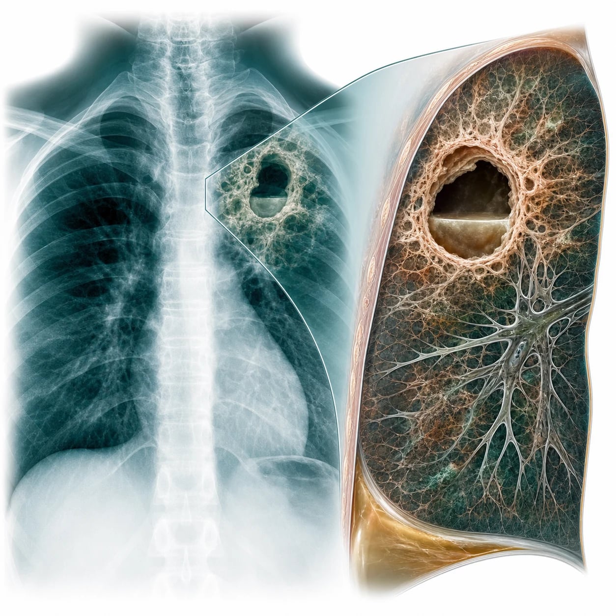
The pathogen and the transmission
The Mycobacterium tuberculosis is a slow-growing, obligate-aerobic, non-motile, intracellular, rod-shaped bacillus of the genus Mycobacterium. It is neither Gram-positive nor Gram-negative in the routine sense: its cell wall is rich in mycolic acid, arabinogalactan and lipoarabinomannan, which resist the decolourisation by the acid-alcohol after the carbol-fuchsin stain — the basis of the acid-fast (the Ziehl–Neelsen) property and of the auramine-rhodamine fluorescence stain. The waxy mycolic-acid wall also explains the slow growth (the colony on the Lowenstein-Jensen in 2 to 8 weeks), the resistance to the drying and the disinfectants, and the survival inside the macrophage.[1]
The transmission is by the airborne route — the aerosol of the droplet nuclei (1 to 5 micrometres) generated by the coughing, the sneezing, the singing or the forceful expiratory effort of the infectious case. The droplet nuclei remain suspended in the air for hours and travel on the air currents, which is the reason for the negative-pressure room and the N95 respirator (a surgical mask does NOT filter the droplet nuclei). The transmission requires a close, prolonged, indoor exposure; the brief ED contact is low-risk but the unrecognised infectious patient in the open ward or the waiting room is the classic outbreak seed. The infectiousness falls rapidly within 2 weeks of the effective therapy, which underlies the de-isolation criteria.[1][3]
The TB transmission and the burden
The immunopathology — the granuloma and the latency
The inhaled bacillus reaches the alveolus and is taken up by the alveolar macrophage. The bacillus survives and replicates inside the macrophage by blocking the phagolysosome fusion (the lipoarabinomannan and the secretion system). The infected macrophage secretes the cytokines and recruits the monocytes, the T-cells and the epithelioid cells, forming the caseating granuloma — the organised structure that contains but does not always kill the bacillus. The central caseous necrosis (the cheese-like, the avascular) may liquefy and cavitate into the airway, releasing the bacilli and producing the contagious cavitary disease. [1]
The cell-mediated (the Th1) immunity develops over 2 to 10 weeks — the CD4+ T-cell, the interferon-gamma, the tumour-necrosis-factor-alpha and the macrophage activation. This is the basis of the tuberculin skin test (the Mantoux) conversion and the interferon-gamma release assay (the IGRA). The containment produces the latent tuberculosis infection (the LTBI) — a viable but dormant bacillus walled in the granuloma, infectious neither to the host nor to others, yet capable of the reactivation years to decades later. Any state that weakens the cell-mediated immunity — the HIV, the anti-TNF therapy, the transplant, the malnutrition, the diabetes, the chronic renal failure, the malignancy, the silicosis — raises the reactivation risk and is examined.[1]
The classification — the primary and the reactivation
The primary tuberculosis is the initial infection, often subclinical, producing the Ghon focus (the subpleural lower or middle-lobe lesion) and the hilar lymphadenopathy, which together calcify into the Ghon complex. The cell-mediated immunity contains it in the majority, but the latent infection persists.[1]
The reactivation (the secondary) tuberculosis is the reactivation of the latent infection years later, typically in the upper lobe (the high oxygen tension), with the cavitation, the fibrosis, and the caseating granulomata, and it is the contagious form. The risk factors are the HIV, the immunosuppression, the malnutrition, the diabetes, the chronic renal failure, the migrant or the travel from the endemic area, the alcohol, and the smoking.[1]
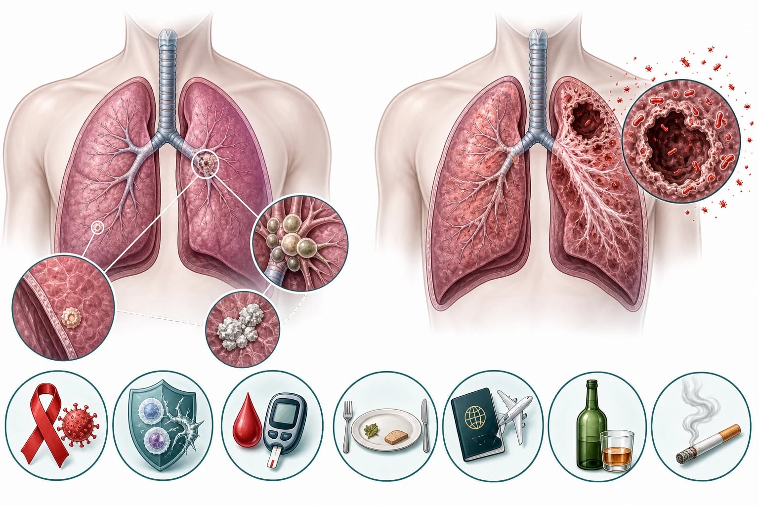
Primary TB
- The first infection; subclinical in most
- The Ghon focus (subpleural lower/middle lobe) + hilar LN
- Calcifies to the Ranke/Ghon complex
- May progress: the primary progressive, the miliary, the TB meningitis in the child
Latent TB (LTBI)
- Viable but dormant; not contagious, not symptomatic
- Positive TST or IGRA; normal chest radiograph
- ~10% lifetime reactivation (higher in HIV/immunosuppressed)
- Treat to prevent the reactivation (the 3HP, the 4R, the 6–9H)
Reactivation (secondary) TB
- Years to decades later; the contagious form
- Upper-lobe cavity, fibrosis, caseating granulomata
- Chronic cough, haemoptysis, night sweats, weight loss
- Triggered by the weakened cell-mediated immunity
Disseminated / miliary TB
- The haematogenous spread; the millet-seed CXR
- Fever, wasting, pancytopenia, hepatosplenomegaly
- Higher in the immunocompromised, the elderly, the child
- Meningitis co-exists — always the lumbar puncture
The risk factors — the conditions that weaken the cell-mediated immunity
The risk factors are examined because they raise the reactivation probability and they shape the index of suspicion. The strongest single risk factor is the HIV — the reactivation rate rises from 10 per cent over a lifetime to roughly 10 per cent per year, and the TB is the commonest opportunistic infection and a leading cause of the death in the HIV globally. The anti-TNF-alpha agents (the infliximab, the adalimumab), the transplant immunosuppression, the chronic steroids (the equivalent of the prednisolone 15 mg for a month) and the chemotherapy all weaken the granuloma. The silicosis damages the macrophage directly; the diabetes, the chronic renal failure, the malnutrition and the smoking impair the cell-mediated immunity. The migrant and the travel from the high-burden country (the sub-Saharan Africa, the South Asia, the South-East Asia, the Western Pacific) raise the prior exposure probability.[1]
High-yield risk factors
- HIV (the single strongest)
- Anti-TNF-alpha therapy (infliximab, adalimumab)
- Solid-organ or stem-cell transplant
- Silicosis — macrophage damage
- Chronic steroids >prednisolone 15 mg/day for >1 month
Moderate risk factors
- Diabetes mellitus (poor control)
- Chronic renal failure / dialysis
- Malnutrition, low body-mass index
- Smoking, alcohol misuse
- Malignancy and chemotherapy
Epidemiologic risk
- Born in / travel to a high-burden country
- Household or close contact of an infectious case
- Homelessness, overcrowding, incarceration
- Residential-care / aged-care facility
- Healthcare worker in a high-burden setting
Paediatric / perinatal
- Young child (<5 yr) — high disseminated risk
- Gastrectomy, jejunoileal bypass
- BCC-deficient mother — neonatal exposure
- The BCG protects the child from the miliary and the meningitis
- Screen the household contacts of a child case
The presentation
The pulmonary presentation is the chronic cough (more than 3 weeks), the haemoptysis, the night sweats, the weight loss, the fever, and the malaise. The extrapulmonary tuberculosis — the cervical lymphadenitis (the scrofula), the tuberculous meningitis (the chronic meningitis, the cranial-nerve palsy), the miliary (the disseminated, the millet-seed chest radiograph, the pancytopenia, the hepatosplenomegaly), the spinal (the Pott disease), and the pericardial and the gastrointestinal.[1]
The cardinal symptoms — the screening threshold
The pleural TB presents as a unilateral pleural effusion in the young adult (the pleurisy of the primary infection); the pleural fluid is a lymphocytic exudate (the protein above 30 g/L, the lymphocyte predominance), and the biopsy with the caseating granulomata is more sensitive than the fluid culture. The pericardial TB presents with the pericardial effusion and the constriction; the TB meningitis presents with the subacute headache, the fever, the meningism and the cranial-nerve (the VI, the III) palsy, and the CSF shows the lymphocytic pleocytosis, the low glucose and the high protein, with the basilar exudate on the imaging. The abdominal TB mimics the Crohn disease, the obstruction or the ascites (the lymphocytic, the high-SAAG-equivalent exudate). The Pott disease (the spinal TB) affects the lower thoracic and the lumbar spine, erodes the disc and the adjacent vertebral bodies with the paraspinal cold abscess (the Pott abscess), and presents with the back pain and the neurological deficit. The scrofula (the cervical lymphadenitis) is the commonest extrapulmonary form, painless and matted, sometimes with the overlying skin discolouration and the sinus tract. [1]
The investigations
The chest radiograph shows the upper-lobe cavitation, the fibrosis, the nodules, and the pleural effusion. The three sputum samples — the morning and the spot — for the acid-fast bacilli (the AFB) smear, the culture (the gold standard, the slow, the weeks), and the nucleic-acid amplification test (the NAAT, the GeneXpert, the rapid, the rifampicin-resistance). The interferon-gamma release assay (the IGRA, the QuantiFERON) supports the latent infection. The biopsy with the caseating granulomata and the AFB for the extrapulmonary. The bloods (the FBC, the liver function, the renal, the HIV).[1][3]
The sputum — the collection, the smear, the culture, the NAAT
The three sputum samples, at least one a morning specimen, are collected over at least 8 to 24 hours in the airborne-isolation room. A good specimen is the mucopurulent, not the saliva; the induced (hypertonic saline) or the bronchoscopic lavage specimen is taken if the patient cannot expectorate. Each is sent for the AFB smear (the Ziehl–Neelsen or the fluorescence — the rapid, the cheap, but a poor sensitivity at the low bacillary load and unable to distinguish the M. tuberculosis from the non-tuberculous mycobacteria), the culture (the Lowenstein-Jensen, the MGIT — the gold standard, the species confirmation, the sensitivity and the drug-susceptibility, but the 2 to 8 weeks), and the NAAT.[1][4]
The GeneXpert MTB/RIF (the Xpert) and the Xpert Ultra are the cartridge-based, closed, real-time PCR platforms that simultaneously detect the M. tuberculosis complex DNA and the rifampicin resistance (the rpoB gene) within 2 hours. The sensitivity exceeds the smear (especially in the smear-negative and the HIV-co-infected), approaches the culture, and is the WHO-recommended first test for the suspected TB. The Xpert Ultra adds the sensitivity for the low-bacillary-load and the paediatric disease. A single negative Xpert does NOT exclude the TB; the three-sputum protocol stands, and the smear-negative infectiousness, though lower, is not zero.[4]
The IGRA and the tuberculin skin test — for the latent infection
The tuberculin skin test (the Mantoux) injects the purified protein derivative intradermally and reads the induration at 48 to 72 hours; the cut-off is 5, 10 or 15 mm depending on the risk (the 5 mm for the HIV and the immunosuppressed, the 10 mm for the moderate risk, the 15 mm for the low risk). It is confounded by the prior BCG vaccination and by the non-tuberculous mycobacteria. The interferon-gamma release assay (the QuantiFERON-TB Gold, the T-SPOT.TB) measures the interferon-gamma released by the T-cells exposed to the TB-specific antigens (the ESAT-6 and the CFP-10); it is a single blood test, it does NOT cross-react with the BCG, and it does NOT distinguish the latent from the active disease. The reproducibility is imperfect, and neither test alone rules the TB in or out — the active TB is diagnosed by the microbiology, not by the IGRA.[7]
Mantoux (TST)
- 5 TU PPD intradermal; read induration at 48–72 h
- Cut-off 5/10/15 mm by the risk
- Confounded by the prior BCG
- Cheap; needs two visits and the trained reader
IGRA (QuantiFERON/T-SPOT)
- Single blood test; IFN-γ to ESAT-6/CFP-10
- Does NOT cross-react with the BCG
- Does NOT distinguish latent from active
- Preferred in the BCG-vaccinated and the serial testing
Active disease
- Diagnosed by the MICROBIOLOGY, not the IGRA
- Sputum smear + culture + GeneXpert
- Tissue biopsy (caseating granulomata + AFB) for extrapulmonary
- IGRA/TST supportive of prior exposure only

The ED role — the suspect, the isolate, the notify
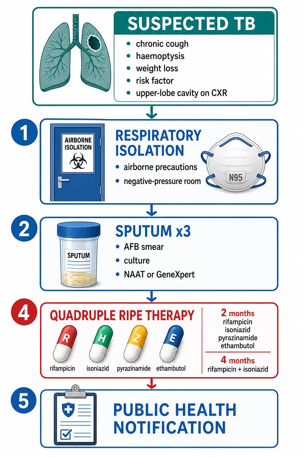
The ED role is the recognition and the containment.[1][3]
- The suspect — the chronic cough, the haemoptysis, the night sweats, the weight loss, the risk factor, the upper-lobe cavity on the radiograph.
- The isolate — the airborne precautions, the negative-pressure room, the N95 mask for the staff, the applied at the suspicion and BEFORE the confirmation.
- The sputum — the three samples for the AFB, the culture, and the NAAT.
- The public-health notification — the TB is the notifiable disease; the contact tracing and the public-health team.
- The disposition — the admission for the unwell, the immunocompromised, the MDR-suspected, the socially-unstable; the discharge with the follow-up for the well, with the isolation instruction and the public-health handover.
The airborne isolation — the engineering and the respirator
The airborne-infection isolation room (the AIIR) is a single-occupancy, negatively pressurised room (the air flows in, not out), with at least 6 to 12 air changes per hour, the high-efficiency particulate air (the HEPA) filtration, and the anteroom. The door stays closed. The N95 respirator (or the higher — the P2/P3, the FFP2/FFP3) is worn by every entrant; the surgical mask is for the patient on transport, NOT for the staff protection (the surgical mask does not filter the droplet nuclei and does not seal). The patient wears the surgical mask when outside the room (the CT, the transport), and the staff wear the N95 throughout.[3]
The de-isolation is considered after the patient is on the effective therapy for at least 2 weeks, is clinically improving (the reduced cough, the defervescence, the reduced sputum), AND has the three negative sputum smears (or the negative Xpert). The single negative Xpert in the low-risk, clinically improving patient is used in some protocols to shorten the isolation.[3]
Airborne precautions
- Negative-pressure AIIR; door closed
- N95 respirator (or P2/P3, FFP2/FFP3) for staff
- HEPA filtration, >6–12 air changes/hour
- For TB, measles, varicella — the droplet nuclei
Droplet precautions
- Single room; surgical mask for staff within 1 m
- For the respiratory viruses, meningococcus, pertussis
- Large droplets >5 µm, short range
- NOT sufficient for the TB
Surgical mask (patient)
- On the patient during the transport
- Source control — catches the large droplets
- Does NOT protect the staff from the droplet nuclei
- Staff protection needs the N95
The ED pathway — the suspect, the isolate, the investigate, the treat, the notify
1 — Suspect
The chronic cough >3 wk, the haemoptysis, the night sweats, the weight loss, the risk factor (HIV, anti-TNF, migrant, diabetic). Triage flag to the airborne room.
2 — Isolate (before the confirmation)
Place in the negative-pressure AIIR. N95 respirator for every entrant. Door closed. Patient in a surgical mask if outside the room.
3 — Investigate
Three sputum samples (the morning + the spot, over 8–24 h) for the smear, the culture, and the GeneXpert. The chest radiograph (apical cavity). Bloods — the FBC, the LFT, the U&E, and the HIV. The IGRA is NOT the active-disease test.
4 — Treat (with the TB team)
The quadruple RIPE for the 2 months then the RH for the 4 months, with the pyridoxine and the baseline LFT/visual acuity/urate. The MDR-suspected → the specialist, do NOT start blindly.
5 — Notify
Notify the public health within the statutory window. The contact tracing, the source-case investigation, and the public-health handover before the discharge.
The management — the RIPE therapy
The quadruple RIPE therapy is the first-line — the rifampicin, the isoniazid, the pyrazinamide, the ethambutol for the 2 months (the intensive phase), then the rifampicin and the isoniazid for the 4 months (the continuation). The total of the 6 months. The direct observation in the non-adherent. The drug toxicity:[1][2]
- The rifampicin — the liver-function derangement, the orange-red discoloration of the urine and the secretions (the benign, the warn the patient), the induction of the cytochrome P450 (the contraceptive and the warfarin failure).
- The isoniazid — the hepatitis, the peripheral neuropathy (the prevent with the pyridoxine or the vitamin B6).
- The pyrazinamide — the hepatitis, the hyperuricaemia and the gout.
- The ethambutol — the optic neuritis (the red-green colour blindness, the visual acuity — the baseline and the monitoring), especially at the higher dose and the renal impairment.
Rifampicin (R)
- Bactericidal; kills the rapidly dividing bacilli
- Orange-red urine/tears/sweat — warn the patient (benign)
- Hepatitis — monitor the LFT
- Induces CYP450 — the OCP and the warfarin FAIL; check the interactions
Isoniazid (H)
- Bactericidal; kills the intracellular, slow bacilli
- Hepatitis — the age-related risk
- Peripheral neuropathy — prevent with pyridoxine (B6) 10–25 mg
- Lupus-like syndrome (rare)
Pyrazinamide (Z)
- Sterilises — kills the semi-dormant bacilli in the acidic centre
- Hepatitis (the dose-related)
- Hyperuricaemia and the gout — avoid in the acute gout
- Stopped after the 2-month intensive phase
Ethambutol (E)
- Added to cover the possible resistance until the sensitivities return
- OPTIC NEURITIS — the red-green and the acuity
- Baseline and the monthly acuity/colour; stop on the symptom
- Renal dose — the drug is renally excreted
The RIPE regimen — the numbers
The MDR-TB (the resistance to the rifampicin and the isoniazid) and the XDR-TB require the specialist regimen with the second-line drugs (the fluoroquinolone, the injectable, the newer agents) and the prolonged course.[2]
The drug resistance — the classification and the newer agents
The resistance is defined by the susceptibility pattern and it dictates the regimen, the duration and the public-health urgency. The isoniazid-monoresistant TB extends the rifampicin-ethambutol continuation. The rifampicin-monoresistant TB (the rr-TB, the rpoB mutation detected by the Xpert) is treated as the MDR-equivalent in much of the world. The MDR-TB (the rifampicin and the isoniazid resistance) and the pre-XDR (the MDR plus the resistance to a fluoroquinolone or an injectable) and the XDR (the MDR plus the resistance to a fluoroquinolone AND an injectable, and now a bedaquiline or a linezolid in the updated definition) need the prolonged, individualised, second-line regimen.[2][6]
The contemporary MDR regimen has shifted from the long injectable-based course toward the shorter, all-oral regimens built on the bedaquiline (the diarylquinoline, the ATP-synthase inhibitor), the delamanid and the pretomanid (the nitroimidazoles), the linezolid (the oxazolidinone) and a fluoroquinolone. The bedaquiline, validated in the phase-2 trial of Diacon and colleagues, transformed the MDR outcomes and the tolerability, and it is now the cornerstone of the WHO-recommended shorter MDR regimen.[6]
Drug-susceptible TB
- RIPE 2 months + RH 4 months (total 6)
- The cure rate >95% with the adherence
- Oral, well-tolerated, low-cost
- Public-health: standard notification + contacts
MDR-TB
- Resistant to rifampicin AND isoniazid
- Shorter all-oral (bedaquiline-based) or longer regimen
- 9–18 months; lower cure, higher mortality
- Specialist-only; full resistance testing; strict isolation
Pre-XDR / XDR-TB
- MDR + fluoroquinolone and/or injectable resistance
- XDR: + bedaquiline or linezolid resistance
- Individualised, prolonged, low-cure regimens
- Highest public-health priority — refer urgently
Boehme 2010 — GeneXpert MTB/RIF (NEJM)
New England Journal of Medicine
PMID 20825313
Key finding
A multicentre evaluation of the Xpert MTB/RIF cartridge on a single sputum, in 1730 patients with the suspected drug-resistant or the HIV-associated TB. The sensitivity for the culture-positive TB was over 98 per cent in the smear-positive and over 72 per cent in the smear-negative disease (rising to over 90 per cent with a second sample), with the rifampicin-resistance detection in under 2 hours.
Practice change
Established the Xpert MTB/RIF as the rapid, closed, PCR-based first test for the suspected TB and the rifampicin resistance — the foundation of the modern WHO-endorsed molecular-first strategy, replacing the smear as the initial diagnostic.
Dorman 2021 — TBTC Study 31 / AIDS Clinical Trials Group A5349 (NEJM)
New England Journal of Medicine
PMID 33951360
Key finding
A phase-3 non-inferiority trial of 2516 patients with the drug-susceptible pulmonary TB, comparing a four-month rifapentine-moxifloxacin regimen and a four-month rifapentine regimen against the standard six-month control. Both four-month regimens were non-inferior for the 12-month culture-negative outcome, with a similar safety profile.
Practice change
Shortened the treatment of the selected drug-susceptible TB from six to four months — the first successful phase-3 shortening trial in decades, and an option for the selected patient, though the six-month RIPE remains the default in much of the world.
Diacon 2009 — bedaquiline for MDR-TB (NEJM)
New England Journal of Medicine
PMID 19494215
Key finding
A placebo-controlled phase-2 trial of the bedaquiline (the diarylquinoline TMC207) added to the standard second-line regimen in 47 patients with the MDR-TB. The bedaquiline raised the 8-week culture-conversion rate from 9 per cent to 48 per cent, with a manageable adverse-event profile (the QT prolongation was the principal concern).
Practice change
The first-in-class evidence that transformed the MDR-TB therapy, paving the way to the all-oral, shorter, bedaquiline-based regimens that replaced the long injectable courses.
Tagmouti 2014 — IGRA reproducibility (Ann Am Thorac Soc)
Annals of the American Thoracic Society
PMID 25188809
Key finding
A systematic review of the reproducibility of the interferon-gamma release assays across the serial testing. The agreement between the repeated tests was imperfect, with the reversions and the conversions around the cut-off, and the variability higher near the threshold.
Practice change
The IGRA is a useful single test for the latent TB, but a borderline result near the cut-off should NOT be over-interpreted in the serial screening; the active TB is still diagnosed by the microbiology, never by the IGRA alone.
Colditz 1995 — BCG efficacy meta-analysis (Pediatrics)
Pediatrics
PMID 7596718
Key finding
A meta-analysis of the published BCG-vaccination trials, examining the protection against the tuberculosis in the newborns and the infants. The overall protection against the meningeal and the miliary TB was high (around 80 per cent), while the protection against the pulmonary disease was variable and lower across the populations.
Practice change
Established that the neonatal BCG protects principally against the severe disseminated and the meningeal TB of the childhood — the basis for the selective neonatal BCG policy in the low-burden countries and the universal policy in the high-burden countries.
The latent tuberculosis infection (the LTBI)
The latent TB infection is the viable but dormant bacillus in the immune host — the positive TST or IGRA, the normal chest radiograph, the no symptoms and the no contagiousness. It is treated to prevent the future reactivation, and the treatment choice is graded by the reactivation risk. The 3HP (the rifapentine plus the isoniazid weekly for 3 months, the direct observation historically, now self-administered), the 4R (the rifampicin daily for 4 months) and the 6H to 9H (the isoniazid daily for 6 to 9 months, with the pyridoxine) are the principal regimens; the shorter rifamycin-based regimens are preferred for the completion and the lower hepatotoxicity, and the isoniazid is the historic but longer and more hepatotoxic option. The active TB is ALWAYS excluded (the chest radiograph, the symptom screen, the sputum if any feature) before the LTBI treatment, to avoid treating an active case with a single drug and breeding the resistance.[1][5]
The TB and the HIV — the special rules
The TB and the HIV co-infection is a two-way accelerator: the HIV drives the TB reactivation and the progression, and the TB accelerates the HIV. The presentation may be atypical — the smear-negative, the extrapulmonary, the miliary, the lower-lobe or the intrathoracic-lymph-node pattern, and the TB meningitis. The GeneXpert (the Xpert Ultra) is the first test (the higher yield than the smear in the paucibacillary disease). The timing of the antiretroviral therapy is critical — start early (within the first 2 weeks for the CD4 under 50) but watch for the immune reconstitution inflammatory syndrome (the IRIS, the TB-IRIS), the paradoxical worsening of the TB as the immunity recovers. The rifampicin interacts with the antiretrovirals (the protease inhibitors are the worst); coordinate with the HIV team. The MAC prophylaxis with the azithromycin does not treat the TB.[1][2]
The TB in the pregnancy, the child and the renal failure
In the pregnancy, the TB is treated with the standard RIPE — the rifampicin, the isoniazid (with the pyridoxine) and the ethambutol are safe; the pyrazinamide is traditionally avoided in the first trimester in some guidelines (the limited data) though the WHO includes it throughout. Untreated TB harms the mother and the foetus more than the treatment. In the child (under 5), the disease is often paucibacillary and the disseminated or the meningeal; the sputum is hard to obtain (the gastric aspirate, the induced sputum, the Xpert Ultra), and the disseminated and the meningeal TB are the indications for the LP and the high index of suspicion. The renal failure and the dialysis patient has a high reactivation risk and a delayed clearance of the ethambutol and the pyrazinamide — dose-adjust and monitor the LFT, the vision and the urate.[1]
The complications and the prognosis
The complications are the massive haemoptysis, the bronchiectasis, the chronic pulmonary aspergillosis (the fungal ball in the old cavity), the bronchopleural fistula, the Addison disease (the adrenal), and the death. The prognosis is the generally favourable with the completion of the therapy, but the MDR-TB carries the high mortality.[1][2]
[1]The public health and the notifiable disease
The tuberculosis is a notifiable disease in virtually every jurisdiction, and the ED clinician has the statutory duty to notify the public-health unit within the local window (often within 1 working day for the suspected, and immediately for the MDR). The notification triggers the contact tracing (the household and the close contacts, the concentric-circle approach, with the TST/IGRA and the symptom screen, and the LTBI treatment of the infected contacts), the source-case investigation for the paediatric case, and the public-health oversight of the therapy (the directly-observed treatment in the non-adherent). The failure to notify is both a clinical and a legal failure.[1]
The prevention — the BCG and the infection control
The BCG vaccine (the live-attenuated Mycobacterium bovis) protects principally against the disseminated, the miliary and the meningeal TB of the childhood (around 80 per cent), with a variable and lower protection against the pulmonary disease; it is given at the birth in the high-burden countries and selectively (the high-risk neonate) in the low-burden countries.[8] The infection control — the early recognition, the prompt airborne isolation, the N95 respirator, the negative-pressure room, the rapid molecular diagnosis, and the adequate therapy of the infectious case — is the foundation of the TB control in the hospital. The latent-TB screening and treatment of the high-risk groups (the migrant, the HIV, the dialysis, the transplant, the anti-TNF) prevents the future reactivation. The high-burden countries pursue the active case-finding and the shorter regimens.
Common pitfalls
The recurring errors are: the failure to suspect (the chronic cough dismissed); the delay of the isolation (the staff and the patient exposure); the single sputum instead of the three; the omission of the HIV test; the under-recognition of the drug toxicity (the ethambutol optic neuritis, the isoniazid neuropathy, the rifampicin hepatitis); the failure to notify the public health; and the non-adherence with the prolonged therapy (the MDR). [1]
[1] [1] [1] [1] [1] [1] [1] [1] [1]SAQ — Smear-positive pulmonary tuberculosis in the ED
10 minutes · 10 marks
A 34-year-old man who arrived from India 8 months ago presents to the ED with a 6-week history of productive cough, haemoptysis for 3 days, night sweats and a 6 kg weight loss. He is a non-smoker. T 37.9, RR 22, SpO2 95 per cent on room air. Chest radiograph shows a right upper-lobe cavity with surrounding infiltrate.
SAQ — Tuberculous meningitis in the immunocompromised host
10 minutes · 10 marks
A 42-year-old man with HIV (CD4 80, not on antiretroviral therapy) presents with a 2-week history of headache, fever, confusion and a single generalised seizure. GCS 13, neck stiffness, no focal deficit. CT head shows communicating hydrocephalus with basal enhancement.
Red flags
The following features identify the TB or the dangerous situation, in which the isolation and the sputum and the notify are the priority: [1]
[1]Exam pearls
- The ED mantra: suspect, isolate, sputum, notify — the isolation is the clinical decision made at the suspicion, before any test.
- The airborne precautions: the negative-pressure AIIR and the N95 respirator. The surgical mask is for the patient, NOT for the staff.
- The three sputum samples (at least one a morning, over 8–24 h) for the smear, the culture and the GeneXpert — never the single sputum.
- The GeneXpert MTB/RIF (the Xpert Ultra) detects the TB AND the rifampicin resistance in 2 hours — the WHO-recommended first test, replacing the smear.[4]
- The IGRA does NOT distinguish the latent from the active disease — the active TB is diagnosed by the microbiology. The IGRA/TST is for the LTBI and the prior exposure.[7]
- The upper-lobe cavity = the reactivation TB; the lower-lobe + the hilar node = the primary (the Ghon focus).
- The RIPE: rifampicin, isoniazid, pyrazinamide, ethambutol for 2 months, then the RH for 4 months — total 6 months. The pyrazinamide is dropped after the intensive phase.
- The pyridoxine (B6) 10–25 mg co-prescribed with the isoniazid to prevent the peripheral neuropathy — mandatory in the pregnant, the diabetic, the HIV, the alcoholic, the renal-failure.
- The rifampicin induces the CYP450 — the OCP fails, the warfarin dose rises, the protease-inhibitor antiretrovirals need adjustment.
- The ethambutol optic neuritis — the red-green colour blindness and the acuity; the baseline Ishihara and the acuity, the renal dose-adjust.
- The pyrazinamide — the hepatitis and the hyperuricaemia/gout; stopped after 2 months.
- The HIV test is mandatory in every suspected TB.
- The TB meningitis — the lymphocytic CSF, the low glucose, the high protein; treat empirically on the suspicion.
- The pleural TB — the lymphocytic exudate, the high ADA; the pleural biopsy (the Abrams) is more sensitive than the fluid culture.
- The miliary TB — the millet-seed CXR, the marrow pancytopenia, the choroidal tubercles; look for the co-existent meningitis.
- The LTBI — the positive IGRA/TST, the normal CXR, the no symptoms; treat with the 3HP, the 4R or the 6–9H to prevent the reactivation; exclude the active TB first.[5]
- The MDR-TB — the rifampicin + the isoniazid resistance; the bedaquiline-based all-oral shorter regimen.[6]
- The BCG protects the child against the miliary and the meningeal TB (around 80 per cent), not reliably against the pulmonary disease.[8]
- The notification is statutory — notify the public health for the contact tracing and the oversight.
- The de-isolation needs the effective therapy for at least 2 weeks, the clinical improvement, and the three negative smears (or the negative Xpert).[3]
References
- [1]Furin J, Cox H, Pai M. Tuberculosis Lancet, 2019.PMID 30904262
- [2]Lange C, Dheda K, Chesov D, Mandalakas AM, Udwadia Z. Management of drug-resistant tuberculosis Lancet, 2019.PMID 31526739
- [3]Menon LJB, Feliciano CS, de Campos MR, Bollela VR. Decision making to discharge patients from airborne infection isolation rooms: The role of a single GeneXpert MTB/RIF strategy in Brazil Infect Control Hosp Epidemiol, 2020.PMID 32299529
- [4]Boehme CC, Nabeta P, Hillemann D, et al. Rapid molecular detection of tuberculosis and rifampin resistance N Engl J Med, 2010.PMID 20825313
- [5]Dorman SE, Nahid P, Kurbatova EV, et al. Four-Month Rifapentine Regimens with or without Moxifloxacin for Tuberculosis N Engl J Med, 2021.PMID 33951360
- [6]Diacon AH, Pym A, Grobusch M, et al. The diarylquinoline TMC207 for multidrug-resistant tuberculosis N Engl J Med, 2009.PMID 19494215
- [7]Tagmouti S, Slater M, Benedetti A, et al. Reproducibility of interferon gamma (IFN-γ) release Assays. A systematic review Ann Am Thorac Soc, 2014.PMID 25188809
- [8]Colditz GA, Berkey CS, Mosteller F, et al. The efficacy of bacillus Calmette-Guérin vaccination of newborns and infants in the prevention of tuberculosis: meta-analyses of the published literature Pediatrics, 1995.PMID 7596718