ICU · Anatomy
Airway & Respiratory Anatomy
Also known as Airway anatomy · Respiratory anatomy · Larynx · Laryngeal cartilages · Thyroid cartilage · Cricoid cartilage · Arytenoid cartilage · Epiglottis · Vocal cords · Recurrent laryngeal nerve · Superior laryngeal nerve · Cricothyroid membrane · Trachea · Carina · Right main bronchus · Bronchopulmonary segments · Lung lobes · Pleura · Diaphragm · Phrenic nerve · Alveolus · Alveolar-capillary membrane · Surfactant · Type II pneumocyte · Pulmonary vascular tree · Diaphragm innervation
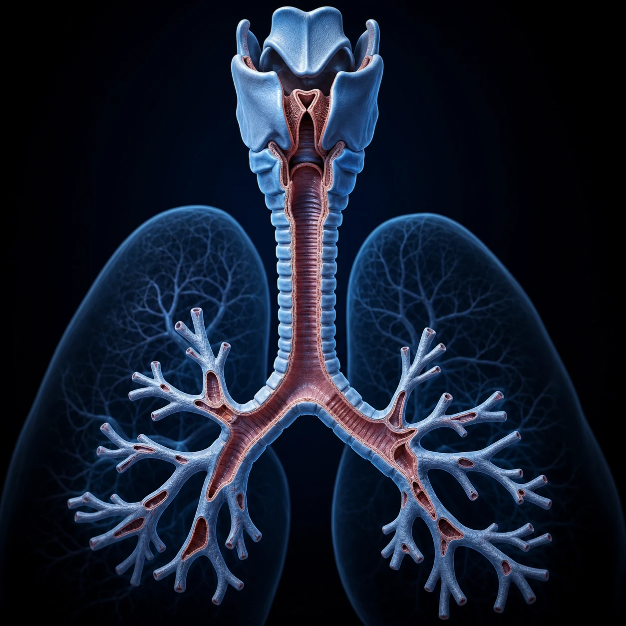
Airway and respiratory anatomy for the ICU First Part: the upper airway (nose, naso-/oro-/laryngopharynx, larynx and its cartilages — thyroid, cricoid as the only complete ring, arytenoid, epiglottis — vocal cords, and the superior and recurrent laryngeal nerves), the lower airway (the trachea 10-12 cm with C-shaped rings, the carina at T4/T5, the right main bronchus wider/shorter/more vertical as the aspiration path), the bronchopulmonary segments (ten right, eight to ten left), lung lobes and hilum, the pleura (visceral and parietal, the pleural space and fluid), the conducting vs respiratory zones (generations 0-23), the alveolar-capillary membrane, the pulmonary vascular tree, and the muscles of breathing (the diaphragm via the phrenic nerve C3-C5).
On this page & tools
Your progress
Saved locally on this device.
8 MCQs with explanations
Target exams
Overview
The airway is described from outside in as the upper airway (nose, mouth, pharynx, larynx) and the lower airway (trachea, bronchi, bronchioles, alveoli). Anatomical detail matters in ICU because it governs airway manoeuvres, the site of tube placement, the route of aspiration, and nerve injuries from intubation or surgery.[1]
In the fellowship viva the airway is best delivered as a continuous journey of a gas molecule — nose → nasopharynx → oropharynx → laryngopharynx → larynx (glottis) → trachea → carina → main bronchi → lobar → segmental → subsegmental bronchi → bronchioles → terminal bronchiole (end of conducting zone) → respiratory bronchiole → alveolar duct → alveolar sac → alveolus — naming the level, the structure that matters there, and the clinical correlate at each step.[1]
The upper airway
Nose and nasal cavity
- The nose warms, humidifies, and filters inspired gas; the vascular mucosa bleeds readily (relevant to nasal intubation).[1]
- The three turbinates (superior, middle, inferior conchae) create a turbulent laminar flow that maximises contact with the mucosa and deposits particles >10 µm; air is warmed to ~32°C and fully humidified by the time it reaches the nasopharynx.[1]
- Kiesselbach's plexus (Little's area) on the anterior nasal septum is the confluence of the sphenopalatine, anterior and posterior ethmoidal, superior labial and greater palatine arteries — the source of >90% of epistaxes and the site traumatized by a nasal tube or nasal airway.[1]
- The paranasal sinuses (frontal, ethmoidal, maxillary, sphenoidal) lighten the skull and resonate the voice; a nasotracheal tube can obstruct their ostia and precipitate sinusitis in the ventilated patient.[1]
Nasopharynx, oropharynx and laryngopharynx
- The pharynx (naso-, oro-, laryngo-) is a shared air and food passage; the soft palate and epiglottis separate the streams during swallowing.[1]
- Nasopharynx (skull base to soft palate): the Eustachian tube opening and the adenoids (pharyngeal tonsil) lie here — relevant to nasogastric/nasotracheal placement and to upper-airway obstruction in children.
- Oropharynx (soft palate to vallecula): contains the palatine tonsils and the base of tongue; the vallecula (anterior to the epiglottis) and piriform fossae (lateral to the laryngeal inlet) are the recesses where secretions pool and where a foreign body or food bolus lodges.
- Laryngopharynx (hyoid to cricoid, continuous with the oesophagus): the piriform fossae here are innervated by the internal laryngeal nerve — a "lump in the throat" (globus) and referred ear pain via the vagus arise from this region.[1]
The larynx
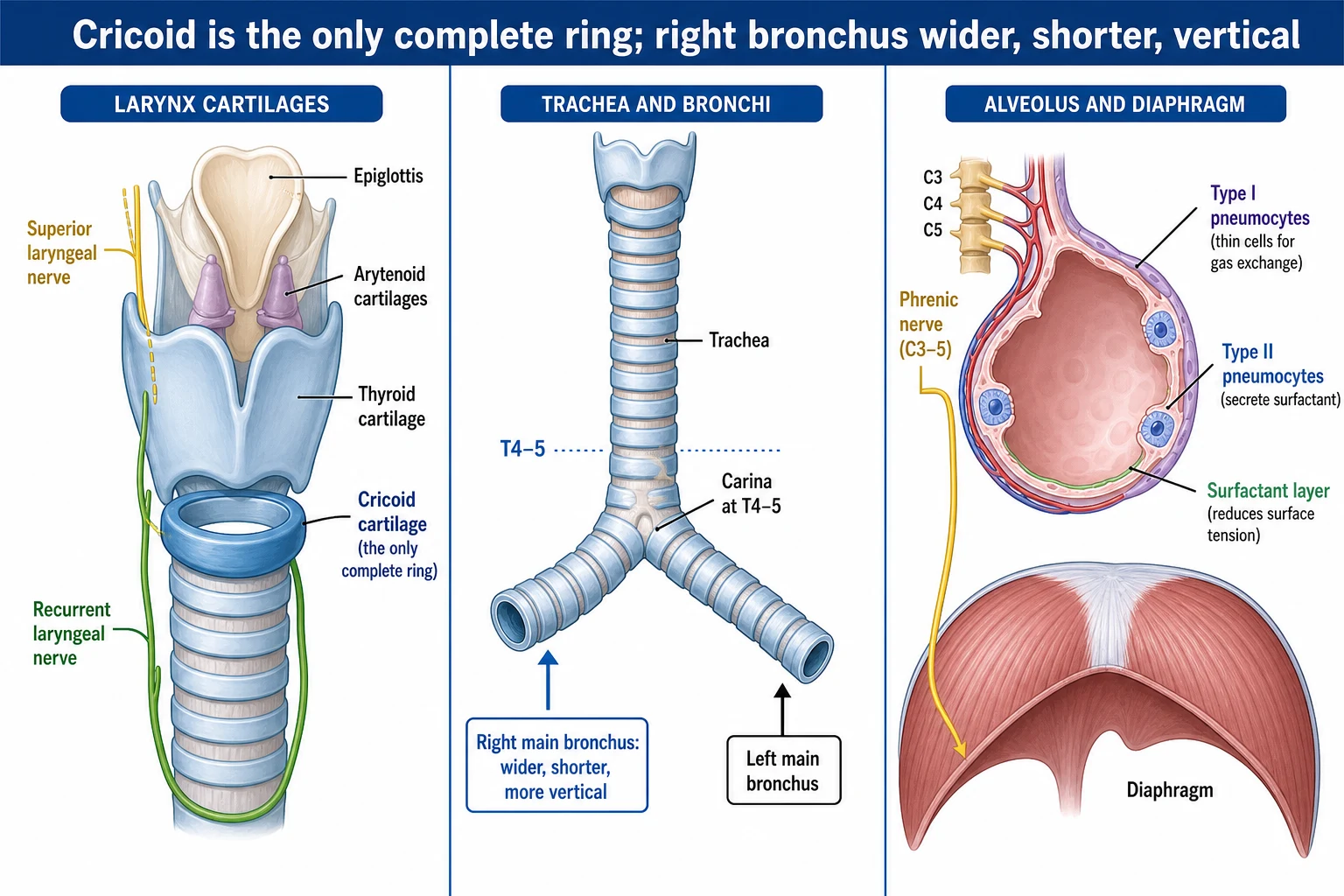
- Cartilages: thyroid, cricoid (the only complete ring - the basis for cricoid pressure and the Sellick manoeuvre), arytenoid, corniculate, cuneiform, and the epiglottis.[1]
- The vocal folds (cords) define the glottis - the narrowest part of the adult airway (in a child the narrowest part is the subglottic cricoid cartilage).[1]
The laryngeal cartilages — unpaired vs paired, and the one fact examiners want from each
| Cartilage | Number | Shape / location | The exam point |
|---|---|---|---|
| Thyroid | Unpaired (largest) | Shield-like; two laminae fuse anteriorly at ~90° (men → laryngeal prominence, "Adam's apple") | Superior & inferior horns; the cricothyroid membrane stretches between its inferior border and the cricoid below — the target of cricothyroidotomy |
| Cricoid | Unpaired | Signet-ring; the ONLY complete cartilaginous ring, narrow anteriorly (arch) and broad posteriorly (lamina) | The basis for cricoid pressure (Sellick) and the paediatric narrowest point of the airway (subglottic) — why uncuffed tubes were historically used in children <8 y |
| Epiglottis | Unpaired | Leaf-shaped elastic cartilage behind the tongue root | Diverts food into the piriform fossae; attached to the thyroid by the thyroepiglottic ligament and to the tongue by the glossoepiglottic fold |
| Arytenoid | Paired | Pyramidal; sits on the cricoid lamina at the cricoarytenoid joint | Vocal process (anterior — the vocal ligament attaches here) and muscular process (lateral — intrinsic muscles attach); rocking/ gliding the arytenoid abducts and adducts the cords |
| Corniculate | Paired | Small; sits on the apex of each arytenoid | Form the swellings of the interarytenoid notch seen at laryngoscopy |
| Cuneiform | Paired | Small, rod-like, in the aryepiglottic fold | The other pair of swellings beside the corniculates |
The laryngeal membranes and ligaments — the anatomy behind airway procedures
| Membrane / ligament | Between | Clinical relevance |
|---|---|---|
| Thyrohyoid membrane | Superior thyroid horn ↔ hyoid | Pierced by the superior laryngeal nerve (internal branch) and superior laryngeal artery — a nerve block target for awake intubation |
| Cricothyroid (median cricothyroid) membrane | Inferior thyroid border ↔ cricoid arch | The target of surgical cricothyroidotomy and of transtracheal needle cannula; palpable as the dip between the firm cricoid and the thyroid prominence in the midline |
| Conus elasticus (cricothyroid membrane proper) | Cricoid arch ↔ vocal ligaments above | Its free upper margin thickens into the vocal ligament (the skeleton of the true cord) |
| Quadrangular membrane | Lateral epiglottis/arytenoids ↔ false cords | Its free lower edge = the vestibular ligament (skeleton of the false cord); upper edge = the aryepiglottic fold |
| Vocal ligament | Vocal process of arytenoid ↔ thyroid cartilage (anterior) | The fibrous core of the true vocal fold; tension set by the cricothyroid muscle |
The vocal cords (true and false)
- The true vocal folds comprise the vocal ligament, the vocalis muscle (medial thyroarytenoid) and overlying mucosa; vibration of their free edge produces voice. They define the rima glottidis (the glottis).[1]
- The false (vestibular) folds lie superiorly, play no part in phonation, and protect the glottis.[1]
- The glottis is the narrowest part of the adult airway; in a child the narrowest point is the subglottic cricoid (a circular, rigid, conical segment).[1]
- A cuff leak that appears only as the cuff pressure falls below the glottic-tissue compression pressure, or a cuff that needs to be over-inflated to seal, suggests the tube is too large for the subglottic lumen — a cause of post-extubation stridor, especially in children.[7]
Laryngeal innervation (examinable)
- Superior laryngeal nerve (a branch of the vagus): provides sensation to the mucosa above the cords, and motor supply to the cricothyroid muscle (which tenses the cords).[1]
- Recurrent laryngeal nerve (also vagus): provides sensation below the cords and motor supply to all the other intrinsic laryngeal muscles. The left nerve loops under the aortic arch (and so is vulnerable to mediastinal or thoracic pathology); the right loops under the right subclavian artery. Unilateral injury causes a hoarse voice; bilateral injury causes stridor and may require intubation or tracheostomy.[1]
The two laryngeal nerves — the branch, the territory, the injury, the cause
| Feature | Superior laryngeal nerve (SLN) | Recurrent laryngeal nerve (RLN) |
|---|---|---|
| Origin | Vagus (CN X), high in the neck | Vagus; left loops under the aortic arch, right under the right subclavian artery |
| Branches | Internal (sensory, above cords; pierces thyrohyoid membrane) and external (motor) | One trunk; sensory (below cords) + motor |
| Motor supply | The cricothyroid muscle only (the tensor — tightens the cord for high-pitched voice) | All other intrinsic muscles — thyroarytenoid, posterior & lateral cricoarytenoid, transverse & oblique arytenoids, vocalis (i.e. abduction, adduction, relaxation) |
| Sensory supply | Mucosa above the cords (vallecula, piriform fossa, supraglottic larynx) | Mucosa below the cords (subglottis, trachea) |
| Effect of injury | Loss of high-pitched voice; aspiration on fluids (lost supraglottic sensation) | Unilateral → hoarse voice (cord paralysed in paramedian position); bilateral → stridor (cords apposed in midline) |
| Typical cause | Thyroid surgery (external branch runs on the inferior constrictor close to the superior thyroid artery) | Thyroid surgery, cardiac/thoracic surgery (left nerve under the aortic arch — CABG, mitral), high ETT cuff, mediastinal/Pancoast tumour, aortic aneurysm |
The trachea and bronchial tree
- The trachea is 10-12 cm long, supported by 16-20 C-shaped cartilaginous rings open posteriorly (closed by the trachealis muscle). It bifurcates at the carina at vertebral level T4/T5 (the sternal angle), opposite the angle of Louis.[1]
- The right main bronchus is wider, shorter, and more vertical than the left, so an aspirated object or a too-deep endotracheal tube usually enters the right (typically the right upper or lower lobe).[1]
- The lungs divide into bronchopulmonary segments: the right lung has three lobes (upper, middle, lower) and ten segments; the left lung has two lobes (upper, including the lingula, and lower) with eight to ten segments. Each segment can be individually resected.[1]
Tracheal anatomy — the numbers the examiner wants verbatim
| Parameter | Value | Why it matters |
|---|---|---|
| Length | 10-12 cm in the adult | Sets the distance from cords to carina; the working length for a tracheostomy tract |
| Diameter | ~2.0-2.5 cm | An 8.0 mm ETT occupies much of the lumen — leak and airflow matter |
| Number of rings | 16-20 C-shaped rings | The "C" is open posteriorly (flat against the oesophagus), closed by the trachealis smooth muscle — a posterior tracheal tear (intubation injury) is through this membrane |
| Origin | Lower border of the cricoid at C6 | The landmark for cricothyroidotomy and for surgical cricothyroid access |
| Bifurcation (carina) | T4/T5 vertebral level — the sternal angle, angle of Louis | The fixed keel-shaped ridge the ETT tip must sit 2-3 cm above; a carinal tumour or widening is a red flag |
| Mucosa | Ciliated pseudostratified columnar epithelium, goblet cells, submucosal seromucous glands | The mucociliary escalator — paralysis (intubation bypass) + ciliary damage (high FiO2/smoke) cause retained secretions and atelectasis |
Right vs left main bronchus — the single most examined airway comparison
| Feature | Right main bronchus | Left main bronchus |
|---|---|---|
| Width | Wider | Narrower |
| Length | Shorter (~2.5 cm) | Longer (~5 cm) |
| Angle / verticality | More vertical (continues almost straight down from the trachea, ~25° from vertical) | More horizontal (~45° from vertical) |
| Consequence — aspiration | An aspirated tooth, vomitus or foreign body enters the RIGHT — classically the right lower lobe (most dependent) or the right upper lobe (the RUL bronchus comes off almost horizontally just below the carina) | The left is spared because of its angle |
| Consequence — tube placement | A too-deep ETT enters the right main bronchus → ventilates only the right lung → left lung collapse, ↑ peak pressure | A left double-lumen tube must negotiate this longer, narrower, more horizontal path |
| Origin of upper-lobe bronchus | RUL bronchus arises ~1 cm below the carina — an ETT advanced only slightly too far can occlude it | LUL bronchus arises further out |
Bronchopulmonary segments
Each lung is subdivided into bronchopulmonary segments, each aerated by its own tertiary (segmental) bronchus and each with a separate arterial supply — so each can be individually resected (segmentectomy) without violating its neighbours.[1]
The bronchopulmonary segments — ten on the right, eight-to-ten on the left
| Lobe | Right lung segments | Left lung segments |
|---|---|---|
| Upper lobe | Apical (1), Posterior (2), Anterior (3) | Apicoposterior (1+2), Anterior (3), Superior lingular (4), Inferior lingular (5) |
| Middle / lingula | Lateral (4), Medial (5) | (Lingula forms part of LUL — see above) |
| Lower lobe | Superior / apical (6), Medial basal (7), Anterior basal (8), Lateral basal (9), Posterior basal (10) | Superior / apical (6), Anteromedial basal (7+8), Lateral basal (9), Posterior basal (10) |
| Total | 10 segments | 8-10 segments (8 if apicoposterior and anteromedial are fused; 10 if split) |
The naming and numbering convention — say it cleanly in the viva
Lung anatomy — lobes, fissures and the hilum
- Right lung: three lobes — RUL, RML, RLL — separated by the horizontal fissure (between RUL and RML) and the oblique fissure (between RLL and RUL/RML). The horizontal fissure runs at the level of the 4th rib anteriorly.[1]
- Left lung: two lobes — LUL (with the lingula) and LLL — separated only by the oblique fissure (the left has no horizontal fissure; the lingula is the LUL segment that corresponds to the RML).[1]
- Each lung has a costal surface (ribs), a mediastinal surface (heart, great vessels), a diaphragmatic surface (base) and an apex (extends ~2-3 cm above the medial third of the clavicle into the neck — above the first rib, vulnerable to iatrogenic pneumothorax from a central line).[1]
The hilum (root of the lung)
- The hilum transmits the main bronchus, the pulmonary artery, the two pulmonary veins, the bronchial arteries and veins, lymphatics and autonomic nerves, all enclosed in a sleeve of mediastinal pleura.[1]
- The arrangement differs side to side. On both sides the bronchus is posterior, the pulmonary artery is superior-anterior, and the pulmonary veins are inferior — the classic "B-A-V" (bronchus-posterior, artery-superior, vein-inferior).[1]
- The right main bronchus lies posterior and slightly superior to the right pulmonary artery (eparterial position on the right is classically described for the RUL bronchus); on the left the pulmonary artery arches over the left main bronchus (hyperterial).[1]
The pleura
The pleura is a thin serous membrane that invests each lung and lines the hemithorax, creating the pleural space — a potential space whose negative pressure is what keeps the lung expanded against the chest wall.[1]
Visceral vs parietal pleura — the differences that explain symptoms and signs
| Feature | Visceral pleura | Parietal pleura |
|---|---|---|
| What it covers | The lung surface and dips into the fissures | Lines the chest wall (costal), the diaphragm (diaphragmatic), the mediastinum (mediastinal), and the lung apex (cervical / cupula — rises above the 1st rib into the neck) |
| Sensory innervation | Autonomic (visceral) — INSENSITIVE to pain and touch (no somatic pain fibres) | Somatic — pain-sensitive: costal via intercostal nerves, diaphragmatic (central) via the phrenic nerve (C3-5 → referred shoulder-tip pain), peripheral diaphragmatic via intercostal nerves |
| Why pleuritic chest pain is localised | — | Because the costal parietal pleura is somatic; pleurisy hurts sharply on inspiration at a point you can point to |
| Why a basal pleural problem refers to the shoulder | — | Irritation of the central diaphragmatic parietal pleura (phrenic C3-5) → referred pain to the shoulder (C3-4 dermatomes via the supraclavicular nerves) |
The pleural space and fluid
- The pleural space is a potential space containing only 5-10 mL of serous fluid that couples the lung to the chest wall by surface tension (the visceral and parietal layers never normally touch).[1]
- Intrapleural pressure is negative — about -5 cmH2O at end-expiration and -8 cmH2O at end-inspiration — generated by the inward elastic recoil of the lung pulling against the outward recoil of the chest wall. A pneumothorax equilibrates this to atmospheric pressure and the lung collapses.[1]
- Pleural fluid is secreted by the parietal pleura (mainly via the intercostal arteries' Starling filtration) and drained by the parietal pleural lymphatics (stomata → intercostal lymphatics); turnover is several hundred mL/day. An effusion forms when production (↑ capillary pressure in CCF, ↑ permeability in inflammation/infection) exceeds drainage.[1]
- Pleural recesses — the costodiaphragmatic recess (the lowest point; a chest drain placed too low or fluid drained too fast risks injuring the diaphragm/liver or spleen) and the costomediastinal recess (anterior, beside the sternum).[1]
The conducting and respiratory zones
- Conducting zone (airway generations 1-16, from the trachea to the terminal bronchioles): moves air but takes no part in gas exchange - the "anatomical dead space" (~2 mL/kg).[1]
- Respiratory zone (generations 17-23, from respiratory bronchioles to alveolar ducts and sacs): the site of gas exchange. The acinus is the unit distal to a terminal bronchiole.[1]
Airway generations 0-23 — the conducting vs respiratory zones, structure and function
| Generation | Structure | Zone | Cross-sectional area | Function / clinical point |
|---|---|---|---|---|
| 0 | Trachea | Conducting | Smallest total area | The "pipe" — bulk flow |
| 1-4 | Main, lobar, segmental bronchi | Conducting | Small | Cartilage-supported; site of lumbar aspirate/foreign body (gen 1, R main) |
| 5-16 | Subsegmental bronchi → bronchioles → terminal bronchiole (gen 16) | Conducting | Rising | Terminal bronchiole = last strictly conducting airway; end of dead space |
| 17-19 | Respiratory bronchioles | Respiratory (transition) | Large | Alveoli bud off the wall — gas exchange begins here |
| 20-22 | Alveolar ducts | Respiratory | Very large | Lined entirely by alveoli |
| 23 | Alveolar sacs (terminal) | Respiratory | ~Total cross-sectional area of the tract is ~10⁴× the trachea | The gas-exchange end-point |
Dead space — three types, all derived from this anatomy
| Type | Definition | Volume / fraction | Example in the ICU |
|---|---|---|---|
| Anatomical dead space | The conducting zone (gen 0-16) — ventilated but not perfused | ~2 mL/kg (~150 mL), ~30% of Vt | Doubles with a large ETT and circuit extension; reduced by tracheostomy (bypasses upper-airway dead space) |
| Alveolar dead space | Alveoli ventilated but not perfused | Normally negligible | Large in pulmonary embolism (ventilated but unperfused alveoli) and high PEEP over-distension |
| Physiological dead space | Anatomical + alveolar | Vd/Vt ~0.3-0.4 normally | A rising Vd/Vt (Bohr, Enghoff) >0.6 predicts failed weaning and mortality in ARDS/ventilated patients |
The alveolus and the alveolar-capillary membrane
- There are roughly 300 million alveoli, giving a gas-exchange surface of about 70 m squared.[1]
- Type I pneumocytes cover about 95 per cent of the alveolar surface and form the thin blood-gas barrier; they cannot divide.[1]
- Type II pneumocytes produce surfactant (dipalmitoylphosphatidylcholine, which lowers surface tension and prevents atelectasis) and are the stem cells that repopulate the epithelium after injury.[1]
- Alveolar macrophages clear debris and microbes.[1]
The cells of the alveolus — who does what, and the numbers
| Cell | % of alveolar surface | % of alveolar cells | Shape / role | Clinical correlate |
|---|---|---|---|---|
| Type I pneumocyte | ~95% | ~25% | Extremely thin, squamous; forms the blood-gas barrier | Cannot divide — injured by hyperoxia, smoke, high tidal volumes (volutrauma); the cell that fails in ARDS |
| Type II pneumocyte | ~5% | ~60% | Cuboidal; produces surfactant (DPPC + surfactant proteins A-D); the stem cell that differentiates into type I after injury | Surfactant production falls from day 1 of ventilation → atelectasis; surfactant proteins also mediate innate host defence (opsonisation of pathogens)[8] |
| Type III (brush) cell | <1% | rare | Microvilli; likely a sensory/chemoreceptor cell | Obscure; rarely examined |
| Alveolar macrophage (dust cell) | — | numerous | Phagocytoses particulates and microbes; migrates to the bronchiole to be carried up the mucociliary escalator | The cell that fails in immunosuppression and chronic smoke exposure; "heart-failure cells" when they have haemosiderin in pulmonary oedema |
The alveolar-capillary membrane (the blood-gas barrier)
- The barrier a gas molecule crosses has, from alveolus to red cell: surfactant + type I pneumocyte + fused basal lamina (alveolar and capillary) + capillary endothelium + plasma + erythrocyte membrane. At its thinnest it is ~0.2-2.5 µm.[1]
- Diffusing capacity falls whenever this barrier is thickened (pulmonary fibrosis, pulmonary oedema, ARDS, pneumocystis) or the surface area is reduced (pneumonectomy, emphysema, collapse, PE).[1]
- The membrane is asymmetric: on one side the capillary bulges into the alveolus — a thin side (gas exchange) and a thick side (fluid/solute exchange, where oedema first accumulates in the interstitium).[1]
The pores of Kohn and the channels of Lambert and Martin — collateral ventilation
| Channel | Connects | Function |
|---|---|---|
| Pores of Kohn | Adjacent alveoli | Collateral ventilation between alveoli; develop with age — absent in neonates (a reason neonatal atelectasis is hard to re-expand) |
| Channels of Lambert | Pre-terminal bronchiole ↔ adjacent alveoli | Collateral drift between a bronchiole and the alveoli of a neighbouring unit |
| Channels of Martin | Adjacent terminal bronchioles | Collateral ventilation at the bronchiolar level |
The pulmonary vascular tree
- Pulmonary circulation: the pulmonary artery carries deoxygenated blood from the right ventricle → right and left pulmonary arteries → lobar/segmental arteries → arterioles → the dense capillary network in the alveolar wall → venules → the four pulmonary veins → left atrium (oxygenated). It is a low-pressure (mean PAP 12-15 mmHg), low-resistance system that can recruit and distend capillaries as flow rises.[1]
- Hypoxic pulmonary vasoconstriction (HPV): pulmonary arterioles constrict in regions of low alveolar oxygen, diverting flow to better-ventilated alveoli — the lung's built-in V/Q optimiser. HPV is attenuated by vasodilators (nitric oxide is inhaled, so it does NOT abolish it; but systemic vasodilators — GTN, SNP, Ca-blockers, volatile anaesthetics — do), worsening shunt.[1]
- Bronchial circulation (the systemic supply of the airway itself): two left bronchial arteries arise from the descending thoracic aorta and one right from a right intercostal artery (usually the 3rd posterior intercostal). It nourishes the airway down to the respiratory bronchioles. Its venous return is partly to the azygos/hemiazygos (bronchial veins) and partly to the pulmonary veins — the latter is the anatomical (normal) shunt of ~1-2% of cardiac output.[1]
- The double circulation means a pulmonary embolus usually spares airway viability (the bronchial circulation keeps the parenchyma alive) — but it stops gas exchange (no perfusion), producing dead space.[1]
Pulmonary vs bronchial circulation — the two systems of the lung
| Feature | Pulmonary circulation | Bronchial circulation |
|---|---|---|
| Origin | Right ventricle (deoxygenated) | Aorta / systemic (oxygenated) |
| Pressure | Low (mean PAP 12-15 mmHg) | Systemic (high) |
| Gas exchange | Yes — the functional circulation | No — nourishes the airway wall to the respiratory bronchiole |
| Volume / shunt | ~100% of CO; no shunt normally | ~1-2% of CO drains to the pulmonary veins → the normal anatomical shunt |
| In pulmonary embolism | Blocked → dead space | Usually intact → keeps the lung viable, so infarction is the exception not the rule |
| Origin of massive haemoptysis | Less common source | The bronchial arteries are the usual source of massive haemoptysis (hypertrophied, high-pressure systemic) → bronchial artery embolisation is the treatment |
Muscles of breathing
- Inspiration is active: the diaphragm (the principal muscle, innervated by the phrenic nerve C3-5) descends and expands the chest; the external intercostals and scalenes assist. Accessory muscles (sternocleidomastoid, pectorals) recruit in distress ("3-5 keeps the diaphragm alive").[1]
- Expiration is passive at rest (elastic recoil); the internal intercostals and abdominal muscles drive forced expiration (and cough).[1]
The diaphragm — the principal inspiratory muscle
- The diaphragm is a musculofibrous dome separating the thorax from the abdomen; its central tendon is the aponeurotic core and the muscular fibres radiate from it to the xiphoid, the lower six ribs and the upper three lumbar vertebrae (the right and left crura).[1]
- It is innervated by the phrenic nerves (C3, C4, C5 — "C3, 4, 5 keeps the diaphragm alive") arising chiefly from C4; a spinal cord injury above C3 abolishes all breathing, at C3-4 the diaphragm may partially survive, and below C5 the diaphragm is spared but intercostal and abdominal muscles are lost.[1]
- In quiet breathing the diaphragm descends 1-2 cm; on deep inspiration up to 10 cm. Descent pushes the abdominal viscera down and raises intra-abdominal pressure; when the abdomen is splinted (obesity, pregnancy, tense ascites) the diaphragm is less effective and the accessory muscles compensate.[1]
- The three principal apertures transmit the IVC (T8, right crus), oesophagus (T10, right crus — the phreno-oesophageal ligament and the crural fibres form the lower oesophageal sphincter mechanism), and aorta (T12, behind/ between the crura).[1]
The other respiratory muscles
- External intercostals (T1-T11, intercostal nerves): elevate the ribs in inspiration (bucket-handle and pump-handle movements widen the chest).[1]
- Internal intercostals: the interosseous part depresses the ribs (forced expiration); the intercartilaginous part (parasternal) is inspiratory.[1]
- Scalenes (anterior, middle, posterior): elevate the first two ribs and are active even in quiet breathing — a common misconception is that they are only accessory muscles.[1]
- Accessory muscles of inspiration (recruited in distress): sternocleidomastoid (elevates the sternum), pectoralis major/minor, serratus anterior, latissimus dorsi, upper trapezius.[1]
- Muscles of expiration: the abdominal muscles (rectus abdominis, external/internal oblique, transversus abdominis) — by raising intra-abdominal pressure they push the diaphragm up; essential for cough and for the patient with a high spinal cord lesion, who can inspire but cannot cough effectively.[1]
The diaphragm vs the accessory muscles — and what fails in each injury
| Muscle group | Innervation | Action | Clinical point |
|---|---|---|---|
| Diaphragm | Phrenic C3-5 | Inspiration (primary) | C3+ cord injury → no breathing; unilateral phrenic palsy → raised hemidiaphragm, paradoxical movement on fluoroscopy ("sniff test") |
| External intercostals | Intercostal T1-T11 | Inspiration | Lost in lower cervical / thoracic cord injury → reduced chest expansion; paradoxic (flail) movement with multiple rib fractures |
| Sternocleidomastoid | Spinal accessory (CN XI) + C2-3 | Accessory inspiration | Recruited first in respiratory distress — the most visible sign of increased work of breathing |
| Abdominal muscles | T6-L1 | Forced expiration, cough, Valsalva | Lost in cord injury → ineffective cough → sputum retention, the commonest cause of respiratory failure in acute quadriplegia |
Exam practice — SAQs
SAQ — Predicted difficult airway in a hypoxic, obese patient with reflux
10 minutes · 10 marks
A 58-year-old obese man (BMI 42) with severe community-acquired pneumonia is admitted to ICU. He is increasingly hypoxic (SpO2 88 percent on 15 L oxygen via non-rebreather), RR 32 with accessory-muscle use and stertorous breathing. Examination: thick neck, full beard, receded mandible, inter-incisor distance 2.5 cm, thyromental distance 5 cm, Mallampati III. Known hiatus hernia with reflux; not fasted. You are called to intubate.
SAQ — Reduced compliance in ventilated ARDS
10 minutes · 10 marks
A 62-year-old woman (height 165 cm, weight 60 kg) with severe influenza A pneumonia is intubated and ventilated for ARDS. Volume-control ventilation: Vt 360 mL (6 mL/kg predicted body weight), PEEP 12 cmH2O, FiO2 0.8, set RR 28. Peak inspiratory pressure 42 cmH2O, plateau pressure (inspiratory hold) 32 cmH2O. Arterial blood gas: pH 7.28, PaO2 61 mmHg, PaCO2 52 mmHg. Chest X-ray shows bilateral diffuse alveolar infiltrates. No pneumothorax or pleural effusion.
Clinical pearls — exam-exhaustive airway and respiratory anatomy
FlowSteps — the clinical anatomy workflows
Localising a vocal-cord palsy — the anatomical reasoning from bedside sign to lesion site
Detect the sign: hoarse voice, weak cough, or inspiratory stridor
Hoarseness or a "bovine" cough suggests a unilateral cord palsy; biphasic or inspiratory stridor in a recently extubated or post-thyroid/cardiac-surgery patient suggests bilateral cord palsy — an airway emergency.
Confirm with nasendoscopy
A paralysed cord sits in the paramedian position (unilateral) or both apposed in the midline (bilateral). The cricothyroid-innervated cord still tenses in unilateral RLN injury but cannot abduct.
Map the nerve course to a lesion site
RLN: skull base → neck (thyroid surgery) → thorax. The LEFT loops under the aortic arch (mitral/CABG, aortic aneurysm, mediastinal mass); the RIGHT under the right subclavian. A LEFT cord palsy with no neck operation demands a chest CT.
Distinguish RLN from SLN (external branch) injury
RLN → cord paralysed in paramedian, abduction lost. SLN (external) → cord moves normally but cannot be tensed for high pitch; internal SLN loss → aspiration on fluids (lost supraglottic sensation).
Secure the airway if bilateral
Bilateral RLN palsy = stridor = intubate (small tube) or tracheostomy. Do not wait — the cords sit apposed and the airway will close.
Confirming endotracheal tube position — anatomy at every step
Pass the tube through the glottis under direct vision
The glottis is the narrowest part of the adult airway; the ETT cuff sits in the subglottic trachea below the cords. In a child the cricoid (subglottic) is the narrowest — use an uncuffed or carefully sized cuffed tube.
Bilateral chest rise + auscultate axillae and epigastrium
Equal air entry both sides, no epigastric gurgling. Absent left-sided air entry with preserved right = a too-deep tube in the right main bronchus (wider, shorter, more vertical) — withdraw 1-2 cm and recheck.
Quantitative waveform capnography
Sustained CO2 over six breaths excludes oesophageal intubation. The oesophagus lies immediately posterior to the trachea (against the open back of the C-rings) — the anatomical reason an oesophageal intubation can masquerade briefly.
Chest X-ray: tube tip 2-3 cm above the carina
The carina is at T4/T5 (angle of Louis). A tip too low enters the right main bronchus with head flexion; too high risks extubation with extension. Allow for neck movement shifting the tip ~2 cm.
Interpreting a raised hemidiaphragm on CXR — the anatomical differential
Confirm it is truly raised and paralysed
A raised dome can be due to low lung volume (atelectasis, pleural disease) without paralysis. Fluoroscopic sniff test: paradoxical ascent = phrenic nerve palsy. Ultrasound of diaphragm thickening fraction is the bedside alternative.
Side matters
Right: think cervical (C3-5) cord or nerve-root lesion, internal-jugular line trauma, phrenic in the pericardium (post-cardiac-surgery ice slush), or a subpulmonic process. Left: ALSO consider the recurrent-laryngeal-style mediastinal causes — an enlarged LA, aortic aneurysm, or esophageal/mediastinal mass compressing the phrenic or vagus.
Assess impact
Unilateral paralysis is often tolerated; bilateral or in obesity/COPD causes orthopnoea, sleep-disordered breathing and failure to wean. Measure sniff nasal inspiratory force and FVC in supine vs erect.
Treat if symptomatic and persistent
Surgical diaphragmatic plication (durable benefit, Hüttl 2004) for unilateral; pacing may help selected bilateral cases with intact lower-motor-neurons; mechanical ventilation / NIV for bilateral paralysis.
The cricoid pressure evidence — what the trials actually showed
Bhatia & Bhagat — Cricoid pressure: Where do we stand? (PMID 24574584)
Source
Journal of Anaesthesiology and Clinical Pharmacology, 2014 — narrative review of the evidence for and against cricoid pressure
Context
Sellick described cricoid pressure in 1961 to prevent regurgitation during RSI; it became near-universal dogma despite thin evidence
Key points
Cricoid pressure (a) occludes the oesophagus (which lies immediately posterior to the open tracheal rings) between the firm cricoid ring and the C6 vertebra; (b) is misapplied in up to 50% of cases; (c) can worsen laryngoscopic view (especially in the obese) and may itself cause airway obstruction if too forceful
Bottom line
Cricoid pressure is reasonable in the patient at high aspiration risk, but it is NOT mandatory; release it if it impairs the laryngoscopic view or ventilation. The modern trend is modified or no cricoid pressure.
Tessarolo et al. — Effectiveness and risks of cricoid pressure during RSI (PMID 35577760)
Source
Emergency Medicine Australasia, 2022 — ANZ review directly relevant to CICM/FFICM practice
Question
Does cricoid pressure prevent aspiration, and at what cost?
Findings
No randomised trial powered for the rare outcome of aspiration exists; observational data suggest cricoid pressure can IMPAIR laryngoscopic view, increase difficulty and intubation attempts, and may cause upper-airway obstruction. Evidence for preventing aspiration is largely inferential (cadaver/oesophageal-pressure studies)
Bottom line
Use cricoid pressure selectively in high-risk patients; if it impedes the airway manoeuvre, release or reduce it. The 'cricoid-or-nothing' reflex has no evidential basis.
Dunn — Cricoid Pressure: Contradictory Evidence Regarding a Standard Practice (PMID 35476194)
Source
AORN Journal, 2022
Theme
Surveys the contradictory evidence behind a long-standing perioperative 'standard' and the trend toward modified/abandoned use
Practical implication
When cricoid pressure is used, 10 N (1 kg) in the awake patient rising to 30-40 N after loss of consciousness is the textbook force; forces >40 N distort the airway and impair intubation
Bottom line
A practice of 'apply, but release if it impairs the view' is defensible; routine blind application is increasingly hard to justify.
Mahfood et al. — Reexpansion pulmonary edema (PMID 3279931)
Source
Annals of Thoracic Surgery, 1988 — the classic case series establishing the predictable complication of rapid lung re-expansion
Mechanism
A lung collapsed for days develops capillary leak, interstitial oedema and surfactant loss; rapid re-expansion floods the interstitium and alveoli
Risk factors
Drainage of >1.5 L in one sitting, large pneumothorax of >3 days' duration, application of high negative suction, young age
Clinical features
Cough, chest tightness, hypoxia and frothy/pink sputum developing within 1-24 h of drainage, on the DRAINED side; mortality historically ~20%
Bottom line
Drain slowly — stop at 1.5 L or if the patient coughs/complains; avoid routine high negative suction; this anatomical physiology underpins safe pleural drainage.
Weibel ER — Lung morphometry: the link between structure and function (PMID 27981379)
Source
Cell and Tissue Research, 2017 — definitive synthesis from the founder of lung stereology
Core facts
The human lung has ~300 million alveoli and a gas-exchange surface of ~70-85 m2; the airway tree branches over 23 generations from trachea to alveolar sacs; the conducting zone (gen 0-16) narrows and lengthens until total cross-sectional area rises ~10,000-fold at the respiratory zone
Structure-function link
The enormous terminal cross-sectional area means flow velocity falls to near-zero at the alveolar ducts — gas moves by DIFFUSION in the respiratory zone, not convection; this is the morphometric basis of gas exchange
Bottom line
The numbers examiners cite (300 million alveoli, 70 m2, 23 generations) all derive from the Weibel morphometric school — quote the source.
Nazer — Topical Ice Slush and diaphragmatic function after CABG (PMID 28473213)
Source
Heart, Lung and Circulation, 2018
Finding
Topical ice slush used for myocardial protection during CABG measurably reduced sniff nasal inspiratory force postoperatively — objective evidence of phrenic nerve cold injury
Anatomical basis
The phrenic nerve runs anterior to the root of the lung in the pericardium, directly exposed to the iced saline bathed over the heart
Clinical implication
Suspect phrenic injury in the post-cardiac-surgery patient who fails to wean, especially after topical hypothermia; raised hemidiaphragm on CXR + sniff test confirm. Most recover; persistent symptomatic cases need plication.
Whitsett — Surfactant homeostasis and innate host defence of the lung (PMID 20351134)
Source
Innate Immunity, 2010 — review
Key teaching
Surfactant is NOT just an anti-atelectasis agent. The surfactant proteins A and D are collectins — pattern-recognition molecules that opsonise bacteria and viruses, regulate alveolar macrophages, and are an integral part of pulmonary innate immunity
ICU relevance
Type II pneumocyte injury (from high-stretch ventilation, oxygen toxicity, infection) depletes both the surface-active lipid and the host-defence proteins — surfactant dysfunction is both a cause and consequence of ARDS
Bottom line
When you explain 'surfactant lowers surface tension', add that its proteins A and D are innate-immunity molecules — the exam-exhaustive answer.
Menon et al. — Laryngeal injury following endotracheal intubation (PMID 36168788)
Source
Anaesthesia and Intensive Care, 2023 — ANZ review
Spectrum of injury
Mucosal oedema, granuloma, ulceration, vocal-cord palsy (RLN compression by a high cuff or tube tip), arytenoid dislocation (traumatic intubation), subglottic stenosis (long-term)
Novel angle
Gastro-oesophageal reflux around the tube is increasingly implicated in peri-glottic injury — refluxate pools in the glottis because the ETT stents the cords open and the cuff lies BELOW the cords
Bottom line
The larynx is the narrowest adult airway and is anatomically fixed — minimise cuff pressure (20-30 cmH2O), avoid oversized tubes, limit time intubated, and consider reflux control in unexplained post-extubation laryngeal dysfunction.
Hüttl et al. — Laparoscopic diaphragmatic plication for phrenic nerve palsy (PMID 15108692)
Source
Surgical Endoscopy, 2004
Intervention
Laparoscopic plication of the paralysed hemidiaphragm (plication makes the dome taut so the abdominal viscera no longer push it up into the hemithorax)
Outcome
Durable long-term improvement in FVC, dyspnoea and exertional capacity in symptomatic unilateral paralysis
Bottom line
Most phrenic palsies recover spontaneously; plication is reserved for persistent, symptomatic unilateral paralysis. An option to know for the post-cardiac-surgery patient with a chronically raised hemidiaphragm.
Red flags
Mnemonics
The four 'complete ring' rules of the airway — what is circular, what is open
The phrenic nerve — 'C3, 4, 5 keeps the diaphragm alive'
The recurrent laryngeal nerve asymmetry — 'Left under the arch, right under the subclavian'
Common viva questions and model answers
Anatomy viva — the recurring questions and the one-line model answer
| Question | Model answer |
|---|---|
| "What is the narrowest part of the adult airway? And in a child?" | Adult = the glottis (rima glottidis, between the true vocal cords). Child = the subglottic cricoid cartilage (the only complete ring — circular, rigid, conical) — the basis for uncuffed tubes historically in children <8 years.[1] |
| "Why does an aspirated foreign body usually lodge in the right lung?" | The right main bronchus is wider, shorter (~2.5 cm), and more vertical than the left, so it is the line of least resistance from the trachea; the RUL bronchus comes off almost horizontally just below the carina and is a second common trap.[1] |
| "What is the only complete cartilaginous ring in the airway, and why does it matter?" | The cricoid — the basis for cricoid pressure (Sellick) during RSI and for the paediatric narrowest point being subglottic.[2] |
| "At what vertebral level does the trachea bifurcate?" | The carina at T4/T5 — the sternal angle, angle of Louis. The ETT tip should sit 2-3 cm above it.[1] |
| "Which nerve supplies the cricothyroid muscle, and which supplies all the others?" | The external branch of the superior laryngeal nerve supplies cricothyroid (the tensor). The recurrent laryngeal nerve supplies all other intrinsic laryngeal muscles (abductors and adductors).[1] |
| "A patient is hoarse after thyroid surgery. What has happened?" | A unilateral recurrent laryngeal nerve injury — the cord is paralysed in the paramedian position. Confirm with nasendoscopy. Most recover; persistent cases may need vocal-fold medialisation. Bilateral injury is an airway emergency (stridor).[1] |
| "A patient is hoarse but has never had neck surgery. Where is the lesion?" | Especially on the LEFT — a mediastinal cause until proven otherwise: aortic aneurysm/dissection, enlarged LA (mitral), bronchial/Pancoast tumour. Order a chest CT.[1] |
| "Why does a basal pneumonia give shoulder-tip pain?" | Irritation of the central diaphragmatic parietal pleura (innervated by the phrenic nerve C3-5) → referred pain to the supraclavicular dermatomes (C3-4). The visceral pleura is insensitive.[1] |
| "What is the alveolar-capillary barrier made of, and how thick?" | Surfactant + type I pneumocyte + fused basal lamina + capillary endothelium, ~0.2-2.5 µm thick. It is thickened in fibrosis/oedema/ARDS and its area is lost in emphysema/pneumonectomy — both reduce DLCO.[1] |
| "What is the source of massive haemoptysis — pulmonary or bronchial circulation?" | The bronchial arteries (systemic, high-pressure) are the usual source; treat with bronchial artery embolisation. The pulmonary circulation is low-pressure and rarely bleeds massively.[1] |
| "Name the conducting zone and the respiratory zone by airway generation." | Conducting = generations 0-16 (trachea to terminal bronchiole) = anatomical dead space ~2 mL/kg. Respiratory = generations 17-23 (respiratory bronchioles to alveolar sacs) = gas exchange. The acinus is distal to a terminal bronchiole.[1] |
| "How many alveoli, and what is the gas-exchange surface area?" | ~300 million alveoli, ~70-85 m2 (Weibel morphometry).[1] |
The structures crossing each diaphragmatic aperture — the three holes of the diaphragm
| Structure | Level | What passes | Clinical correlate |
|---|---|---|---|
| Caval opening (central tendon) | T8 | Inferior vena cava (+ right phrenic nerve fibres) | A large IVC that cannot be clamped intrathoracically at T8 — relevant to hepatic transplantation and trauma |
| Oesophageal hiatus (right crus) | T10 | Oesophagus, left and right vagi, oesophageal branches of the left gastric vessels | Hiatus hernia (sliding) enlarges this; the crural fibres form the functional lower oesophageal sphincter — the anatomical basis of reflux and of crural compression in RSI |
| Aortic hiatus (behind/between the crura) | T12 | Aorta, azygos/hemiazygos veins, thoracic duct | Behind the diaphragm — not torn by diaphragmatic contraction; the aorta is fixed here |
The bottom line — the structures that change ICU practice
From anatomy to ICU decision — the load-bearing facts
| Anatomical fact | The ICU decision it drives |
|---|---|
| Cricoid = only complete ring | Cricoid pressure (RSI); paediatric subglottic narrowest point (tube sizing, post-extubation stridor) |
| Right main bronchus wider, shorter, vertical | Too-deep ETT → right mainstem; aspirated material → right; double-lumen tube design |
| R carina T4/T5 | ETT tip 2-3 cm above carina on CXR; allow for neck movement |
| RLN: left under aortic arch, right under subclavian | Hoarse after thyroid = RLN; hoarse with no neck surgery (left) = chest CT |
| Phrenic C3-5, anterior to lung root | Cord level predicts breathing; post-cardiac-surgery diaphragm palsy; raised hemidiaphragm workup |
| Visceral pleura insensitive, parietal pleura somatic | Pleural biopsy/drain needs parietal anaesthesia only; shoulder-tip = diaphragmatic irritation |
| Alveolar type II = surfactant + stem cell | Surfactant loss in ventilation → atelectasis; SP-A/D are innate-immunity molecules |
| HPV optimises V/Q | Avoid systemic vasodilators in hypoxia; inhaled NO is selective |
| Bronchial arteries = source of massive haemoptysis | Bronchial artery embolisation is first-line |
| Re-expansion physiology | Limit thoracentesis to <1.5 L; avoid high suction |
References
- [1]Weibel ER Lung morphometry: the link between structure and function Cell Tissue Res, 2017.PMID 27981379
- [2]Bhatia N, Bhagat H Cricoid pressure: Where do we stand? J Anaesthesiol Clin Pharmacol, 2014.PMID 24574584
- [3]Tessarolo E, Marello S, McAlister S, et al. Review article: Effectiveness and risks of cricoid pressure during rapid sequence induction for endotracheal intubation in the emergency department: A systematic review Emerg Med Australas, 2022.PMID 35577760
- [4]Dunn D Cricoid Pressure: Contradictory Evidence Regarding a Standard Practice AORN J, 2022.PMID 35476194
- [5]Mahfood S, Hix WR, Aaron BL, et al. Reexpansion pulmonary edema Ann Thorac Surg, 1988.PMID 3279931
- [6]Nazer RI Topical Ice Slush Adversely Affects Sniff Nasal Inspiratory Force After Coronary Bypass Surgery Heart Lung Circ, 2018.PMID 28473213
- [7]Menon R, Whitfield K, Russell J, et al. Laryngeal injury following endotracheal intubation: Have you considered reflux? Anaesth Intensive Care, 2023.PMID 36168788
- [8]Whitsett JA Review: The intersection of surfactant homeostasis and innate host defense of the lung: lessons from newborn infants Innate Immun, 2010.PMID 20351134
- [9]Hüttl TP, Wichmann MW, Reichart B, et al. Laparoscopic diaphragmatic plication: long-term results of a novel surgical technique for postoperative phrenic nerve palsy Surg Endosc, 2004.PMID 15108692