ICU · Anatomy
Cardiovascular Anatomy
Also known as Cardiac anatomy · Coronary circulation · Left anterior descending · Conduction system · Heart valves · Aortic arch · Cardiac cycle · Frank-Starling law · Starling forces · Venous return curve · Guytonian physiology · Mean systemic filling pressure · Cardiac chambers · Pulmonary circulation · Great vessels · Peripheral venous anatomy · Central venous catheter anatomy
Cardiovascular anatomy and Guytonian physiology for the ICU First Part: the four chambers and valves, the cardiac wall and microscopic structure, the cardiac cycle, the coronary arteries and their perfusion territories (LAD, circumflex, right; coronary dominance; diastolic flow), the conduction system (SA node to AV node to His-Purkinje), the great vessels and aortic-arch branches, the systemic and pulmonary circulations, the Frank-Starling mechanism, the Starling forces governing transcapillary fluid exchange, the venous-return and cardiac-function curves of Guyton, and the peripheral venous system relevant to central venous catheter insertion.
On this page & tools
Your progress
Saved locally on this device.
8 MCQs with explanations
Target exams
Overview
The heart is a four-chambered muscular pump whose anatomy determines perfusion territory in infarction, the site of line tip placement, and the consequences of valvular and conduction disease. This summary covers the chambers and valves, the cardiac wall and microscopic structure, the cardiac cycle, the coronary circulation, the conduction system, the great vessels, the systemic and pulmonary circulations, the Frank-Starling mechanism, the Starling forces of transcapillary exchange, the venous-return and cardiac-function curves of Guyton, and the peripheral venous system used for central access.[1]
For the First Part examiner the recurring threads are: anatomy that predicts disease (the coronary territory of an infarct, the nodal supply of the right coronary, the isthmus as the site of deceleration injury), anatomy that predicts procedure safety (the carotid-internal-jugular relationship, the cavo-atrial junction as the catheter-tip target), and physiology that explains haemodynamics (why venous return sets cardiac output, why diastole governs coronary flow, why tachycardia ischaemia-is the subendocardium).[1]
Chambers and valves
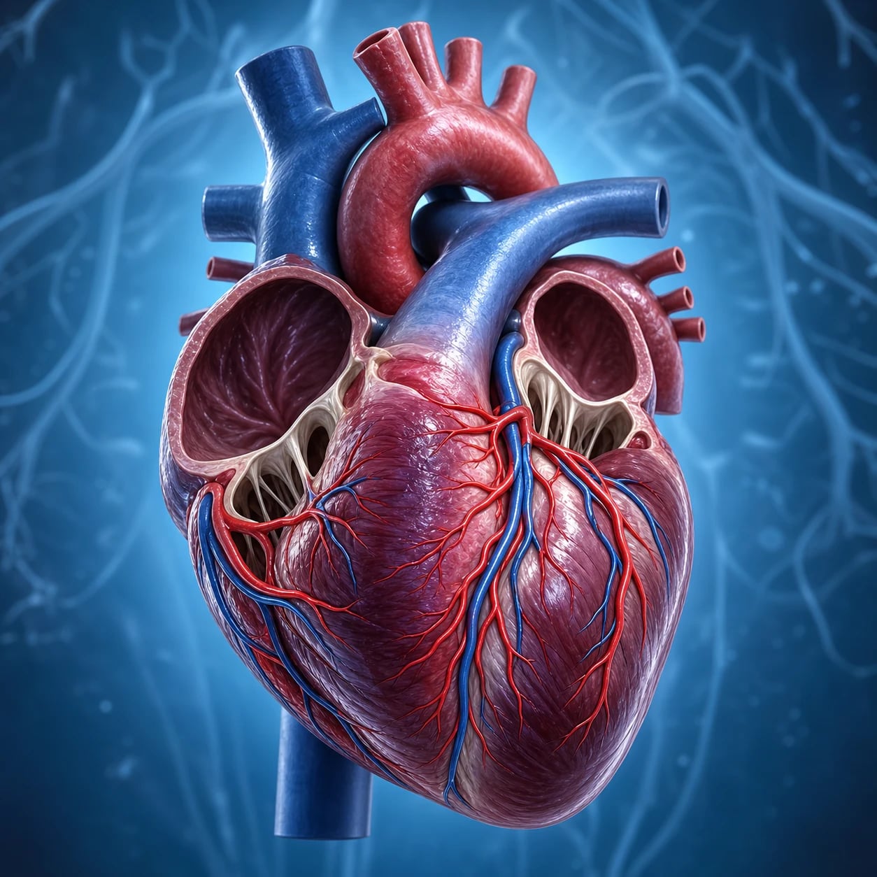
- Right atrium receives the SVC, IVC, and coronary sinus; the fossa ovalis is the remnant of the fetal foramen ovale (a flap-like defect in the interatrial septum). The smooth posterior wall (sinus venarum) receives the great veins; the muscular anterior wall carries the pectinate muscles and the crista terminalis (a vertical ridge marking the embryological junction of sinus venosus and atrium, and the line the catheter must not cross). The Eustachian valve guards the IVC orifice and the Thebesian valve the coronary sinus; in utero the Eustachian valve directs IVC blood across the foramen ovale.[1][1]
- Right ventricle is crescent-shaped and wraps around the LV; its inflow is trabeculated (trabeculae carneae) and its outflow (conus/infundibulum) smooth. The moderator band (septomarginal trabeculation) crosses from the interventricular septum to the base of the anterior papillary muscle and carries the right bundle branch — its anatomy explains why right bundle branch block is common in RV overload. The tricuspid valve (three leaflets: anterior, posterior, septal) guards the right AV orifice and is attached by chordae tendineae to three papillary muscles.[1][1]
- Left atrium is the most posterior chamber; it receives the four pulmonary veins (superior and inferior, right and left) and bears the left atrial appendage — a blind, trabeculated outpouching that is the dominant source of cardiogenic thrombus in atrial fibrillation (because stagnant flow and endothelial dysfunction combine there). The mitral valve (two leaflets: a large anterior/aortic leaflet and a smaller posterior leaflet) guards the left AV orifice and is attached to anterolateral and posteromedial papillary muscles by chordae tendineae.[1]
- Left ventricle is thick-walled (wall thickness ~3× the RV, 8–12 mm) and conical, generating systemic pressures; the aortic valve has three cusps (right, left, and posterior/non-coronary) sitting within dilations called the sinuses of Valsalva, which give origin to the coronary ostia (right cusp → RCA; left cusp → left main). The non-coronary cusp faces the interatrial septum.[1][1]
The four valves are anchored in the fibrous (cardiac) skeleton — a framework of dense connective tissue at the AV and ventriculo-arterial junctions that electrically insulates the atria from the ventricles (so that the AV node/His bundle is the only normal conduction bridge) and provides the mechanical scaffolding for valve closure.[1]
The cardiac wall and microscopic anatomy
The heart wall has three layers, from inside out: the endocardium (a thin layer of endothelium and subendocardial connective tissue continuous with the vessel intima — the subendocardial layer carries the Purkinje fibres and is the most vulnerable zone in ischaemia), the myocardium (the thick muscular middle layer, thickest in the LV), and the epicardium (the visceral layer of serous pericardium, against which the coronary arteries and veins run in the epicardial fat).[1]
The heart sits within the pericardium: an outer fibrous pericardium (dense connective tissue that limits acute over-distension) and a double-layered serous pericardium (parietal layer lining the fibrous pericardium, visceral layer = epicardium, with a thin film of ~15–50 mL of pericardial fluid between). The pericardial cavity has two clinically important recesses: the transverse sinus (between the aorta/pulmonary trunk anteriorly and the SVC/RA/upper left atrium posteriorly — the space a finger can pass behind the aorta during cardiac surgery) and the oblique sinus (a blind cul-de-sac behind the left atrium, bounded by the pulmonary veins).[1]
The cardiac myocyte is a striated, branching cell joined to its neighbours by intercalated discs. Each disc carries two functionally distinct junctions: desmosomes (mechanical adhesion — they stop the cells being pulled apart during contraction) and gap junctions (low-resistance electrical connections made of connexin proteins, which let the action potential pass from cell to cell so the myocardium behaves as a functional syncytium). Internally, T-tubules invaginate the sarcolemma and carry the depolarisation deep into the cell to the sarcoplasmic reticulum (the intracellular calcium store), whose controlled release triggers contraction. The contractile unit is the sarcomere (between two Z-lines), built of thick (myosin) and thin (actin) filaments with the regulatory proteins tropomyosin and the troponin complex (troponin C binds calcium, I inhibits, T binds tropomyosin). Calcium binding to troponin C exposes the actin binding site for the myosin head — excitation-contraction coupling.[1]
Cardiac myocyte vs skeletal muscle vs conduction (Purkinje) tissue
| Feature | Working atrial/ventricular myocyte | Skeletal myocyte | Purkinje fibre (conduction) |
|---|---|---|---|
| Cell shape | Short, branching | Long, cylindrical | Larger, pale-staining |
| Intercalated discs (desmosome + gap junction) | Present | Absent | Present |
| T-tubules | Abundant (ventricle); sparse (atrium) | Abundant | Few/absent |
| Myofibrils | Many | Many | Few (specialised for conduction, not force) |
| Gap junctions | Many (fast cell-to-cell spread) | Few | VERY many (fastest conduction in heart) |
| Conduction velocity | ~0.5 m/s | — | ~4 m/s (fastest) |
| Automaticity | None (normally) | None | Latent (escape pacemaker) |
The cardiac cycle
The cycle is the repeating sequence of electrical and mechanical events that ejects blood. The mechanical phases, with their defining pressure events:[1]
The cardiac cycle — one left-sided cycle (~0.8 s at 75 bpm)
- ATRIAL SYSTOLE (atrial kick): the atria contract (P wave), topping up the ventricles (~20–30 per cent of stroke volume; lost in atrial fibrillation). LV end-diastolic volume ~120 mL, pressure ~5–12 mmHg.[1]
- ISOVOLUMETRIC VENTRICULAR CONTRACTION: ventricular depolarisation (QRS) triggers contraction; all four valves shut, so ventricular volume is constant while pressure rises sharply (the first heart sound, S1 — mitral and tricuspid closure).[1]
- VENTRICULAR EJECTION: when LV pressure exceeds aortic (~80 mmHg), the aortic valve opens; rapid then reduced ejection follows. Peak LV pressure ~120 mmHg. About 70 mL is ejected (stroke volume); the rest (~50 mL) is the end-systolic volume (ejection fraction ~60 per cent).[1]
- ISOVOLUMETRIC VENTRICULAR RELAXATION: the ventricle relaxes with all valves closed; when LV pressure falls below aortic, the aortic valve snaps shut (the dicrotic notch on the aortic pressure trace, and the second heart sound, S2).[1]
- RAPID FILLING then DIASTASIS: the mitral valve opens when LV pressure falls below LA; rapid passive filling first, then slower filling (diastasis), until the next atrial systole. Coronary perfusion occurs predominantly in this diastolic filling phase.[1]
Typical right-sided pressures are much lower than left (RA 0–8 mmHg, RV 15–30/0–8 mmHg, PA 15–30/4–12 mmHg), reflecting the low-resistance pulmonary circuit; left-sided pressures (LA ~8 mmHg, LV 100–140/5–12 mmHg, aorta 100–140/70–90 mmHg) reflect the high-resistance systemic circuit.[1]
Normal intracardiac pressures (right vs left)
| Site | Right side (mmHg) | Left side (mmHg) | Comment |
|---|---|---|---|
| Atrium (mean) | RA 0–8 | LA (or PCWP) 6–12 | PCWP is the surrogate for LA pressure |
| Ventricle (systolic/diastolic) | RV 15–30 / 0–8 | LV 100–140 / 5–12 | RV generates ~1/5 systemic pressure |
| Great artery (systolic/diastolic) | PA 15–30 / 4–12 | Aorta 100–140 / 70–90 | PA is a low-pressure, low-resistance circuit |
| Pulmonary vs systemic vascular resistance | PVR ~80–120 dyn·s·cm⁻⁵ | SVR ~800–1200 dyn·s·cm⁻⁵ | PVR ~1/10 of SVR |
Coronary circulation
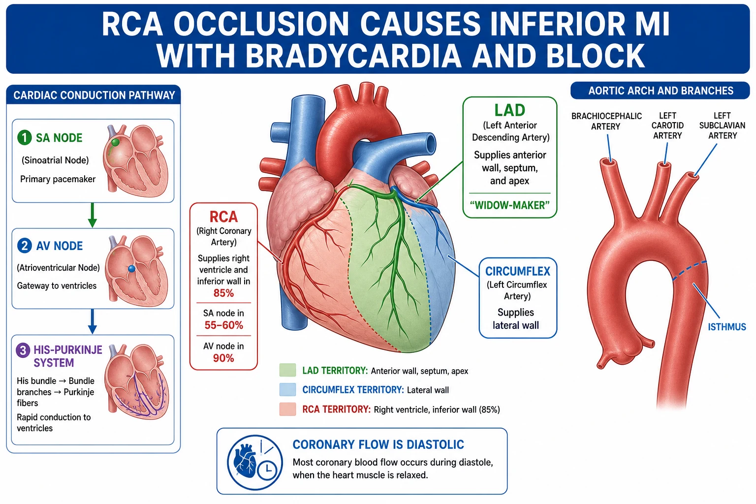
- The left main coronary artery arises from the left sinus of Valsalva and divides after 0.5–2 cm into the left anterior descending (LAD) and the left circumflex (LCx). The LAD runs in the anterior interventricular groove to the apex, giving septal perforators (the main supply of the interventricular septum and its conduction tissue) and diagonal branches (anterior LV wall).[1][1]
- The left circumflex (LCx) runs in the left AV groove, giving obtuse marginal branches to the lateral LV wall.
- The right coronary artery (RCA) arises from the right sinus of Valsalva and runs in the right AV groove. It gives the conus branch (supplying the RV outflow), the SA nodal artery (in ~55–60 per cent), acute marginal branches to the RV free wall (critical in RV infarction), and — when dominant — the posterior descending artery (PDA) and posterior left ventricular (PLV) branches supplying the inferior wall and posterior septum.[1][1]
- Perfusion territory:
- LAD — the anterior wall, the interventricular septum (septal perforators), and the apex; occlusion causes anterior infarction and is the highest-risk territory ("widow-maker" when proximal).[1]
- LCx — the lateral wall of the LV.[1]
- RCA — the right ventricle and, in most people, the inferior wall of the LV.[1]
- Coronary dominance is defined by which artery gives the PDA. Right dominance (PDA from RCA) in about 85 per cent; left dominance (PDA from LCx) in 8–10 per cent; codominant in 5–7 per cent. In a left-dominant system the inferior wall and the AV nodal supply come from the circumflex, so a lateral infarct can behave like an inferior one.[1]
- Nodal supply: the SA node is supplied by the RCA in 55–60 per cent (otherwise the circumflex); the AV node is supplied by the RCA in 90 per cent (otherwise the circumflex). RCA occlusion thus often produces inferior infarction with bradycardia or heart block.[1]
- Coronary flow occurs in diastole; tachycardia (which shortens diastole) and high LV diastolic pressure both reduce coronary perfusion, especially in the subendocardium.[1][2][3]
Coronary venous drainage
Most venous return (~75 per cent) drains via the coronary sinus, which runs in the posterior AV groove and opens into the right atrium between the IVC orifice and the tricuspid valve. Its tributaries are the great cardiac vein (accompanying the LAD/LCx), the middle cardiac vein (in the posterior interventricular groove, accompanying the PDA), and the small cardiac vein (with the RCA). The anterior cardiac veins drain directly into the right atrium, and the smallest cardiac (Thebesian) veins drain directly into all four chambers — the latter accounts for the small physiological shunt (deoxygenated blood entering the left side) and is why coronary sinus blood is sampled to estimate global myocardial oxygen balance.[1]
Collaterals
The coronary circulation is anatomically end-arterial (few functional collaterals at baseline), which is why acute occlusion causes infarction. However, with chronic ischaemia an arteriolar collateral network develops over weeks to months that can progressively protect ischaemic territory — the anatomical basis for the "developed collaterals" seen on angiography in chronic total occlusions and the mismatch between severe coronary disease and preserved LV function in some patients.[1]
Coronary artery territories and ECG lead correlations
| Artery | Branch | Territory supplied | Typical ECG changes |
|---|---|---|---|
| LAD | Septal perforators | Anterior 2/3 of interventricular septum + bundle branches | V1–V2 (septal) |
| LAD | Diagonals | Anterior LV wall, apex | V3–V4 (anterior), V1–V4 (extensive anterior) |
| LAD | (proximal) | All of above — "widow-maker" | V1–V4 ± high risk of cardiogenic shock, block |
| LCx | Obtuse marginals | Lateral LV wall | I, aVL, V5–V6 (lateral) |
| RCA | Acute marginals | RV free wall | Right-sided leads V3R–V4R |
| RCA (dominant) | PDA + PLV | Inferior wall, posterior septum | II, III, aVF (inferior); V7–V9 (posterior) |
| RCA | SA / AV nodal branches | SA node (55–60%), AV node (90%) | Sinus bradycardia, AV block with inferior MI |
Coronary blood flow — why it is diastolic and why the subendocardium ischaemia-is first
Coronary blood flow is unique because the heart must perfuse itself while it generates systemic pressure. Two features make LV coronary flow predominantly diastolic:[2][3]
- During systole, the contracting LV myocardium generates tissue pressures that exceed intramural coronary pressure, compressing the vessels that course through the wall — so systolic flow to the LV (especially the deep, subendocardial layers) is near zero. The RV, which develops much lower pressures (~25 mmHg), is perfused throughout both systole and diastole.[3]
- During diastole, the myocardium relaxes, tissue pressure falls, and the pressure gradient from aorta to myocardium favours flow. Flow is therefore timed to diastole.[2]
The clinical consequences are the exam staples:[2]
- Tachycardia is ischaemia-prone because it preferentially shortens diastole (the perfusing phase) while lengthening systole (the non-perfusing phase). At very high heart rates the time available for coronary flow collapses.
- Raised LV end-diastolic pressure (aortic stenosis, LV failure) raises subendocardial tissue pressure, opposing coronary inflow — the anatomical basis for subendocardial ischaemia in aortic stenosis and LV failure.
- The subendocardium is the watershed zone: it sits at the end of the transmural pressure gradient, is most compressed in systole, and therefore ischaemia-is first. This is why subendocardial (non-Q-wave) infarction is the common pattern in demand ischaemia and tachyarrhythmias.[2]
Coronary flow is regulated locally by metabolic vasodilation (adenosine released when ATP is consumed, plus CO₂, K⁺, hypoxia), endothelial factors (nitric oxide, prostacyclin), myogenic autoregulation (constant flow over a perfusion pressure range of ~60–140 mmHg), and sympathetic β-mediated dilation. The supply:demand ratio (diastolic pressure-time index over systolic tension-time index, Buckberg) summarises the balance; a falling ratio predicts subendocardial ischaemia.[2][3]
Conduction system
- The impulse originates in the sinoatrial (SA) node in the high right atrium at the junction with the SVC (the crista terminalis), the body's dominant pacemaker (intrinsic rate ~60–100/min under autonomic tone; the denervated rate is ~100/min). It spreads through atrial muscle — partly via the internodal tracts (anterior/middle/posterior) and across the atria to the left atrium via Bachmann's bundle — to reach the atrioventricular (AV) node in the floor of the right atrium near the coronary sinus.[1][1]
- The AV node is the only normal electrical connection between atria and ventricles (the fibrous skeleton insulates the rest). It introduces the physiological conduction delay (the PR interval, ~0.12–0.20 s), which lets atrial systole complete ventricular filling before the ventricles contract. It has the slowest conduction velocity in the heart (~0.05 m/s) and is the substrate for re-entrant AV nodal tachycardias.[1]
- From the AV node the impulse enters the bundle of His, which divides into the right and left bundle branches (the left further into anterior and posterior fascicles), then into the subendocardial Purkinje fibres — the fastest conducting tissue (~4 m/s) — which depolarise the ventricles from apex to base, septum to free wall.[1]
- Escape pacemakers: the AV node/junction fires at ~40–60/min; the Purkinje/ventricular myocardium at ~20–40/min. When the SA node fails, these define the rate of junctional and idioventricular escape rhythms.[1]
Action potentials — fast (Na⁺) vs slow (Ca²⁺)
Two cell types underlie cardiac electricity. Fast-response cells (working atrial and ventricular myocytes, His-Purkinje) have a rapid phase 0 upstroke driven by Na⁺ influx, a plateau (phase 2, Ca²⁺ influx balanced by K⁺ efflux), and a long refractory period. Slow-response cells (SA and AV nodes) lack stable Na⁺ channels: their phase 0 is Ca²⁺-driven (slow), and — crucially — they show spontaneous phase 4 depolarisation (the "funny current" I_f, a Na⁺/K⁺ mixed current activated by hyperpolarisation) which is the molecular basis of automaticity. This is why calcium-channel blockers (verapamil, diltiazem) selectively slow the SA/AV nodes (the rate-dependent tissue) without much effect on working myocardium.[1]
Blood supply to the conduction system
Blood supply of the conduction system — the anatomical reason nodal block follows RCA occlusion
| Structure | Blood supply | Consequence of occlusion |
|---|---|---|
| SA node | RCA 55–60%, LCx 40–45% | Sinus bradycardia, sinus arrest, atrial arrhythmias |
| AV node | RCA 90% (via the posterior descending / AV nodal branch), LCx 10% | First-/second-/third-degree AV block with inferior MI |
| Bundle of His | Dual supply (AV nodal artery + proximal LAD septal perforators) | Relatively protected — proximal block less common |
| Right bundle branch + left anterior fascicle | LAD septal perforators | Bifascicular block (RBBB + LAFB) in anterior MI — high grade block risk |
| Left posterior fascicle | Dual supply (RCA + LCx) | Posterior fascicular block is rare (protected) |
| Purkinje fibres | Subendocardial — most ischaemia-prone zone | Ventricular ectopy in ischaemia |
Great vessels and the aortic arch
- The aorta is divided into the ascending aorta (no branches except the coronary ostia just above the aortic valve), the aortic arch (which gives, from right to left, the brachiocephalic (innominate) trunk, the left common carotid, and the left subclavian arteries), the isthmus (the slightly narrowed segment just distal to the left subclavian, where the mobile arch meets the tethered descending aorta), and the descending thoracic aorta (intercostal, bronchial, oesophageal branches) continuing as the abdominal aorta.[1]
- The isthmus is the typical site of traumatic aortic injury from rapid deceleration — the mobile arch swings forward while the descending aorta is fixed, shearing the wall at the junction.[1][4]
- The SVC forms from the two brachiocephalic veins and drains the upper body into the right atrium; its junction with the RA (the cavo-atrial junction) is the target for central venous catheter tips. The IVC drains the lower body (receiving the hepatic veins just below the diaphragm) into the RA.[1]
- The pulmonary trunk carries deoxygenated blood from the RV to the lungs, dividing into right and left pulmonary arteries; the four pulmonary veins (two from each lung) return oxygenated blood to the left atrium. This means the pulmonary arteries are the only arteries that carry deoxygenated blood, and the pulmonary veins the only veins that carry oxygenated blood.[1]
- The ligamentum arteriosum is the fibrous remnant of the fetal ductus arteriosus (which shunted RV output into the descending aorta in utero, bypassing the lungs). It is the structure around which the left recurrent laryngeal nerve loops under the aortic arch to ascend into the neck — the anatomical reason a left-sided mediastinal mass or aortic aneurysm causes hoarseness ( Ortner's cardinia).[1]
Aortic arch variants
The classic three-vessel arch (brachiocephalic, left carotid, left subclavian) is present in ~70 per cent. The commonest variant is the "bovine" arch — the brachiocephalic and left common carotid share a common origin (~20 per cent) — which matters for carotid cannulation and aortic-arch vessel stenting. An aberrant right subclavian artery (arteria lusoria) arises as the last branch of a left arch and passes behind the oesophagus, causing dysphagia lusoria; a right-sided aortic arch (with mirror-image branching or an aberrant left subclavian) is associated with congenital heart disease (especially tetralogy variants). A coarctation classically sits at the isthmus, with rib notching (collateral intercostal enlargement) and radio-femoral delay on examination.[1]
Systemic vs pulmonary circulation
The cardiovascular system is two circulations in series: the systemic (left heart → body → right heart) and the pulmonary (right heart → lungs → left heart), joined at the heart.[1]
Systemic vs pulmonary circulation
| Feature | Systemic circulation | Pulmonary circulation |
|---|---|---|
| Driving pump | Left ventricle (thick, ~8–12 mm wall) | Right ventricle (thin, ~3–4 mm wall) |
| Pumping pressure | High (aorta 100–140/70–90 mmHg) | Low (PA 15–30/4–12 mmHg) |
| Vascular resistance | High (SVR ~800–1200 dyn·s·cm⁻⁵) | Low (PVR ~80–120 dyn·s·cm⁻⁵, ~1/10 of SVR) |
| Vessel structure | Thick muscular arteries, prominent elastic laminae | Thin-walled, highly distensible arteries |
| Capillary exchange | Systemic capillaries — Starling forces at all beds | Pulmonary capillaries — gas exchange; lower hydrostatic pressure |
| Transit time at capillary | ~1 s (systemic) | ~0.75 s (rest), ~0.3 s (exercise) |
| Effect of gravity | Marked regional variation (orthostatic pooling) | Marked — West's zones (apex zone 1, base zone 3) |
| Oxygenation of blood carried | Oxygenated (arteries) / deoxygenated (veins) | Deoxygenated (arteries) / oxygenated (veins) |
The LV is built for pressure; the RV is a volume pump coupled to a low-impedance circuit. Because the RV generates only ~1/5 of LV pressure, it is exquisitely afterload-sensitive: a sudden rise in PVR (massive PE, ARDS, hypoxia) can precipitate acute RV failure. The thin RV free wall also explains why RV infarction is preload-dependent — it needs adequate filling to generate forward flow, which is why nitrates are harmful and fluids may help in RV infarction.[1]
The Frank-Starling mechanism
The Frank-Starling law of the heart states that, within the physiological range, the force of ventricular contraction is proportional to the end-diastolic fibre length (preload) — the heart pumps what it receives. The cellular basis is the length-tension relationship of the sarcomere: stretch to an optimal length (~2.0–2.2 μm) brings actin and myosin filaments into optimal overlap and increases calcium sensitivity of troponin C, so more cross-bridges form at any given calcium concentration. Beyond ~2.2 μm, filaments are overstretched and force falls (the descending limb — the basis of decompensated heart failure).[1]
Clinically, preload is approximated by ventricular end-diastolic volume or pressure (clinically by RA pressure for the RV, by PCWP for the LV). Afterload is the load against which the ventricle ejects (clinically by SVR/systemic pressure for the LV, PVR for the RV). A family of ventricular function (Starling) curves relates stroke volume (or cardiac output) to filling pressure; increased contractility (inotropy) — sympathetic stimulation, catecholamines — shifts the curve up and to the left, while decreased contractility (heart failure, negative inotropes) shifts it down and to the right.[1]
Two related intrinsic phenomena deserve mention: the Anrep effect (an increase in afterload modestly increases contractility) and the Bowditch (Treppe) effect (an increase in heart rate increases contractility via intracellular calcium accumulation) — both intrinsic, myogenic, and independent of nerves or hormones.[1]
Starling forces and transcapillary fluid exchange
The same Ernest Starling described the forces governing fluid movement across capillaries. The classic Starling equation is:[1]
Jv = Kf [(Pc − Pi) − σ(πc − πi)]
where Jv is net fluid flux out of the capillary; Pc is capillary hydrostatic pressure (favours filtration); Pi is interstitial hydrostatic pressure (opposes filtration); πc is plasma colloid osmotic (oncotic) pressure, mainly from albumin (favours reabsorption); πi is interstitial oncotic pressure (favours filtration); Kf is the capillary filtration coefficient (surface area × permeability); and σ is the reflection coefficient of proteins (how well the membrane holds them back; 1 = impermeable, 0 = freely permeable).[1]
The four Starling forces and their clinical drivers
| Force | Direction of fluid movement | Increased by | Decreased by |
|---|---|---|---|
| Pc — capillary hydrostatic (favours filtration OUT) | Filtration | Venous pressure rise (heart failure), arteriolar dilation, fluid | Dehydration, arteriolar constriction |
| Pi — interstitial hydrostatic (opposes filtration) | Reabsorption | Tight tissues, negative pressure | Loose tissues (oedema more likely) |
| πc — plasma oncotic (favours reabsorption IN) | Reabsorption | Albumin / colloid | Hypoalbuminaemia (nephrotic, liver failure, burns, sepsis) |
| πi — interstitial oncotic (favours filtration) | Filtration | Inflammation, proteolysis (burns) | — |
In the classic model, filtration predominates at the arterial end of a systemic capillary (Pc ~30–35 mmHg exceeds the net oncotic pull ~20–25 mmHg) and reabsorption at the venous end (Pc ~12–15 mmHg). The revised Starling model (with the endothelial glycocalyx layer, EGL) refines this: the EGL is the true semipermeable membrane sitting just inside the endothelium, and the relevant oncotic pressure is that under the glycocalyx (sub-glycocalyx), which is low because filtered protein is washed away by the low-pressure interstitium. The result is that many capillary beds filter along their entire length at a low rate, with return by lymphatics; oedema arises when the EGL is degraded (sepsis, ischaemia-reperfusion, major surgery) or when capillary pressure or protein leak rises.[1]
The clinical take-home: oedema results from (i) raised capillary hydrostatic pressure (heart failure, venous obstruction, fluid overload), (ii) low plasma oncotic pressure (hypoalbuminaemia), (iii) raised capillary permeability (SIRS/sepsis, burns, anaphylaxis — "leaky capillaries"), (iv) lymphatic obstruction (lymphoedema), and (v) high interstitial oncotic pressure (myxoedema). The choice of resuscitation fluid — crystalloid vs colloid — turns on these forces, because a colloid's volume-expanding effect depends on πc, which is reduced when the glycocalyx is damaged (protein leaks out, blunting the oncotic advantage).[1][1]
Venous return and Guytonian physiology — what sets cardiac output
Arthur Guyton's reframing is the cornerstone of ICU haemodynamics: cardiac output is determined by the venous return to the heart, not primarily by the heart's intrinsic pumping power, because at normal filling the healthy heart pumps whatever the systemic circulation delivers to it. The right atrium is simply the meeting point of two systems, and the operating cardiac output is the intersection of a cardiac function curve and a vascular function (venous return) curve.[1]
The two curves
The cardiac function curve (Starling) plots cardiac output against right atrial pressure: as RAP rises, the heart pumps more (up to a plateau, where the curve flattens). The vascular function (venous return) curve plots venous return against RAP: as RAP rises, the gradient driving venous return falls, so venous return falls; when RAP equals the mean systemic filling pressure (MSFP), the gradient is zero and venous return is zero; when RAP falls to/below zero, the great veins collapse and venous return plateaus (circulatory independence — the venous "waterfall").[1][1]
The intersection of these two curves is the operating point — the actual cardiac output and RAP. Guyton's equation makes this explicit: [1]
VR = (MSFP − RAP) / Rv [1]
where MSFP is the mean systemic filling pressure (the pressure everywhere in the systemic circulation if the heart were stopped and flow were zero — ~7 mmHg normally, set by blood volume and venous tone), RAP is right atrial pressure (back-pressure), and Rv is the resistance to venous return (mostly in the post-capillary venules, because resistance is concentrated where the pressure drop occurs).[1]
Determinants of cardiac output — the Guytonian view
| Intervention | Effect on MSFP | Effect on Rv (venous resistance) | Effect on the curves | Net effect on cardiac output |
|---|---|---|---|---|
| Volume / transfusion | ↑ (more volume stretches veins) | — | Vascular curve shifts right/up | ↑ CO |
| Sympathetic stimulation (venoconstriction) | ↑ (veins squeeze blood centrally) | — | Vascular curve shifts right/up | ↑ CO (mobilises unstressed volume) |
| Vasodilators (nitroprusside, anaesthesia) | ↓ | ↓ | Vascular curve shifts down/left | Variable; CO often ↑ from lower Rv, ↓ from lower MSFP |
| Venodilators (nitroglycerine) | ↓ | — | Vascular curve shifts down/left | ↓ preload → ↓ CO |
| Positive-pressure ventilation / PEEP | (raises) intrathoracic pressure → ↑ measured CVP | — | Effectively ↑ RAP, ↓ gradient | ↓ venous return → ↓ CO |
| Spontaneous inspiration | ↓ intrathoracic pressure | — | RA pressure falls → ↑ gradient | ↑ venous return (right-sided preload) |
| Inotrope (dobutamine) | — | — | Cardiac curve shifts up/left | ↑ CO at same RAP |
| Heart failure | — | — | Cardiac curve shifts down/right | ↓ CO; congestion at high RAP |
The pivotal implications for ICU practice: (i) raising RAP beyond a point does not increase CO — once on the flat part of the Starling curve, more preload does not help (and causes congestion); (ii) PEEP and positive-pressure ventilation reduce venous return by raising intrathoracic (hence RAP) pressure — the basis for haemodynamic compromise in volume-depleted, ventilated patients; (iii) volume responsiveness is fundamentally about whether the heart is on the ascending limb of its Starling curve, which is why dynamic indices (pulse-pressure variation, passive leg raise, end-expiratory occlusion) outperform static CVP; (iv) venoconstriction (sympathetic tone, noradrenaline) mobilises unstressed volume into the stressed compartment, raising MSFP and supporting venous return — a key, under-appreciated effect of vasopressors.[1][1][1]
Vascular wall structure — arteries, capillaries, and veins
The vessel wall has three concentric layers — the tunica intima (endothelium on a basement membrane, plus in arteries an internal elastic lamina), the tunica media (smooth muscle and elastic lamellae — the thickest layer in arteries, dominant in controlling diameter and resistance), and the tunica externa (adventitia) (loose connective tissue with the vasa vasorum that nourish the outer wall of large vessels, and an external elastic lamina in larger arteries).[1]
Vessel types — wall structure and function
| Vessel | Wall | Lumen/diameter | Key function | Notes |
|---|---|---|---|---|
| Elastic artery (aorta, brachiocephalic, carotid, subclavian, pulmonary trunk) | Thick, many elastic lamellae, thin media smooth muscle | Large | Conductance; Windkessel effect (elastic recoil sustains diastolic flow) | Stores systolic energy, releases in diastole — keeps coronary flow diastolic |
| Muscular (distributing) artery (radial, femoral, coronary, splenic, renal) | Prominent smooth muscle, prominent IEL | Medium | Distribute flow; vasoconstriction/dilation | Site of atherosclerosis |
| Arteriole | 1–2 layers smooth muscle | Small (~10–100 μm) | Resistance vessels (~60–70% of SVR drop) | The main regulators of blood pressure and flow distribution |
| Capillary | Single endothelial layer + basement membrane | ~5–10 μm | Exchange (Starling forces, gas exchange) | Three types: continuous (most), fenestrated (gut, glomerulus), sinusoid (liver, marrow) |
| Venule | Thin, post-capillary endothelium ± pericytes | Small | Exchange + collection | Post-capillary venules — major site of inflammatory exudation |
| Vein | Thin media, little elastic, valves; adventitia dominant | Large, compliant | Capacitance reservoir (~60–70% of blood volume) | Valves (absent in SVC/IVC/portal) aid return; "capacitance vessels" |
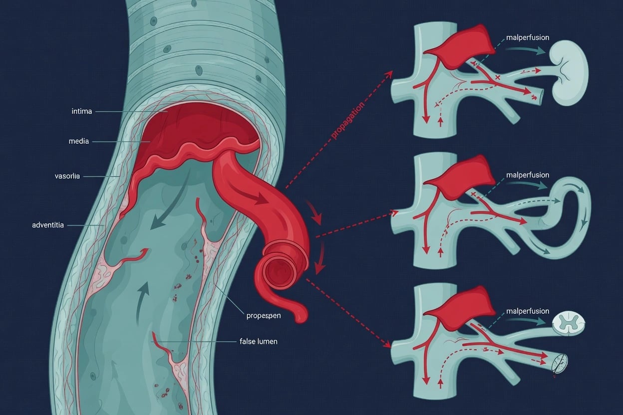
The clinical anatomy that matters: the arterioles set resistance (hence vasopressors targeting them raise systemic pressure); the veins hold the reservoir (hence venoconstriction mobilises blood to support preload); the endothelium is the largest endocrine organ in the body (nitric oxide, prostacyclin, endothelin, von Willebrand factor), and its glycocalyx governs the Starling forces above. The vasa vasorum supply the outer wall of large vessels — the inner wall is nourished directly from the lumen — which is why aortic dissection (which splits the media along the vasa vasorum plane) devascularises the outer wall and risks rupture.[1]
Cardiac innervation
The heart is richly innervated. Parasympathetic fibres reach it via the vagus nerve (CN X) — the right vagus preferentially to the SA node, the left to the AV node — releasing acetylcholine on M2 receptors that slow SA nodal firing and AV conduction (the basis of vagal manoeuvres for SVT and of vagally-mediated bradycardias). The resting heart is under tonic vagal dominance — that is why the resting rate (~60–80) is below the intrinsic SA rate (~100).[1]
Sympathetic fibres arise from T1–T5, synapse in the cervical and upper thoracic sympathetic ganglia (cervical cardiac nerves and thoracic cardiac splanchnic nerves), and join the vagal fibres in the cardiac plexus (between the aortic arch, tracheal bifurcation, and pulmonary trunk) before distributing to the SA/AV nodes, atria, and ventricles. Noradrenaline on β1 receptors increases rate (chronotropy), conduction (dromotropy), and contractility (inotropy). The baroreceptor reflex (carotid sinus and aortic arch afferents via CN IX and X) modulates both limbs beat-to-beat.[1]
Pericardium and cardiac lymphatics
The pericardium limits acute cardiac over-distension and fixes the heart in the mediastinum. The fibrous pericardium fuses inferiorly with the central tendon of the diaphragm (so the heart moves with respiration) and superiorly with the adventitia of the great vessels. The serous pericardium's reflections create the transverse sinus (between aorta/PA anteriorly and SVC/upper LA posteriorly) and the oblique sinus (behind the LA, bounded by the pulmonary veins). The pericardial sac normally holds ~15–50 mL of fluid; rapid accumulation of as little as 100–200 mL can cause tamponade (because the fibrous pericardium cannot stretch acutely), whereas chronic effusions of litres may be tolerated.[1]
Cardiac lymphatics drain via epicardial trunks that follow the coronary arteries to mediastinal nodes and ultimately the thoracic duct. Lymphatic obstruction (rare) causes refractory pericardial effusion, and the lymphatic pathway is implicated in myocardial oedema and inflammation resolution.[1]
Peripheral venous system for central venous catheter insertion
Central venous access relies on the anatomy of the great veins returning to the right atrium. The relevant vessels:[1][5]
- Internal jugular vein (IJV) — runs in the carotid sheath deep to sternocleidomastoid, lateral and superficial to the carotid artery (medial and deep) with the vagus nerve posteriorly between them; it joins the subclavian behind the sternoclavicular joint to form the brachiocephalic (innominate) vein. The right IJV has a straight course to the SVC and right atrium, making it the first-choice site.[1][5]
- Subclavian vein — lies immediately below the clavicle, anterior to (and separate from) the subclavian artery (which sits posterosuperiorly, separated by scalenus anterior), and below the pleural apex. Its fixed position makes it easy to cannulate blind but it is non-compressible (bleeding risk in coagulopathy) and risks pneumothorax and subclavian vein stenosis (which destroys future AV fistula options).[5]
- Brachiocephalic (innominate) veins — the right is short and vertical; the left is longer and crosses to join the right behind the sternum, which is why left-sided catheters can kink or malposition (tip flicking up the contralateral IJV).[1]
- Superior vena cava — formed by the union of the two brachiocephalics; drains into the right atrium at the cavo-atrial junction, the target catheter-tip position. A tip that lies too proximally (in the brachiocephalic) erodes the vein wall; too distally (in the RA) risks perforation and arrhythmia.[5]
- Azygos vein — arches over the right main bronchus to drain into the SVC just above the RA — an important collateral when the SVC is obstructed (SVC syndrome) and the route by which misplaced lines sometimes curl in the upper mediastinum.[1]
- Femoral vein — the continuation of the external iliac under the inguinal ligament; medial in the femoral sheath (lateral-to-medial: nerve, artery, vein — NAVEL). It is compressible and away from the thorax (no pneumothorax) but has the highest infection rate and DVT risk.[5]
- Basilic, cephalic, and median cubital veins — arm veins used for peripherally inserted central catheters (PICCs); the basilic is preferred (straighter course to the axillary/subclavian) and the cephalic often angles sharply at the clavipectoral fascia (causing passage difficulty).[1]
Site selection — anatomy-driven trade-offs
The 3SITES trial (Parienti, 2015) randomised CVC site and quantified these trade-offs: subclavian had the lowest bloodstream infection and DVT but the most pneumothorax; femoral had the most infection and DVT; jugular sat in between. The choice is individualised — subclavian when infection risk dominates and clotting is normal, jugular when ultrasound access and compressibility matter (most ICU lines), femoral for emergency access, severe coagulopathy, or when the upper body is excluded.[6]
CVC site selection — anatomy-driven trade-offs (3SITES context)
| Site | Anatomical advantages | Anatomical disadvantages | When preferred |
|---|---|---|---|
| Right IJV | Straight to RA; ultrasound-guided; compressible | Pneumothorax (low); central-site infection risk | FIRST CHOICE for most ICU CVCs, CRRT, dialysis |
| Subclavian | Lowest infection; comfortable; vein fixed (does not collapse) | Pneumothorax (1–5%); non-compressible (coagulopathy risk); subclavian stenosis (destroys future AV fistula) | Long-term access, normal clotting, no future fistula |
| Left IJV | Available when right thrombosed | Tortuous course through left brachiocephalic — kinking, malposition; thoracic duct on left | Second-line |
| Femoral | Compressible; no pneumothorax; away from mediastinum | Highest infection; DVT; immobile; recirculation (dialysis) | Arrest, catastrophic bleeding, severe coagulopathy |
| PICC (basilic/cephalic) | Bedside, no pneumothorax, lower infection than CVC | Smaller lumen; arm immobility; cephalic angle difficulty | Medium-term access, antibiotics/Tpn |
The central-line bundle and tip position
Ultrasound-guided right IJV cannulation and the insertion bundle (Pronovost/Keystone)
- POSITION: head-down (Trendelenburg 15–20°) to distend the vein; head turned slightly away (30–45°) — over-rotation stretches the vein over the carotid and narrows it.[5]
- ULTRASOUND SURVEY: high-frequency linear probe transverse at the cricoid level — identify the IJV (oval, compressible) lateral to the carotid (round, pulsatile, non-compressible); confirm patency and exclude thrombus.[5]
- MAXIMAL STERILE BARRIERS: cap, mask, sterile gown and gloves, full-body drape; 2% chlorhexidine skin prep.[7]
- IN-PLANE NEEDLE ADVANCEMENT: puncture the anterior wall only (avoid through-and-through); aspirate dark, non-pulsatile venous blood; confirm venous (not arterial).[5]
- SELDINGER: pass the J-wire; confirm wire-in-vein on ultrasound in short- and long-axis; the wire should NOT cross the midline (which would indicate oesophageal/carotid misplacement); dilate and insert to the correct depth (right IJV ≈ 15 cm).[5]
- CONFIRM AND SECURE: aspirate and flush all lumina; chest X-ray to confirm the tip at the cavo-atrial junction and exclude pneumothorax.[5]
- THE BUNDLE (Keystone): hand hygiene, maximal sterile barriers, chlorhexidine, avoid femoral, daily review with prompt removal — these reduce catheter-related bloodstream infection.[7]
Vascular variants that catch the unwary
A persistent left superior vena cava (PLSVC) (~0.3–0.5 per cent of the population) results from failure of the left anterior cardinal vein to regress; it drains the left upper body via the coronary sinus into the right atrium (in ~90 per cent — haemodynamically silent, but a left-sided CVC tip ends up curled in the coronary sinus on chest X-ray, often outside the cardiac silhouette to the left). A double SVC (persistent left + normal right) is also possible. A left-sided IVC drains via the hemiazygos and azygos into the right SVC. These are the reason to confirm line position and orientation when the catheter does not follow the expected course.[1]
Exam practice
SAQ — Coronary anatomy applied to inferior STEMI with right ventricular involvement
10 minutes · 10 marks
A 62-year-old man presents with 40 minutes of crushing central chest pain radiating to the jaw, diaphoresis and nausea. ECG shows 2 mm ST elevation in II, III, aVF with reciprocal depression in I and aVL. Right-sided leads V3R–V4R show 1 mm ST elevation. BP 86/52, HR 38 (sinus bradycardia). Chest is clear; JVP is raised at 8 cm. Past history: hypertension, no prior aspirin.
SAQ — Central venous catheter site selection and anatomy in a coagulopathic septic patient
10 minutes · 10 marks
A 70-year-old woman with severe COPD on home oxygen is admitted with community-acquired pneumonia and septic shock. BP 76/40, lactate 4.8, mottled peripheries. INR 2.2 (not on warfarin), platelets 75 × 10⁹/L, prior right mastectomy with axillary node clearance. She requires central access for noradrenaline infusion and the team is debating subclavian versus right internal jugular versus femoral routes.
Clinical pearls
Key trials and evidence
Guyton AC — Venous-return and cardiac-output curves (1955, Physiol Rev)
Study type
Classical integrative physiology / theoretical framework (dog open-chest preparations + mathematical equating of independent curves)
Concept
Cardiac output is set by the intersection of a cardiac function (Starling) curve and a vascular function (venous return) curve; defined mean systemic filling pressure (MSFP) and the equation VR = (MSFP − RAP) / Rv
Key insight
At normal filling the healthy heart pumps whatever the systemic circulation delivers — venous return, not intrinsic cardiac power, is the prime determinant of cardiac output
Clinical bottom line
The foundation of modern ICU haemodynamics: explains why PEEP cuts venous return, why volume responsiveness matters more than static CVP, and why venoconstriction (vasopressors) supports cardiac output
Hoffman & Buckberg — Myocardial supply:demand ratio (1978, Am J Cardiol)
Study type
Critical review integrating regional coronary flow physiology
Concept
The ratio of diastolic pressure-time index (supply, DPTI) to systolic tension-time index (demand, TTI) — the 'endocardial viability ratio' — predicts subendocardial ischaemia
Key insight
LV coronary flow is diastolic; tachycardia, anaemia, hypotension, and raised LVEDP all lower the DPTI/TTI ratio and favour subendocardial ischaemia
Clinical bottom line
Explains why tachyarrhythmias, aortic stenosis, and anaemia are ischaemia-prone, and why rate control and maintaining diastolic pressure protect the subendocardium
Buckberg — Heart rate and regional LV blood flow (1975, Cardiovasc Res)
Study type
Experimental (anaesthetised dogs, microsphere flow measurement)
Intervention
Pacing-induced variation in heart rate
Key finding
As heart rate rises, total LV flow may rise but the **endocardial/epicardial flow ratio falls** — tachycardia preferentially threatens the subendocardium despite maintaining aggregate flow
Clinical bottom line
Experimental demonstration of why tachycardia ischaemia-is the subendocardium first — the anatomical-physiological basis for rate control in ischaemia
McGee & Gould — Preventing complications of central venous catheterization (2003, NEJM review)
Study type
Authoritative clinical review
Scope
Mechanical (arterial puncture, pneumothorax, haematoma), infectious (catheter-related bloodstream infection), and thrombotic complications of CVC insertion by site
Key message
Complication rates are site-dependent and anatomically driven; ultrasound guidance, maximal sterile barriers, and site selection are the main preventive levers
Clinical bottom line
The reference for CVC technique and the anatomical safety rules (carotid medial/deep to IJV; subclavian pneumothorax and stenosis; femoral infection)
3SITES trial — CVC insertion site (Parienti 2015, NEJM)
Study design
Multicentre randomised controlled trial — ~3,027 patients needing a CVC for ≥3 days across 18 ICUs
Intervention
Subclavian vs jugular vs femoral site — randomly assigned
Primary outcome
Composite of catheter-related bloodstream infection AND symptomatic DVT: lowest with SUBCLAVIAN (subclavian 1.5%, jugular 3.3%, femoral 4.0%)
Mechanical risk
Symptomatic pneumothorax HIGHEST with SUBCLAVIAN (1.1%) vs jugular (0.3%) vs femoral (0%)
Clinical bottom line
Subclavian minimises infection/thrombosis but maximises pneumothorax; choose subclavian when infection risk and clean clotting dominate, IJV when ultrasound access and compressibility dominate, femoral for emergency/coagulopathic access
Pronovost / Keystone — Central-line bundle (2006, NEJM)
Study design
Prospective cohort / quasi-experimental — 103 ICUs in Michigan, USA
Intervention
Five-component bundle: hand hygiene, maximal sterile barriers, chlorhexidine skin prep, avoid femoral site, daily review with prompt removal
Primary outcome
Median catheter-related bloodstream infection rate fell from 2.7 to 0 per 1,000 catheter-days (up to 66% reduction), sustained at 18 months
Clinical bottom line
A simple anatomically-grounded bundle (avoid the high-infection femoral site; use maximal sterile barriers at the chosen site) dramatically reduces CRBSI — the foundation of every ICU line protocol
Pate — Traumatic rupture of the aortic isthmus (1999, World J Surg)
Study type
Surgical series / selective-management program
Concept
The isthmus (just distal to the left subclavian) is the dominant site of blunt traumatic aortic injury because of the shear between the mobile arch and the tethered descending aorta
Key message
Deceleration injury demands prompt aortic imaging (CT angiography) and selective operative vs endovascular management; mediastinal widening on chest X-ray is the screening clue
Clinical bottom line
The anatomical reason the isthmus tears, and the rationale for imaging any major deceleration injury
Red flags
Cross-cutting principles — anatomy predicts disease, procedure, and haemodynamics
The one-table revision — anatomy → clinical consequence
| Anatomical fact | Clinical consequence |
|---|---|
| RCA dominant in 85%; supplies AV node 90%, SA node 55–60% | Inferior MI → bradycardia/AV block; possible RV infarction (preload-sensitive) |
| Coronary flow is diastolic; subendocardium most compressed in systole | Tachycardia, raised LVEDP → subendocardial ischaemia; rate control protects |
| Moderator band carries RBB; LAD septal supply to LBB anterior fascicle | RV stretch → RBBB; anterior MI → bifascicular block |
| SA/AV nodes are calcium-channel (slow) tissue; working myocardium is sodium (fast) | Verapamil/diltiazem slow the node, are useless/dangerous in VT and WPW |
| Fibrous skeleton insulates atria from ventricles | AV node is sole bridge; accessory pathways (WPW) cause pre-excitation |
| LV thick (pressure pump); RV thin (volume pump) coupled to low-PVR circuit | RV exquisitely afterload-sensitive; sudden PVR rise (PE, ARDS) → acute RV failure |
| Aortic isthmus = mobile arch meets fixed descending aorta | Deceleration shears it; widened mediastinum → CT angiography |
| Left recurrent laryngeal nerve loops under ligamentum arteriosum | Aortic/mediastinal pathology → hoarseness (Ortner's) |
| LAA is a trabeculated, blind-ending pouch | Dominant source of cardiogenic thrombus in AF |
| MSFP (~7 mmHg) set by volume + venous tone; VR = (MSFP − RAP)/Rv | Venous return sets CO; PEEP lowers it; vasopressors raise it (venoconstriction) |
| Glycocalyx is the true semipermeable membrane | Sepsis strips it → leak, loss of oncotic advantage of colloid |
| Right IJV → straight to cavo-atrial junction; carotid medial/deep | Right IJV first choice; aim lateral to carotid; confirm venous before dilating |
| Subclavian vein fixed, non-compressible; femoral high-infection | Site trade-off (3SITES): subclavian for low infection if clotting normal; femoral for emergency/coagulopathy |
Sample exam question — worked answer
[1]Worked answer. The left main coronary artery arises from the left sinus of Valsalva and divides into the LAD (anterior wall, septum via septal perforators, and apex — the "widow-maker" if proximal) and the left circumflex (lateral wall via obtuse marginals). The right coronary arises from the right sinus, runs in the right AV groove, gives the conus branch, the SA nodal artery (in ~55–60 per cent), acute marginals to the RV, and — in the 85 per cent who are right-dominant — the posterior descending artery and posterior left ventricular branches to the inferior wall; it also supplies the AV node in 90 per cent. Coronary venous drainage is predominantly via the coronary sinus (great, middle, small cardiac veins) into the right atrium. Coronary dominance is defined by which artery gives the PDA.[1][1]
Left ventricular coronary flow is diastolic because during systole the contracting LV generates intramural tissue pressures that exceed coronary pressure, compressing the vessels coursing through the wall — so systolic flow to the LV is near zero. In diastole the myocardium relaxes, tissue pressure falls, and the aorta-to-myocardium gradient favours flow. The right ventricle, which develops much lower pressure (~25 mmHg), is perfused throughout the cycle. Tachycardia threatens the subendocardium because it shortens diastole (the perfusing phase) while lengthening systole (the non-perfusing phase); the subendocardium, sitting at the deepest, most-compressed end of the transmural gradient, ischaemia-is first. Raised LV end-diastolic pressure (aortic stenosis, LV failure) raises subendocardial tissue pressure and produces the same effect. The supply:demand ratio (DPTI/TTI, Buckberg/Hoffman) formalises this: tachycardia, anaemia, hypotension, and high LVEDP all lower the ratio and favour subendocardial (non-Q-wave) ischaemia.[2][3]
Guyton's framework holds that cardiac output is set by the intersection of the cardiac function curve and the vascular (venous-return) curve, summarised by VR = (MSFP − RAP) / Rv — the gradient between mean systemic filling pressure (~7 mmHg, set by blood volume and venous tone) and right atrial pressure, divided by the resistance to venous return. Positive-pressure ventilation raises intrathoracic pressure, which raises measured right atrial pressure (RAP); with MSFP and Rv held constant, the (MSFP − RAP) gradient narrows and venous return falls, and so cardiac output falls. In the hypovolaemic patient (low stressed volume, low MSFP) the gradient is already marginal, and the additional rise in RAP from induction and PEEP can precipitate cardiovascular collapse. The treatment is to raise stressed volume (fluid) and venous tone (vasopressors), restoring MSFP and the venous-return gradient, and to use the lowest effective PEEP. This is the anatomical-physiological reason a well-filled patient tolerates positive-pressure ventilation and a depleted one does not.[1][1][1]
Bottom line. The coronary territory of an infarct, the diastolic timing of coronary flow and subendocardial vulnerability, and the Guytonian determinants of cardiac output are three faces of the same cardiovascular anatomy — and each translates directly into an ICU management decision (rate control in ischaemia, fluid in RV infarction, vasopressors and cautious PEEP in shock).[1]
References
- [1]Guyton AC Determination of cardiac output by equating venous return curves with cardiac response curves Physiol Rev, 1955.PMID 14356924
- [2]Hoffman JIE, Buckberg GD The myocardial supply:demand ratio--a critical review Am J Cardiol, 1978.PMID 146425
- [3]Buckberg GD Variable effects of heart rate on phasic and regional left ventricular muscle blood flow in anaesthetized dogs Cardiovasc Res, 1975.PMID 123481
- [4]Pate JW Traumatic rupture of the aortic isthmus: program of selective management World J Surg, 1999.PMID 9841764
- [5]McGee DC, Gould MK Preventing complications of central venous catheterization N Engl J Med, 2003.PMID 12646670
- [6]Parienti JJ, Mongardon N, Megarbane B, et al. (3SITES Study Group) Intravascular Complications of Central Venous Catheterization by Insertion Site N Engl J Med, 2015.PMID 26398070
- [7]Pronovost P, Needham D, Berenholtz S, et al. An intervention to decrease catheter-related bloodstream infections in the ICU N Engl J Med, 2006.PMID 17192537