ICU · Anatomy
Gastrointestinal Anatomy
Also known as GI anatomy · Oesophagus · Stomach · Small intestine · Large intestine · Liver blood supply · Pancreas · Spleen · Portocaval anastomoses · Couinaud liver segments · Splanchnic circulation · Biliary tree · Sphincter of Oddi · Portal triad
Gastrointestinal anatomy from mouth to anus for the ICU First Part: the oesophagus and its three sphincters/constrictions, the stomach and its secretory cells, the small intestine (duodenum at the ampulla of Vater, jejunum, ileum — villi and microvilli), the large intestine (caecum, colon, rectum), the liver's dual blood supply and eight Couinaud segments, the portal triad, the biliary tree and sphincter of Oddi, the exocrine and endocrine pancreas, the spleen, and the splanchnic circulation (coeliac trunk, SMA, IMA) with its portocaval anastomoses.
On this page & tools
Your progress
Saved locally on this device.
8 MCQs with explanations
Target exams
Overview
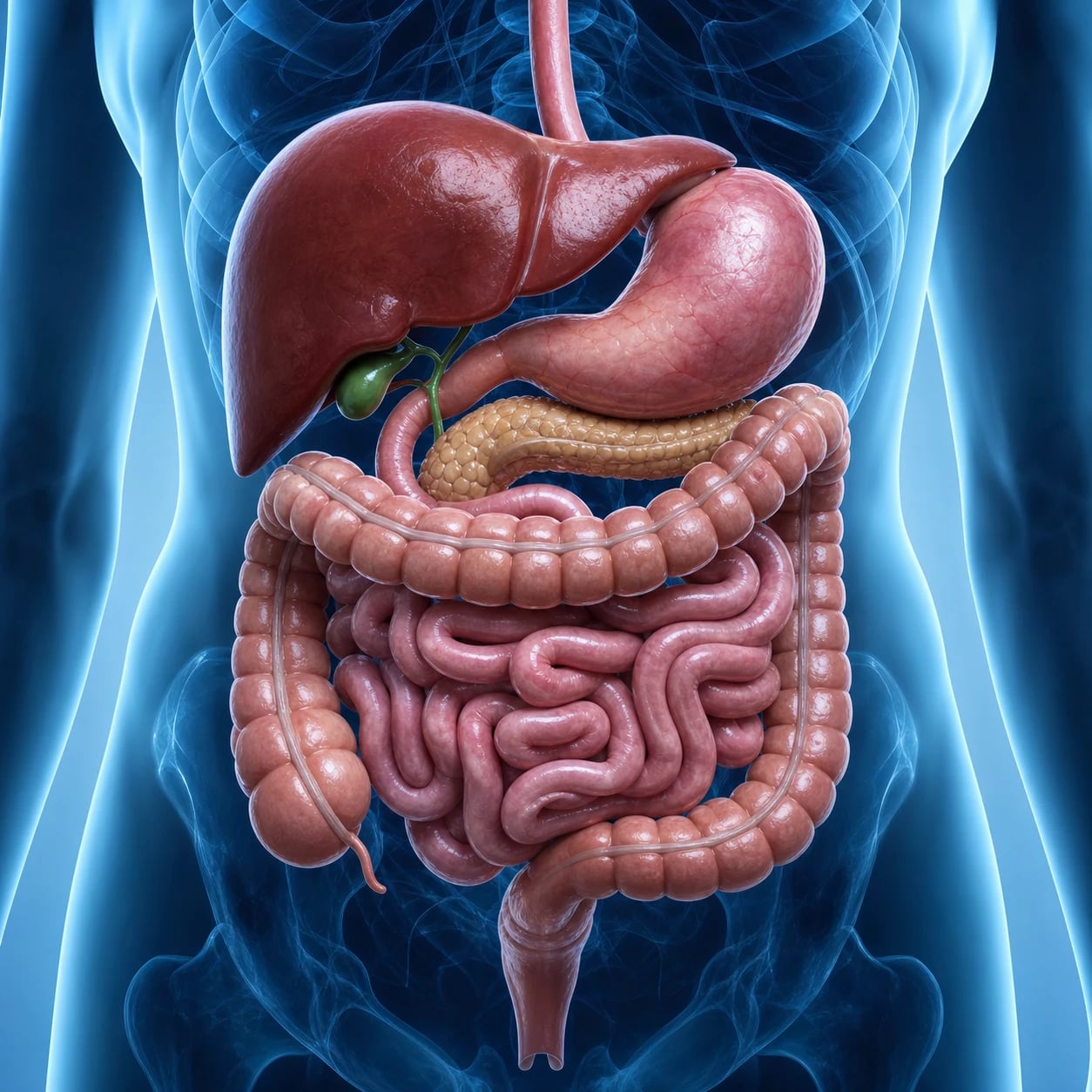
The gut runs from mouth to anus and is paralleled by the accessory organs of digestion - the liver, gallbladder, and pancreas. Anatomical detail matters in the ICU for nasogastric and feeding-tube placement, the interpretation of imaging, the site of upper GI bleeding, and the consequences of mesenteric and biliary obstruction.[1]
The oesophagus
- A muscular tube about 25 cm long with an upper oesophageal sphincter (cricopharyngeus) and a lower oesophageal sphincter at the diaphragmatic hiatus.[1]
- Three natural constrictions where foreign bodies and tumours lodge: the cricopharyngeus (about 15 cm), the aortic/bronchial crossing (about 25 cm), and the diaphragmatic hiatus (about 40 cm).[1]
The stomach
- Divided into cardia, fundus, body, antrum, and pylorus; the muscular pyloric sphincter controls gastric emptying.[1]
- Secretory cells: parietal (oxyntic) cells secrete hydrochloric acid and intrinsic factor; chief cells secrete pepsinogen; G cells in the antrum secrete gastrin; mucous neck cells and surface mucus protect the epithelium.[1]
Small intestine
- Duodenum (about 25 cm, C-shaped) receives bile and pancreatic juice at the ampulla of Vater (the major duodenal papilla), where the common bile duct and pancreatic duct unite.[1]
- Jejunum (about 2.5 m) is the main site of nutrient absorption; ileum (about 3.5 m) absorbs vitamin B12 and reclaims bile salts and contains Peyer patches.[1]
- Villi and microvilli enlarge the surface about 600-fold, supporting the gut's absorptive mass.[1]
Large intestine
- The caecum (with the appendix) gives rise to the ascending, transverse, descending and sigmoid colon and the rectum.[1]
- The colon absorbs water and electrolytes and hosts the bacterial flora that synthesise vitamin K; the appendix is the classic site of appendicitis, and the sigmoid the common site of diverticular disease.[1]
Liver and biliary tree
- The liver has a dual blood supply: the hepatic artery (about 25 per cent of flow, oxygen-rich) and the portal vein (about 75 per cent, nutrient-rich from the gut).[1]
- The functional unit is the acinus (or lobule); bile drains from canaliculi into ducts, the common hepatic duct, the common bile duct, and to the ampulla of Vater.[1]
- Portal hypertension (cirrhosis) opens portocaval anastomoses - oesophageal, paraumbilical (caput medusae), and rectal varices.[1]
Pancreas
- Retroperitoneal, with head, neck, body, and tail; the head nestles in the C-loop of the duodenum.[1]
- Exocrine acini secrete digestive enzymes (amylase, lipase, and the proteases as inactive precursors such as trypsinogen) into the duodenum via the pancreatic duct.[1]
- Endocrine islets of Langerhans secrete insulin (beta cells), glucagon (alpha), somatostatin (delta), all into the portal circulation.[1]
Red flags
The gut tube — the examiner's mental map
The gastrointestinal tract is a continuous muscular tube about 9 metres long (shorter in life, where smooth-muscle tone keeps it about 6-7 m) running from the mouth to the anus, with the accessory organs of digestion — the liver, gallbladder, pancreas, and salivary glands — emptying into its lumen. For the ICU First Part examiner the tract is best learned as four repeating facts at every level: (1) the length and course of that segment, (2) the blood supply (an artery of supply and a vein of drainage — usually to the portal system), (3) the histological layers (mucosa, submucosa, muscularis externa with its two layers and the myenteric/Auerbach and submucosal/Meissner plexuses, and the serosa/adventitia), and (4) the clinical surface — what is absorbed, what bleeds, what obstructs, and what the bedside procedure (NG, ERCP, surgery) needs to know. Holding these four facts at every level makes any segment answer complete.[1][1]
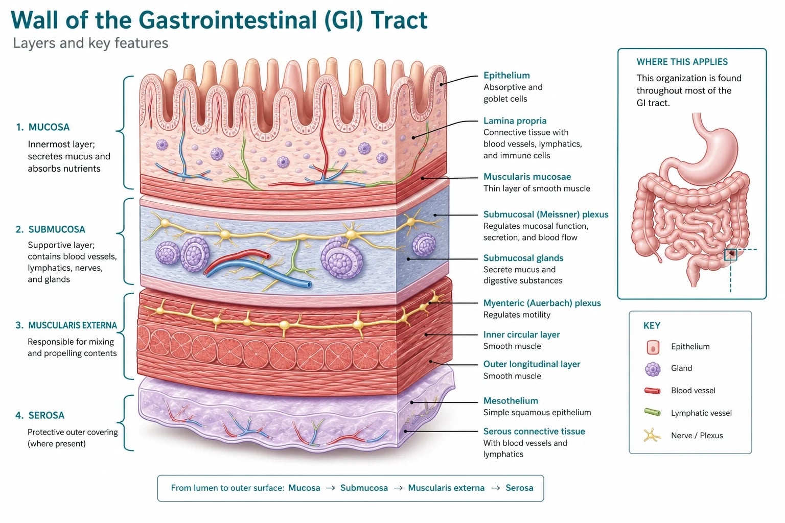
The gut wall has a consistent four-layered architecture throughout. From within outwards these are the mucosa (epithelium, lamina propria, muscularis mucosae), the submucosa (the strongest layer, carrying the submucosal/Meissner plexus and the major vessels), the muscularis externa (inner circular and outer longitudinal smooth muscle, with the myenteric/Auerbach plexus between them — the enteric nervous system's motor layer), and the outermost serosa (visceral peritoneum) where the gut is intraperitoneal, or adventitia (connective tissue) where it is retroperitoneal. Only the oesophagus has skeletal (striated) muscle in its upper third; from the oesophagus onwards smooth muscle dominates, and the enteric plexuses (Auerbach and Meissner) carry the local reflexes that govern peristalsis and secretion independently of the central nervous system — the "gut brain."[1][1]
The oesophagus in depth — three sphincters and three constrictions
The oesophagus is a muscular tube about 25 cm long (measured from the upper incisor teeth) that conveys a bolus from the pharynx to the stomach by peristalsis. It begins at the level of the cricoid cartilage (C6) — the pharyngo-oesophageal junction — and ends where it passes through the diaphragm at the oesophageal hiatus (T10) to join the stomach at the cardia. Anatomically it has cervical (C6 to T1, related to the trachea, thyroid, and recurrent laryngeal nerve in the tracheo-oesophageal groove), thoracic (T1 to the diaphragm — related to the aortic arch, the left main bronchus, the left atrium, and the descending aorta), and abdominal (about 2 cm, from the hiatus to the cardia) parts.[1]
Two functionally distinct sphincters control the tube — the examiner's classic "three sphincters" frames the upper oesophageal sphincter (UES), the lower oesophageal sphincter (LES), and (sometimes added) the cricopharyngeal sphincter, which is in fact the muscular component of the UES itself.[1][1]
The oesophageal sphincters
| Feature | Upper oesophageal sphincter (UES) | Lower oesophageal sphincter (LES) |
|---|---|---|
| Anatomical substrate | The cricopharyngeus part of the inferior pharyngeal constrictor, attached to the cricoid cartilage | A physiological (not anatomically discrete) high-pressure zone, 2-4 cm long, at the gastro-oesophageal junction |
| Level | C6 (cricoid cartilage) | T10-T11, at the diaphragmatic hiatus |
| Muscle type | Skeletal (striated) muscle — tonic contraction driven by the recurrent laryngeal nerve | Smooth muscle — tonic contraction by vagal (cholinergic) tone |
| Resting state | TONICALLY CLOSED (high pressure ~40-100 mmHg); relaxes briefly during the swallowing reflex | TONICALLY CLOSED (high pressure ~10-30 mmHg above gastric); relaxes on swallowing and on transient LES relaxations |
| Main role | Prevents air entry into the oesophagus during breathing; opens only to let a bolus through | Prevents gastro-oesophageal reflux of acidic gastric contents |
| Clinical failure | Aspiration in neurogenic dysphagia (stroke, bulbar palsy) | GORD, Barrett's oesophagus, oesophageal varices form at this junction |
The three anatomical constrictions are the points where foreign bodies, pills, and tumours lodge, and where a swallowed corrosive does most damage. They are best given as distances from the upper incisor teeth:[1][1]
The three oesophageal constrictions (from the incisors)
| Constriction | Distance from incisors | Cause | Clinical significance |
|---|---|---|---|
| 1. Upper — cricopharyngeus/UES | 15 cm | The cricopharyngeus muscle (pharyngo-oesophageal junction, C6) | Narrowest point; foreign bodies and meat impaction lodge here; the site of Zenker's diverticulum (through Killian's dehiscence, above the cricopharyngeus) |
| 2. Middle — aortic/bronchial | 25 cm | Crossing of the aortic arch and the left main bronchus | Coin/food impaction; an enlarging left atrium (mitral stenosis) can also narrow here, displacing the oesophagus backwards |
| 3. Lower — diaphragmatic | 40 cm | The diaphragmatic hiatus (oesophageal opening in the diaphragm at T10) | Hiatus hernia; the ampulla of Vater is NOT here (it is in the duodenum at about 60 cm) |
15-25-40 — the three oesophageal constrictions from the incisors
The swallowing reflex and the role of the sphincters
- ORAL PREPARATION: mastication and bolus formation; the tongue pushes the bolus to the oropharynx.[1]
- PHARYNGEAL PHASE (involuntary, about 1 s): the soft palate elevates (closes the nasopharynx), the larynx rises and the epiglottis tips (protects the airway), and the superior/middle/inferior pharyngeal constrictors propel the bolus downwards.
- UES OPENS: the cricopharyngeus relaxes (corticobulbar and vagal input via the recurrent laryngeal nerve) to let the bolus enter the oesophagus, then snaps shut — the upper sphincter at work.
- PRIMARY PERISTALSIS: a coordinated wave of circular-then-longitudinal smooth-muscle contraction (Auerbach's plexus plus vagovagal reflex) sweeps the bolus down at 2-5 cm/s.
- SECONDARY PERISTALSIS: local distension triggers further waves to clear any residue.
- LES RELAXES (receptive relaxation of the GOJ, vagal non-adrenergic non-cholinergic / nitric-oxide-mediated) to admit the bolus to the stomach, then re-contracts to form the anti-reflux barrier (LES tone + the diaphragmatic crural pinch + the angle of His + the mucosal rosette).[1]
The stomach in depth — five regions and the gastric gland
The stomach is a J-shaped reservoir in the left upper quadrant under the left costal margin, continuous above with the oesophagus at the cardia and below with the duodenum at the pylorus. It is divided into five anatomical regions:[1][1]
The five regions of the stomach
| Region | Location | Function / contents |
|---|---|---|
| Cardia | Immediately below the GOJ (2-3 cm) | Mucous-secreting; the landmark dividing oesophagus from stomach |
| Fundus | The dome above and to the LEFT of the cardia (fills with gas on an erect CXR — the gastric air bubble) | Stores food and gas; contains the parietal (oxyntic) cell mass |
| Body (corpus) | The large central portion, between fundus and antrum | The main secretory region — parietal, chief, and ECL cells |
| Antrum | The distal funnel-shaped part, narrowing toward the pylorus | Contains the G cells (gastrin); the site of grinding and mixing |
| Pylorus | The thick muscular outlet guarded by the pyloric sphincter | Controls gastric emptying; the tumour/ulcer site of gastric outlet obstruction |
The mucosa of the body and fundus is thrown into gastric pits leading into gastric glands (oxyntic glands), each lined by a cast of specialised secretory cells. The examiner wants each cell, its product, and its stimulus.[1][1]
The secretory cells of the gastric gland
| Cell | Location in gland | Product | Principal stimulus / role |
|---|---|---|---|
| Parietal (oxyntic) cells | Upper/mid body and fundus (the "oxyntic mucosa") | Hydrochloric acid (HCl, to pH ~1-2) and intrinsic factor | Stimulated by gastrin (G cells), histamine (ECL cells, via H₂ receptors), and vagal acetylcholine (M₃ receptors); inhibited by somatostatin and by proton-pump inhibitors (which block the H⁺/K⁺-ATPase) |
| Chief (peptic) cells | Base of the glands, body and fundus | Pepsinogen (the inactive zymogen), converted to active pepsin by gastric acid | Stimulated by vagal input and acid; pepsin begins protein digestion |
| G cells | The antrum (and duodenum) | Gastrin (a peptide hormone) | Stimulated by peptides/amino acids in the lumen, by vagal GRP, and by gastric distension; inhibited by low antral pH (negative feedback via somatostatin) |
| Enterochromaffin-like (ECL) cells | Body and fundus | Histamine | Stimulated by gastrin; histamine then paracrine-stimulates the parietal cell (the "histamine amplifier") |
| D cells | Antrum and body | Somatostatin | The "brake" — released when antral pH falls below 3, inhibiting gastrin, histamine, and acid |
| Mucous neck cells / surface mucus cells | Neck of the gland and surface | Mucus and bicarbonate (the "mucus-bicarbonate barrier") | Protect the epithelium from acid and pepsin; the barrier is breached in peptic ulceration |
Acid secretion by the parietal cell (the H⁺/K⁺-ATPase — the PPI target)
- CO₂ + H₂O are combined by carbonic anhydrase inside the parietal cell to form carbonic acid (H₂CO₃), which dissociates into H⁺ and HCO₃⁻.[1]
- The H⁺ is actively pumped into the gland lumen in exchange for K⁺ by the H⁺/K⁺-ATPase (the proton pump) on the apical (canalicular) membrane — the target of proton-pump inhibitors (omeprazole).
- Cl⁻ follows H⁺ into the lumen (through chloride channels), so the secretion is HCl; the HCO₃⁻ exits the basolateral membrane in exchange for Cl⁻ (the "chloride shift"), alkalinising the blood (the "gastric alkaline tide").
- Three stimuli converge on the parietal cell: acetylcholine (vagal, M₃), histamine (ECL, H₂), and gastrin (G cell, CCK₂) — histamine being the final common amplifier.
- Somatostatin (D cell, SST₂) and the prostaglandins inhibit each step — the basis for somatostatin analogues and misoprostol.[1]
The gastric gland cast — the only source of intrinsic factor
Small intestine in depth — duodenum, jejunum, ileum
The small intestine is about 6 m long in life (up to 7 m in the cadaver, where smooth muscle is flaccid) and is the principal site of digestion and absorption. It runs from the pylorus to the ileocaecal valve and is divided, proximally to distally, into duodenum, jejunum, and ileum.[1][1]
The duodenum is the first 25 cm, C-shaped, and largely retroperitoneal; it curves around the head of the pancreas (the "C-loop"). Its descending (second) part bears the major duodenal papilla (ampulla of Vater), where the common bile duct and main pancreatic duct unite through the sphincter of Oddi to empty into the gut. The minor papilla (about 2 cm proximal) drains the accessory pancreatic duct of Santorini. The duodenum ends at the duodenojejunal flexure, suspended by the ligament of Treitz — the radiological and surgical landmark dividing upper from lower GI bleeding.[1][1]
The jejunum (about 2.5 m) occupies the left upper quadrant and is the main site of nutrient absorption (carbohydrate, protein, fat, folate, iron, calcium). The ileum (about 3.5 m) occupies the right lower quadrant and has the specific absorptive tasks: vitamin B₁₂ (with intrinsic factor) and bile salts (enterohepatic recirculation), and it carries the Peyer patches (aggregated lymphoid tissue, the gut's immune sentinels, hypertrophied in typhoid and the lead-point of intussusception in children).[1]
Duodenum vs jejunum vs ileum
| Feature | Duodenum | Jejunum | Ileum |
|---|---|---|---|
| Length | ~25 cm | ~2.5 m | ~3.5 m |
| Location | C-loop around the pancreatic head, mostly retroperitoneal | LUQ, intraperitoneal | RLQ, intraperitoneal |
| Lumen | Widest proximal segment | Wide, thick wall (valves of Kerckring tall) | Narrower, thinner wall |
| Villi / plicae | Short; receives bile + pancreatic juice | Tall, numerous plicae circulares (valves of Kerckring) — gives the "feathery" look on barium | Shorter, sparse plicae; fat in wall (pink) |
| Specific absorption | Iron (apical), calcium | Most nutrients, folate, iron, fat (as chylomicrons via lacteals) | Vitamin B₁₂ (with IF), bile salts |
| Lymphoid tissue | — | Sparse | Peyer patches (aggregated lymphoid nodules) |
| Vascular arcades | — | Few long vasa recta (few arcades) | Many short arcades (many vasa recta) — a denser mesenteric vascular pattern |
The mucosa of the entire small intestine is enormously amplified by three sequential folds: the plicae circulares (valves of Kerckring) — permanent circular mucosal folds; the villi (~1 mm finger-like projections, each with an arteriole, a venule, and a central lacteal); and the microvilli of each absorptive enterocyte (the brush border, carrying the disaccharidases — lactase, sucrase, maltase — and peptidases). Together these multiply the surface area about 600-fold (to about 30 m²), which is why even modest mucosal loss (coeliac, rotavirus, short bowel) devastates absorption.[1][1]
Absorption of a meal across the small-intestinal enterocyte
- LUMINAL digestion: pancreatic amylase, lipase, and trypsin/chymotrypsin/elastase break starch, fat, and protein to oligomers; the brush-border disaccharidases (lactase, sucrase-isomaltase) finish carbohydrates to monosaccharides.[1]
- TRANSPORT into the enterocyte: glucose and galactose via SGLT1 (Na⁺-dependent); fructose via GLUT5; amino acids and di-/tripeptides via Na⁺- and H⁺-dependent transporters; fatty acids plus monoglycerides (with bile salts) form micelles that diffuse in.
- INTRACELLULAR processing: monosaccharides and amino acids pass through; fatty acids are re-esterified into triglycerides, coated with apoproteins to form chylomicrons.
- BASOLATERAL exit: glucose and amino acids via GLUT2 and amino-acid transporters into the portal blood; chylomicrons are too large for capillaries and enter the central lacteal → lymphatics (thoracic duct) — the route by which lipid reaches the systemic circulation, bypassing the first-pass liver.
- BILE SALT reclamation: bile salts are reabsorbed in the terminal ileum, return to the liver via the portal vein, and are resecreted — the enterohepatic circulation (~6 cycles/day). Terminal-ileum disease or resection breaks this loop → bile-salt diarrhoea, malabsorption of fat and of fat-soluble vitamins (A, D, E, K), and gallstones.[1]
What the ileum specifically absorbs — 'B12 and Bile'
Large intestine in depth — caecum, colon, rectum, anal canal
The large intestine is about 1.5 m long and frames the abdomen, running from the ileocaecal valve to the anus. It is identified at laparotomy/laparoscopy by three external features absent from the small bowel: the taeniae coli (three longitudinal muscle bands — the longitudinal muscle gathered into strips rather than a continuous sheet), the haustra (saccular outpouchings formed because the taeniae are shorter than the bowel), and the appendices epiploicae (fat-filled tags of serosa). It absorbs water and electrolytes (concentrating ~1-2 L of ileal effluent into ~150-200 g of faeces), hosts the colonic bacterial flora that synthesise vitamin K and some B vitamins, and stores and expels faeces.[1][1]
The segments are the caecum (with the vermiform appendix at its base — the classic site of appendicitis, pain beginning periumbilical then migrating to McBurney's point), the ascending colon (right flank, retroperitoneal), the transverse colon (intraperitoneal, on its mesocolon — the most mobile part), the descending colon (left flank, retroperitoneal), the sigmoid colon (S-shaped on its mesentery — the commonest site of diverticular disease and volvulus), the rectum, and the anal canal.[1]
Small intestine vs large intestine
| Feature | Small intestine | Large intestine |
|---|---|---|
| Length | ~6 m | ~1.5 m |
| External features | Smooth; no taeniae/haustra/epiploicae | Taeniae coli, haustra, appendices epiploicae |
| Longitudinal muscle | Continuous layer | Gathered into three taeniae |
| Wall | Thin; plicae + villi + microvilli | Thick; no villi, lots of goblet cells |
| Main function | Digestion and absorption of nutrients | Water and electrolyte absorption; bacterial flora |
| Vitamin synthesis | — | Vitamin K (and some B vitamins) by gut flora |
| Luminal content | Chyme (liquid) | Faeces (semi-solid to solid) |
The anal canal has a key surgical landmark — the pectinate (dentate) line, the wavy line at the bases of the anal columns marking the embryological junction of endoderm (hindgut, above) and ectoderm (proctodeum, below). Above and below the line the epithelium, blood supply, lymphatic drainage, innervation, and pain sensation all differ — the basis for distinguishing internal (above the line, painless, from the superior rectal vein / portal system → may be a varix) from external (below the line, painful, from the inferior rectal vein / systemic) haemorrhoids.[1]
Above vs below the pectinate (dentate) line
| Feature | Above the line (endoderm) | Below the line (ectoderm) |
|---|---|---|
| Epithelium | Columnar (gut mucosa) | Stratified squamous (skin) |
| Arterial supply | Superior rectal (terminal branch of IMA) | Inferior rectal (internal pudendal, systemic) |
| Venous drainage | Superior rectal → portal system | Inferior rectal → systemic (caval) system |
| Lymphatics | Internal iliac and inferior mesenteric nodes | Superficial inguinal nodes |
| Pain | Visceral — INSENSITIVE (autonomic) | Somatic — VERY SENSITIVE (inferior rectal nerve) |
| Haemorrhoids | Internal — painless, may bleed; above this is rectal varix territory (portal) | External — painful |
The liver in depth — eight Couinaud segments, the portal triad, and the functional unit
The liver is the largest gland and the largest visceral organ (~1.5 kg), lying in the right upper quadrant under the diaphragm. It has a dual blood supply: the hepatic artery (proper hepatic artery, a branch of the common hepatic from the coeliac trunk) delivers ~25-30% of inflow (oxygen-rich), and the portal vein delivers ~70-75% (nutrient-rich, from the gut, spleen, and pancreas). These two supplies merge in the portal tracts, branch together down to the sinusoids, and drain centrally into the hepatic veins → IVC.[1][1]
The functional unit is described two ways. The classic hepatic lobule is a hexagon with a central vein at its core and portal triads at the corners. The acinus of Rappaport (the functional/physiological unit) is a diamond oriented around the distributing branches of the portal venules and hepatic arterioles, divided into zones 1, 2, 3 radiating outward; zone 3 (centrilobular, nearest the central vein) is the least oxygenated and the first to die in ischaemia and the first to lay down fibrosis.[1]
Each portal triad (at the corner of every lobule, visible on histology and on a liver biopsy) carries, in a single connective-tissue sheath, three structures:[1]
The portal triad — the three structures in every portal tract
| Component | Origin | Function in the triad |
|---|---|---|
| Branch of the hepatic artery | Coeliac trunk → common hepatic → proper hepatic artery | Oxygenated systemic blood (~25%) |
| Branch of the portal vein | Splenic + superior mesenteric veins | Nutrient-rich venous blood from the gut (~75%) |
| Bile ductule ( + lymphatic) | Bile canaliculi → ducts | Drains bile OUT (the only outward flow of the triad — blood flows IN) |
The modern surgical Couinaud classification divides the liver into eight functionally independent segments, each with its own portal triad inflow and hepatic venous outflow, permitting segmental resection. The segments are numbered clockwise in the frontal plane, viewed as if the liver were held in front of you with the IVC at the top.[1][1]
Couinaud liver segments — the eight resectable units
| Segment | Location | Common name | Notes |
|---|---|---|---|
| I | Posterior, between IVC and ligamentum venosum | Caudate lobe | Embryologically distinct; drains directly to the IVC (bypasses hepatic veins) — hypertrophies in Budd-Chiari |
| II, III | Left lateral, superior + inferior | Left lateral segment (II = superior, III = inferior) | Lateral part of the left lobe |
| IV | Left medial (between falciform and umbilical fissure) | Left medial segment / quadrate lobe | IVa superior, IVb inferior |
| V, VIII | Right anterior, inferior + superior | Right anterior segment | Anterior to the right hepatic vein |
| VI, VII | Right posterior, inferior + superior | Right posterior segment | Posterior to the right hepatic vein |
The midline plane is defined by the middle hepatic vein (running in the main fissure / Cantlie's line from the gallbladder fossa to the IVC) — it divides the right (segments V-VIII) from the left (segments I-IV) liver. The right hepatic vein divides right anterior (V, VIII) from right posterior (VI, VII); the left hepatic vein divides left lateral (II, III) from left medial (IV).[1]
Couinaud segments — 'I-caudate, then clockwise II-VIII'
Two non-parenchymal liver cell populations matter to the ICU:[1]
- Kupffer cells — the fixed macrophages of the liver, anchored to the endothelial lining of the sinusoids. They are part of the mononuclear phagocyte system and clear endotoxin (from portal venous blood), bacteria, and aged red cells from the circulation; they are central to the hepatic response in sepsis and to the hyperbilirubinaemia of sepsis.
- Hepatic stellate (Ito) cells — the vitamin-A-storing cells of the space of Disse. When activated (by injury, alcohol, viral hepatitis, NASH) they transform into myofibroblasts and lay down collagen — the cell that drives liver fibrosis and cirrhosis.[1]
The biliary tree in depth — from canaliculus to ampulla
Bile is secreted by hepatocytes into the bile canaliculi (intercellular channels between adjacent hepatocytes), flows into the ducts of Hering → interlobular ducts (in the portal triad), then the larger right and left hepatic ducts (draining the right and left liver), which unite to form the common hepatic duct. The cystic duct from the gallbladder joins the common hepatic duct to form the common bile duct (CBD). The CBD descends behind the first part of the duodenum and through the head of the pancreas to join the main pancreatic duct (of Wirsung) at the ampulla of Vater (hepatopancreatic ampulla), opening into the duodenum at the major duodenal papilla, the whole outlet controlled by the sphincter of Oddi.[1][1]
The gallbladder is a pear-shaped reservoir under the liver, divided into fundus, body, and neck (the neck narrows into the cystic duct; a dilatation at the neck — Hartmann's pouch — is where gallstones lodge). It stores and concentrates bile (up to 10-fold, by water absorption) between meals; after a fatty meal, cholecystokinin (CCK) from the duodenal I-cells causes the gallbladder to contract and the sphincter of Oddi to relax, delivering concentrated bile to the gut for fat emulsification.[1]
Calot's (cystohepatic) triangle — bounded by the cystic duct (inferiorly), the common hepatic duct (medially), and the liver edge (superiorly) — contains the cystic artery (usually a branch of the right hepatic artery) and is the critical anatomy of laparoscopic cholecystectomy: identifying and clipping the cystic duct and artery within this triangle prevents the catastrophic error of injuring the common hepatic/CBD.[1]
Bile flow — from hepatocyte to duodenum (and back to the liver)
- HEPATOCYTE secretion into bile canaliculi (bile-salt-dependent and -independent flow).[1]
- DUCTULES → interlobular ducts (portal triad) → right and left hepatic ducts → COMMON HEPATIC DUCT.
- Between meals, the sphincter of Oddi is CLOSED: bile is diverted UP the cystic duct into the gallbladder for storage and concentration.
- After a meal, CCK (from duodenal I-cells, released by fat/protein) → gallbladder contracts plus sphincter of Oddi relaxes → concentrated bile flows via the CBD → ampulla → duodenum.
- Bile salts emulsify fat and form micelles; they are then reabsorbed in the terminal ileum → portal vein → back to the liver → resecreted (enterohepatic circulation, ~6 cycles/day, ~95% reclamation).[1]
EPISOD — sphincterotomy for suspected sphincter of Oddi dysfunction (Cotton 2014, JAMA)
Study design
Multicentre, randomised, SHAM-controlled trial — 213 post-cholecystectomy patients with suspected sphincter of Oddi dysfunction (types II and III) across 7 US centres
Intervention
Endoscopic sphincterotomy vs sham (manometry-directed for type II)
Primary outcome
Pain-related disability at 12 months — NO benefit of sphincterotomy over sham, and a high rate of post-ERCP pancreatitis (~11%)
Clinical bottom line
The sphincter of Oddi is a real anatomical structure but 'sphincter of Oddi dysfunction' as a cause of pain is rarely (if ever) improved by sphincterotomy, while the procedure carries a serious pancreatitis risk — reshaped practice away from ERCP for type III suspected SOD
The pancreas in depth — exocrine and endocrine, head to tail
The pancreas is a retroperitoneal organ lying transversely across the posterior abdominal wall at L1-L2, behind the stomach and in front of the IVC, aorta, and left kidney. It is divided into head (nestled in the C-loop of the duodenum, with the uncinate process hooking behind the SMA and SMV), neck (over the SMV-portal vein confluence), body, and tail (reaching the hilum of the spleen). Its duct system — the main pancreatic duct of Wirsung running the length of the gland to the ampulla of Vater, and the accessory duct of Santorini opening separately at the minor papilla — drains its exocrine secretion into the duodenum.[1][1]
The exocrine and endocrine pancreas
| Feature | Exocrine pancreas | Endocrine pancreas (islets of Langerhans) |
|---|---|---|
| Cell type | Acinar cells (and duct cells) | Islet cells (scattered among the acini) |
| Product | Digestive enzymes + bicarbonate-rich fluid | Hormones (insulin, glucagon, etc.) |
| Destination | The duodenal lumen (via the pancreatic ducts) | The bloodstream (islets drain into the portal vein — so insulin reaches the liver first) |
| Mass | ~98-99% of the gland | ~1-2% of the gland (but about 10% of pancreatic blood flow) |
| Control | Secretin (ducts, bicarbonate) + CCK (acini, enzymes) + vagal input | Nutrients (glucose), neural, and hormonal inputs |
| Failure | Malabsorption (steatorrhoea, fat-soluble-vitamin deficiency), seen in chronic pancreatitis and after pancreatectomy | Diabetes (type 1 if beta cells destroyed; type 3c in pancreatic disease) |
The exocrine acinar cells synthesise the digestive enzymes, most as inactive zymogens to prevent autodigestion:[1]
- Proteases — secreted as trypsinogen, chymotrypsinogen, procarboxypeptidase, proelastase; trypsinogen is activated to trypsin in the duodenum by enterokinase (enteropeptidase) on the duodenal brush border, and trypsin then activates all the other zymogens (and itself — autocatalysis). Premature intracellular activation (e.g. in pancreatitis) is the mechanism of autodigestion.
- Lipase (and colipase, phospholipase) — fat digestion.
- Amylase — starch digestion.
- The duct cells secrete a bicarbonate-rich, alkaline fluid (driven by secretin) that neutralises acidic gastric chyme — without it, duodenal enzymes cannot work and the duodenum is damaged by acid. [1]
The endocrine islets contain four principal cell types, each with its hormone:[1]
The islet cell types of Langerhans
| Cell (% of islet) | Location within islet | Hormone | Principal action |
|---|---|---|---|
| Beta (β) ~70% | Core | Insulin | Lowers blood glucose (anabolic — drives glucose into cells) |
| Alpha (α) ~20% | Mantle | Glucagon | Raises blood glucose (glycogenolysis, gluconeogenesis) |
| Delta (δ) ~5-10% | Mantle | Somatostatin | Inhibits both insulin and glucagon (the "islet brake") |
| PP (F) cells | Mantle | Pancreatic polypeptide | Inhibits pancreatic exocrine secretion and gallbladder contraction |
Islet cells — 'BAD' (Beta in the core, Alpha and Delta around the edge)
The spleen in detail
The spleen is the largest lymphoid organ (~150 g), tucked under the left costal margin in the left hypochondrium between the 9th and 11th ribs, related to the fundus of the stomach (gastric impression), the splenic flexure of the colon (colic impression), and the left kidney (renal impression). Its blood supply is the splenic artery (a tortuous branch of the coeliac trunk) and its drainage the splenic vein (which runs with the artery and unites with the superior mesenteric vein behind the pancreatic neck to form the portal vein).[1]
Functionally the spleen has two compartments: the red pulp (cords and sinusoids — the "filter" that removes aged/abnormal red cells, and the site of extravascular haemolysis and of Howell-Jolly body removal), and the white pulp (the lymphoid tissue — B-cell follicles and T-cell zones, mounting the immune response to encapsulated organisms). It is supported by accessory spleens (splenunculi) in about 10-20% of people, usually near the hilum — relevant because they can hypertrophy after splenectomy and cause recurrence of immune cytopenia.[1]
Splanchnic circulation — coeliac trunk, SMA, IMA
The foregut, midgut, and hindgut are each supplied by one of the three unpaired midline arteries arising from the abdominal aorta; the venous drainage of all three converges on the portal vein. This is the splanchnic (visceral) circulation, and its arterial territories and their watershed points are central to mesenteric ischaemia.[1][1]
The three unpaired gut arteries and their territories
| Artery (origin from aorta) | Vertebral level | Embryological gut | Supplies |
|---|---|---|---|
| Coeliac trunk | T12 | Foregut | Lower oesophagus, stomach, 1st and 2nd parts of duodenum, liver, gallbladder, pancreas, spleen — via left gastric, splenic, common hepatic branches |
| Superior mesenteric artery (SMA) | L1 | Midgut | 3rd and 4th parts of duodenum, jejunum, ileum, caecum, ascending and proximal two-thirds of transverse colon |
| Inferior mesenteric artery (IMA) | L3 | Hindgut | Distal third of transverse colon, descending and sigmoid colon, upper rectum (via left colic, sigmoid, superior rectal branches) |
The coeliac trunk is a short (~1-2 cm) artery dividing almost immediately into three: the left gastric (lesser curve of stomach and lower oesophagus), the splenic (pancreas, spleen, fundus via short gastrics, and greater curve via the left gastroepiploic), and the common hepatic (which gives the gastroduodenal and the right gastric before continuing as the proper hepatic to the liver, giving the right gastroepiploic from the gastroduodenal). This rich anastomotic network around the stomach (the two gastroepiploic arcades on the greater curve, the two gastric arteries on the lesser curve) explains why the stomach tolerates gastric artery embolisation and why vagotomy alone rarely causes ischaemia.[1]
The SMA arises at L1 and passes anterior to the uncinate process of the pancreas and the left renal vein (an important surgical landmark — the SMV is to its right), supplying the entire midgut. The IMA arises at L3 and supplies the hindgut. The two crucial watershed zones between territories are:[1]
- Griffith's point — the junction of the SMA (middle colic) and the IMA (left colic) supply, at the splenic flexure (the junction of the proximal two-thirds and the distal third of the transverse colon). The splenic flexure is the classic site of ischaemic colitis because it sits at this marginal watershed with a variable and often poor collateral supply.
- Sudeck's point — the junction of the sigmoid branches and the superior rectal (IMA) supply with the middle/inferior rectal (internal iliac) supply, at the rectosigmoid junction. This is vulnerable after IMA ligation in aortic or colorectal surgery. [1]
Coeliac-SMA-IMA levels — 'T12, L1, L3 (the boat sail)'
From gut lumen to liver — the portal venous system
- The veins draining the gut (the superior mesenteric vein from the midgut, the inferior mesenteric vein from the hindgut, the splenic vein from the foregut/spleen) all converge on the portal vein.[1]
- The portal vein forms BEHIND the neck of the pancreas by the union of the superior mesenteric and splenic veins (the inferior mesenteric usually joins the splenic).
- The portal vein runs up in the hepatoduodenal ligament (the free edge of the lesser omentum), posterior to the hepatic artery and the CBD (the portal triad — CBD on the right, hepatic artery on the left, portal vein posterior), to the porta hepatis.
- In the liver it divides into right and left branches, distributing to the sinusoids where nutrients are processed, then drains via the hepatic veins to the IVC.
- The whole system is a portal-systemic (portocaval) circuit — two capillary beds (gut then liver) in series, with no valves; when portal pressure rises, blood escapes through the four portocaval anastomoses (below).[1]
Portocaval (portosystemic) anastomoses in depth
The portal venous system has no valves and meets the systemic venous system at four characteristic sites where, in portal hypertension, the porto-systemic pressure gradient dilates the connecting veins into clinically important collaterals. These are the portocaval anastomoses.[1][2]
The portocaval anastomoses (portal to systemic escape routes)
| Site | Portal vein | Systemic vein | Clinical sign when dilated |
|---|---|---|---|
| 1. Lower oesophagus (GOJ) | Left gastric (oesophageal veins) | Azygos (systemic) | Oesophageal/gastric varices — the catastrophic bleeder |
| 2. Umbilicus | Paraumbilical veins (along ligamentum teres) | Superior and inferior epigastric (systemic) | Caput medusae — radiating periumbilical veins |
| 3. Rectum | Superior rectal (IMA → portal) | Middle and inferior rectal (internal iliac → systemic) | Rectal varices (distinct from internal haemorrhoids — varices are a portocaval sign) |
| 4. Retroperitoneum / bare areas | Retroperitoneal veins, veins of Retzius | Lumbar, phrenic, renal (systemic) | Usually silent; seen on imaging as collaterals |
Natural history of variceal bleeding — Graham & Smith (1981, Gastroenterology)
Study type
Landmark observational cohort — patients followed after a first variceal haemorrhage, defining the natural history before modern therapy
Key findings
A first variceal bleed carries a very high early mortality (~30-40% per episode in the era before banding/terlipressin); survivors are at high risk of rebleeding (~60-70% within 1 year), with each rebleed carrying similar mortality
Anatomical basis
The bleeder is the submucosal venous plexus of the GOJ — a portocaval anastomosis — thin-walled, unsupported by surrounding tissue, and exposed to the full portal-systemic pressure gradient
Clinical bottom line
Established the imperative for PRIMARY prevention (non-selective beta-blockers / endoscopic banding in cirrhosis with varices) and for definitive risk reduction after a bleed
Clinical correlations for the ICU
Nasogastric and feeding-tube placement. The three oesophageal constrictions (15-25-40 cm) and the ligament of Treitz (~60 cm to the DJ flexure) determine where an NG tube tip sits: a standard NG drains the stomach (tip 45-60 cm at the nose), a post-pyloric nasojejunal tube must cross the pylorus and the ligament of Treitz (~80-100 cm) to deliver feed distal to the pylorus — essential when pancreatic rest or aspiration risk matters (severe acute pancreatitis, gastric outlet obstruction, high aspiration risk).[1]
Mapping the site of upper GI bleeding. Haematemesis of bright red blood implies an active arterial source (often a high-pressure varix or a spurting duodenal-ulcer artery — the gastroduodenal artery posteriorly). "Coffee-ground" vomit implies slower exposure to acid. Melaena (black, tarry, foul-smelling stool) forms when blood is digested as it transits the gut, and indicates bleeding proximal to the ligament of Treitz. The Rockall and Glasgow-Blatchford scores stratify risk and need for intervention.[3][4]
Rockall vs Glasgow-Blatchford — the two upper-GI-bleed risk scores
| Feature | Rockall score | Glasgow-Blatchford score (GBS) |
|---|---|---|
| Purpose | Predict mortality (and identifies low-risk) | Predict need for intervention (transfusion, endoscopy, surgery) |
| Timing | Can be calculated after endoscopy (full Rockall) or before (admission Rockall) | Before endoscopy — purely clinical/lab |
| Inputs | Age, shock (HR/BP), comorbidity, endoscopic diagnosis and stigmata of recent haemorrhage | Urea, Hb, systolic BP, pulse, presentation with syncope/melaena, hepatic disease, cardiac failure |
| Low-risk / safe discharge | Score 0-1 | GBS 0-1 — very low risk, candidate for outpatient management |
| Strength | Inclusion of endoscopic findings improves mortality prediction | No endoscopy needed; better at identifying who NEEDS an intervention |
Glasgow-Blatchford score — predicting need for treatment (Blatchford 2000, Lancet)
Study type
Development and validation cohort — consecutive admissions with upper GI bleeding
The score
Urea, haemoglobin, systolic BP, pulse, syncope, melaena, hepatic disease, cardiac failure — all available at the bedside, no endoscopy
Key result
A score of 0 identified patients who almost never needed intervention and could be considered for safe outpatient management; high scores strongly predicted need for transfusion/endoscopy/surgery
Clinical bottom line
The GBS is the standard pre-endoscopy triage tool; it anatomically reflects that shock (from a large bleed) and uraemia (from digested blood → protein load) signal a bleeding source needing intervention
Rockall score — risk assessment after acute upper GI haemorrhage (Rockall 1996, Gut)
Study type
Large multicentre UK audit of admissions with acute upper GI bleeding (the 1993 audit, ~4185 patients)
The score
Age, shock, comorbidity, endoscopic diagnosis, and stigmata of recent haemorrhage — maximum 11 points (admission sub-score up to 7)
Key result
Mortality rose steeply with score (near 0% at 0-1; up to ~40% or more at the highest); rebleeding risk similarly tracked the score
Clinical bottom line
Gives a reproducible estimate of rebleeding and death after an upper GI bleed; the endoscopic component encodes the anatomy of the lesion (e.g. a visible vessel in a DU = high-risk stigmata)
ERCP and the ampulla. Endoscopic retrograde cholangiopancreatography reaches the ampulla of Vater (major papilla, ~60 cm from the incisors) to extract CBD stones (choledocholithiasis), stent biliary strictures, or place pancreatic-duct stents. The shared anatomy of the CBD and pancreatic duct at the ampulla is why a gallstone (gallstone pancreatitis) or an ampullary tumour obstructs BOTH systems, and why ERCP itself can precipitate pancreatitis (instrumentation of the papilla / contrast under pressure in the pancreatic duct).[5]
Acute mesenteric ischaemia — anatomy of the territory. Occlusion of the SMA (embolus in AF, or thrombosis of an atherosclerotic origin) devascularises the entire midgut (jejunum to proximal transverse colon) — a surgical emergency presenting as pain out of proportion to examination, metabolic acidosis, and (late) peritonism. Non-occlusive mesenteric ischaemia (NOMI) reflects low-flow splanchnic vasoconstriction in shock (often with vasopressors). Ischaemic colitis preferentially strikes the splenic flexure (Griffith's point) watershed — presenting with left-lower-quadrant pain and bloody diarrhoea, usually self-limiting.[1]
Exam practice — SAQs
SAQ — Gastrointestinal anatomy applied to emergency upper GI endoscopy for variceal haemorrhage
10 minutes · 10 marks
A 62-year-old man with alcohol-related cirrhosis and known oesophageal varices presents to the emergency department with three large-volume episodes of haematemesis. He is pale and diaphoretic: HR 124, BP 88/52, Hb 62 g/L, INR 1.9. He is resuscitated with balanced crystalloid, packed red cells and fresh frozen plasma, given intravenous terlipressin 2 mg, a broad-spectrum antibiotic (ceftriaxone 1 g) and a proton-pump inhibitor, and taken to the endoscopy suite for emergency oesophagogastroduodenoscopy. You are asked to describe the anatomy that guides the procedure.
SAQ — Vascular supply of the GI tract applied to acute mesenteric ischaemia
10 minutes · 10 marks
A 68-year-old woman with chronic atrial fibrillation (on no anticoagulation) presents with sudden onset severe periumbilical abdominal pain that is markedly out of proportion to her abdominal examination. She has vomited once. On examination: HR 132 (irregularly irregular), BP 90/54, RR 28, SpO2 95 per cent on room air; the abdomen is soft with minimal tenderness despite the patient reporting severe pain. Lactate 6.4 mmol/L, venous pH 7.21, creatinine 168 (baseline 88), amylase mildly raised. CT angiography demonstrates an embolic occlusion of the superior mesenteric artery 4 cm distal to its origin, with gas in the small-bowel wall (pneumatosis intestinalis).
Clinical pearls
Sample exam question — worked answer
[1]Worked answer. The abdominal gut tube and its accessory organs are supplied by three unpaired midline branches of the abdominal aorta, each corresponding to an embryological gut division.[1][1]
The coeliac trunk arises at T12 and supplies the foregut — the distal oesophagus, stomach, the proximal duodenum (first and second parts), the liver, gallbladder, pancreas, and spleen. It is a short trunk (~1-2 cm) dividing into the left gastric (lesser curve and lower oesophagus), the splenic (pancreas, spleen, fundus via short gastrics, greater curve via the left gastroepiploic), and the common hepatic (giving the gastroduodenal and right gastric, then continuing as the proper hepatic to the liver and giving the right gastroepiploic). The stomach is encircled by anastomotic arcades on both curves, which is why it tolerates arterial occlusion.[1]
The superior mesenteric artery (SMA) arises at L1 and supplies the midgut — the distal duodenum (third and fourth parts), jejunum, ileum, caecum and appendix, ascending colon, and the proximal two-thirds of the transverse colon. It passes anterior to the uncinate process of the pancreas and the left renal vein (a key surgical landmark), with the superior mesenteric vein on its right.[1]
The inferior mesenteric artery (IMA) arises at L3 and supplies the hindgut — the distal third of the transverse colon, the descending and sigmoid colon, and the upper rectum (via the left colic, sigmoid, and superior rectal branches). The lower rectum is supplied by the middle and inferior rectal arteries from the internal iliac (systemic) circulation, creating a porto-systemic anastomosis at the rectum.[1]
The venous drainage of all three territories converges on the portal vein — formed behind the neck of the pancreas by the union of the superior mesenteric and splenic veins (the inferior mesenteric usually joins the splenic). The portal vein runs in the hepatoduodenal ligament posterior to the hepatic artery and CBD (the portal triad) to the porta hepatis, where its blood is processed by the liver before draining via the hepatic veins to the IVC. The whole system is a valveless, two-capillary-bed circuit — and when portal pressure rises (cirrhosis, portal-vein thrombosis), blood escapes through the portocaval anastomoses.[1][2]
The four classic portocaval sites are: (1) the lower oesophagus — left gastric (portal) to azygos (systemic) veins → oesophageal/gastric varices, the catastrophic bleeder; (2) the umbilicus — paraumbilical veins to the superior/inferior epigastric veins → caput medusae; (3) the rectum — superior rectal (portal/IMA) to middle/inferior rectal (systemic/internal iliac) veins → rectal varices (distinct from haemorrhoids); and (4) the retroperitoneum and bare areas (veins of Retzius) — usually clinically silent, seen on imaging. The clinical consequences are variceal haemorrhage (~15-20% mortality per bleed), hepatic encephalopathy (portosystemic shunting of gut-derived neurotoxins like ammonia, bypassing the liver), and the radiological signs of portal hypertension (splenomegaly, ascites, collaterals).[2]
Bottom line. Three unpaired arteries (coeliac T12, SMA L1, IMA L3) supply the foregut, midgut, and hindgut; their watersheds (Griffith's point at the splenic flexure, Sudeck's point at the rectosigmoid) are the sites of mesenteric ischaemia. All venous blood converges on the portal vein, and a raised portal pressure opens four portocaval anastomoses — the anatomical substrate of varices, caput medusae, rectal varices, and encephalopathy.[1][1]
References
- [1]Bismuth H Surgical anatomy and anatomical surgery of the liver World J Surg, 1982.PMID 7090393
- [2]Graham DY, Smith JL The course of patients after variceal hemorrhage Gastroenterology, 1981.PMID 6970703
- [3]Rockall TA, Logan RF, Devlin HB, Northfield TC Risk assessment after acute upper gastrointestinal haemorrhage Gut, 1996.PMID 8675081
- [4]Blatchford O, Murray WR, Blatchford M A risk score to predict need for treatment for upper-gastrointestinal haemorrhage Lancet, 2000.PMID 11073021
- [5]Cotton PB, Durkalski V, Romagnuolo J, et al. Effect of endoscopic sphincterotomy for suspected sphincter of Oddi dysfunction on pain-related disability following cholecystectomy: the EPISOD randomized clinical trial JAMA, 2014.PMID 24867013