ICU · Anatomy
Neuroanatomy — Brainstem, Cranial Nerves & Spinal Cord
Also known as Neuroanatomy · Brainstem · Cranial nerves · Spinal cord tracts · Corticospinal tract · Spinothalamic tract · Artery of Adamkiewicz · Brainstem reflexes · Circle of Willis · Cerebral cortex · Basal ganglia · Blood-brain barrier · Ventricular system
Neuroanatomy for the ICU First Part: the cerebral cortex (frontal, parietal, temporal, occipital lobes and their functions), the basal ganglia and their direct/indirect pathways, the thalamus and hypothalamus, the limbic system, the brainstem (midbrain, pons, medulla) and its vital centres, the twelve cranial nerves with their main functions, the brainstem reflexes used in coma and brain-death assessment (pupillary, corneal, gag/cough, oculocephalic), the cerebellum, the ventricular system and CSF flow, the Circle of Willis and the MCA/ACA/PCA territories, the major spinal cord tracts (corticospinal, spinothalamic, dorsal columns) and their blood supply, the blood-brain barrier, and the autonomic nervous system.
On this page & tools
Your progress
Saved locally on this device.
8 MCQs with explanations
Target exams
Overview
The brainstem houses the vital cardiorespiratory centres, the cranial nerve nuclei, and the ascending and descending tracts. Its anatomy underpins the neurological examination of coma (which cranial nerve sign localises the lesion) and the bedside reflexes used in brain-death testing.[1]
This topic covers the central nervous system from the cortex down to the conus: the four cortical lobes and their functions, the basal ganglia and their motor loops, the thalamus and hypothalamus, the limbic system, the brainstem and its cranial nerves, the cerebellum, the ventricular system and the flow of cerebrospinal fluid, the Circle of Willis and the cerebral arterial territories, the three long spinal tracts and their blood supply, the blood-brain barrier, and the autonomic nervous system. The exam asks for structure (name it), function (what does it do), and the clinical correlate (what sign does a lesion produce) - so each section below is built around that triad. [1]
The cerebral cortex
The cerebral cortex is the 2-4 mm grey-matter mantle of the cerebrum, thrown into gyri and sulci that triple its surface area. It is organised into four paired lobes (frontal, parietal, temporal, occipital) plus the insula (deep within the lateral sulcus) and the limbic lobe. Function is localised to gyri, so a focal lesion produces a focal deficit - the basis of cortical localisation.[1]
The cortex is built on the columnar principle: vertically oriented arrays of neurons (the cortical columns) are the functional units. The neocortex (six layers) makes up 90 per cent; the older allocortex (hippocampus, olfactory cortex) is three-layered. Brodmann mapped 52 cytoarchitectonic areas; the handful an intensivist must know are the primary motor (area 4, precentral gyrus), the primary somatosensory (areas 3, 1, 2, postcentral gyrus), the primary visual (area 17, calcarine cortex), the primary auditory (areas 41, 42, superior temporal), Broca's motor speech (areas 44, 45, inferior frontal, dominant hemisphere) and Wernicke's receptive language (area 22, posterior superior temporal, dominant hemisphere).[1]
Frontal lobe
- Primary motor cortex (precentral gyrus, area 4): contralateral voluntary motor control, laid out as the motor homunculus - leg medial, face lateral, hand and lips disproportionately large.
- Premotor and supplementary motor areas: motor planning and programming of complex movements; supplementary area (medial) programs sequential movement.
- Frontal eye field (area 8): voluntary conjugate gaze to the contralateral side - a lesion drives the eyes toward the lesion (the intact opposite field wins).
- Broca's area (44, 45, dominant hemisphere): motor speech / expressive language; a lesion gives non-fluent, effortful speech with preserved comprehension.
- Prefrontal cortex (most anterior): executive function, judgement, personality, social behaviour, working memory, bladder control - a lesion gives abulia, disinhibition, urinary incontinence, grasp reflexes.
Parietal lobe
- Primary somatosensory cortex (postcentral gyrus, areas 3, 1, 2): contralateral touch, pain, temperature, proprioception, laid out as the sensory homunculus (mirror of the motor map).
- Superior parietal lobule (association): stereognosis, graphesthesia, two-point discrimination, body schema - a lesion gives astereognosis and agraphesthesia.
- Non-dominant inferior parietal lobule: spatial awareness - a lesion gives contralateral hemi-neglect (denial of the left side, dressing apraxia, constructional apraxia).
- Dominant inferior parietal lobule (angular gyrus, area 39): a lesion gives Gerstmann syndrome (agraphia, acalculia, finger agnosia, right-left disorientation) and alexia with agraphia.
Temporal lobe
- Primary auditory cortex (41, 42, Heschl gyrus): bilateral tonotopic hearing (unilateral lesion is clinically silent because of bilateral representation).
- Wernicke's area (22, dominant hemisphere): receptive language / comprehension - a lesion gives fluent but empty speech (word salad), impaired comprehension, and unawareness of the deficit.
- Hippocampus and parahippocampal gyrus: consolidation of new declarative (explicit) memory; bilateral lesions abolish new learning.
- Amygdala: emotional valence and fear conditioning; the optic radiation of Meyer loop passes through - a temporal lesion gives a contralateral superior quadrantanopia ("pie in the sky").
Occipital lobe
- Primary visual cortex (area 17, striate cortex, banks of the calcarine fissure): retinotopic map of the contralateral visual field.
- Visual association cortex (18, 19): interpretation, colour, motion; ventral stream (the "what", temporal) and dorsal stream (the "where", parietal).
- A unilateral occipital lesion gives a contralateral homonymous hemianopia; macular sparing occurs because the macula has a large, bilateral cortical representation.
The motor and sensory homunculi (Penfield's somatotopic maps) are exam staples. Both are drawn on the central sulcus: motor on the precentral, sensory on the postcentral. The body is inverted - the leg and foot are on the medial surface (paracentral lobule), the arm and hand over the convexity, the face and tongue most laterally and inferiorly. The areas devoted to the hand, lips and tongue are vastly enlarged relative to their size (cortical magnification), reflecting the density of their innervation. This map predicts the deficit: a small cortical stroke over the hand area weakens only the hand (a pure motor monoparesis that the unwary may attribute to a peripheral nerve).[1]
The basal ganglia
The basal ganglia are a group of deep grey-matter nuclei that initiate and smooth voluntary movement, control muscle tone and posture, and contribute to procedural (habit) learning. They do not themselves produce movement; they modulate the cortical motor areas through the thalamus in a loop (cortex → striatum → globus pallidus → thalamus → cortex).[1]
The components are: the striatum (caudate nucleus + putamen), the globus pallidus (internal and external segments, GPi and GPe), the subthalamic nucleus, and the substantia nigra (pars compacta SNc, pars reticulata SNr). The putamen + globus pallidus together form the lentiform nucleus. The caudate and putamen are the input zone (they receive from cortex); the GPi and SNr are the output zone (they inhibit the thalamus). [1]
Direct pathway (facilitates movement)
- Cortex excites the striatum; striatum inhibits the GPi/SNr; the GPi/SNr normally inhibit the thalamus, so inhibiting them disinhibits the thalamus.
- Net effect: the thalamus is released, the motor cortex is facilitated, movement is initiated.
- Driven by D1 (excitatory) dopamine receptors in the striatum - so dopamine from the SNc promotes movement via the direct pathway.
Indirect pathway (suppresses unwanted movement)
- Cortex excites the striatum; striatum inhibits the GPe; the GPe normally inhibits the subthalamic nucleus, so inhibiting the GPe releases the subthalamic nucleus.
- The subthalamic nucleus excites the GPi/SNr, increasing thalamic inhibition and suppressing movement.
- Driven by D2 (inhibitory) dopamine receptors - so dopamine from the SNc suppresses the indirect pathway, again promoting movement.
The clinical payoff: Parkinson disease is degeneration of the SNc dopaminergic neurons - dopamine is lost, so the indirect pathway runs unchecked and the direct pathway is underactive; movement is globally suppressed (bradykinesia, rigidity, tremor). Huntington disease is degeneration of the striatal neurons of the indirect pathway (the GABA-ergic medium spiny neurons); suppression is lost, so excess, unwanted movement appears (chorea). A lesion of the subthalamic nucleus (classically a lacunar stroke) releases the most violent form - hemiballismus, wild flinging of the contralateral limb. Wilson disease (copper deposition in the basal ganglia, especially the putamen) gives a parkinsonian or choreiform picture with the pathognomonic copper ring at the corneoscleral limbus (the Kayser-Fleischer ring).[1]
The thalamus and hypothalamus
The thalamus is the great sensory and motor relay of the brain. It is a paired egg-shaped mass of grey matter that forms the lateral wall of the third ventricle (the two thalami are joined across the midline by the interthalamic adhesion). Every major sensory modality except olfaction synapses in the thalamus before reaching the cortex, and the cerebellum and basal ganglia relay through it on their way to the motor cortex. The intensivist must know the functional nuclei: [1]
- VPL (ventral posterolateral): body sensation (the medial lemniscus and spinothalamic tract terminate here) - the relay for contralateral body touch, pain, temperature, proprioception.
- VPM (ventral posteromedial): face sensation - the relay for the trigeminal system.
- VL (ventral lateral) and VA (ventral anterior): motor - the relay for the cerebellum and basal ganglia to the motor cortex.
- Lateral geniculate nucleus (LGN): vision (optic tract).
- Medial geniculate nucleus (MGN): hearing (inferior colliculus).
- Anterior nucleus: limbic (memory, emotion; part of the Papez circuit).
- Dorsomedial (DM) nucleus: prefrontal / executive. [1]
A unilateral thalamic lesion classically gives a thalamic (Dejerine-Roussy) syndrome: transient contralateral hemiparesis, then a distressing, persistent, treatment-resistant contralateral pain (thalamic pain) with allodynia, dysaesthesia and hemisensory loss.[1]
The hypothalamus is the master homeostatic regulator, sitting below the thalamus and forming the floor and walls of the third ventricle. Its nuclei control the autonomic nervous system, body temperature (the anterior hypothalamus dissipates heat - sweating, vasodilation; the posterior conserves it - shivering, vasoconstriction), hunger and satiety (ventromedial = satiety; lateral = hunger), thirst and water balance (supraoptic and paraventricular nuclei make ADH, via the supraopticohypophyseal tract to the posterior pituitary), the sleep-wake cycle and circadian rhythm (suprachiasmatic nucleus), and the endocrine axis (releasing hormones to the anterior pituitary via the hypophyseal portal system; oxytocin and ADH to the posterior pituitary). Hypothalamic lesions therefore cause a bewildering catalogue - diabetes insipidus (ADH loss), SIADH, poikilothermia or hyperthermia, hyperphagia and obesity, insomnia or somnolence, and (when severe) hypothalamic coma with autonomic storms, hyperthermia and cardiovascular instability.[1]
The limbic system
The limbic system is the ring of cortex and deep nuclei around the corpus callosum and diencephalon that governs emotion, motivation, memory and olfaction. Its core is the Papez circuit, the closed loop that encodes emotional memory: hippocampal formation → fornix → mammillary body → anterior nucleus of the thalamus → cingulate gyrus → parahippocampal gyrus → back to the hippocampus.[1]
The components to name: the hippocampal formation (dentate gyrus, hippocampus proper, subiculum) - consolidation of new declarative memory; the amygdala - fear and emotional valence; the mammillary bodies (on the floor of the hypothalamus); the anterior thalamic nucleus; the cingulate gyrus (above the corpus callosum); the parahippocampal gyrus (medial temporal); the fornix (the C-shaped output fibre bundle of the hippocampus); and the septal area. Olfaction is the only sensory modality that bypasses the thalamus on its first synapse - it reaches the cortex (the piriform cortex) directly, which is why smell has such immediate emotional power. [1]
The clinical correlates are exam favourites: Korsakoff syndrome (damage to the mammillary bodies and dorsomedial thalamus from thiamine deficiency in alcoholism) gives anterograde amnesia with confabulation; temporal lobe epilepsy arises in the hippocampus; Kluver-Bucy syndrome (bilateral amygdala damage) gives hyperphagia, hypersexuality, visual agnosia and emotional blunting; and hippocampal sclerosis is the commonest pathology in mesial temporal lobe epilepsy.[1]
The brainstem
- Midbrain - contains the cerebral peduncles, the substantia nigra, the red nucleus, and cranial nerves III and IV nuclei. The Edinger-Westphal nucleus (parasympathetic to the pupil) lies here.[1]
- Pons - contains CN V, VI, VII and VIII nuclei and the corticospinal fibres before they decussate.[1]
- Medulla - contains CN IX, X, XI and XII nuclei and the cardiorespiratory control centres (the dorsal respiratory group, ventral respiratory group, and the nucleus tractus solitarius), plus the pyramidal decussation of the corticospinal tracts.[1]
Each brainstem level carries a signature cluster of cranial nerve nuclei, which is why a brainstem lesion localises to its level on the cranial nerve examination alone: [1]
Midbrain
- CN III (oculomotor) nucleus at the level of the superior colliculus; CN IV (trochlear) at the inferior colliculus - the only nerve to exit dorsally and decussate.
- Edinger-Westphal nucleus (parasympathetic): pupillary constriction via the third nerve.
- Red nucleus and substantia nigra: extrapyramidal motor.
- A lesion gives Weber syndrome (ipsilateral III palsy + contralateral hemiparesis, as the cerebral peduncle is caught) and a fixed dilated pupil from third-nerve compression.
Pons
- CN V (trigeminal) motor and principal sensory nuclei; CN VI (abducens) nucleus; CN VII (facial) nucleus winds around VI (the internal genu) - a VI nucleus lesion also weakens the face.
- CN VIII (vestibulocochlear) nuclei at the pontomedullary junction.
- Paramedian pontine reticular formation (PPRF): the horizontal gaze centre.
- A lesion gives Millard-Gubler (ipsilateral VI and VII palsy + contralateral hemiparesis), a horizontal gaze palsy, and the locked-in syndrome when bilateral ventral pons is infarcted.
Medulla
- CN IX, X, XI, XII nuclei; the nucleus ambiguus (IX, X, XI - swallowing, phonation, palate); the dorsal motor nucleus of X (parasympathetic to thoracic and abdominal viscera); the solitary nucleus (visceral afferent, taste, baroreceptor).
- The pyramidal decussation of the corticospinal tracts (motor fibres cross here).
- The dorsal and ventral respiratory groups and the chemosensitive areas that drive ventilation.
- A lateral medullary lesion (posterior inferior cerebellar artery territory) gives Wallenberg syndrome - ipsilateral Horner, facial sensory loss, ataxia and bulbar weakness with contralateral body pain/temperature loss.
The cranial nerves
| CN | Name | Main function |
|---|---|---|
| I | Olfactory | Smell |
| II | Optic | Vision |
| III | Oculomotor | Most extraocular muscles, eyelid (levator palpebrae), parasympathetic pupillary constriction |
| IV | Trochlear | Superior oblique (eye down and in) |
| V | Trigeminal | Facial sensation; muscles of mastication |
| VI | Abducens | Lateral rectus (eye abduction) |
| VII | Facial | Muscles of facial expression; taste anterior two-thirds of tongue; lacrimal and salivary glands |
| VIII | Vestibulocochlear | Hearing and balance |
| IX | Glossopharyngeal | Sensation posterior tongue; swallowing; carotid body chemo/baroreceptor afferent |
| X | Vagus | Parasympathetic to thoracic and abdominal viscera; swallowing, phonation |
| XI | Accessory | Sternocleidomastoid and trapezius |
| XII | Hypoglossal | Tongue movement |
A CN III palsy gives a "down and out" eye with a dilated pupil (parasympathetic loss); an abducens (VI) palsy gives failure of abduction (a false-localising sign in raised intracranial pressure).[1]
A useful grouping for the viva: CN I and II are not true peripheral nerves - they are extensions (tracts) of the brain, myelinated by oligodendrocytes and wrapped in meninges, which is why multiple sclerosis (an oligodendrocyte disease) commonly causes optic neuritis. CN III, IV, VI, XI (motor) and the motor branch of V are the somatic motor group. CN V (sensory), VII, IX, X carry the branchial arch derivatives. CN VII, IX, X are the parasympathetic secretomotor group (lacrimal, salivary and thoracic/abdominal viscera). [1]
Brainstem reflexes (coma and brain-death assessment)
- Pupillary light reflex (afferent CN II, efferent CN III) - tests midbrain and the third nerve; a fixed dilated pupil suggests third-nerve compression (uncal herniation).[1]
- Corneal reflex (afferent CN V, efferent CN VII) - tests pons.[1]
- Gag reflex (afferent CN IX, efferent CN X) and cough reflex (CN X) - test medulla.[1]
- Oculocephalic (doll's-eye) reflex (afferent CN VIII, efferent CN III, IV, VI) - absent in brainstem injury; preserved in metabolic coma with an intact brainstem.[1]
The formal brain-death examination - the order of testing
Establish the prerequisites first
Before any reflex is tested, four prerequisites must be met: (1) a known, irreversible cause of coma; (2) exclusion of confounders - core temperature above 36 C, no central nervous system depressant drug on board (check a drug level if needed), no severe metabolic/endocrine derangement, no severe electrolyte/acid-base disturbance; (3) the patient is normotensive; (4) sufficient observation time since the insult. Without all four, the test is invalid.
Test the pupillary light reflex (II/III, midbrain)
Use a bright light in a dim room; both pupils should constrict briskly and symmetrically. A fixed dilated pupil in brain death is the loss of the parasympathetic III outflow. Note: drugs (atropine, adrenaline) and trauma can confound the pupils.
Test the corneal reflex (V/VII, pons)
Touch the cornea (not the sclera) with a wisp of cotton or saline drop; both eyes should blink. Absent in brain death. Test gently to avoid abrasion in an organ donor.
Test the gag and cough reflexes (IX/X, medulla)
Stimulate the posterior pharynx for the gag; pass the endotracheal suction catheter to the carina for the cough. Both absent in brain death. The central drive to breathe is tested next by the apnoea test.
Perform the apnoea test (medullary respiratory centre)
Pre-oxygenate with 100% oxygen for 10 min; disconnect from the ventilator and deliver oxygen at 6 L/min via a tracheal catheter. Watch for any spontaneous respiratory effort over 8-10 min while the PaCO2 climbs to 60 mmHg (or rises 20 mmHg above baseline). No effort = absent respiratory drive = confirms brain death. Abort and re-oxygenate if the patient destabilises.
Two assessments, by two clinicians
The whole examination is performed twice, separated by an interval, and documented by two qualified practitioners (varies by jurisdiction). Ancillary testing (four-vessel angiography, cerebral scintigraphy, EEG, transcranial Doppler) is reserved for when the clinical test cannot be completed or interpreted.
The cerebellum
The cerebellum ("little brain") sits in the posterior cranial fossa beneath the tentorium, comprising only 10 per cent of brain volume but more than half of all its neurons. It does not initiate movement and is not consciously perceived; it coordinates and calibrates movement and posture by comparing the intended movement (from the cortex, via the pontine nuclei) with the actual movement (via the spinocerebellar proprioceptive input), then correcting the cortex and the descending motor tracts.[1]
Anatomically it has a midline vermis and two hemispheres, divided by the primary fissure into an anterior lobe and a much larger posterior lobe, with the small flocculonodular lobe (the vestibulocerebellum) tucked underneath. Three fibre bundles connect it to the brainstem: the superior cerebellar peduncle (the main outflow, to the red nucleus and thalamus - carries the dentatorubrothalamic tract), the middle cerebellar peduncle (the main inflow, the corticopontocerebellar fibres), and the inferior cerebellar peduncle (spinocerebellar and vestibulocerebellar inflow). [1]
Functionally there are three longitudinal zones: the vestibulocerebellum (flocculonodular - balance, eye movement; lesion = truncal ataxia and nystagmus), the spinocerebellum (vermis and paravermis - posture, gait and limb synergy; lesion = gait and trunk ataxia, dysmetria), and the cerebrocerebellum (the lateral hemispheres - planning and timing of skilled movement; lesion = dysmetria, dysdiadochokinesia, intention tremor, decomposition of movement). The cerebellar cortex has three layers - molecular, Purkinje, granular - and the deep cerebellar nuclei (fastigial, interposed, dentate) give the output. Every cerebellar pathway is double-crossed - the input crosses once to reach the cerebellum and the output crosses again to reach the thalamus - so a cerebellar lesion gives ipsilateral signs.[1]
The cerebellar sign cluster (the exam answer): ataxia (wide-based, staggering gait), dysmetria (past-pointing on finger-nose and heel-shin), intention tremor (worse as the target is approached), dysdiadochokinesia (impaired rapid alternating movement), nystagmus (coarse, toward the side of the lesion), scanning (staccato) dysarthria, rebound phenomenon (the outstretched limb swings wildly when released), and hypotonia with pendular reflexes. The lateral signs are ipsilateral; midline (vermian) lesions give truncal and gait ataxia out of proportion to the limbs. [1]
The ventricular system and CSF flow
The ventricular system is the cerebrospinal-fluid (CSF)-filled cavity within the brain, lined by ependyma. CSF is produced by the choroid plexus (the highly vascular tuft in the roofs of the lateral and fourth ventricles) at about 0.3-0.4 mL/min (roughly 500 mL/day); the total volume in the system at any moment is only about 150 mL (30 mL in the ventricles, 120 mL in the subarachnoid space), so it turns over about three times a day.[1]
The flow of CSF - the pathway you must recite verbatim
Production
CSF is made by the choroid plexus of the two lateral ventricles (the bulk) and of the third and fourth ventricles. It is an ultrafiltrate of plasma, actively secreted - the choroid epithelium tight-junctions form a blood-CSF barrier.
Lateral ventricles to the third
CSF flows from each lateral ventricle through the interventricular foramen of Monro into the single midline third ventricle.
Third to the fourth
It then passes through the narrow cerebral aqueduct of Sylvius (in the midbrain - a common site of obstructive block, e.g. by a pineal or tectal tumour) into the fourth ventricle.
Fourth to the subarachnoid space
It exits the fourth ventricle through three openings: the single midline foramen of Magendie into the cisterna magna, and the two lateral foramina of Luschka into the cerebellopontine angle cisterns. It now bathes the brain and cord in the subarachnoid space.
Reabsorption into the venous system
CSF is reabsorbed by the arachnoid granulations (and the spinal arachnoid villi) into the dural venous sinuses - mainly the superior sagittal sinus - driven by the pressure gradient between the subarachnoid space and the venous sinus.
The blockages that matter: obstruction within the ventricular system (aqueduct, fourth ventricle outlets, posterior fossa tumour) causes non-communicating (obstructive) hydrocephalus - the ventricles proximal to the block dilate and intracranial pressure rises. Block at the level of the arachnoid granulations (after subarachnoid haemorrhage, meningitis, or tumour) causes communicating hydrocephalus. The special syndrome to know is normal-pressure hydrocephalus - a communicating hydrocephalus with intermittently raised pressure - whose triad (gait apraxia, dementia, urinary incontinence) is potentially reversible by a ventricular shunt.[1]
The Circle of Willis
The Circle of Willis is the polygonal anastomotic ring at the base of the brain that interconnects the anterior (internal carotid) and posterior (vertebrobasilar) circulations. It sits in the interpeduncular cistern, surrounding the optic chiasm and the pituitary stalk. When complete it provides collateral flow: an occlusion of one feeding vessel can be offset by flow around the ring. [1]
Drawing the Circle of Willis - the named vessels, anterior to posterior
The anterior (carotid) limb
Each internal carotid artery (ICA) divides into the anterior cerebral artery (ACA, medially) and the middle cerebral artery (MCA, laterally). The two ACAs are joined across the midline by the anterior communicating artery (ACoA).
The posterior communicating arteries
From each ICA, just before its bifurcation, a posterior communicating artery (PCoA) runs backward to join the ipsilateral posterior cerebral artery (PCA), completing each side of the ring.
The posterior (vertebrobasilar) limb
The two vertebral arteries join on the clivus to form the basilar artery; the basilar terminates by dividing into the two posterior cerebral arteries (PCAs).
The completed ring
The complete ring is therefore: ICA - ACoA - ICA (anteriorly), ICA - PCoA - PCA (each side), and the basilar feeding the two PCAs (posteriorly).
The ring is complete in only about half of people - the ACoA, PCoA or one PCA is hypoplastic or absent in the rest. The named sites of aneurysm formation (where the bifurcation haemodynamics stress the wall) are, in descending order of frequency, the anterior communicating artery (the single commonest site, classically presenting with a bitemporal hemianopia from chiasmal compression), the posterior communicating artery (the classic surgical third-nerve palsy - the dilated pupil and ptosis, because the parasympathetic fibres ride on the surface of CN III where the PCoA aneurysm compresses them), and the middle cerebral artery bifurcation (M1-M2, the commonest site for an embolic stroke).[1][6]
The small perforating vessels off the circle supply the deep structures and are themselves high-yield: the medial and lateral lenticulostriate arteries (off the MCA and ACA) supply the basal ganglia and the internal capsule; the recurrent artery of Heubner (off the ACA) supplies the head of the caudate and the anterior limb of the internal capsule; the thalamoperforating and thalamogeniculate arteries (off the PCA) supply the thalamus; and the anterior choroidal artery (off the ICA) supplies the posterior limb of the internal capsule, the optic tract and the hippocampus - its occlusion gives the classic triad of hemiplegia, hemisensory loss and homonymous hemianopia. [1]
The cerebral artery territories
The cortical territory each artery supplies predicts the deficit from its occlusion, which is why the vascular map is examined as hard as the cortical map. The internal carotid terminally divides into the ACA and the MCA; the vertebrobasilar system ends in the two PCAs.[1]
Anterior cerebral artery (ACA)
- Supplies the medial frontal and parietal lobes and the anterior two-thirds of the corpus callosum - i.e. the leg area of the motor and sensory homunculi.
- A complete occlusion gives contralateral leg-dominant weakness and sensory loss (leg worse than arm, arm worse than face), abulia, urinary incontinence and a grasp reflex.
- Segments A1 (precommunicating) through A4/A5 (callosal); the recurrent artery of Heubner comes off A1/A2 and supplies the internal capsule.
Middle cerebral artery (MCA) - the commonest stroke
- Supplies the lateral frontal, parietal and temporal cortex (face and arm of the homunculus, Broca and Wernicke on the dominant side, the frontal eye field, the insula) and, via the lenticulostriates, the basal ganglia and internal capsule.
- A complete dominant-hemisphere MCA stroke gives contralateral face-and-arm-dominant hemiparesis, hemisensory loss, a homonymous hemianopia, a gaze deviation toward the lesion, and global aphasia (Broca + Wernicke). The nondominant gives the same minus the aphasia plus hemi-neglect.
- Segments M1 (horizontal, gives the lenticulostriates) through M4 (cortical); MCA bifurcation aneurysm and MCA embolus are both common.
- The malignant MCA syndrome (oedema of the infarcted territory, 1-5 days) is the indication for decompressive hemicraniectomy.
Posterior cerebral artery (PCA)
- Supplies the occipital lobe (primary visual cortex), the inferior temporal lobe, and via perforators the thalamus and midbrain.
- A complete occlusion gives a contralateral homonymous hemianopia (often with macular sparing, because the macula has a dual PCA/MCA borderzone representation), and a contralateral hemisensory loss if the thalamus is involved.
- The calcarine cortex is supplied by the PCA alone, with little collateral - so PCA infarction is often a dense, congruous field defect.
Watershed (borderzone) territories
- The strips of cortex between adjacent arterial territories - the ACA/MCA borderzone over the high convexity (the "man in a barrel" pattern - bilateral arm and leg weakness sparing the face, from profound hypoperfusion), and the MCA/PCA borderzone in the parieto-occipital region.
- Watershed infarcts are the signature of systemic hypotension / cardiac arrest, not of a single vessel occlusion.
The arterial territories explain why the time-critical treatments work the way they do: a proximal large-vessel occlusion in the MCA (M1) or internal carotid is the lesion amenable to mechanical thrombectomy, because the clot is retrievable and the territory is large; small-vessel (lacunar) occlusions deep in the basal ganglia are not. Reperfusion in the right window rescues the ischaemic penumbra (the hypoperfused but not yet infarcted tissue around the core).[1][2][3][4]
Spinal cord tracts
- Lateral corticospinal tract (motor): fibres cross in the medullary pyramidal decussation, so a cord lesion causes ipsilateral upper-motor-neuron weakness below the level.[1]
- Spinothalamic tract (pain and temperature): fibres cross within one or two segments at the cord level, so a lesion causes contralateral loss of pain and temperature below the level (Brown-Sequard pattern in hemi-cord injury).[1]
- Dorsal (posterior) columns (proprioception, vibration, fine touch): fibres cross in the medulla (the medial lemniscus), so a cord lesion causes ipsilateral loss below the level.[1]
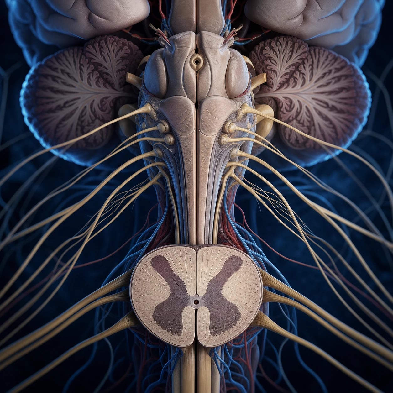
In cross-section the cord is an H-shaped (butterfly) core of grey matter surrounded by white matter. The grey matter has a posterior (dorsal) horn (sensory, where the dorsal roots synapse) and an anterior (ventral) horn (lower motor neurons, where the ventral roots exit); the lateral horn (T1-L2) carries the sympathetic preganglionic neurons. The white matter is divided into dorsal columns (gracile fasciculus medially for the lower limb, cuneate laterally for the upper limb), the lateral columns (corticospinal laterally, spinothalamic anterolaterally, plus the spinocerebellar tracts), and the ventral columns. There are 31 pairs of spinal nerves (8 cervical, 12 thoracic, 5 lumbar, 5 sacral, 1 coccygeal); note there are 8 cervical roots but only 7 cervical vertebrae (C1 exits above C1; C8 exits below C7; below that every root exits below its vertebra). [1]
Cord blood supply
- The anterior spinal artery (from the vertebrals) supplies the anterior two-thirds of the cord, including the corticospinal and spinothalamic tracts - so anterior spinal artery syndrome spares the dorsal columns (proprioception and vibration intact) but causes bilateral motor and pain/temperature loss.[1]
- Two posterior spinal arteries supply the posterior third (the dorsal columns).[1]
- The artery of Adamkiewicz (a large segmental feeder, usually left, T9-L2) reinforces the anterior supply to the lower two-thirds; injury during aortic surgery or aortic dissection can infarct the cord.[1]
- The adult cord ends as the conus medullaris at L1-L2; below this the cauda equina is a collection of lumbosacral roots (a lumbar puncture below L3 avoids the cord).[1]
The classic cord syndromes, each a distinct lesion pattern:
- Complete cord transection - loss of everything (motor, sensory, autonomic) below the level, with initial spinal shock (flaccidity, areflexia) then spasticity.
- Anterior spinal artery syndrome - motor and pain/temperature lost bilaterally, dorsal columns spared (vibration and proprioception intact); usually vascular (aortic surgery, dissection, hypotension).
- Brown-Sequard (hemicord) syndrome - ipsilateral motor (corticospinal) and proprioception (dorsal columns) loss, contralateral pain and temperature loss (spinothalamic); the textbook lesion of a stab wound or hemitumour.
- Central cord syndrome - the commonest incomplete cord injury; weakness greater in the arms than the legs (the arm fibres lie centrally in the corticospinal tract), with variable sensory loss and bladder dysfunction; typically a hyperextension injury in an older patient with cervical spondylosis.
- Posterior cord syndrome - rare; isolated loss of proprioception and vibration (the dorsal columns), as in B12 deficiency (subacute combined degeneration) or tabes dorsalis. [1]
The blood-brain barrier
The blood-brain barrier (BBB) is the specialised interface that strictly limits what crosses from the blood into the brain extracellular fluid. Its structural basis is the tight junctions (zonulae occludentes) between the cerebral capillary endothelial cells - these seal the paracellular space and force solutes to cross the cells themselves. The endothelium is invested by a basement membrane, by pericytes (contractile cells that regulate flow and permeability), and by the astrocyte end-feet that ensheath over 99 per cent of the capillary surface. It is the tight junction, not the astrocyte, that is the barrier itself - but the astrocytes induce and maintain it.[1]
What crosses and what does not:
- Crosses freely: gases (oxygen, carbon dioxide), water, small lipophilic molecules (most anaesthetic agents, ethanol, barbiturates), and substances carried by specific transporters - glucose (GLUT1), amino acids (L-type for large neutral amino acids such as L-DOPA, the basis of Parkinson therapy since dopamine itself does not cross).
- Excluded: large, polar, or charged molecules; plasma proteins; most drugs that are highly protein-bound; many antibiotics (penicillin enters poorly and is concentrated only when the meninges are inflamed). [1]
A handful of circumventricular organs sit outside the barrier - the area postrema (the chemoreceptor trigger zone that initiates vomiting, e.g. to opioids and digoxin), the organum vasculosum of the lamina terminalis (the osmoreceptor), the subfornical organ, the median eminence and posterior pituitary (for hormone release into blood), and the pineal gland. These need to sample the blood directly, so their capillaries are fenestrated.[1]
Clinically, the BBB matters in three ways: (1) drug delivery - you must design a drug to cross it (levodopa for Parkinson, prodrugs such as enalapril, or bypass it by intrathecal injection of, say, baclofen or methotrexate); (2) meningeal inflammation opens the barrier, which is why penicillin reaches therapeutic CSF levels in bacterial meningitis but not in health, and why gadolinium contrast enhances an abscess or tumour ring; and (3) breakdown underlies cerebral oedema in infection, tumour, ischaemia and trauma - the vasogenic oedema that raises intracranial pressure. [1]
The autonomic nervous system
The autonomic nervous system (ANS) is the involuntary motor division that controls smooth muscle, cardiac muscle and glands. It has three parts: the sympathetic (thoracolumbar), the parasympathetic (craniosacral), and the enteric (the intrinsic plexuses of the gut).[1]
The sympathetic outflow is thoracolumbar - the preganglionic neurons lie in the lateral horn of the spinal cord from T1 to L2/L3. Their fibres exit via the ventral roots, enter the sympathetic chain (paravertebral ganglia) via the white ramus communicans, and there either synapse (at the same level, a higher level, or a lower level) or pass straight through to a prevertebral (collateral) ganglion (the coeliac, superior mesenteric and inferior mesenteric). The result is the classic "fight or flight" response: tachycardia, increased contractility, bronchodilation, pupillary dilation, sweating, piloerection, splanchnic vasoconstriction, hepatic glycogenolysis and adrenal medullary secretion. The preganglionic fibre is short (it synapses close to the cord), the postganglionic fibre is long (it travels to the target). The adrenal medulla is a modified sympathetic ganglion: it is driven directly by preganglionic fibres and secretes adrenaline (~80%) and noradrenaline (~20%) directly into the blood.[1]
The parasympathetic outflow is craniosacral. The cranial part travels in CN III (the Edinger-Westphal pupillary constrictor), CN VII (lacrimal and submandibular/sublingual salivary), CN IX (parotid) and the mighty CN X (vagus), which supplies the heart, the airways and most of the gut down to the splenic flexure. The sacral part arises from S2-S4 (the pelvic splanchnic nerves) and supplies the distal colon, bladder and the pelvic viscera. The preganglionic fibre is long (it runs all the way to a ganglion in or near the target organ) and the postganglionic fibre is short.[1]
Sympathetic (thoracolumbar)
- Preganglionic neurons in the lateral horn T1-L2/L3; short preganglionic, long postganglionic fibres.
- Preganglionic neurotransmitter: acetylcholine on nicotinic receptors in the ganglion.
- Postganglionic neurotransmitter: noradrenaline on alpha/beta adrenergic receptors - the exception is sweat glands, which use acetylcholine on muscarinic receptors.
- Functions: the fight-or-flight response - tachycardia, bronchodilation, mydriasis, sweating, splanchnic vasoconstriction, ejaculation, adrenal medullary secretion.
Parasympathetic (craniosacral)
- Preganglionic neurons in the brainstem nuclei of CN III, VII, IX, X and in the sacral cord S2-S4; long preganglionic, short postganglionic fibres.
- Preganglionic neurotransmitter: acetylcholine on nicotinic receptors.
- Postganglionic neurotransmitter: acetylcholine on muscarinic (M1-M5) receptors.
- Functions: rest and digest - bradycardia, bronchoconstriction, miosis, lacrimation, salivation, increased gut motility and secretions, bladder contraction, penile erection.
The receptor pharmacology is the bridge to the drugs the intensivist uses every day. Alpha-1 (Gq-coupled) causes vasoconstriction, mydriasis, prostatic contraction - phenylephrine, noradrenaline. Alpha-2 (Gi-coupled, presynaptic) inhibits further noradrenaline release - clonidine, dexmedetomidine. Beta-1 (Gs-coupled) increases heart rate and contractility and renin release - isoprenaline, dobutamine. Beta-2 (Gs-coupled) bronchodilates, uterine-relaxes and shifts potassium into cells - salbutamol. The muscarinic receptors (M2 slows the heart, M3 contracts the bladder and gut and bronchoconstricts) are the targets of atropine (antagonist) and neostigmine (indirect, via acetylcholinesterase inhibition). The enteric nervous system - the myenteric (Auerbach) plexus between the longitudinal and circular muscle and the submucosal (Meissner) plexus in the submucosa - is the brain of the gut, capable of peristalsis even when denervated; the vagus and the splanchnic sympathetic fibres merely modulate it.[1]
[1]Exam practice — SAQs
SAQ — Neuroanatomical basis of brainstem death testing after cardiac arrest
10 minutes · 10 marks
A 54-year-old man is day 4 in ICU after an out-of-hospital ventricular fibrillation cardiac arrest with a down-time of 25 minutes. Targeted temperature management at 36 C has been completed and he has been rewarmed for 24 hours. He remains GCS 3 with absent brainstem reflexes on a fixed midazolam and fentanyl infusion; noradrenaline 0.15 mcg/kg/min, MAP 75, core temperature 36.8 C. The intensive care consultant asks you to outline the anatomical basis of the bedside brain-death examination and the confounders you must exclude before testing. Blood alcohol is undetectable; midazolam level is within sedative range.
SAQ — Cerebral autoregulation curve in severe traumatic brain injury
10 minutes · 10 marks
A 28-year-old man is admitted to ICU after a high-speed motorbike crash with a severe diffuse traumatic brain injury (initial GCS 6) and an intracranial pressure (ICP) monitor reading 24 mmHg. He is intubated, sedated and ventilated (PaCO2 38 mmHg). Blood pressure: systolic 96, MAP 68. CT brain shows diffuse axonal injury with effacement of the basal cististerns but no surgical mass. The nurse asks whether to start noradrenaline to push the MAP higher; the registrar is worried about worsening cerebral oedema. You are asked to explain the cerebral autoregulation curve and how it should guide the CPP target.
Clinical pearls
Evidence and trials
NINDS rt-PA Stroke Study (1995) - the foundation of acute stroke thrombolysis
Design
Two-part randomised, double-blind, placebo-controlled trial of intravenous alteplase vs placebo in 624 patients with acute ischaemic stroke
Intervention
Intravenous alteplase (rt-PA) within 3 hours of symptom onset
Result
Significantly more patients had a favourable outcome at 3 months (Part 2); symptomatic intracerebral haemorrhage was more frequent with alteplase (6.4% vs 0.6%) but overall mortality was not increased
Bottom line
Established IV thrombolysis within 3 hours as standard care for acute ischaemic stroke - the licensing basis for alteplase worldwide
ECASS III (Hacke 2008) - extending the thrombolysis window to 4.5 hours
Design
Randomised, double-blind, placebo-controlled trial in 821 patients with acute ischaemic stroke
Intervention
Intravenous alteplase 3 to 4.5 hours after symptom onset
Result
Alteplase gave a significantly higher rate of favourable outcome (52.4% vs 45.2%); symptomatic intracranial haemorrhage was more frequent (2.4% vs 0.3%); no significant mortality difference
Bottom line
Extended the licensed thrombolysis window from 3 to 4.5 hours for selected patients - the second pillar of the modern acute stroke pathway
DAWN (Nogueira 2018) and DEFUSE 3 (Albers 2018) - thrombectomy out to 16-24 hours with imaging selection
Design
Two independent randomised trials of mechanical thrombectomy vs standard care in patients selected by CT or MRI perfusion imaging (a clinical-imaging mismatch), 6 to 24 hours (DAWN) and 6 to 16 hours (DEFUSE 3) after onset
Intervention
Endovascular thrombectomy plus standard medical therapy vs standard medical therapy alone
Result
Both trials were stopped early for overwhelming benefit: thrombectomy roughly doubled the rate of functional independence at 90 days
Bottom line
Reframed acute stroke as a thrombectomy disease: a proximal large-vessel (ICA/M1) occlusion with a favourable penumbra-core mismatch is retrievable many hours after onset, provided the patient is selected by perfusion imaging
ISAT (Molyneux 2002) - endovascular coiling vs surgical clipping of ruptured aneurysms
Design
Multicentre randomised trial in 2143 patients with a ruptured intracranial aneurysm (subarachnoid haemorrhage) suitable for either treatment
Intervention
Neurosurgical clipping vs endovascular coil embolisation
Result
At one year, death or dependency was significantly lower with coiling (23.5% vs 30.9%); the benefit was largely sustained at long-term follow-up, at the cost of a slightly higher late re-bleed risk
Bottom line
Established endovascular coiling as first-line for most ruptured aneurysms amenable to either technique - the anatomical relevance is the Circle of Willis, where most saccular aneurysms arise
AAN brain-death guideline (Wijdicks 2010) - the bedside standard
Source
Evidence-based guideline update from the Quality Standards Subcommittee of the American Academy of Neurology
Purpose
To update and reaffirm the 1995 AAN practice parameter for determining brain death in adults
Key content
Defines the prerequisites (irreversible cause, no confounders, normothermia, normotension), the three findings (coma, absence of brainstem reflexes, apnoea), and the apnoea test threshold (PaCO2 60 mmHg or a 20 mmHg rise)
Bottom line
The reference protocol for the clinical determination of brain death - the brainstem reflexes tested are the pupillary (II/III), corneal (V/VII), gag and cough (IX/X) and the apnoea test (medullary drive)
Red flags
References
- [1]National Institute of Neurological Disorders and Stroke rt-PA Stroke Study Group Tissue plasminogen activator for acute ischemic stroke N Engl J Med, 1995.PMID 7477192
- [2]Hacke W, Kaste M, Bluhmki E, et al (ECASS Investigators) Thrombolysis with alteplase 3 to 4.5 hours after acute ischemic stroke N Engl J Med, 2008.PMID 18815396
- [3]Nogueira RG, Jadhav AP, Haussen DC, et al (DAWN Investigators) Thrombectomy 6 to 24 Hours after Stroke with a Mismatch between Deficit and Infarct N Engl J Med, 2018.PMID 29129157
- [4]Albers GW, Marks MP, Kemp S, et al (DEFUSE 3 Investigators) Thrombectomy for Stroke at 6 to 16 Hours with Selection by Perfusion Imaging N Engl J Med, 2018.PMID 29364767
- [5]Wijdicks EF, Varelas PN, Gronseth GS, Greer DM (American Academy of Neurology) Evidence-based guideline update: determining brain death in adults: report of the Quality Standards Subcommittee of the American Academy of Neurology Neurology, 2010.PMID 20530327
- [6]Molyneux A, Kerr R, Stratton I, et al (ISAT Collaborative Group) International Subarachnoid Aneurysm Trial (ISAT) of neurosurgical clipping versus endovascular coiling in 2143 patients with ruptured intracranial aneurysms: a randomised trial Lancet, 2002.PMID 12414200