ICU · Anatomy
Renal & Genitourinary Anatomy
Also known as Renal anatomy · Nephron · Glomerulus · Juxtaglomerular apparatus · Ureter · Renal blood supply · Loop of Henle
Renal and genitourinary anatomy for the ICU First Part: the kidney (cortex and medulla, the nephron segments from glomerulus to collecting duct), the dual-capillary renal blood supply (afferent then efferent arteriole), the juxtaglomerular apparatus and the renin-angiotensin trigger, and the ureteric constrictions relevant to obstruction.
On this page & tools
Your progress
Saved locally on this device.
8 MCQs with explanations
Target exams
Overview
The kidneys are paired retroperitoneal organs that filter blood, regulate fluid and electrolytes, and excrete nitrogenous waste. Their microanatomy - two capillary beds in series and a countercurrent system - underlies glomerular filtration, tubular reabsorption, and urine concentration.[1]
The kidney
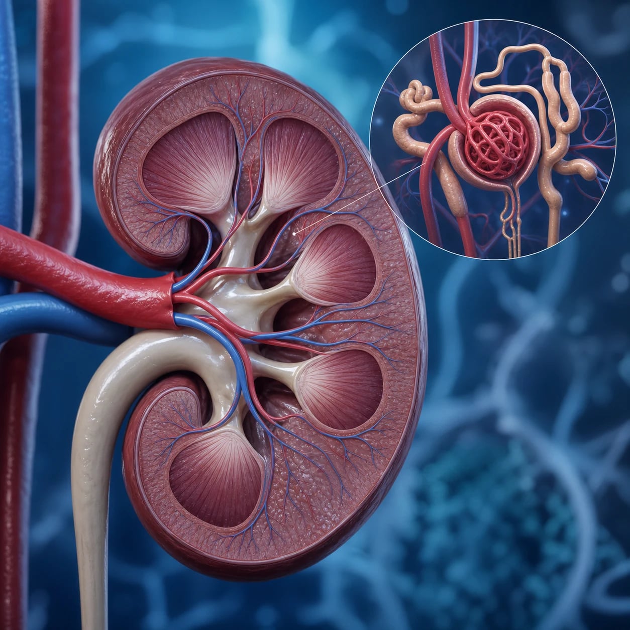
- The kidneys lie retroperitoneally at T12-L3; the right sits lower than the left (under the liver). Each has an outer cortex and an inner medulla of 8-18 renal pyramids, whose tips (papillae) drain into minor then major calyces and the renal pelvis.[1]
- A nephron is the functional unit (about one million per kidney).[1]
The nephron segments
- Glomerulus - a capillary tuft invaginated into Bowman's capsule; the filtration barrier (fenestrated endothelium, basement membrane, podocyte foot processes) holds back cells and large proteins.[1]
- Proximal convoluted tubule - reabsorbs the bulk of the filtrate: all glucose and amino acids, about 65 per cent of sodium and water, and secretes organic acids and drugs.[1]
- Loop of Henle - the descending limb is water-permeable (passive water reabsorption into the hypertonic medulla) and the thick ascending limb actively pumps sodium, potassium and chloride (the Na-K-2Cl cotransporter, the loop diuretic target) and is impermeable to water - the countercurrent multiplier that builds medullary hypertonicity.[1]
- Distal convoluted tubule - fine sodium and calcium tuning (the Na-Cl cotransporter, the thiazide target; calcium reabsorption under parathyroid hormone).[1]
- Collecting duct - aldosterone-driven sodium retention (the ENaC, the potassium-sparing diuretic target) and antidiuretic-hormone-driven water reabsorption (aquaporin-2 channels), setting the final urine volume and osmolality.[1]
Renal blood supply - a portal system
Blood passes through two capillary beds in series, unique to the kidney:[1]
- The afferent arteriole feeds the glomerular capillaries (where filtration occurs); the efferent arteriole drains them.
- The efferent arteriole then forms the peritubular capillaries (cortex) and the vasa recta (medulla), which reabsorb what the tubule has reclaimed.
This arrangement lets the kidney regulate glomerular pressure by adjusting afferent and efferent arteriolar tone (the basis of renal perfusion in shock and of angiotensin-II-mediated efferent constriction in renal hypoperfusion).[1]
The juxtaglomerular apparatus
- Where the distal tubule meets its own glomerulus, the macula densa (specialised distal-tubule cells) senses distal sodium/chloride delivery and signals the juxtaglomerular cells on the afferent arteriole to release renin.[1]
- Renin activates angiotensinogen to angiotensin I, then II, which constricts the efferent arteriole to maintain glomerular pressure when renal perfusion falls - and drives aldosterone and sodium retention.[1]
Ureters and lower tract
- Each ureter runs 25-30 cm retroperitoneally; its three natural constrictions - the pelviureteric junction, the pelvic brim where it crosses the iliac vessels, and the vesicoureteric junction - are the sites where stones lodge.[1]
- The bladder holds 400-600 mL; the male urethra is about 20 cm (prostatic, membranous, spongy) and the female about 4 cm - the short female urethra explains the higher urinary-tract infection risk.[1]
Red flags
Macroscopic (gross) anatomy of the kidney
- Position and level. The kidneys lie retroperitoneally on the posterior abdominal wall, spanning T12 to L3; the right kidney sits lower (a half-vertebra) because the liver occupies the right upper quadrant. The left kidney is more medial and lies between T12 and L3, with its hilum at about L1.[1][1]
- Surface relations. Anterior relations differ by side: the right kidney is related to the liver (hepatic impression), the second part of the duodenum (medially, over the hilum), the hepatic flexure of the colon, and the suprarenal gland superomedially; the left kidney is related to the stomach (upper pole, separated by the lesser sac), spleen, tail of the pancreas (across the hilum), splenic flexure and descending colon, and the left suprarenal gland. The pancreas tail and duodenum are the operative danger - mobilising these organs risks injury during retroperitoneal access.[1][1]
- Posterior relations. Both kidneys rest on the diaphragm (lower), quadratus lumborum and psoas major; the subcostal nerve (T12), iliohypogastric and ilioinguinal nerves (L1) run obliquely across the lower pole beneath the fascia - a pleural reflection (costodiaphragmatic recess) crosses the upper pole and is the reason a high flank incision or a mal-placed nephrostomy causes a hydrothorax.[1]
- Three layers of investing fascia and fat. From within out: (1) the fibrous capsule (capsula fibrosa) - thin, invests the parenchyma, strips easily and is the surgical plane for partial nephrectomy and decapsulation (used to relieve intrarenal tamponade); (2) the perirenal fat (adipose within the perirenal space); (3) the renal fascia of Gerota (perirenal fascia), which envelops kidney, adrenal and perirenal fat, closes inferiorly (the inferior cone is open - the route of pancreatic fluid tracking to the groin); (4) the pararenal fat external to Gerota's fascia. These planes matter for nephrostomy, abscess, and the spread of retroperitoneal haemorrhage in anticoagulated patients.[1][1]
- Hilum and sinus. The renal hilum is the medial concavity; from anterior to posterior lie the renal vein, renal artery, and renal pelvis (mnemonic V-A-P); the ureter is most posterior and inferior. From superior to inferior at the hilum: artery-vein-artery-ureter is variable, but vein anterior, artery middle, pelvis posterior is constant and is the order encountered at nephrectomy. The renal sinus houses the pelvis, calyces, vessels and fat.[1][1]
- Internal architecture. Section shows an outer cortex (granular, contains glomeruli and convoluted tubules) and an inner medulla of 8 to 18 renal pyramids (one per lobe); the cortex dips between pyramids as the columns of Bertin. Each pyramid tapers to a renal papilla that projects into a minor calyx; 7 to 13 minor calyces fuse into 2 to 3 major calyces, which drain into the renal pelvis. The pelvis narrows at the pelviureteric junction to become the ureter.[1][1]
The numbers examiners ask for verbatim
The renal arterial tree - segmental, end-arterial, and the reason for lobar infarction
The kidney's blood supply is the most surgically and radiologically important branch of the aorta, and its segmental, end-arterial organisation explains why a focal renal infarct is wedge-shaped and why a partial nephrectomy follows avascular planes.[1][1]
Blood from aorta to glomerulus - the full named chain
Renal artery
Lateral aortic origin at the level of L1-L2 (between superior and inferior mesenteric arteries). The RIGHT is longer and passes BEHIND the IVC; the LEFT is shorter and crosses the aorta anteriorly as its vein.
Anterior and posterior divisions
Just before or within the hilum the renal artery divides into an anterior trunk (supplying upper, middle and lower poles) and a smaller posterior trunk (posterior segment).
Five segmental arteries (apical, upper, middle, lower, posterior)
Each supplies a self-contained Brodel segment. They are END ARTERIES (no anastomosis) - the basis of the avascular Brödel/Brodel incision and of segmental infarction when one is occluded or embolised.
Interlobar arteries
Travel in the columns of Bertin between adjacent pyramids, giving capsular and perforating branches (the latter anastomose - the source of capsular collateral flow that may keep a totally obstructed kidney viable).
Arcuate arteries
Arch OVER the bases of the pyramids at the corticomedullary junction - they do not enter the pyramid, marking the boundary seen on Doppler as the corticomedullary transition.
Interlobular (cortical radiate) arteries
Rise radially through the cortex toward the capsule; an afferent arteriole arises from each.
Afferent arteriole -> glomerulus -> efferent arteriole
The dual-capillary portal system: afferent feeds the glomerular tuft (filtration); efferent drains it and forms the second capillary bed.
Peritubular capillaries (cortex) and vasa recta (medulla)
Cortical peritubular capillaries reclaim PCT/DCT reabsorbate; the straight, hairpin VASA RECTA (from the juxtamedullary efferent arterioles) run alongside the loops of Henle and are the anatomical basis of the countercurrent exchanger that preserves medullary hypertonicity.
- Venous drainage largely mirrors the arteries but, unlike the arteries, the veins freely anastomose - which is why the kidney tolerates segmental venous ligation but not segmental arterial interruption. The left renal vein is longer and crosses between the superior mesenteric artery and the aorta - the nutcracker (SMA) space whose compression produces left renal vein entrapment (nutcracker syndrome: haematuria, left flank pain, varicocele). It receives the left adrenal vein and the left gonadal (testicular/ovarian) vein, then drains to the IVC; the right renal vein is short and drains directly into the IVC. The left gonadal vein drains into the left renal vein whereas the right drains directly into the IVC - the anatomical reason a left varicocele may signal left renal vein obstruction or tumour, while a right varicocele (draining directly to IVC) more often indicates a retroperitoneal mass.[1][1]
- Lymphatics drain via nodes at the hilum to para-aortic (lumbar) nodes; renal-cell carcinoma therefore spreads to para-aortic nodes first (the rationale for nodal dissection at nephrectomy).[1]
The filtration barrier - three layers, two selectivities
Glomerular filtration is the kidney's first and most selective step. The barrier holds back cells and nearly all protein while passing water and small solutes at ~180 L/day. It has three structural layers and a shared charge.[1][1]
Fenestrated endothelium
Innermost (capillary lumen)
- Capillary endothelial cells perforated by ~70 nm fenestrations - block cells and platelets, pass water and solutes
- Carries a negative surface charge (sialoproteins/glycocalyx) that repels anionic proteins
Glomerular basement membrane
Middle (fused basal laminae)
- A trilaminar sheet of type IV collagen (network), laminin, nidogen and proteoglycans (heparan sulfate)
- Main SIZE and CHARGE barrier; heparan sulfate gives a strong negative charge that repels albumin (also anionic)
- Anti-GBM antibody (Goodpasture) and Alport mutations (type IV collagen alpha chains) strike here
Podocyte slit diaphragm
Outermost (Bowman's space)
- Visceral epithelial cells (podocytes) send interdigitating foot processes bridged by a 30-40 nm slit diaphragm
- Nephrin and podocin are the structural proteins; their genes (NPHS1, NPHS2) mutate in congenital nephrotic syndrome
- Effacement of foot processes (flattening) is the universal finding in proteinuric states - minimal change disease, FSGS, diabetic nephropathy
- Mesangial cells sit between capillary loops: contractile, phagocytic, supportive, and they secrete and turnover the matrix. They respond to angiotensin II (contraction reduces filtration surface area) - one reason mesangial IgA deposition and mesangial proliferative diseases disturb filtration.[1]
The nephron - cortical versus juxtamedullary, and the drug targets along its length
- There are about one million nephrons per kidney, formed entirely before birth; loss is irreversible and accelerates with age and disease. Nephrons fall into two types distinguished by glomerular depth and loop length, with direct consequences for concentrating ability.[1][1]
- Cortical nephrons (~85 per cent) sit in the outer cortex; their loops of Henle dip only to the outer medulla and their efferent arterioles feed cortical peritubular capillaries - they do most of the bulk reabsorption.
- Juxtamedullary nephrons (~15 per cent) have glomeruli at the corticomedullary junction and long loops of Henle that reach the tip of the papilla; their efferent arterioles give rise to the vasa recta. Although few, they generate the entire medullary osmotic gradient - the concentrating engine of the kidney.
Proximal convoluted tubule
Bulk reabsorption (~65%)
- Isosmotic reabsorption of ~65% filtered Na+ and water; ~100% of glucose and amino acids (SGLT2 and SGLT1)
- Secretes organic anions/cations - the route by which furosemide, penicillin and radiocontrast reach their luminal targets; blocked by probenecid
- Carbonic anhydrase here and in the proximal straight tubule - acetazolamide site; also the main Ca2+/PO4 handling site (PO4 via NaPi, regulated by PTH/FGF-23)
Loop of Henle
Countercurrent multiplier
- Thin descending limb - water-permeable (AQP1), urea and Na+ enter, fluid equilibrates with hypertonic medulla
- Thick ascending limb - Na-K-2Cl cotransporter (NKCC2), K+ backleak (lumen-positive), IMPERMEABLE to water - dilutes tubular fluid and builds medullary interstitial hypertonicity
- LOOP DIURETIC target (furosemide, bumetanide, torsemide); also reabsorbs ~25% filtered Ca2+ and Mg2+ (paracellular, driven by the lumen-positive potential - why loops waste Ca2+/Mg2+)
Distal convoluted tubule
Fine tuning (Na, Ca)
- Na-Cl cotransporter (NCC) - the THIAZIDE target; thiazides cause mild hypercalcaemia because the same mechanism enhances Ca2+ reabsorption
- Ca2+ reabsorption via TRPV5, up-regulated by parathyroid hormone - the distal tubule is where PTH acts on calcium
- Impermeable to water (no ADH effect yet) - continues diluting the urine; the 'cortical diluting segment'
Collecting duct
Final volume and K+/acid
- Principal cells - ENaC (aldosterone-driven, the amiloride/triamterene/spironolactone site) reabsorbs Na+ and secretes K+ (the potassium-sparing diuretic site)
- Principal cells - AQP2 (ADH-driven) and AQP3/4 (constitutive basolateral) insert to reabsorb water; the final determinant of urine osmolality (50-1200 mOsm/kg)
- Intercalated cells - H+ secretion (type A, via H+-ATPase) and HCO3- handling (type B); the final acid-base tuning site; also K+ reabsorption in hypokalaemia (H-K-ATPase)
The juxtaglomerular apparatus and the renin-angiotensin-aldosterone system
- At the vascular pole, where the distal tubule returns to its own glomerulus, three cell types form the juxtaglomerular apparatus (JGA): the macula densa (a plaque of specialised, densely-packed distal-tubule cells sensing tubular Na+/Cl- and osmolality via the NKCC2 transporter); the juxtaglomerular (granular) cells - modified smooth-muscle cells in the afferent arteriole wall that store and release renin; and the extraglomerular mesangial (Lacis) cells, which couple the two.[1][1]
- Three stimuli for renin release: (1) reduced stretch of the afferent arteriole (a baroreceptor, falls in perfusion pressure); (2) reduced NaCl delivery sensed at the macula densa (tubuloglomerular feedback); (3) increased sympathetic tone (beta-1 effect on JG cells). Renin cleaves circulating angiotensinogen (liver-derived) to angiotensin I, which ACE (mostly pulmonary, on endothelium) converts to angiotensin II.[1][1]
- Angiotensin II has four renal effects that all protect GFR when perfusion falls: it constricts the efferent arteriole (more than afferent) to maintain intraglomerular pressure; it stimulates aldosterone (zona glomerulosa) for distal Na+ retention; it directly stimulates proximal Na+ reabsorption; and it stimulates thirst and ADH. It also constricts mesangial cells (reducing filtration surface).[1]
The JGA - the three components and the renin triggers
JGA & 3S
Modified afferent-arteriolar smooth muscle - store and secrete renin
Lacis/extraglomerular mesangial cells couple macula densa to the arteriole
Specialised distal-tubule cells sensing luminal NaCl via NKCC2
FALL in afferent arteriolar stretch -> renin
FALL in macula-densa NaCl delivery -> renin (tubuloglomerular feedback)
RISE in sympathetic tone -> renin - blocked by beta-blockers
AIPRI - Angiotensin-Converting-Enzyme Inhibition in Progressive Renal Insufficiency
N Engl J Med 1996 (Maschio et al)
Multicentre double-blind RCT, 583 patients with non-diabetic chronic renal insufficiency (creatinine 1.5-4 mg/dL), benazepril vs placebo.
Key finding
Benazepril reduced the composite of doubling serum creatinine or needing dialysis by 53% - independent of the small blood-pressure fall - establishing that ACE inhibition is renoprotective through glomerular-haemodynamic (efferent dilatation lowering intraglomerular pressure) as well as antihypertensive effects.
Practice change
ACE inhibition became first-line renoprotection in proteinuric non-diabetic renal disease - the anatomical basis being preferential efferent arteriolar dilatation lowering intraglomerular pressure.
REIN follow-up - Ramipril Efficacy In Nephropathy
Lancet 1999 (Ruggenenti et al)
Randomised, placebo-controlled, stratified by baseline proteinuria; non-diabetic, proteinuric chronic nephropathies; ramipril vs placebo plus conventional antihypertensives.
Key finding
In patients with non-nephrotic proteinuria, ramipril slowed GFR decline and reduced the risk of end-stage renal failure, with the benefit exceeding that predicted by blood-pressure reduction alone and greatest in the highest proteinuria stratum.
Practice change
Confirmed proteinuria stratification of benefit and cemented RAAS blockade (efferent arteriolar effect) as standard for proteinuric chronic kidney disease.
Autonomic and sensory innervation of the kidney, ureter and bladder
- Sympathetic preganglionic fibres arise from T10 to L1 (greater, lesser and least splanchnic nerves) and synapse in the aorticorenal and renal ganglia; postganglionic fibres travel with the renal artery in the renal plexus to the vessels, tubules and juxtaglomerular cells. Effects: afferent and efferent arteriolar vasoconstriction (alpha-1), renin release (beta-1), and proximal Na+ reabsorption. Renal denervation (catheter-based renal artery ablation) exploits the fact that these nerves run in the renal artery adventitia.[1][1]
- Parasympathetic supply is vagal and weak; its functional role in the kidney is minor. The clinically important parasympathetics are the pelvic splanchnic nerves S2-S4 (nervi erigentes), which supply the bladder detrusor (via the pelvic nerves) and the distal bowel and genitalia.
- Pain fibres from the kidney, renal pelvis and upper ureter travel with the sympathetic nerves to T10-L1, so renal pain is referred to the flank, loin and lower abdomen (the classic loin-to-groin radiation of ureteric colic follows the genitofemoral and ilioinguinal nerves, L1-L2). Lower ureteric and bladder pain is referred via S2-S4 to the suprapubic region, perineum and groin.[1][1]
- The renal capsule is richly innervated and stretched by swelling (acute pyelonephritis, obstruction, haemorrhage) producing a dull, aching flank pain distinct from the colic of ureteric spasm. Cystoscopy, ureteric stents and bladder distension below a cord lesion can trigger autonomic dysreflexia (lesions at/above T6) through unopposed sympathetic discharge.[1]
Kidney and upper ureter
Visceral afferent T10-L1
- Sympathetic T10-L1 via splanchnic nerves and renal plexus
- Pain referred to flank/loin/lower abdomen; renal capsule stretch is dull and aching
- Parasympathetic (vagal) minor; no somatic supply
Bladder and lower ureter
Mixed T11-L2 and S2-S4
- Detrusor: parasympathetic S2-S4 (pelvic nerves, cholinergic) - the target of anticholinergics and beta-3 agonists for overactive bladder
- Internal sphincter (smooth): sympathetic T11-L2 (hypogastric, alpha-1) - alpha-blockers relax it in urinary retention/benign prostatic obstruction
- External sphincter (skeletal): somatic via pudendal nerve S2-S4 (Onuf's nucleus) - voluntary control; the lesion in spinal shock is areflexic
Urethra
Pudendal S2-S4
- Somatic sensation and external sphincter via the pudendal nerve
- Membranous urethra encircled by the external sphincter (the only voluntary GU control)
- Female urethra and distal male spongy urethra carry the highest bacterial colonisation -> ascending UTI risk
The ureter - course, constrictions and blood supply
- Each ureter is 25-30 cm long and runs retroperitoneally from the renal pelvis to the bladder, crossing the pelvic brim at the bifurcation of the common iliac artery. It is crossed anteriorly by the gonadal vessels and, on the right, by the second part of the duodenum; on the left it is crossed by the vas deferens (male) or uterine artery (female) - the mnemonic 'water under the bridge' (ureter under the uterine artery/vas) is the surgical warning during hysterectomy or colectomy.[1][1]
- Three natural constrictions where calculi lodge (in descending order of frequency): the pelviureteric junction; the pelvic brim where the ureter crosses the iliac vessels; and the vesicoureteric junction (the narrowest, ~3 mm). The vesicoureteric junction's oblique, tunnelled entry through the bladder wall is the anti-reflux mechanism; its shortening or malformation causes vesicoureteric reflux (and recurrent pyelonephritis).[1][1]
- Segmental blood supply (surgical relevance): the upper third from the renal artery; the middle third from the gonadal and common iliac arteries; the lower third from the superior and inferior vesical arteries. The vessels enter on the medial side and there is a relative watershed at the pelviureteric junction - the reason ureteric ischaemia and stricture form after retroperitoneal surgery, aortic grafting or prolonged stenting, and why ureteric mobilisation must preserve the adventitia.[1]
The bladder and urethra
- The bladder sits behind the pubic symphysis, extraperitoneal, with peritoneum covering only its dome (hence a distended bladder pushes peritoneum up - the basis of safe suprapubic aspiration/cystostomy above the pubic symphysis to avoid bowel). Capacity is 400-600 mL; first urge is felt at ~150 mL and fullness at ~400 mL.[1][1]
- The trigone is the smooth triangular area between the two ureteric orifices and the internal urethral meatus; its interureteric ridge (Mercier's bar) is embryologically mesodermal (unlike the rest of the detrusor, endodermal) and is a fixed landmark at cystoscopy. The detrusor is interlacing smooth muscle; contraction (parasympathetic M3) raises intravesical pressure and opens the bladder neck.[1]
- Two sphincters: the internal urethral sphincter (smooth muscle, involuntary, sympathetic alpha-1, T11-L2) at the bladder neck - the site of physiological continence; and the external urethral sphincter (skeletal, voluntary, somatic pudendal S2-S4 - Onuf's nucleus) in the urogenital diaphragm - the site of socially appropriate continence. The male urethra is ~20 cm (prostatic 3 cm, membranous 1-2 cm - the narrowest and least distensible - and spongy ~15 cm); the female is ~4 cm, opening into the vestibule, which is why women have more UTIs and why catheterisation is anatomically straightforward.[1][1]
Internal sphincter
Smooth, involuntary
- Bladder neck; sympathetic T11-L2, alpha-1 adrenergic
- Maintains continence at rest and during ejaculation (closes bladder neck to prevent retrograde ejaculation)
- Relaxed by alpha-blockers (tamsulosin) - used for urinary retention and to facilitate catheterisation
External sphincter
Skeletal, voluntary
- Urogenital diaphragm/membranous urethra; somatic pudendal nerve, Onuf's nucleus S2-S4
- Voluntary continence and start-stop of micturition
- In spinal shock it is flaccid (areflexic retention); with reflex recovery it becomes spastic (detrusor-sphincter dyssynergia)
Renal replacement therapy access - the anatomy of dialysis
Dialysis access is a recurring CICM/FFICM viva topic because the choice and the complications flow directly from vascular anatomy.[1][1]
Arteriovenous fistula (AVF)
Best long-term
- Radiocephalic (Brescia-Cimino): radial artery anastomosed to cephalic vein at the wrist/anatomical snuffbox
- Brachiocephalic: brachial artery to cephalic vein at the antecubital fossa; brachiobasilic (transposed) when cephalic is unsuitable
- Vein ARTERIALISES over 6-12 weeks (wall thickens, dilates) - the maturation period; needels access at least 2-3 cm apart (rope-ladder technique)
- Lowest infection and longest patency of any access - the gold standard
Arteriovenous graft (AVG)
Prosthetic bridge
- PTFE loop between an artery and a vein, usually forearm loop graft (brachial artery-to-antecubital vein) or upper-arm straight graft
- Used when native veins are exhausted (small, thrombosed, previous cannulation)
- Higher infection and thrombosis than AVF but can be used sooner (2-4 weeks to incorporate)
Tunnelled central catheter
Permanent CVC
- Right internal jugular preferred (straight to the cavoatrial junction); tip at the cavoatrial junction under fluoroscopy
- Tunnelled subcutaneously from an exit site on the chest wall to a venotomy - cuff in-growth secures it and reduces infection vs non-tunnelled
- Highest infection and central-venous-stenosis risk; used when AVF/AVG not feasible or while awaiting maturation
Creation and maturation of a radiocephalic AV fistula
Vessel assessment
Clinical exam and venous mapping - cephalic vein calibre >2 mm, continuous on tourniquet, no segmental stenosis; radial artery palpable with Allen test confirming ulnar dominance so the hand is not rendered ischaemic.
Anastomosis at the anatomical snuffbox / wrist
Local or regional block; the cephalic vein (superficial, on the radial side of the dorsum) is anastomosed end-to-side (or side-to-side) to the radial artery just proximal to the wrist.
Arterialisation of the vein
Exposure to arterial pressure thickens the wall and dilates the lumen over 6-12 weeks; the vein becomes palpable as a thrill and audible as a bruit, and robust enough to take two large-bore (15-17 G) needles each dialysis.
Cannulation - arterial and venous needles
The 'arterial' needle draws blood from the fistula (pointing toward the anastomosis or away, unit policy) and the 'venous' needle returns it (15 cm apart minimum); the rope-ladder technique rotates sites to prevent aneurysm.
Surveillance
Flow monitoring, recirculation, dynamic/np venous pressure trends, and Doppler - a falling flow or a collapsing segment warns of stenosis at the anastomosis or in the draining (subclavian/brachiocephalic) vein.
3SITES - central venous catheter insertion site
N Engl J Med 2015 (Parienti et al)
Multicentre randomised trial, 3027 patients needing a CVC for at least 3 days; subclavian vs jugular vs femoral.
Key finding
The composite of catheter-related bloodstream infection and symptomatic deep-vein thrombosis was lowest with SUBCLAVIAN (1.5%) vs jugular (3.3%) vs femoral (4.0%); but symptomatic pneumothorax was highest with subclavian (1.1%) vs jugular (0.3%) vs femoral (0%).
Practice change
Subclavian minimises infection and thrombosis but maximises pneumothorax; for dialysis access the trade-off is dominated by subclavian-vein stenosis, so the IJ remains first-choice for RRT catheters while subclavian is preferred for general ICU lines when clotting and infection dominate.
Dialysis Access Consortium (DAC) - clopidogrel for new AV fistulas
JAMA 2008 (Dember et al)
Randomised, double-blind, placebo-controlled trial, 877 patients receiving a new AV fistula; clopidogrel vs placebo for 6 weeks.
Key finding
Clopidogrel reduced the rate of early (6-week) AV-fistula failure from 61.8% to 47.7% (relative risk reduction ~25%), but did NOT increase the proportion of fistulas suitable for dialysis at maturity - thrombosis at the wrist anastomosis is only one of several maturation failure modes.
Practice change
Antiplatelet therapy at fistula creation is reasonable to reduce early thrombosis, but the anatomical bottleneck for useable fistula flow is vein maturation (dilatation and wall thickening), which clopidogrel does not fix.
Surface anatomy and clinical examination landmarks
- Ballottement (renal punch / Murphy's punch sign). With the patient supine, one hand placed in the costovertebral angle (the angle between the 12th rib and the erector spinae) is struck sharply by the other hand; transmitted tenderness suggests renal capsule inflammation (pyelonephritis). The kidney is ballotable only when enlarged to at least twice normal size (polycystic, hydronephrotic, tumour) or displaced.[1][1]
- The lumbar triangle of Petit - the weak area bounded by the external oblique anteriorly, latissimus dorsi posteriorly and the iliac crest inferiorly, with internal oblique as its floor - is the portal for the muscle-splitting (lumbodorsal) approach to the kidney (open nephrectomy, percutaneous nephrostomy, retroperitoneal abscess drainage) and the route through which a lumbar hernia (Petit's hernia) may protrude.[1]
- Renal percussion and the relationship to the 12th rib defines the safe upper limit for a flank incision and the risk of pleural breach - the costodiaphragmatic recess crosses the 12th rib, so a supra-12th-rib incision risks a hydrothorax.[1]
Embryology - why ectopic, horseshoe and pelvic kidneys occur
- The definitive kidney (metanephros) develops from two primordia: the ureteric bud (a diverticulum of the mesonephric/Wolffian duct) which branches to form the ureter, renal pelvis, calyces and collecting ducts; and the metanephric blastema (sacral intermediate mesoderm) which forms the nephrons (glomerulus to DCT). The two must meet and induce each other - failure produces renal agenesis, multicystic dysplastic kidney or ectopia.[1]
- The kidneys ascend from the pelvis to T12-L3 during weeks 6-9, acquiring successively higher segmental arteries; failure to ascend gives a pelvic kidney, and fusion before ascent produces a horseshoe kidney (typically fused at the lower poles, caught under the inferior mesenteric artery and supplied by multiple anomalous vessels). The horseshoe kidney is the most common fusion anomaly (~1 in 400) and predisposes to ureteropelvic junction obstruction, stones and infection.[1][1]
Exam practice — SAQs
SAQ — Renal vascular anatomy applied to dialysis access in a septic, access-less patient
10 minutes · 10 marks
A 62-year-old woman with end-stage kidney disease secondary to diabetic nephropathy is admitted to ICU with septic shock from a urinary source, requiring noradrenaline 0.25 mcg/kg/min and urgent continuous renal replacement therapy (CRRT). She has no established dialysis access. She has a non-matured left radiocephalic arteriovenous fistula created 8 weeks ago and a left-sided permanent pacemaker. The team is debating right internal jugular, subclavian, and femoral routes for a temporary haemodialysis catheter.
SAQ — Genitourinary anatomy applied to obstructive uropathy with sepsis
10 minutes · 10 marks
A 68-year-old man with known bilateral renal calculi presents to ICU with anuric acute kidney injury (creatinine 540 micromol/L, potassium 6.8 mmol/L) and septic shock from pyelonephritis (lactate 4.6, noradrenaline 0.3 mcg/kg/min). He has a palpable distended bladder. CT abdomen shows a 9 mm stone at the left vesicoureteric junction, right hydronephrosis with a staghorn calculus, and a markedly enlarged prostate with a thickened, trabeculated bladder wall. Bilateral percutaneous nephrostomies and a suprapubic catheter are being considered.
Clinical pearls for the CICM / FFICM / EDIC exam
Renal anatomy mnemonics
VAP & WATER
At the hilum: Vein-Artery-Pelvis, anterior to posterior
Segmental arteries are END arteries - wedge infarcts
Renal pelvis and ureter are the most posterior hilar structures
Left renal vein (and ureter) pass UNDER the SMA / uterine artery / vas deferens
Two capillary beds in series - afferent -> glomerulus -> efferent -> peritubular/vasa recta
Ureter: PUJ, iliac crossing, VUJ (narrowest) - where stones lodge
Renal segmental arteries don't anastomose; veins do (tolerate ligation)
Straight to cavoatrial junction; avoid subclavian (stenosis)
Glossary of anatomical terms an examiner may press
- Columns of Bertin - extensions of renal cortex between the medullary pyramids; sometimes hypertrophied (septum of Bertin) and mistaken for a tumour on imaging.[1]
- Brodel's line / white line - the relatively avascular plane on the convex renal border between anterior and posterior segmental arterial territories - the incision line for anatrophic nephrolithotomy and partial nephrectomy.[1]
- Renal fascia of Gerota - the perirenal fascia enclosing kidney, adrenal and perirenal fat; defines the perirenal space and the route of spread of retroperitoneal collections.[1][1]
- Vasa recta - straight, hairpin capillaries from the juxtamedullary efferent arterioles that run with the loops of Henle - the countercurrent exchanger that preserves medullary hypertonicity.[1]
- Macula densa / Lacis cells - the sensor and the coupler of the JGA; Lacis (extraglomerular mesangial) cells transmit the macula-densa signal to the JG cells.[1][1]
- Trigone of the bladder - the smooth triangular region between the ureteric orifices and the internal meatus; embryologically mesodermal (unlike the endodermal detrusor) and the site where ureteric ectopia and reflux are anatomically determined.[1]
References
- [1]Maschio G, Alberti D, Janin G, et al Effect of the angiotensin-converting-enzyme inhibitor benazepril on the progression of chronic renal insufficiency. The Angiotensin-Converting-Enzyme Inhibition in Progressive Renal Insufficiency Study Group N Engl J Med, 1996.PMID 8596594
- [2]Ruggenenti P, Perna A, Gherardi G, et al Renoprotective properties of ACE-inhibition in non-diabetic nephropathies with non-nephrotic proteinuria Lancet, 1999.PMID 10437863
- [3]Dember LM, Beck GJ, Allon M, et al Effect of clopidogrel on early failure of arteriovenous fistulas for hemodialysis: a randomized controlled trial JAMA, 2008.PMID 18477783
- [4]Parienti JJ, Mongardon N, Megarbane B, et al Intravascular Complications of Central Venous Catheterization by Insertion Site N Engl J Med, 2015.PMID 26398070