ICU · Anatomy
Vascular-Access Anatomy — Central & Arterial
Also known as Vascular access anatomy · Central venous catheter · Internal jugular vein · Subclavian vein · Carotid sheath · Radial artery · Allen test · Femoral vein
Vascular-access anatomy for the ICU First Part: the central venous routes (internal jugular in the carotid sheath, subclavian below the clavicle, femoral in the femoral sheath) with their risks and the tip position at the cavoatrial junction, and the arterial-line sites (radial with the Allen test for ulnar collateral flow, femoral, dorsalis pedis).
On this page & tools
Your progress
Saved locally on this device.
8 MCQs with explanations
Target exams
Overview
Safe vascular access depends on knowing the relations of the target vessel and the structure of the sheath it lies in. Central venous lines are placed at the internal jugular, subclavian, or femoral veins; arterial lines most often at the radial artery. Each site trades convenience against a distinctive risk profile.[1]
Central venous access
Internal jugular vein
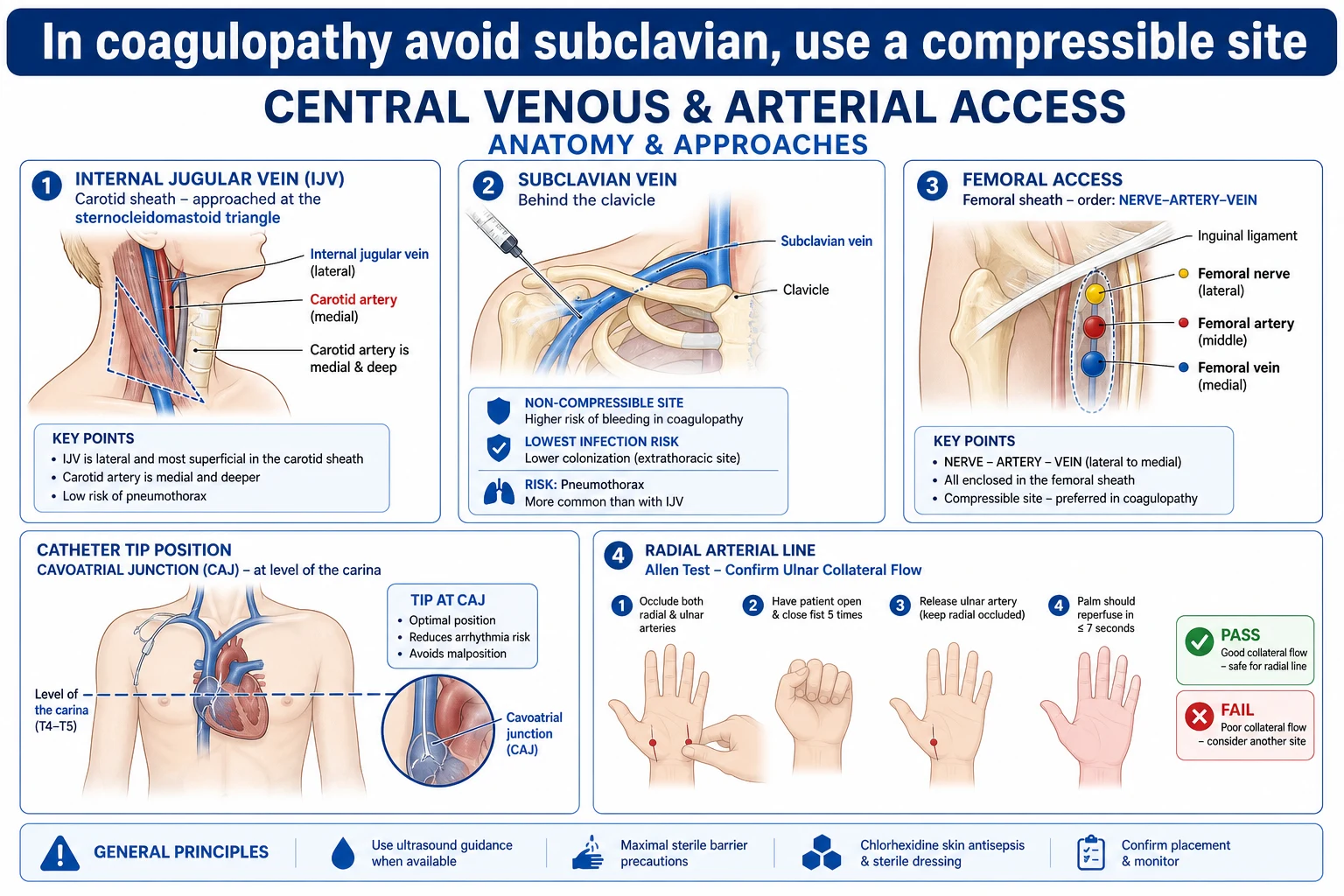
- Lies within the carotid sheath, with the common carotid artery medial and deep and the vagus nerve between them; the sheath runs deep to sternocleidomastoid.[1]
- The landmark approach targets the apex of the triangle between the two heads of sternocleidomastoid (Sedillot triangle), aiming toward the ipsilateral nipple; ultrasound is now standard.[1]
- Advantages: a straight, short course to the SVC (especially on the right), and the vein is compressible. Risks: carotid artery puncture and, on the left, thoracic duct injury; pneumothorax risk is lower than for the subclavian route.[1]
Subclavian vein
- Runs just below the clavicle, between it and the first rib, and joins the internal jugular to form the brachiocephalic (innominate) vein.[1]
- Advantages: the lowest infection rate and patient comfort for longer-term use. Risks: a relatively high pneumothorax rate and that the vessel is not compressible (avoid in coagulopathy); stenosis can complicate future fistula use.[1]
Femoral vein
- Lies in the femoral sheath; from lateral to medial the contents are nerve, artery, vein (the mnemonic N-A-V), with the vein medial to the artery at the inguinal ligament.[1]
- Advantages: compressible, no pneumothorax risk, useful in an emergency or cardiac arrest. Risks: a higher infection and deep-vein-thrombosis rate, and reduced mobility for the patient.[1]
Catheter-tip position
- A central line tip should lie in the superior vena cava just above the cavoatrial junction (the junction of the SVC and right atrium); a tip in the right atrium or right ventricle risks arrhythmia and perforation.[1]
- Confirm position on a chest X-ray: a right internal jugular line runs straight down to the SVC; the carina sits roughly level with the cavoatrial junction on a chest film.[1]
Arterial access
- The radial artery at the wrist is the commonest site because it is superficial and the hand usually has dual circulation through the superficial palmar arch (predominantly ulnar); a positive Allen test (filling of the hand from the ulnar artery after radial compression) confirms adequate collateral flow before cannulation.[1]
- Alternative sites: femoral (accurate central pressure but higher infection and retroperitoneal-bleed risk), dorsalis pedis and axillary/brachial arteries.[1]
- The radial is a terminal branch of the brachial artery; it lies lateral to the flexor carpi radialis tendon at the wrist.[1]
Red flags
Vessel wall anatomy - why a vein differs from an artery
Every vascular access device sits inside a blood-vessel wall, and the wall architecture of a vein and an artery is what makes cannulation, dilation, and complications differ. Both are built from three tunicae, but the proportions are reversed.[1]
- Tunica intima - the innermost layer: a single endothelium on a basement membrane, over sub-endothelial connective tissue and the internal elastic lamina. In arteries the internal elastic lamina is a distinct, wavy, refractile layer; in veins it is thin or absent. Endothelial injury (from the needle, wire, catheter, or an irritant infusate) is the seed of catheter-related thrombosis and stenosis.[1]
- Tunica media - the muscular layer: smooth muscle and elastic tissue. In arteries the media is the thickest layer (a high-pressure conduit needs a muscular wall); in veins the media is thin because venous pressure is low. This single fact explains why an artery is round, thick-walled, pulsatile, and NON-compressible on ultrasound, while a vein is oval, thin-walled, and COMPRESSIBLE - the distinction examiners ask you to make at the probe.[1][4]
- Tunica externa (adventitia) - the outer connective-tissue layer carrying the vasa vasorum (vessels supplying the wall) and nervi vasorum. In veins the adventitia is the thickest layer; in arteries it is thinner than the media. The tough adventitia is what the dilator and catheter cuff grip, and the layer a suture anchors when a line is tunnelled.[1]
Arterial vs venous wall - the three-tunica comparison
| Feature | Artery | Vein |
|---|---|---|
| Dominant layer | Tunica media (muscular) - thickest | Tunica adventitia (connective tissue) - thickest |
| Internal elastic lamina | Prominent, wavy | Thin or absent |
| Lumen : wall thickness | Small lumen, thick wall | Large lumen, thin wall |
| Shape on ultrasound | Round | Oval (collapsible) |
| Compressibility | Non-compressible | Compressible (collapses under probe pressure) |
| Pulsatility | Pulsatile (systolic expansion) | Non-pulsatile (gentle, respiratory variation) |
| Valves | Absent (except at cardiac outflow) | Present - bicuspid, every 2-4 cm in limb veins |
| Pressure | High (systemic) | Low (return by capacitance + muscle pump + one-way valves) |
| Wall : lumen ratio | High | Low |
| Vasa vasorum | Present, sparse | Present, abundant in large veins |
Why this matters at the bedside
- A vein's thin media and large lumen accept a large-bore catheter (triple-lumen, sheath, dialysis line) with little resistance and little wall trauma; an artery's muscular media grips a small cannula and spasm is common (radial, brachial, femoral arterial lines).[6]
- The arterial muscular media is the substrate for vasospasm and for pseudoaneurysm when the intima/media is breached and the adventitia contains the pulsatile haematoma.[6]
- The venous valves (see below) are why a wire or catheter occasionally meets resistance and must be withdrawn, rotated, and re-advanced rather than forced.
Valve anatomy relevant to line insertion
Venous valves are bicuspid (two crescentic leaflets) and are ubiquitous in the peripheral and central veins of the limbs and neck - present roughly every 2-4 cm in the upper- and lower-limb veins. Arteries do not have valves (with the single exception of the aortic and pulmonary valves at cardiac outflow).[1]
Where valves matter for vascular access
- Internal jugular vein: a valve sits at the lower end of the IJV just before it joins the subclavian (the valve of the jugulo-subclavian confluence), with further valves along its course. A J-tip guidewire negotiates them, but a straight wire or a stiff dilator can catch a leaflet and fold it, deflecting the wire into a side-branch or abutting the wall. Remedy: withdraw slightly, rotate the wire 180 degrees, re-advance.[1]
- Subclavian vein: a valve is classically described at the lateral border of the first rib; the wire usually passes it but resistance here is expected and not forced.[1]
- Femoral vein: the femoral and great saphenous veins are richly valved (the saphenofemoral junction has a prominent terminal valve). A catheter advanced against a closed valve can "tent" the wall or enter the great saphenous; retracting and redirecting medially fixes it.
- PICC / peripheral veins: the basilic, cephalic, and brachial veins are densely valved; a PICC can be held up at a valve and the tip can sit on a leaflet, predisposing to thrombosis. This is one reason a PICC is advanced to the cavoatrial junction, where central veins are valve-less.[1]
- Direction of flow: valves enforce unidirectional, cardiac-ward flow. The SVC, IVC, and right atrium are valve-less, which is why central tips and PA-catheter balloons float freely to the cavoatrial junction.[7]
Central venous access - the internal jugular vein in depth
The carotid sheath
The carotid sheath is a condensation of the deep cervical fascia that invests three structures and runs from the skull base to the root of the neck, deep to sternocleidomastoid. The contents, in their constant relationship, are:[1]
- the internal (or common) carotid artery - medial and posterior (deep);
- the internal jugular vein - lateral and superficial (anterolateral);
- the vagus nerve (CN X) - posteriorly, between the two, in the posterior angle of the sheath. [1]
This constant triad is the safety rule for IJV cannulation: aim lateral to the palpated carotid pulse, and the round, pulsatile, non-compressible artery stays medial and deep to the oval, compressible vein.[1][4]
Outside the sheath but intimately related: the ansa cervicalis (motor to strap muscles) lies on the anterior sheath wall; the cervical sympathetic chain and stellate ganglion lie posteromedial (a high/lateral needle deviation causes Horner's); the phrenic nerve runs on scalenus anterior, deep and lateral (injury paralyses the hemidiaphragm); the brachial plexus lies posterolateral between scalenus anterior and medius (avoid the posterior/interscalene approach).[1]
Sternocleidomastoid and the Sedillot triangle
The sternocleidomastoid (SCM) has two heads - a sternal head (medial, to the manubrium) and a clavicular head (lateral, to the medial third of the clavicle) - which diverge inferiorly to bound a surface triangle, the lesser supraclavicular fossa (Sedillot's triangle). The IJV runs deep to SCM throughout; at the apex of the two-head triangle (around the level of the cricoid cartilage, C6) the vein is most superficial and accessible, which is why this is the standard ultrasound-guided puncture point.[1][4]
Ultrasound anatomy
On a transverse high-frequency linear probe at the cricoid level:[4]
- the IJV appears as an oval, anechoic, thin-walled, COMPRESSIBLE structure that varies with respiration (collapses on inspiration/upright, distends on expiration/Valsalva/Trendelenburg);
- the carotid appears as a round, anechoic, thick-walled, pulsatile, NON-compressible structure, medial and deep;
- colour Doppler shows flow in the carotid (pulsatile, high-velocity) and slower, phasic flow in the IJV;
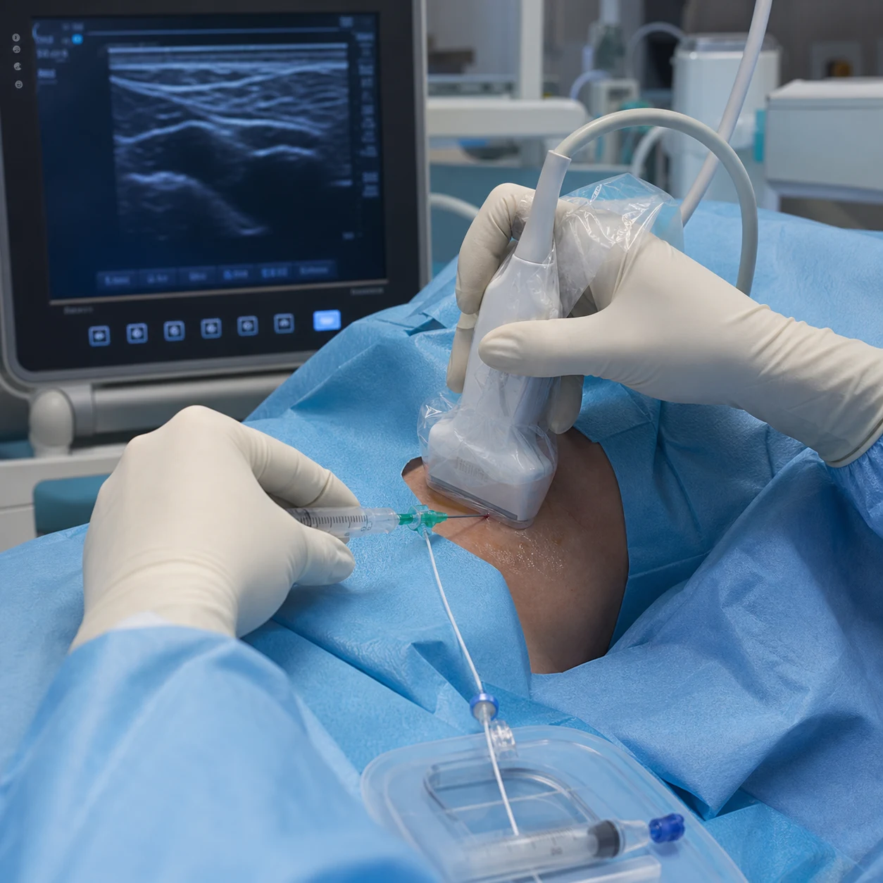
- the needle is tracked in-plane (long axis) or out-of-plane (short axis), puncturing only the anterior wall to avoid a through-and-through injury of the underlying carotid.[4]
Right vs left, and the thoracic duct
On the right, the brachiocephalic (innominate) vein and SVC run a short, straight course to the right atrium - the right IJV is the preferred site (straight path, lowest tip malposition). On the left, the left brachiocephalic vein crosses the midline behind the sternum to join the right, a tortuous course that predisposes to kinking and to the tip pointing into the contralateral IJV or the internal mammary vein.[1]
The thoracic duct ascends in the left chest, arches in the left neck, and drains into the junction of the left IJV and left subclavian - left IJV cannulation can lacerate it, causing chylothorax. There is no equivalent risk on the right (the right lymphatic duct is small).[1]
Central venous access - the subclavian vein in depth
Course and relations
The subclavian vein is the continuation of the axillary vein at the lateral border of the first rib; it runs arch-like across the top of the first rib, in the groove for the subclavian artery but separated from the artery by the scalenus anterior muscle (the vein lies anterior/inferior to scalenus anterior, the artery posterior/superior). It passes behind the sternoclavicular joint to join the IJV, forming the brachiocephalic (innominate) vein.[1]
The key relations for the infraclavicular approach:[1][1]
- Superficially: skin, subcutaneous fat, the clavicle (the needle passes beneath the middle third of the clavicle).
- The vein sits in the groove between the clavicle and the first rib - the target for the landmark technique (aim toward the sternal notch, just under the clavicle, advancing in the plane just deep to the clavicle).
- Sibson's fascia (suprapleural membrane) and the apex of the lung / cupula of pleura lie deep to the vein - a needle advanced too deeply or too steeply perforates Sibson's fascia and the pleura, producing a pneumothorax (the signature subclavian complication).
- The subclavian artery lies posterosuperior, separated by scalenus anterior; a too-posterior puncture hits it. Because the artery and vein lie behind the clavicle, the subclavian vessels are NOT compressible - the basis for avoiding this site in coagulopathy.[1][5]
- The phrenic nerve crosses anterior to scalenus anterior (between the vein and the muscle); the brachial plexus lies posterolateral.
Sibson's fascia and pneumothorax risk
Sibson's fascia is the thickened superior extension of the endothoracic fascia that tethers the cervical dome of pleura (the cupula, rising above the medial third of the clavicle into the root of the neck). The subclavian vein lies just anterior to this pleural dome. The subclavian approach therefore carries the highest pneumothorax rate of the three central sites (around 1-5 per cent, higher than IJV and absent in femoral). The post-procedure chest X-ray is mandatory after subclavian (and low-IJV) insertion, both to confirm the tip and to exclude pneumothorax.[1][5]
Why subclavian has the lowest infection and the highest stenosis risk
The subclavian site has the lowest catheter-related bloodstream infection rate of the three central sites (3SITES confirmed subclavian less than jugular less than femoral for the infection/DVT composite), because it is a clean, immobile, easily-prepped area with a flat, easily-dressed exit site. The trade-offs are pneumothorax, non-compressibility, and subclavian-vein stenosis - a particular problem in patients who may need a future arteriovenous fistula for dialysis, because subclavian stenosis destroys the downstream arm veins the fistula needs; dialysis guidelines therefore advise against subclavian CVCs in anyone who might become dialysis-dependent.[3][5]
Central venous access - the femoral vein in depth
Course and the femoral sheath (N-A-V)
The femoral vein is the continuation of the popliteal vein at the adductor hiatus; it enters the thigh posterior to the femur, then runs deep to the inguinal ligament to become the external iliac vein. In the groin it lies within the femoral sheath, a fascial condensation with the femoral artery. From lateral to medial at the inguinal ligament the contents are:[1]
- the femoral nerve (lateral, outside the sheath proper);
- the femoral artery;
- the femoral vein (medial);
- the great saphenous vein drains into the femoral vein just below the inguinal ligament (the saphenofemoral junction). [1]
The mnemonic is N-A-V (nerve, artery, vein, lateral to medial) - or "NAVEL" including lymphatics most medially. The vein lies immediately medial to the femoral artery pulsation at the inguinal ligament; the puncture is made 1-2 cm below the inguinal ligament, just medial to the arterial pulse, at 45 degrees, aiming cephalad.[1][5]
Why femoral has the highest infection and DVT risk
The femoral site is compressible (a major advantage in coagulopathy or cardiac arrest) and has no pneumothorax risk, but it sits in a moist, hair-bearing, contaminated intertriginous area close to the perineum, giving it the highest catheter-related bloodstream infection rate of the three central sites. The 3SITES trial and the Merrer JAMA RCT both showed more infection and more deep-vein thrombosis with the femoral route. It also restricts patient mobility and hip flexion, and the catheter is uncomfortable and hard to dress securely.[3][5]
The three central venous access sites - the exam trade-off table
| Site | Course / landmark | Key relation | Compressible | Pneumothorax | Infection | Special risk |
|---|---|---|---|---|---|---|
| Internal jugular (IJV) | Carotid sheath, deep to SCM; apex of two SCM heads at C6 | Carotid medial/posterior, vagus between | Yes | Low (<1%) | Intermediate | Left side: thoracic duct (chylothorax); carotid puncture |
| Subclavian | Groove between clavicle and first rib, behind sternoclavicular joint | Sibson's fascia / pleura deep; artery posterosuperior | No | High (1-5%) | Lowest | Stenosis (kills future AV fistula); haemothorax |
| Femoral | Femoral sheath, N-A-V; 1-2 cm below inguinal ligament, medial to pulse | Artery lateral; nerve lateral to artery | Yes | None | Highest | DVT; immobility; groin contamination |
Catheter tip position - the cavoatrial junction
A central venous catheter tip should lie in the distal SVC, just above the cavoatrial junction (where the SVC enters the right atrium), parallel to the long axis of the vein. A tip in the right atrium or right ventricle risks arrhythmia (RV endocardial contact - ectopy, VT), cardiac perforation and tamponade (the thin-walled atrium/RV perforates), and tricuspid-valve damage.[1][7]
The carina as the radiological landmark
On an erect chest X-ray the carina (T4/T5) lies approximately level with, or just above, the cavoatrial junction (which sits at the pericardial reflection, about 2 cm below the carina on average). The Stonelake and Bodenham cadaver and radiological study established that the carina is a safe surrogate for the upper limit of the pericardial reflection - a catheter tip positioned at or above the carina is almost certainly outside the pericardium, removing the risk of fatal tamponade from intra-pericardial perforation. This is why tip position is reported relative to the carina on the post-procedure film.[7]
Lumen and depth rules
- Right IJV: tip at about 15 cm at the skin (short, straight path to the SVC).
- Left IJV: about 17-20 cm (longer, tortuous course through the left brachiocephalic vein).
- Subclavian: about 15-17 cm; the tip should lie parallel to the SVC long axis (not abutting the wall at an acute angle, which erodes the wall).
- Femoral: a long catheter (20-30 cm) advanced so the tip lies in the IVC just below the right atrium; shorter catheters may sit in the common iliac vein and give unreliable central venous pressures.[1]
Arterial access - the radial artery in depth
Anatomy of the radial and ulnar arteries and the palmar arches
The brachial artery divides in the cubital fossa (opposite the neck of the radius) into its two terminal branches:[1][6]
- the radial artery - the smaller branch; it descends under brachioradialis, then at the wrist lies superficially lateral to the tendon of flexor carpi radialis (FCR) and medial to abductor pollicis longus. It crosses the scaphoid in the anatomical snuffbox and enters the palm dorsally to form the deep palmar arch (predominantly radial). It is the commonest arterial-line site because it is superficial, palpable, and the hand usually has dual circulation.
- the ulnar artery - the larger branch; it descends medial to flexor carpi ulnaris, crosses the wrist in Guyon's canal, and forms the superficial palmar arch (predominantly ulnar). The superficial arch is the dominant supply to the digits in most people. [1]
The two arches anastomose across the palm, so that occlusion of one artery is normally compensated by flow from the other through the arch - the anatomical basis of the Allen test.[1]
The Allen test (and modified Allen test)
The Allen test assesses whether the ulnar artery can perfuse the whole hand alone (i.e. whether the palmar arch is intact) before cannulating the radial artery. If the ulnar supply is inadequate, radial cannulation could devascularise the hand in the (common) event of radial thrombotic occlusion.[6]
The modified Allen test
Exsanguinate the hand
Patient clenches the fist several times to exsanguinate the hand (examiner can assist by milking blood out of the fingers).
Compress BOTH arteries
Examiner compresses both the radial and ulnar arteries at the wrist. Patient opens the hand - it should be pale/white (exsanguinated).
Release the ULNAR only
Examiner releases the ulnar artery, keeping the radial compressed, and times the return of normal colour to the palm.
Interpret
Colour return <5-7 seconds = NORMAL (intact ulnar/palmar arch - radial cannulation safe). 7-15 seconds = equivocal (caution). >15 seconds = ABNORMAL (do NOT cannulate the radial on this side - the hand depends on it).
Objective variant
The modified Allen test uses pulse oximetry or Doppler on the thumb/finger to detect flow objectively (more reliable than visual colour return).
Other arterial access sites
Arterial line sites - anatomy and trade-offs
| Site | Anatomy / landmark | Advantage | Disadvantage / risk |
|---|---|---|---|
| Radial (1st choice) | Lateral to FCR tendon at wrist; collateral via ulnar (Allen test) | Superficial, palpable, dual circulation; safe | Small calibre; thrombosis/occlusion (~20% transient, ~1% permanent); ischaemia in low-flow/vasopressors/Raynaud's |
| Femoral | Below inguinal ligament, lateral to femoral vein (N-A-V); 1-2 cm below ligament | Large; easy in profound shock/arrest; reflects central aortic pressure | Atherosclerosis; retroperitoneal haematoma; infection; contaminated site |
| Dorsalis pedis | Lateral to extensor hallucis longus tendon, between 1st and 2nd metatarsals | Alternative when upper-limb sites unavailable | Small; peripheral waveform damped; unreliable in peripheral vascular disease; collateral via plantar arch (verify flow) |
| Posterior tibial | Posterior to medial malleolus | Alternative foot site | Small; damped waveform |
| Brachial (usually avoided) | Medial to biceps tendon in antecubital fossa | Good waveform; reflects central pressure | End-artery (no collateral) - thrombosis devascularises forearm; median nerve injury; compartment syndrome |
| Axillary | High, in the axilla; left side reflects central aorta | Large; tolerated long-term | Near brachial plexus - haematoma compresses plexus; sheath tip near aortic arch |
Peripherally inserted central catheters (PICCs)
A PICC is a long, thin central catheter inserted through a peripheral arm vein and advanced so the tip lies at the cavoatrial junction, in the distal SVC - the same target as a tunnelled CVC. The basilic, cephalic, and brachial veins of the upper arm are the entry choices, with a clear order of preference.[1]
Vein preference: basilic > cephalic > brachial
- Basilic vein (preferred): runs along the medial aspect of the arm, is usually larger in calibre than the cephalic, has a straighter course into the axillary vein, and is easier to navigate despite its valves. It is the first-choice PICC entry vein.
- Cephalic vein (second choice): runs along the lateral aspect of the arm and dips between deltoid and pectoralis major (the deltopectoral groove) to join the axillary vein. It is often smaller, more tortuous, and prone to spasm, and the angle at the clavipectoral junction can deflect the wire; access success is lower than basilic.
- Brachial vein (third choice, often paired with basilic): the deep venae comitantes running with the brachial artery; they are deeper, smaller, and lie adjacent to the artery and median nerve (risk of arterial puncture and nerve injury), so they are the third choice but are commonly used under ultrasound when the superficial veins are unsuitable.[1]
Tip position
The PICC tip should lie at the cavoatrial junction (distal SVC, carina level on chest X-ray), where the vein is wide and the tip floats freely - the same Stonelake/Bodenham landmark as for any central catheter. A tip too proximal (in the subclavian/brachiocephalic) is associated with thrombosis and unreliable central venous pressure; a tip in the right atrium risks arrhythmia and perforation.[7]
PICC vs tunnelled CVC vs non-tunnelled CVC
| Feature | PICC | Non-tunnelled CVC (IJV/SC/fem) | Tunnelled CVC (Hickman/Broviac) |
|---|---|---|---|
| Entry | Peripheral arm vein | Central vein at bedside | Central vein + subcutaneous tunnel to exit site on chest wall |
| Tip | Cavoatrial junction (distal SVC) | Cavoatrial junction | Cavoatrial junction |
| Indwelling time | Weeks-months | Days-weeks | Months-years |
| CRBSI risk | Lower than non-tunnelled | Highest of the three | Low (Dacron cuff + tunnel) |
| Insertion setting | Bedside/IR, sterile | ICU bedside, sterile bundle | Theatre or IR, aseptic |
| Common use | Antibiotics, mid-term TPN, difficult access | Resuscitation, CRRT, short-term CVP/drugs | Long-term chemotherapy, TPN, transplantation |
Dialysis catheters - tunnelled vs non-tunnelled
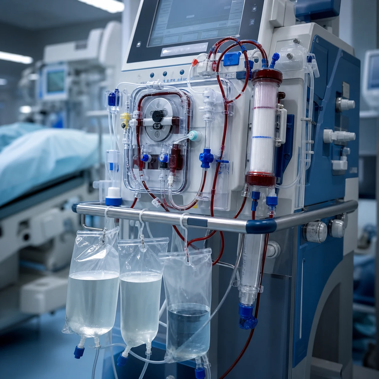
Vascular access for haemodialysis/CRRT uses larger-bore, dual-lumen catheters (afferent and efferent lumina). The two configurations differ in design and intended duration.[1]
- Non-tunnelled (temporary) dialysis catheter: a stiff dual-lumen catheter placed at the bedside in a central vein (right IJV preferred, then femoral; subclavian avoided if a future fistula is possible). Used for short-term dialysis (days-weeks), CRRT in acute kidney injury, or bridging to definitive access. Higher infection rate; removed as soon as definitive access (fistula or graft) is ready.
- Tunnelled (long-term) dialysis catheter: a cuffed dual-lumen catheter (e.g. Permcath) placed in a central vein (usually right IJV), then passed through a subcutaneous tunnel on the anterior chest wall to an exit site remote from the venotomy. A Dacron cuff sits in the tunnel and is ingrown by fibrous tissue, anchoring the catheter and forming a mechanical/antimicrobial barrier. Used for months-years when a fistula/graft is not yet usable or not possible. Lower infection than non-tunnelled because of the cuff and the remote exit site.[1]
Tunnelled vs non-tunnelled dialysis catheters
| Feature | Non-tunnelled | Tunnelled (cuffed) |
|---|---|---|
| Setting | ICU/ward bedside | Theatre or interventional radiology |
| Anaesthesia | Local +/- sedation | Local +/- sedation or general |
| Duration | Days-weeks | Months-years |
| Cuff | None | Dacron cuff in subcutaneous tunnel |
| Exit site | At the venipuncture (neck/groin) | Remote, on chest wall |
| CRBSI rate | Higher | Lower |
| Preferred central vein | Right IJV > femoral (avoid subclavian if fistula possible) | Right IJV (long straight path to cavoatrial junction) |
| Removal | Bedside pull | Surgical/IR dissection of cuff |
The pulmonary artery (Swan-Ganz) catheter
The pulmonary artery catheter (PAC) is a flow-directed balloon-tipped catheter inserted through a large-bore introducer sheath in a central vein, then "floated" through the right heart into the pulmonary artery to measure right-heart pressures, pulmonary artery occlusion ("wedge") pressure, and cardiac output.[1]
The route through the right heart
From the central venous sheath, the balloon-tipped PAC passes, in order:[1]
- Central vein (typically right IJV or right subclavian, for the straightest path; femoral possible but longer) ->
- Superior vena cava ->
- Right atrium (pressure trace: low, a and v waves, x and y descents) ->
- Tricuspid valve ->
- Right ventricle (pressure trace rises sharply: systolic about 25 mmHg, diastolic about 5) ->
- Pulmonary valve ->
- Pulmonary artery (pressure trace: systolic about 25 mmHg but a diastolic step-UP to about 10-12, distinguishing it from the RV) ->
- With the balloon inflated, the catheter occludes a distal pulmonary artery branch ->
- Pulmonary artery occlusion ("wedge") pressure trace (low, about 6-12 mmHg, reflecting left atrial pressure via the static column through the pulmonary capillary bed).[1]
Anatomical and safety points
- The balloon is inflated only during flotation and wedge measurement, and deflated immediately afterwards - prolonged wedging risks pulmonary infarction and rupture (especially with pulmonary hypertension, the elderly, and anticoagulation).[1]
- A PAC advanced too far loops in the right ventricle (ventricular ectopy, VT) or over-wedges (rupture risk); the waveform guides correct position.
- Insertion complications mirror those of any central line (pneumothorax for subclavian/IJV, arterial puncture, arrhythmia) plus right bundle-branch block (the catheter traumatising the right bundle - problematic in a patient with pre-existing LBBB, producing complete heart block).[1]
Extracorporeal membrane oxygenation (ECMO) cannulae - VV vs VA
ECMO drains venous blood, oxygenates and decarboxylates it in an external membrane lung, and returns it; the cannulation strategy (veno-venous vs veno-arterial) is defined by which vessels are cannulated and where the oxygenated blood is returned.[1]
Veno-venous ECMO (VV-ECMO) - for respiratory failure
VV-ECMO bypasses only the lungs (the heart still pumps the systemic circulation). Deoxygenated blood is drained from the central venous system and returned, oxygenated, to the right atrium.[1]
- Drainage (venous): the femoral vein (or IJV) -> IVC -> right atrium.
- Return (oxygenated): the right IJV (right atrium) - the classic femoro-jugular configuration.
- Single dual-lumen cannula (Avalon/Y-cannula): a single large cannula in the right IJV with one drainage port in the IVC/RA and one return jet directed across the tricuspid valve toward the RV outflow - avoids recirculation and allows single-site cannulation and patient mobility.[1]
- Recirculation is the VV-specific problem: some returned oxygenated blood is immediately re-drained, reducing effective oxygenation; configuration and flow rate minimise it.
- The heart is native - VV-ECMO provides no circulatory support, so it is used only where the circulation is adequate (isolated respiratory failure).
Veno-arterial ECMO (VA-ECMO) - for cardiac (plus or minus respiratory) failure
VA-ECMO bypasses both the heart and the lungs, providing circulatory support. Venous blood is drained and returned directly into the arterial system.[1]
- Drainage (venous): the femoral vein -> IVC -> right atrium.
- Return (arterial): the femoral artery, delivering oxygenated blood retrograde up the aorta (the classic femoro-femoral peripheral configuration).
- Central VA-ECMO (in a sternotomy, post-cardiotomy): drainage from the right atrium, return to the ascending aorta.
- Harlequin (north-south) syndrome: with femoro-femoral VA-ECMO, the native heart ejects poorly oxygenated blood (from failing lungs) into the aortic root and arch, while retrograde ECMO blood fills the descending aorta. If lung oxygenation is poor, the upper body (coronaries, brain) receives desaturated blood while the lower body is well oxygenated - monitored by a right-radial arterial line and cerebral oximetry, and managed by adding venous drainage or converting to a hybrid (VA-V) configuration.[1]
- Limb ischaemia: the large femoral arterial return cannula can occlude the common femoral artery, so a distal perfusion cannula (perfusing the superficial femoral artery) is routinely placed to prevent leg ischaemia.
VV-ECMO vs VA-ECMO - cannulation and support
| Feature | VV-ECMO | VA-ECMO |
|---|---|---|
| Indication | Isolated respiratory failure (severe ARDS, asthma) | Cardiac failure plus or minus respiratory (severe cardiogenic shock, arrest, myocarditis) |
| Circulatory support | None (heart native) | Full cardiopulmonary bypass |
| Drainage | Femoral vein or IJV -> IVC/RA | Femoral vein (peripheral) or RA (central) |
| Return | IJV (RA) - femoro-jugular, or single dual-lumen cannula | Femoral artery (retrograde aorta) or ascending aorta (central) |
| Typical configuration | Femoro-jugular, or single dual-lumen (Avalon) | Femoro-femoral (peripheral); RA-to-aorta (central) |
| Signature problem | Recirculation (re-drainage of returned blood) | Harlequin syndrome; limb ischaemia (distal perfusion cannula) |
| Arterial line for monitoring | Any | Right radial (to detect upper-body hypoxaemia) |
Insertion bundles and infection prevention - the anatomy-grounded bundle
The Pronovost/Keystone intervention showed that a simple, anatomy-driven bundle collapses catheter-related bloodstream infection (CRBSI) rates. The anatomy-relevant components are: avoid the femoral site (high bacterial load, contaminated intertriginous skin), maximal sterile barriers (cap, mask, sterile gown and gloves, full-body drape), 2% chlorhexidine skin antisepsis (allowed to dry), and daily review with prompt removal.[2]
The central-line insertion bundle (Pronovost/Keystone, adapted)
Hand hygiene
Alcohol-based hand rub before the procedure and before each glove change.
Maximal sterile barriers
Cap, mask covering nose and mouth, sterile gown, sterile gloves, AND a full-body sterile drape over the patient (not just a small drape).
Chlorhexidine skin antisepsis
2% chlorhexidine in 70% alcohol, applied with friction and ALLOWED TO DRY (do not wipe). Chlorhexidine superior to povidone-iodine for CVC skin antisepsis.
Optimal site selection
Prefer the subclavian or IJV over the femoral site to minimise bacterial colonisation and infection; weigh against pneumothorax and coagulopathy.
Daily review with prompt removal
Every day ask whether the line is still needed; remove it as soon as it is not. Duration of catheterisation is a linear risk for CRBSI.
Key trials and evidence
3SITES trial (Parienti 2015, NEJM) - central venous catheter insertion site
Design
Multicentre randomised controlled trial; 3,027 patients needing a central venous catheter for at least 3 days across 18 ICUs in France
Intervention
Subclavian vs jugular vs femoral insertion site - randomly assigned
Primary outcome
Composite of catheter-related bloodstream infection AND symptomatic deep vein thrombosis: lowest with SUBCLAVIAN (1.5%) < jugular (3.3%) < femoral (4.0%)
Mechanical risk
Symptomatic pneumothorax HIGHEST with SUBCLAVIAN (1.1%) vs jugular (0.3%) vs femoral (0%)
Clinical bottom line
The definitive site-selection trial: subclavian minimises infection/thrombosis but maximises pneumothorax. Choose subclavian when infection risk and clean clotting dominate, IJV when ultrasound access and compressibility dominate, femoral for emergency/coagulopathic access
Pronovost / Keystone (2006, NEJM) - the central-line bundle
Design
Prospective cohort / quasi-experimental study in 103 ICUs in Michigan, USA
Intervention
A five-component bundle: hand hygiene, maximal sterile barriers, chlorhexidine skin prep, avoidance of the femoral site, and daily review with prompt removal
Primary outcome
Median catheter-related bloodstream infection rate fell from 2.7 to 0 per 1,000 catheter-days (up to 66% relative reduction), sustained at 18 months
Clinical bottom line
A simple, anatomy-grounded bundle (avoid the high-infection femoral site; maximal sterile barriers at the chosen site; daily review) dramatically reduces CRBSI - the foundation of every ICU line protocol
Merrer 2001 (JAMA) - femoral vs subclavian catheterisation RCT
Design
Multicentre randomised controlled trial; 289 patients in 8 French ICUs randomised to femoral vs subclavian central venous catheterisation
Primary outcome
Mechanical, infectious, and thrombotic complications at 48 h and at catheter removal
Key result
Femoral had MORE infectious complications (femoral 19.8% vs subclavian 4.5%) and MORE asymptomatic deep vein thrombosis (femoral 21.5% vs subclavian 1.9%). Severe mechanical complications (haemothorax, pneumothorax) only with subclavian. Femoral punctures were more likely to be arterial (9.4% vs 3.1%)
Clinical bottom line
Confirmed the site trade-off: femoral trades compressibility/no pneumothorax for significantly higher infection and DVT; subclavian trades the lowest infection for mechanical (pneumothorax/haemothorax) risk. The anatomy-driven site-choice principle
Karakitsos 2006 (Critical Care) - real-time ultrasound-guided IJV catheterisation
Design
Prospective randomised comparison of real-time ultrasound-guided vs landmark-based internal jugular vein catheterisation in 900 critical care patients
Primary outcome
Overall success, first-pass success, number of attempts, and mechanical complications
Key result
Ultrasound guidance INCREASED overall success and first-pass success, REDUCED the number of attempts, and REDUCED mechanical complications (carotid puncture, haematoma, haemothorax, pneumothorax) compared with the landmark technique
Clinical bottom line
Established real-time 2D ultrasound as the standard for IJV cannulation - it converts anatomical knowledge into direct visual guidance and cuts mechanical complications. The basis for guidelines mandating ultrasound for elective CVC insertion
Stonelake and Bodenham 2006 (British Journal of Anaesthesia) - the carina and the pericardial reflection
Design
Cadaver and radiological study defining the relationship of the carina to the pericardial reflection (the cavoatrial junction surrogate)
Key finding
The carina lies at or just above the upper limit of the pericardial reflection on chest X-ray; a catheter tip positioned at or above the carina is almost certainly OUTSIDE the pericardium
Clinical bottom line
Established the carina as the safe radiological landmark for the central catheter tip - a tip above the carina removes the risk of fatal cardiac tamponade from intra-pericardial perforation. The basis for tip-position reporting on the post-line chest film
Exam practice
SAQ — Central venous access anatomy and late complications (cardiac tamponade)
10 minutes · 10 marks
A 72-year-old woman was admitted to the ICU with urosepsis. A right internal jugular triple-lumen catheter was inserted two days ago by the landmark technique without ultrasound. She now complains of chest tightness and breathlessness: HR 128, BP 84/50, JVP distended to the angle of the jaw, muffled heart sounds, and the post-line chest X-ray shows the catheter tip projected over the cardiac silhouette.
SAQ — Arterial line insertion anatomy and site selection in shock
10 minutes · 10 marks
A 64-year-old man on a high-dose noradrenaline infusion (0.6 mcg/kg/min) for septic shock needs beat-to-beat arterial pressure monitoring and frequent blood-gas sampling. The team plans a left radial arterial line, but his peripheries are cool and poorly perfused and he has a history of Raynaud\'s phenomenon.
Clinical pearls
Worked exam question
[1]Worked answer. The three central venous sites each have a constant landmark-target relationship and a distinctive risk profile.[1][1]
Internal jugular vein. The IJV runs in the carotid sheath, deep to sternocleidomastoid, with the common carotid artery MEDIAL and POSTERIOR (deep) and the vagus nerve between them in the posterior angle. The sheath runs from the skull base to the root of the neck; the SCM has two heads (sternal and clavicular) whose apex (Sedillot's triangle, about C6) is the high central landmark where the IJV is most superficial. Ultrasound shows the IJV as an oval, thin-walled, compressible, non-pulsatile structure lateral to the round, thick-walled, pulsatile, non-compressible carotid. On the right the brachiocephalic vein runs a short straight course to the SVC (preferred site); on the left the course is tortuous and the thoracic duct drains into the left jugulo-subclavian junction, so left IJV risks chylothorax. Risks: carotid puncture, pneumothorax (low, <1%), and on the left, thoracic-duct injury.[1][4]
Subclavian vein. The subclavian vein is the continuation of the axillary vein at the lateral border of the first rib; it arches across the top of the first rib in the groove between the clavicle and first rib (anterior/inferior to scalenus anterior, which separates it from the subclavian artery), and passes behind the sternoclavicular joint to join the IJV, forming the brachiocephalic vein. Deep to the vein lie Sibson's fascia (the suprapleural membrane) and the cupula of pleura - the anatomical basis of subclavian's higher pneumothorax rate (1-5%). Because the subclavian vessels lie behind the clavicle and cannot be compressed, subclavian is avoided in coagulopathy (haemothorax risk). It has the lowest infection rate but causes subclavian-vein stenosis, which destroys future arteriovenous-fistula options for dialysis.[1][5]
Femoral vein. The femoral vein lies in the femoral sheath; at the inguinal ligament the contents run N-A-V (nerve, artery, vein) from lateral to medial, so the vein is immediately MEDIAL to the femoral artery pulse. It is punctured 1-2 cm below the inguinal ligament, just medial to the pulse. It is compressible (ideal in coagulopathy and cardiac arrest) and has no pneumothorax risk, but it sits in a contaminated intertriginous area and has the highest catheter-related bloodstream infection and deep-vein-thrombosis rates (3SITES, Merrer).[3][5]
The tip belongs in the distal SVC just above the cavoatrial junction, at the level of the carina on chest X-ray (Stonelake and Bodenham: the carina sits at/above the pericardial reflection, so a tip there is outside the pericardium and free of fatal tamponade risk). A tip in the right atrium or ventricle risks arrhythmia, tricuspid damage, and perforation/tamponade.[7]
Bottom line. The recurring thread is the landmark-danger-safety-rule triplet: IJV (carotid medial/posterior - aim lateral, ultrasound, head-down); subclavian (Sibson's fascia and pleura deep, non-compressible - avoid in coagulopathy and future-dialysis); femoral (N-A-V medial to the artery - compressible, but highest infection/DVT). The tip sits at the carina, outside the pericardium. The 3SITES and Merrer trials quantify the trade-off; the Keystone bundle operationalises infection prevention.[1][2]
References
- [1]McGee DC, Gould MK Preventing complications of central venous catheterization N Engl J Med, 2003.PMID 12646670
- [2]Pronovost P, Needham D, Berenholtz S, et al. An intervention to decrease catheter-related bloodstream infections in the ICU N Engl J Med, 2006.PMID 17192537
- [3]Parienti JJ, Mongardon N, Megarbane B, et al. (3SITES Study Group) Intravascular Complications of Central Venous Catheterization by Insertion Site N Engl J Med, 2015.PMID 26398070
- [4]Karakitsos D, Labropoulos N, De Groot E, et al. Real-time ultrasound-guided catheterisation of the internal jugular vein: a prospective comparison with the landmark technique in critical care patients Crit Care, 2006.PMID 17112371
- [5]Merrer J, De Jonghe B, Golliot F, et al. (French Catheter Study Group) Complications of femoral and subclavian venous catheterization in critically ill patients: a randomized controlled trial JAMA, 2001.PMID 11495620
- [6]Tegtmeyer K, Brady G, Lai S, Braner DA Videos in Clinical Medicine. Placement of an arterial line N Engl J Med, 2006.PMID 16611944
- [7]Stonelake PA, Bodenham AR The carina as a radiological landmark for central venous catheter tip position Br J Anaesth, 2006.PMID 16415318
- [8]Romeu-Bordas O, Ballesteros-Pena S [Reliability and validity of the modified Allen test: a systematic review and metanalysis] Emergencias, 2017.PMID 28825257