ICU · Burns
Burn Assessment — TBSA, Depth & Airway
Also known as Burn assessment · Rule of Nines · Lund-Browder chart · Burn depth · Inhalation injury · Parkland formula · Palm method · TBSA estimation · Superficial partial thickness burn · Deep dermal burn · Full thickness burn · Baux score · Revised Baux · Burn centre referral · Major burn criteria · Chemical burn assessment · Electrical burn assessment · Laser Doppler imaging burn depth · Carbonaceous sputum · Singed nasal hair
The burn the assessment — the the TBSA (the total the body the surface the area — the Rule of the Nines, the Lund-the-Browder, the palm), the the depth (the superficial / the partial / the deep the partial / the full the thickness), and the the airway / the inhalation the injury (the soot, the singed the hair, the carbonaceous the sputum → the early the intubation). The severity the classification. The Parkland the formula (the 4 mL/kg/%TBSA — the half the 8 h, the rest the 16 h). Burn assessment is built on three pillars — the Total Body Surface Area (TBSA), the depth, and the airway/inhalation injury — which together determine fluid resuscitation, the need for transfer to a burns unit, and prognosis. TBSA is estimated by the Rule of Nines (adult), the Lund-Browder chart (the gold standard, age-adjusted for children), or the palm method (the patient's palm including fingers = ~1% TBSA, for small/irregular burns). Only partial-thickness (2nd degree) and full-thickness (3rd degree) burns are counted — superficial (1st degree / erythema / sunburn) is excluded because it inflates the calculation and causes fluid overload. Depth is classified as superficial (epidermal — red, dry, painful, heals in days), superficial partial (upper dermis — red, moist, blistered, exquisitely painful, heals 1-2 weeks), deep partial (deep dermis — pale, mottled, reduced sensation, heals 3-8 weeks with scarring), and full thickness (all skin layers — white/charred, dry, leathery, painless, requires grafting). Inhalation injury doubles mortality and demands early intubation before airway oedema (which peaks at 24-48 h) makes the airway impossible. A major burn (20% TBSA adult, 10% child, or any burn of face/hands/feet/perineum/major joints, electrical, chemical, inhalation, or with comorbidity) mandates transfer to a specialist burns centre. The Parkland formula (Baxter, 4 mL/kg/%TBSA lactated Ringer's, half in the first 8 h from the TIME of the burn) is the historical starting point; modern care titrates to urine output and perfusion rather than slavishly following a formula. Prognosis is captured by the Baux score (age + %TBSA) and the revised Baux (age + %TBSA + inhalation injury).
On this page & tools
Your progress
Saved locally on this device.
Target exams
Red flags
Overview & definition
The burn the assessment — the TBSA (the total the body the surface the area), the depth, and the the airway / the inhalation the injury. The three the pillars the that the determine the fluid the resuscitation (the Parkland), the transfer, and the prognosis. The accurate the assessment the essential.[1][1]
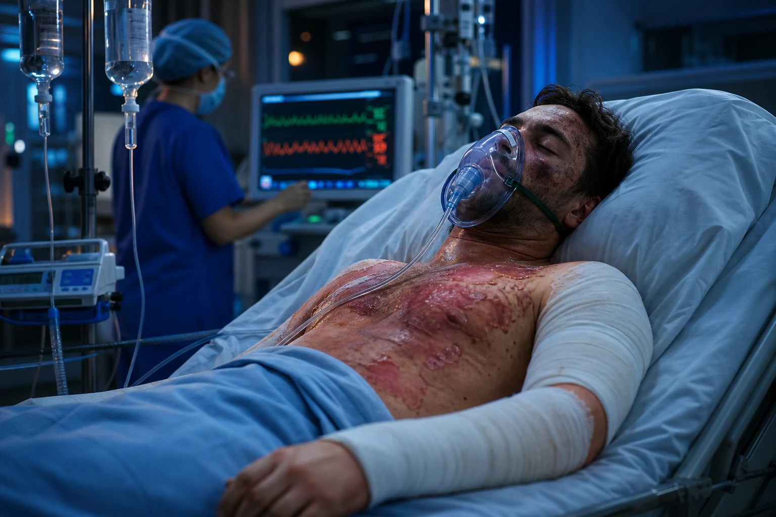
Burn assessment is the single most deterministic early intervention in burn care: the numbers you write down at the bedside in the first hour set the fluid rate for the next 24 hours, decide whether the patient is transferred to a burns centre, and predict survival. Get the assessment wrong — by counting superficial erythema, by using the Rule of Nines in a toddler, by missing an inhalation injury — and every downstream decision is wrong too. The discipline rests on three pillars that the examiner will probe in turn:[1][1][3]
- TBSA — how big? The percentage of total body surface area burned, estimated by the Rule of Nines (adult), the Lund-Browder chart (gold standard, age-adjusted), or the palm method (small/irregular burns). Only partial- and full-thickness burns count.
- Depth — how deep? Superficial (epidermal), superficial partial (upper dermis), deep partial (deep dermis), full thickness (all skin). Depth determines pain, healing potential, the need for grafting, and (together with TBSA) fluid requirements.
- Airway / inhalation injury — is the airway threatened? Soot, singed nasal hair, carbonaceous sputum, hoarse voice, enclosed-space fire → the patient is intubated early, before oedema closes the airway. Inhalation injury doubles mortality. [1]
The TBSA — the total the body the surface the area
The the Rule of the Nines (the adult):[1][1]
- The head + the neck: the 9 per cent.
- The each the arm: the 9 per cent (the 4.5 the anterior, the 4.5 the posterior).
- The each the leg: the 18 per cent (the 9 the anterior, the 9 the posterior).
- The anterior the trunk: the 18 per cent.
- The posterior the trunk: the 18 per cent.
- The perineum: the 1 per cent.
The the Lund-the-Browder the chart — the more the precise, the age-the-adjusted (the children the larger the head, the smaller the legs). The gold the standard.[2][1][1]
The the palm the method — the patient the palm + the digits the = the 1 per cent (the for the small / the irregular the burns).[1]
The count ONLY the partial-the-thickness (the 2nd the degree) and the full-the-thickness (the 3rd the degree). The NOT the superficial (the 1st the degree — the erythema / the sunburn).[1][1]
Why TBSA matters — what it drives
The %TBSA is not a number for its own sake; it directly drives four management decisions, every one of which the examiner will probe:[1][1][3]
- Fluid resuscitation — the Parkland formula multiplies weight × %TBSA × 4. An over-estimate of 10% TBSA inflates fluid by 10% and contributes to fluid creep, abdominal compartment syndrome, and death. An under-estimate under-resuscitates and causes burn shock and AKI.
- Transfer threshold — the major burn criteria (transfer to a burns centre) are partly TBSA-driven (>20% adult, >10% child, >5% full thickness).
- Prognosis — the Baux and revised Baux scores use %TBSA as a core variable.
- Surgical planning — large TBSA burns need staged excision and grafting; the donor-to-recipient ratio is calculated against unburned TBSA. [1]
The Rule of Nines (Wallace) — the bedside adult estimate
Devised by Alexander Wallace in 1951 as a rapid adult bedside estimate, the Rule of Nines divides the body into regions that are multiples of 9%. It is fast, memorable, and adequate for the initial adult resuscitation — but it is grossly inaccurate in children, in whom a proportionally huge head and small legs break the assumption of adult proportions. Always replace it with a Lund-Browder chart as soon as one is available.[1][1]
Rule of Nines — adult body region allocation (Wallace 1951)
| Body region | Anterior | Posterior | Total per region | Running total |
|---|---|---|---|---|
| Head and neck | 4.5% | 4.5% | 9% | 9 |
| Each upper limb (arm) | 4.5% | 4.5% | 9% (×2 = 18) | 27 |
| Each lower limb (leg) | 9% | 9% | 18% (×2 = 36) | 63 |
| Anterior trunk | 18% | — | 18% | 81 |
| Posterior trunk | — | 18% | 18% | 99 |
| Perineum and genitalia | — | — | 1% | 100 |
The numbers add to 100% only when the perineum (1%) is included — a perennial exam trap. The buttocks are counted with the posterior trunk, not the legs. Note also that the Rule of Nines was derived from adults; using it unchanged in a 2-year-old systematically under-estimates head burns and over-estimates leg burns by several percent each.[2]
The Lund-Browder chart — the gold standard, age-adjusted
The Lund and Browder chart (1944), refined from Berkow's 1924 data, is the most accurate of the clinical (non-instrumental) methods because it corrects for the changing body proportions of growth. The head and neck are large at birth (~19%) and shrink to the adult 9% by age 10-15; the legs correspondingly grow from ~13% each in infancy to the adult 18%. Other regions are essentially constant.[2][1][1]
Lund-Browder age-adjusted percentages — head and legs only (everything else as adult)
| Body region | Age 0 (newborn) | Age 1 | Age 5 | Age 10 | Age 15 | Adult |
|---|---|---|---|---|---|---|
| Head and neck (A — half front/back) | 19% | 17% | 13% | 11% | 9% | 7% |
| Each thigh (B) | 5.5% | 6.5% | 8% | 8.5% | 9% | 9.5% |
| Each leg below knee (C) | 5% | 5% | 5.5% | 6% | 6.5% | 7% |
| Each leg total (B+C, front+back) | ~13% | ~14% | ~17% | ~17.5% | ~18% | 18% |
| Neck, anterior trunk, posterior trunk, each arm, buttocks, genitalia | — | — | — | — | — | as adult |
How to use it at the bedside: draw the burn on the standard Lund-Browder body map (front and back), look up the age-appropriate A/B/C values, and total the involved areas. Most burns-unit paper charts and electronic systems have the chart pre-printed. The head is so dominant in infants that a scald to the scalp of a 6-month-old can easily be 15-18% TBSA on its own — a fact that converts a "moderate" scald into a major burn requiring formal resuscitation and transfer.[2]
The palm method — the small/irregular burn tool
For small, scattered, or irregular burns where the Rule of Nines is too coarse, the palm method estimates each patch against the patient's own palm. The palm (including the fingers, i.e. the volar surface from wrist to fingertip) of the patient is ~0.8-1% of their TBSA — conveniently close to 1%, which is the figure used clinically. Count the number of "patient palms" that would cover the burn to get a percentage.[1][3]
Palm method — getting it right
| Question | Answer |
|---|---|
| Whose palm? | The PATIENT'S OWN palm — surface area scales with body size, so the patient's palm is the right denominator. Using the rescuer's palm is a common error. |
| Palm alone, or palm + fingers? | Palm + digits (whole volar hand). Palm alone is ~0.5%; palm + fingers is ~0.8-1%. The clinical convention rounds to 1% for the whole hand. |
| When to use it? | Small burns (<15% TBSA), irregular/scattered burns, paediatric patchy scalds, and as a sanity-check on a Rule of Nines estimate. |
| Accuracy? | Crude but adequate for the small burn. For the larger burn use Rule of Nines or Lund-Browder. |
Worked example — TBSA by Rule of Nines
TBSA estimation — 70 kg adult, flame burn to anterior trunk, anterior right leg, and whole right arm
Identify the regions
Anterior trunk = 18%. Anterior surface of one leg = 9%. Whole of one arm (anterior + posterior) = 9%.
Sum the regions
18 + 9 + 9 = 36% TBSA.
EXCLUDE superficial erythema
If the red patches on the anterior trunk are superficial (1st degree, erythema only, no blister), they are NOT counted. Subtract them. Only count partial-thickness (blistered, moist) and full-thickness (white/charred) areas.
Cross-check with the palm method
Lay the patient's palm over the irregular patches. ~36 palms would cover 36% — consistent with the Rule of Nines estimate.
Apply the Parkland formula
Resuscitation volume = 4 × 70 × 36 = 10,080 mL lactated Ringer's in 24 h. Half (5,040 mL) in first 8 h from TIME OF BURN.
Decide on transfer
36% TBSA is a major burn (>20% adult) → transfer to a burns centre.
Common TBSA errors — the avoidable mistakes
TBSA estimation — the recurring mistakes that cost marks (and lives)
| Error | What happens | How to avoid |
|---|---|---|
| Counting superficial (1st degree) burns | Inflates TBSA → fluid overload → fluid creep, abdominal compartment syndrome | Count ONLY 2nd- and 3rd-degree burns. Erythema without blister is excluded. |
| Using the Rule of Nines in a child | Under-estimates head burns, over-estimates leg burns (~8-10% error) | Use the Lund-Browder chart in every child. |
| Counting the patient's whole front as "18%" when only part is burnt | Over-estimates | Subdivide: only the burnt area of the anterior trunk counts. |
| Using the rescuer's palm in the palm method | Under/over-estimates depending on size mismatch | Use the patient's own palm including fingers (~1%). |
| Forgetting the perineum (1%) | Under-estimates by 1% | Include perineum and genitalia separately. |
| Reckoning time from admission | Mis-times the Parkland first-half window | Always clock from the TIME OF THE BURN. |
| Double-counting overlapping regions | Over-estimates | Chart the burn on a Lund-Browder body map; total once. |
The depth
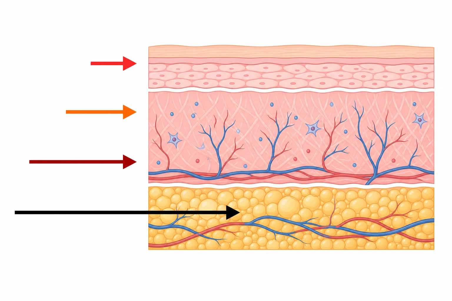
Burn depth determines pain, healing potential, the need for surgery, and contributes (with TBSA) to fluid requirements. Depth is classified into four grades, often expanded to five by separating full-thickness skin burns from deeper 4th-degree burns that involve fascia, muscle, or bone. The examiner expects you to identify each grade at the bedside from appearance, sensation, and blanching, and to state the healing and management implications.[1][1][1]
| Depth | Layer | Appearance | Sensation | Healing |
|---|---|---|---|---|
| Superficial (1st) | Epidermis | Red, dry, no blister | Painful | 3-6 days, no scar |
| Superficial partial (2a) | Upper dermis | Red, moist, blisters | Very painful | 1-2 weeks, minimal scar |
| Deep partial (2b) | Deep dermis | Pale, mottled, blisters | Reduced pain | 3-8 weeks, scar |
| Full thickness (3rd) | All skin | White/charred, dry, leathery | Painless (destroyed nerves) | Skin graft required |
| Full thickness + subcut (4th) | Muscle, bone | Visible tissue | Painless | Surgical reconstruction |
The four depths in clinical detail — what the examiner asks
Burn depth — the four grades, clinical feature by feature
| Feature | Superficial (epidermal / 1st) | Superficial partial (2a) | Deep partial (2b) | Full thickness (3rd) |
|---|---|---|---|---|
| Layer involved | Epidermis only | Epidermis + superficial (papillary) dermis | Epidermis + deep (reticular) dermis | Entire dermis destroyed; may extend to fat |
| Classic cause | Sunburn, minor flash | Scald (hot water), brief flame | Scald (hot oil), flame, contact | Flame, molten metal, electricity, chemical, prolonged contact |
| Appearance | Red, dry, no blisters; blanches with pressure then refills | Red, moist, blisters (fluid-filled), blanches briskly | Pale, mottled, cherry-red fixed staining; blisters; sluggish blanching | White, waxy, charred, dry, leathery (eschar); NO blanching |
| Sensation / pain | Painful (intact nerves) | Exquisitely painful (intact nerve endings) | Reduced sensation — pinprick felt as dull pressure | Painless — nerve endings destroyed |
| Capillary refill | Brisk (<2 s) | Brisk | Sluggish or absent | Absent — thrombosed vessels |
| Hair follicles | Intact; hair pulls easily | Intact | May be damaged | Destroyed; hair pulls out painlessly |
| Healing | 3-6 days, no scar (from basal keratinocytes) | 1-2 weeks, no/minimal scar (from dermal appendages) | 3-8 weeks, hypertrophic scar; may progress to full thickness | Cannot heal spontaneously — skin graft required |
| Management | Oral analgesia, moisturiser; no resuscitation | Topical antimicrobial, non-adherent dressing; outpatient | Surgical assessment — may need excision/grafting | Surgical excision and grafting |
Pathophysiology of each depth — why the features appear
The clinical features of each depth flow directly from the depth of tissue destruction and the survival of skin appendages (hair follicles, sweat glands, sebaceous glands), whose epithelial cells are the reservoir for re-epithelialisation.[1][1]
- Superficial (epidermal): Only the epidermis is injured; the basal layer (stratum basale) survives and re-epithelialises within days. Vasodilation produces erythema and pain (intact nociceptors). No blister, no scar. The classic example is sunburn.
- Superficial partial (2a): The burn extends into the papillary (upper) dermis. The dermal microvasculature is intact enough to briskly blanch and refill; the blisters are the separated necrotic epidermis over a serous exudate. Nerve endings are intact and hyperaemic, hence the exquisite pain. Re-epithelialisation proceeds from the surviving dermal appendages within 1-2 weeks, with minimal scarring.
- Deep partial (2b): The burn extends into the reticular (deep) dermis. Much of the microvasculature is thrombosed → sluggish or absent blanching, a fixed mottled/marbled appearance, and reduced sensation (some nerve endings destroyed). The surviving appendages are sparse; healing takes 3-8 weeks and proceeds with exuberant granulation and hypertrophic scarring. Critically, deep partial burns are unstable — inadequate perfusion, infection, or desiccation can convert them to full thickness within days.
- Full thickness (3rd): The entire dermis is destroyed — all skin appendages, vasculature, and nerve endings are coagulated. The wound is dry, leathery, and insensate; the thrombosed subdermal plexus gives a marbled/charred look with no blanching. Because no epithelial reservoir survives, spontaneous healing is impossible and surgical excision with skin grafting is mandatory. Deep full-thickness burns (4th degree) extend into subcutaneous fat, fascia, muscle, or bone and need flap reconstruction or amputation. [1]
Depth is dynamic — the concept of burn "conversion"
Burn depth is not fixed at the moment of injury. A superficial partial burn can deepen to full thickness over 48-72 h through burn wound progression ("conversion"): ongoing microvascular thrombosis, oedema-mediated perfusion failure, desiccation, and infection all extend the zone of coagulation into the surrounding zone of stasis. The classic Jackson's burn wound model describes three concentric zones: the zone of coagulation (irreversibly necrotic centre), the zone of stasis (salvageable but at risk — the target of good resuscitation), and the zone of hyperaemia (the viable inflamed periphery).[1][1]
Jackson's burn wound zones — what determines conversion
| Zone | Location | Tissue fate | What saves it |
|---|---|---|---|
| Zone of coagulation | Centre of the burn | Irreversibly necrotic (full thickness) | Nothing — excise and graft |
| Zone of stasis | Surrounding ring | Salvageable BUT will convert to necrosis if perfusion fails | Adequate resuscitation, no vasoconstrictors, avoid infection, moist wound care |
| Zone of hyperaemia | Outermost periphery | Viable, inflamed, will recover | Self-limiting; supportive care |
Clinical implication: re-assess burn depth at 48-72 h. What looked like a superficial partial burn on day 1 may declare itself as full thickness by day 3. Decisions about excision and grafting are usually made at 72 h for this reason, supplemented by laser Doppler imaging where available.[1]
Adjuncts to clinical depth assessment — laser Doppler imaging (LDI)
Clinical assessment of burn depth is correct ~70% of the time in experienced hands; the deep partial burn is the hardest call. Laser Doppler imaging (LDI) scans the wound with a low-power laser and maps dermal perfusion in colour-coded flux bands, predicting healing potential within 14-21 days with >90% accuracy. It is the most validated instrumental method (Monstrey 2008) and is increasingly used in burns centres to triage which deep partial burns need early excision. Assessment is performed at 48-72 h post-burn (when perfusion has stabilised); earlier scanning under-estimates depth because of ongoing oedema.[1]
Other adjuncts: indocyanine green (ICG) videoangiography (real-time perfusion), near-infrared spectroscopy, thermography, and high-frequency ultrasound of the dermis. All are research-/specialist-centre tools; the bedside clinical examination (appearance, sensation, blanching) remains the first-line method.[1]
The airway / the inhalation injury
The the inhalation the injury — the doubles the mortality. The recognise the early (the progressive the airway the oedema over the 24 to 48 h).[1][1]
- The the soot the in the mouth / the nose, the the singed the nasal the hairs, the the carbonaceous the sputum.
- The the hoarse the voice, the the stridor (the imminent the airway the loss).[1]
- The the facial the burns, the enclosed-the-space the fire, the prolonged the exposure.[1]
- The CO / the cyanide (the smoke the inhalation — the see the CO / the cyanide the topic).[1][1]
The management. The the early the intubation (the progressive the airway the oedema → the intubate the before the difficult).[1][1]
Inhalation injury — the three distinct mechanisms
The phrase "inhalation injury" bundles three mechanistically distinct injuries that often coexist and each demand specific treatment. Disentangling them is a frequent viva probe.[1][1][4]
The three components of inhalation injury
| Component | Mechanism / site | Hallmark features | Specific treatment |
|---|---|---|---|
| 1. Supraglottic (upper airway) thermal injury | Direct heat injury to the oropharynx, supraglottic larynx — heat is efficiently exchanged in the upper airway so the tracheobronchial tree is usually spared unless steam | Facial/supraglottic oedema, hoarse voice, stridor — progressive airway oedema over 24-48 h | EARLY INTUBATION before the airway is lost; no specific antidote |
| 2. Infraglottic (lower airway) chemical injury | Smoke toxins (aldehydes, ammonia, hydrogen chloride, nitrogen oxides) dissolve in the airway lining → chemical tracheobronchitis, bronchospasm, mucosal sloughing, casts | Wheeze, bronchorrhoea, drop in compliance, atelectasis, ARDS over 24-72 h | Bronchoscopy (diagnostic + therapeutic toilet), bronchodilators, nebulised heparin/N-acetylcysteine (cast prevention), lung-protective ventilation |
| 3. Systemic toxicity — CO and cyanide | Inhaled carbon monoxide binds haemoglobin (CO-Hb); combustion of plastics/wool releases cyanide that inhibits cytochrome c oxidase | Confusion, headache, cherry-red skin (CO); lactate >10 mmol/L, coma (cyanide); pulse oximetry FALSELY normal | 100% O2 (CO; hyperbaric if available & severe); hydroxocobalamin (cyanide) — see CO/cyanide topic |
Why inhalation injury doubles mortality — the pathophysiology
Smoke inhalation independently increases fluid resuscitation requirements by 30-50% (added systemic inflammation and alveolar-capillary leak), worsens pulmonary compliance, predisposes to pneumonia and ARDS, and adds the CO/cyanide burden. The mortality of a 40% TBSA burn roughly doubles when inhalation injury is present. The Revised Baux score (below) quantifies this by adding the inhalation term.[1][4]
Indications for early intubation
Deciding to intubate the burn patient — when to secure the airway EARLY
STRIDOR or respiratory distress
Stridor is imminent airway loss — intubate NOW, ideally by the most experienced operator, with a plan for surgical airway if needed.
Facial/supraglottic burns with soot, singed nasal hair, carbonaceous sputum
Airway oedema will worsen for 24-48 h. Intubate electively BEFORE transport or before swelling makes it impossible. A "wait and see" approach risks a cannot-intubate/cannot-ventilate disaster.
Hoarse voice, drooling, dysphagia
Signs of supraglottic oedema. Intubate.
Enclosed-space fire / prolonged exposure
High pre-test probability of inhalation injury even if signs are subtle. Low threshold to intubate, especially before transfer.
Large TBSA burn (>40%) needing transfer
Facial oedema will distort anatomy for days; secure the airway before transport.
Reduced GCS / CO-Hb / cyanide toxicity
Comatose or severely poisoned patients cannot protect their airway. Intubate.
The severity the classification
The the major the burn (the transfer to the burn the centre):[1]
- The partial-the-thickness the TBSA the above the 25 per cent (the adult) / the 20 per cent (the child).
- The full-the-thickness the above the 10 per cent.
- The burns the involving the face, the hands, the feet, the perineum, the major the joints.
- The inhalation the injury.
- The electrical, the chemical.
- The comorbidities, the age (the very the young / the old).[1][1]
Major burn criteria — the formal thresholds
"Major burn" is a term of art with explicit thresholds that mandate formal resuscitation and referral. The criteria (American Burn Association and ANZBA-aligned) fall into three groups: TBSA-based, special-site-based, and special-circumstance-based.[1][3]
Major burn criteria — when to resuscitate formally and refer
| Category | Threshold | Why it matters |
|---|---|---|
| TBSA — partial thickness | >20% TBSA adult / >10% TBSA child | Triggers formal fluid resuscitation (Parkland) and burns-unit referral |
| TBSA — full thickness | >5-10% TBSA | Always refer — these will need grafting |
| Special sites | Face, hands, feet, perineum, genitalia, major joints | Functional/cosmetic importance; specialist surgery; high risk of disability |
| Inhalation injury | Any documented or suspected | Adds 30-50% fluid; doubles mortality; needs ICU |
| Electrical burns | Any high-voltage (>1000 V) | Deep muscle/cardiac injury invisible from surface; rhabdomyolysis, arrhythmia |
| Chemical burns | Any significant (especially hydrofluoric, strong acid/alkali) | Progressive tissue destruction until neutralised; specific antidotes |
| Comorbidities | Diabetes, immunosuppression, pregnancy, cardiac, renal, COPD | Reduced physiological reserve; higher complication rate |
| Age | <5 or >65 years with any significant burn | Higher surface-area-to-mass, reduced reserve, safeguarding implications in children |
| Circumferential burns | Any limb or chest | Compartment syndrome / ventilatory failure — needs escharotomy capability |
| Concomitant trauma | Fractures, TBI, other injuries | Polytrauma — specialist combined care |
The thresholds matter because they are decision points. An adult with 25% partial-thickness TBSA is a major burn — resuscitate formally and refer. The same patient with 15% TBSA and no special-site or special-circumstance feature may be managed in a general hospital. The boundary is not bureaucratic: it reflects the TBSA above which the systemic capillary leak becomes clinically important and where outcome is worse outside a burns centre.[1]
Referral criteria to a burns unit
Beyond the major-burn thresholds, the American Burn Association (ABA) and the Australian & New Zealand Burn Association (ANZBA) define explicit referral criteria — burns that should be transferred to a specialist burns centre even when the TBSA is modest. These criteria exist because small burns in critical sites (hands, face, perineum) carry disproportionate functional and cosmetic risk, and because electrical, chemical, and inhalation injuries have specific management the general hospital cannot provide.[1][3]
ABA / ANZBA referral criteria to a burns centre — when to transfer
Partial thickness >10% TBSA total
The core TBSA threshold for referral. Adults >20% and children >10% TBSA partial thickness must be resuscitated AND referred.
Burns involving face, hands, feet, genitalia, perineum, or major joints
Regardless of TBSA — these need specialist surgical and functional rehabilitation. Even a 1% palm burn can cost a livelihood.
Any full-thickness burn >5%
Will require excision and grafting — a burns-centre procedure.
Electrical burns, especially high-voltage
Deep tissue injury, cardiac monitoring, rhabdomyolysis, fasciotomy capability needed.
Chemical burns
Especially hydrofluoric acid (systemic fluoride toxicity, calcium gluconate), strong alkali (liquefactive necrosis, ongoing injury).
Inhalation injury
Doubles mortality; needs bronchoscopy, specialist airway, ICU.
Burns in patients with comorbidities or at the extremes of age
Diabetes, immunosuppression, pregnancy, cardiac/renal disease; age <5 or >65.
Burns with concomitant trauma, or circumferential burns
Polytrauma centres or escharotomy capability required.
Burns in children where abuse is suspected
Safeguarding referral plus burns-centre transfer.
Burns that the receiving hospital cannot manage
If in doubt — refer. Burns centres are happy to advise by phone.
Minor burns — what does NOT need transfer
Burns that can be managed as an outpatient / in a general hospital, provided follow-up is assured:[1][1]
- Partial-thickness burns <10% TBSA (adult) or <5% (child), not involving special sites
- Superficial (1st degree) burns — these never need formal management beyond analgesia
- Small full-thickness burns awaiting elective grafting by a visiting surgeon
- No inhalation injury, no comorbidity, no suspicion of abuse [1]
The pitfall: an "outpatient" burn that crosses a joint or involves the hand loses this status and should be referred.[1]
The Parkland the formula (the fluid the resuscitation)
The the 4 mL/kg/%TBSA (the lactated the Ringer's) — the the half the first the 8 h (the from the time of the burn, the NOT the admission), the the rest the next the 16 h. The endpoint (the urine the output the 0.5 mL/kg/h the adult; the 1 mL/kg/h the child).[1][1]
The Parkland formula — history (Baxter, 1968)
Charles Baxter developed the Parkland formula in the 1960s at Parkland Memorial Hospital in Dallas from a series of dog experiments, then validated it in humans, publishing the definitive description in 1974.[1] The derivation was empirical: the volume of lactated Ringer's required to restore and maintain cardiac output and extracellular volume after a major burn was 3.8-4.2 mL per kg per %TBSA — rounded to 4 mL/kg/%TBSA for the first 24 h. The formula was revolutionary because it was crystalloid-only — replacing earlier colloid-heavy regimens (Evans 1952, Brooke 1953) that gave large volumes of colloid in the first 8 h, when the capillary is leaky and colloid simply escapes into the interstitium and worsens oedema.[3][1]
The formula: [1]
First 24 h fluid (mL) = 4 × weight (kg) × %TBSA burn (2nd + 3rd degree only)
   → Half in the first 8 h, from the TIME OF THE BURN
   → Rest over the next 16 h
   → Lactated Ringer's (Hartmann's); NOT 0.9% saline
The modern approach — formula as a starting estimate, not a prescription
The Parkland formula is now recognised as a starting estimate only. Actual volumes given are typically ~50% higher than calculated — a phenomenon Saffle christened fluid creep (2007). The modern standard (ABA, ISBI, ANZBA consensus) is to start with a formula-derived rate and then titrate hourly to urine output and perfusion, escalating or (critically) reducing the rate as the patient dictates. This is "goal-directed resuscitation" — the burn equivalent of the restrictive-fluid movement in sepsis.[3]
Parkland history vs modern goal-directed approach — the evolution
| Aspect | Historical Parkland (Baxter 1968) | Modern goal-directed (ABA/ISBI/ANZBA) |
|---|---|---|
| Crystalloid volume | Fixed: 4 mL/kg/%TBSA LR | Starting estimate only; titrate to UO 0.5 mL/kg/h (adult), 1.0 (child) |
| First-8-h clock | From time of burn | Same — the clock has not changed |
| Colloid | Withheld first 24 h entirely | Added from 8-24 h once capillary leak sealing; selective (SAFE neutral) |
| Resuscitation endpoint | The formula itself | Urine output, MAP, clearing lactate, mental state — multi-endpoint |
| Conceptual model | "Deliver the formula" | "The formula is a guess; the patient is the truth" |
| Risk of error | Fluid creep (give too much) | Over-titration (chase UO) if not disciplined |
| Acceptance | World-standard starting point | World-standard endpoint philosophy |
The full detail of burn resuscitation — the modified Brooke alternative (2 mL/kg/%TBSA), the Rule of 10s (%TBSA × 10 mL/h as a calculator-free combat starting rate), fluid creep and abdominal compartment syndrome, the SAFE trial and the albumin debate, and the post-24-h diuretic phase — is covered in the dedicated burns-resuscitation-fluids topic. The key assessment point for this topic: TBSA × 4 × weight = the 24-h estimate; half in the first 8 h from the burn; titrate to UO.[3]
Prognosis
The mortality the predicted the by the TBSA, the age, the inhalation the injury (the Baux the score — the age + the %TBSA; the revised the Baux — the + the inhalation).[1][1][1]
The Baux score and its revision
The original Baux score (Baux 1961) was the simple sum of age + %TBSA — a 50-year-old with 50% TBSA scored 100, historically a near-uniformly fatal combination. The score correlated well with mortality but ignored inhalation injury, which (as shown above) independently doubles death risk. Osler and colleagues (2010) revised the score by incorporating inhalation injury into a logistic model, producing the Revised Baux:[1]
Revised Baux = age + %TBSA + (17 × inhalation injury [0 or 1])
Predicted mortality (%) is read from a logistic curve
Examples (Revised Baux → approximate mortality):[1]
- Age 30, 30% TBSA, no inhalation → 60 → ~5%
- Age 50, 40% TBSA, no inhalation → 90 → ~25%
- Age 60, 50% TBSA, with inhalation → 60 + 50 + 17 = 127 → ~75%
- Age 70, 60% TBSA, with inhalation → 147 → ~95%
The LA50 (the %TBSA lethal to 50% of patients) has risen from ~35-40% TBSA in the 1950s to >80% TBSA in modern centres — a measure of progress in resuscitation, surgery, and infection control. Death, when it occurs, is usually late — from sepsis and multi-organ failure, not from burn shock. Survival hinges on early adequate resuscitation, prevention of fluid creep, early excision and grafting, infection control, and nutritional support.[1][1]
Revised Baux (Osler 2010) — deriving and validating the mortality model
Design
Retrospective analysis of the US National Burn Repository — >39,000 burn patients used to derive and validate the model
Innovation
Added an inhalation-injury term (17 points) to the original age + %TBSA Baux score, fitted by logistic regression
Result
Revised Baux predicted mortality more accurately than the original; an RB of 110 ≈ 50% mortality; >140 ≈ >90% mortality
Limitation
Derived from US data; less accurate in the elderly and at the extremes; does not capture comorbidity
Bottom line
The Revised Baux is the most widely used bedside burn mortality score — a single number to set expectations and guide goals-of-care discussions
The burn assessment workflow — the first hour
The structured first-hour burn assessment — the order of operations
A — AIRWAY with C-spine control
Look for soot, singed nasal hair, carbonaceous sputum, hoarse voice, stridor, facial burns, enclosed-space fire. If any present, intubate EARLY before oedema closes the airway. Consider CO-Hb and cyanide.
B — BREATHING and ventilation
Oxygen 100% (treat CO), check for circumferential chest burn (prophylactic escharotomy), tension pneumothorax if trauma. Saturation, ABG, CO-Hb, chest X-ray.
C — CIRCULATION with haemorrhage control
Two large-bore IVs. Estimate %TBSA (Rule of Nines/Lund-Browder). Start Parkland if >20% adult / >10% child. Count ONLY partial + full thickness. Clock from TIME OF BURN.
D — DISABILITY (neurological)
GCS, pupils (CO causes confusion/coma). Check glucose (especially in children). AVPU.
E — EXPOSURE + ENVIRONMENT
Remove all clothing (stop ongoing chemical/thermal injury). Cool the burn with running water 15-20 min (within 3 h) — but prevent hypothermia. Estimate weight (weigh or Broselow in children).
FAST / trauma survey
Burns from explosion, fall, MVA, electrical (with possible fall) — exclude concomitant life-threatening trauma.FAST, eFAST, CT as indicated.
ASSESS DEPTH
At 1 h, then re-assess at 48-72 h. Superficial / superficial partial / deep partial / full thickness. Decide on grafting and laser Doppler imaging if available.
CALCULATE and DOCUMENT
TBSA on a Lund-Browder chart. Parkland 24-h volume + first-8-h rate. Urine output target. Analgesia (titrated IV opioid). Tetanus.
REFERRAL decision
Apply the ABA/ANZBA referral criteria. Refer to a burns centre if any criterion met. Photograph the burn for the referral.
SECONDARY survey and ongoing care
NG tube (decompress gastric stasis in major burns). Urinary catheter (titrate UO). Enteral nutrition within 24-48 h. Consider ICU admission for major burns.
Special burn types affecting assessment
Some burns distort the standard assessment framework because their visible surface does not reflect the true extent of injury. The examiner expects you to flag these and adjust.[1][3]
Electrical burns — TBSA under-estimates injury
High-voltage electrical injury (>1000 V) arcs through deep tissues (muscle, nerve, vessel) along the path of least resistance, often with relatively small visible entry/exit wounds. The surface TBSA may be 5% while the actual tissue injury (rhabdomyolysis, compartment syndrome, cardiac arrhythmia) is vast. Adjust by: (1) treating the visible TBSA as a minimum and starting fluid 50-100% higher than calculated; (2) monitoring for myoglobinuria (dark, tea-coloured urine; positive blood on dipstick without RBC) and maintaining UO at 1.0-1.5 mL/kg/h to flush myoglobin; (3) checking ECG for arrhythmia and cardiac enzymes; (4) examining for compartment syndrome in unburned limbs (fasciotomy). See the burns-electrical-chemical topic.[3]
Chemical burns — the injury continues until neutralised
Acid burns coagulate tissue (eschar, self-limiting); alkali burns liquefy tissue (ongoing injury). Hydrofluoric (HF) acid is uniquely dangerous — fluoride ions bind calcium and magnesium, producing severe hypocalcaemia, hypomagnesaemia, and fatal arrhythmia; the burn site needs topical calcium gluconate gel and the patient may need systemic calcium. The assessment priority is decontamination (copious water irrigation, remove clothing) before estimating TBSA — TBSA measured before decontamination over-estimates the final extent. Then estimate and resuscitate as for a thermal burn, with the addition of HF-specific antidote. See the burns-electrical-chemical topic.[1][3]
Tar and bitumen burns
Hot tar adheres to skin and continues to burn. Cool with water, then remove with a lipophilic solvent (mederal/vegetable oil, baby oil, polymedic) — not aggressively, to avoid skin trauma. Estimate TBSA after removal.[1]
Paediatric burn assessment — the additional pitfalls
Children are not small adults in burn assessment. Three differences dominate:[2]
- Body proportions — a toddler's head is ~17% TBSA (vs 9% adult) and the legs ~13% each (vs 18% adult). Always use the Lund-Browder chart.
- Higher surface-area-to-mass ratio — fluid requirements per kg are higher; UO target is 1.0 mL/kg/h (vs 0.5 adult). Add dextrose-containing maintenance (limited glycogen → hypoglycaemia).
- Safeguarding — non-accidental injury must be considered in every paediatric burn. Stocking/glove distribution, sharply demarcated "dunk" lines (immersion scald), bilateral symmetrical burns, unrelated delay in presentation, inconsistent history, and other unexplained injuries all mandate safeguarding referral alongside burn care.[2]
Adult vs paediatric burn assessment — the key differences
| Parameter | Adult | Child |
|---|---|---|
| TBSA estimation method | Rule of Nines (acceptable); Lund-Browder (gold standard) | Lund-Browder chart ONLY — Rule of Nines is wrong |
| Head and neck | 9% | 17-19% (infant), falling with age |
| Each leg | 18% | ~13% (infant), rising with age |
| Resuscitation threshold | >20% TBSA | >10% TBSA |
| Urine output target | 0.5 mL/kg/h | 1.0 mL/kg/h |
| Resuscitation formula | 4 mL/kg/%TBSA LR (Parkland) | 3 mL/kg/%TBSA LR (Shriners/modified Parkland) |
| Maintenance | Usually not needed separately | Dextrose-containing maintenance IN ADDITION (hypoglycaemia risk) |
| Glucose monitoring | Routine | Hourly |
| Safeguarding | Not applicable | Always consider non-accidental injury |
| Revised Baux | Applies | Less accurate (over-predicts in young children) |
Exam practice — SAQs
SAQ — Paediatric scald: burn depth assessment and TBSA estimation in a 2-year-old
10 minutes · 10 marks
A 2-year-old girl (weight 12 kg) is brought to the emergency department 40 minutes after pulling a freshly boiled cup of tea onto herself at a picnic. She is alert and crying. HR 162, RR 38, BP 95/60, capillary refill 3 s. She has blistered, moist, exquisitely tender red patches over the face and anterior scalp, the whole anterior trunk, and the entire right arm; three discrete pale, mottled, dry patches with sluggish capillary refill lie on the anterior chest; the left arm and both legs are spared. Running cool water has been applied for 10 minutes.
SAQ — Parkland fluid resuscitation with inhalation injury in a 45 percent TBSA adult
10 minutes · 10 marks
A 52-year-old man (weight 80 kg) is brought to the emergency department 2 hours after being pulled unconscious from a house fire in an enclosed room. He is groaning; there is soot in his mouth and oropharynx, singed nasal hairs, carbonaceous sputum, and a hoarse voice. He has a 45 percent TBSA mixed partial- and full-thickness flame burn to the face, anterior trunk, both arms, and anterior thighs; the burn is circumferential on the right arm. GCS 13 (E3V4M6), HR 130, BP 88/54 (MAP 65), RR 28, SpO2 96 percent on 15 L via non-rebreather mask, lactate 6.2 mmol/L, carboxyhaemoglobin 24 percent, glucose 7.4 mmol/L. Two large-bore IV cannulae are in situ.
Red flags
Clinical pearls — the exam-tested nuggets
Exam-style cross-questions — what they will probe
Common viva questions and the model answers
| Question | Model answer |
|---|---|
| "How do you estimate TBSA, and what do you exclude?" | Rule of Nines (adult) — head 9, arm 9 each, leg 18 each, trunk 18 front + 18 back, perineum 1; Lund-Browder (gold standard, age-adjusted — child's head ~17%); palm method (patient's own palm + fingers ≈ 1%) for small/irregular burns. Exclude superficial (1st-degree) — count only partial + full thickness. |
| "A 2-year-old has a scald to the scalp and one arm. Estimate TBSA and resuscitate." | Use Lund-Browder (NOT Rule of Nines): scalp ~17%, one arm 9% → ~26% TBSA. Major burn in a child. Resuscitate: 3 mL/kg/%TBSA LR + SEPARATE dextrose maintenance. UO target 1.0 mL/kg/h. Glucose hourly. Refer to burns centre. Consider safeguarding. |
| "Classify burn depth and how each heals." | Superficial (epidermal, painful, days, no scar); superficial partial (upper dermis, blistered, exquisitely painful, 1-2 weeks, minimal scar); deep partial (deep dermis, pale, reduced sensation, 3-8 weeks, scarring, may convert); full thickness (all skin, leathery, painless, graft required). Re-assess at 72 h. |
| "How would you assess a patient with suspected inhalation injury?" | Look for soot, singed nasal hair, carbonaceous sputum, hoarse voice, stridor, facial burns, enclosed-space fire. Bronchoscopy (upper + lower airway). Check CO-Hb (pulse ox falsely normal). Lactate for cyanide. Intubate EARLY — oedema peaks 24-48 h. Give 100% O2; hydroxocobalamin if cyanide. Expect 30-50% more fluid. |
| "What are the criteria for referring a burn to a burns centre?" | Partial >10% TBSA; any full thickness >5%; face, hands, feet, perineum, genitalia, major joints; electrical; chemical; inhalation; comorbidity; age <5 or >65; circumferential; concomitant trauma; suspected abuse. ABA/ANZBA criteria. |
| "Why is the Parkland formula only a starting point?" | Fluid creep — actual volumes given are ~50% higher than calculated (Saffle 2007). The formula assumes average physiology; inhalation injury, electrical injury, delayed resuscitation, and high base deficit all increase requirements. Modern goal-directed care titrates hourly to UO, MAP, and clearing lactate. |
| "A burn patient becomes oliguric on escalating fluids. What do you do?" | STOP escalating. The most likely diagnosis is abdominal compartment syndrome from fluid creep — measure bladder pressure. If IAP >25 with organ failure, reduce fluid, add colloid, decompress. Differential: under-resuscitation (rising lactate → give fluid) vs established AKI (RRT). |
| "What is the Revised Baux score and what does it tell you?" | Age + %TBSA + 17 × (inhalation injury). A score of ~110 = 50% mortality. It is the bedside burn mortality model (Osler 2010), useful for goals-of-care discussions. The LA50 is now >80% TBSA. |
Key takeaways — the one-page summary
- Three pillars: TBSA, depth, airway. State them up front.[1]
- TBSA: Rule of Nines (adult), Lund-Browder (gold standard; mandatory in children — head ~17%), palm method (patient's palm + fingers ≈ 1%). Count only 2nd + 3rd degree.[2][1]
- Depth: superficial / superficial partial / deep partial / full thickness — by appearance, sensation, blanching. Re-assess at 72 h; laser Doppler imaging is >90% accurate.[1][1]
- Airway: soot, singed hair, carbonaceous sputum, hoarse voice, enclosed-space fire → intubate EARLY. Inhalation injury doubles mortality and adds 30-50% to fluids. Always check CO-Hb and consider cyanide.[1][4]
- Major burn / referral: partial >20% adult / >10% child; full thickness >5%; face/hands/feet/perineum/joints; electrical; chemical; inhalation; comorbidity; circumferential; suspected abuse.[1]
- Parkland: 4 mL/kg/%TBSA LR, half in first 8 h from TIME OF BURN. Starting estimate only — titrate to UO 0.5 mL/kg/h adult, 1.0 child. Watch for fluid creep.[3][1]
- Revised Baux = age + %TBSA + 17 × inhalation; ~110 = 50% mortality; LA50 now >80%.[1]
- Re-assess at 24, 48, 72 h. TBSA, depth, and airway evolve; the plan is iterative.[1]
References
- [1]Kamolz LP, et al. [Burns] Ned Tijdschr Geneeskd, 2024.PMID 39699035
- [2]Fidkowski CW, et al. Pediatric Burn Management Emerg Med Clin North Am, 2025.PMID 41106875
- [3]Pham TN, Cancio LC, Gibran NS. Burn resuscitation Burns, 2009.PMID 18539396
- [4]Enkhbaatar P, Pruitt BA Jr, Suman O, Mlcak R, Wolf SE, Sakurai H, Herndon DN. Maximising HIV prevention by balancing the opportunities of today with the promises of tomorrow: a modelling study Lancet HIV, 2016.PMID 27365203