ICU · Burns
Escharotomy & Compartment Syndrome in Burns
Also known as Escharotomy · Circumferential burn · Compartment syndrome burn · Abdominal compartment syndrome burn · Fasciotomy burn · Chest wall escharotomy
The escharotomy — the surgical incision through the full-thickness eschar to release the circumferential constriction (the tourniquet effect) from circumferential burns of the limbs or the chest. The compartment syndrome (the deep muscle oedema, especially electrical — the FASCIOTOMY, not the escharotomy). The abdominal compartment syndrome (the fluid creep). The monitoring (the Doppler pulses, the capillary refill, the pulse oximetry).
On this page & tools
Your progress
Saved locally on this device.
Target exams
Overview & definition
The escharotomy — the surgical incision through the full-thickness eschar (the leathery necrotic skin) to release the constriction from circumferential burns. The eschar is rigid; as the underlying tissues swell, the circumferential eschar acts like a tourniquet, compressing vessels, nerves, and (for the chest) restricting ventilation. The compartment syndrome — the deep muscle oedema (especially electrical) requires a fasciotomy (not just an escharotomy). The abdominal compartment syndrome — from the fluid creep.[1][1]
The escharotomy is a bedside emergency performed on the ICU or the burns unit — not the operating theatre. The diagnosis is clinical; the release is surgical; the delay is the amputation, the nerve damage, the respiratory failure. The intensivist must recognise the circumferential full-thickness burn, anticipate the swelling from the fluid resuscitation (the fluid creep), and incise the eschar BEFORE the perfusion is lost. The escharotomy and the fasciotomy are two DIFFERENT operations for two DIFFERENT pathologies — the eschar cuts the leathery skin, the fasciotomy opens the deep fascial compartments — and the burn patient often needs BOTH.[1][1][1]
The pathophysiology — the tourniquet effect
A full-thickness (third-degree / deep dermal) burn destroys the dermis and the skin appendages; the necrotic skin dries into a rigid, inelastic, leathery eschar. The eschar has lost the elasticity of normal skin — it cannot expand. Beneath it, two processes drive swelling in the first 24–48 hours:[1][1][3]
- The burn oedema — the direct thermal injury → the increased capillary permeability → the protein-rich exudate into the interstitium (maximal at 8–12 hours, resolving over 48–72 hours).
- The fluid resuscitation — the crystalloid (the Parkland, the modified Brooke) → the further interstitial oedema (the "fluid creep" — the resuscitation that exceeds the calculated requirement, common in the large burns, the inhalation injury, the delayed resuscitation, the alcohol-intoxicated patient).[2][3]
When the burn is circumferential (encircling the limb, the chest, the abdomen), the rigid eschar forms a closed, unyielding sleeve. The tissue swells; the eschar cannot expand; the pressure within the compartment beneath the eschar rises. The thin-walled veins compress first (the venous congestion), then the arteries (the ischaemia), then the nerves (the paraesthesia, the paralysis). On the chest the same rigid sleeve encases the thorax like a plaster cast — the chest wall cannot expand, the tidal volume falls, the airway pressure rises.[1][1][1]
This is the tourniquet effect — the central pathological mechanism. The escharotomy interrupts it by converting the closed sleeve into an open one: a single full-length incision through the eschar to the subcutaneous fat releases the constriction and the tissue bulges outward through the defect (the "mushrooming" — the visible confirmation of an adequate release).[1][1]
The problem — the circumferential burn
A circumferential full-thickness burn produces a rigid, non-yielding eschar that constricts the underlying tissues as they swell from the burn oedema:[1][1][1]
- The limb — the arterial/venous compromise (the tourniquet effect): decreased pulses, the cool/pale, the paraesthesia, the pain on passive stretch. The Doppler-pulse loss = the late/urgent.[1]
- The chest — the restricted ventilation: the high airway pressures, the falling tidal volume, the falling SpO2 (the eschar encases the chest like a cast).[1][1]
- The fingers/toes — the perfusion compromise (the pulse oximetry loss).[1]
- The penis (the circumferential → the urinary retention, the ischaemia).[1]
- The abdomen (the circumferential truncal burn → the abdominal wall rigidity → the restricted diaphragmatic excursion; superimposed on the intra-abdominal hypertension from the fluid resuscitation → the combined respiratory and renal compromise).[4]
- The neck (the circumferential → the venous congestion of the head, the compromised airway from the laryngeal oedema; the escharotomy of the neck is high-risk — the structures beneath — the judicious, the midlateral).[1]
Why the full-thickness burn, and not the partial
Only a full-thickness (third-degree) burn forms an inelastic eschar. A superficial or deep partial-thickness burn remains pliable — the underlying viable dermis retains some elasticity — and so does not produce the tourniquet effect. This is the key discriminator at the bedside: the leathery, dry, painless, non-blanching, waxy or charred full-thickness burn is the one that requires the escharotomy consideration; the blistered, painful, blanching partial-thickness burn does not (but it may still need monitoring for the compartment syndrome from the deep muscle oedema).[1][1]
The indications for escharotomy
The escharotomy is indicated for the circumferential or near-circumferential full-thickness burn with EITHER the established compromise OR the anticipated compromise (the prophylactic). The decision is clinical — there is no blood test and no single pressure threshold for the eschar under skin; the intensivist treats the patient, the limb, and the trend.[1][1][1]
The limb — the absolute (established) indications:
- The circumferential full-thickness burn of the limb with the perfusion compromise — the decreased capillary refill, the cool/pale distal limb, the slow venous filling.
- The Doppler signal loss at the radial, ulnar, posterior tibial, or dorsalis pedis artery — the LATE sign (the arterial occlusion); do not wait for this.[1]
- The pulse oximetry loss on the involved digit.
- The paraesthesia, the paralysis (the nerve compromise) — the LATE signs.[1]
- The progressive pain on passive stretch (the earliest reliable sign in the awake patient — though the deep full-thickness burn itself is insensate, the ischaemic muscle beneath is not).[1]
The limb — the relative (anticipatory / prophylactic) indications:
- The circumferential full-thickness burn of the limb in the patient who will require a LARGE fluid resuscitation (the greater than 30% TBSA, the inhalation injury) — the swelling WILL occur; the prophylactic escharotomy at the time of the resuscitation, before the compromise.[1][2]
- The circumferential burn of the digit (the fingers, the toes) — the digital arteries are end-arteries; the compromise is rapid; the prophylactic release.[1]
The chest — the indications:
- The circumferential full-thickness burn of the chest wall with the restricted ventilation — the rising peak airway pressure, the falling tidal volume (the volume-control mode), the falling SpO2, the increasing work of breathing.[1][1]
- The anticipated need for the mechanical ventilation (the major burn, the inhalation injury) in the patient with the circumferential chest eschar — the prophylactic chest wall escharotomy BEFORE the intubation, so that the positive-pressure ventilation does not find an encased, non-compliant thorax.[1]
The abdomen — the indication:
- The circumferential abdominal burn restricting the diaphragmatic excursion with the respiratory compromise (rare as a sole indication; more often combined with the chest escharotomy).[4]
The penis and the neck: the rare, the specialist, the judicious — the midline/midlateral release for the penis (the glans perfusion), the midlateral for the neck (the structures beneath).[1]
When NOT to perform an escharotomy
- The partial-thickness burn (the pliable, the blistered) — no rigid eschar.
- The non-circumferential full-thickness burn (no closed sleeve, no tourniquet effect).
- The deep muscle oedema WITHOUT the overlying constricting eschar — that is the compartment syndrome, and it needs the FASCIOTOMY, not the escharotomy.[1]
- The electrical injury with the deep muscle involvement — the escharotomy is often insufficient; the patient needs the fasciotomy (and the monitoring for the ongoing rhabdomyolysis and the myoglobinuria).[1][1]
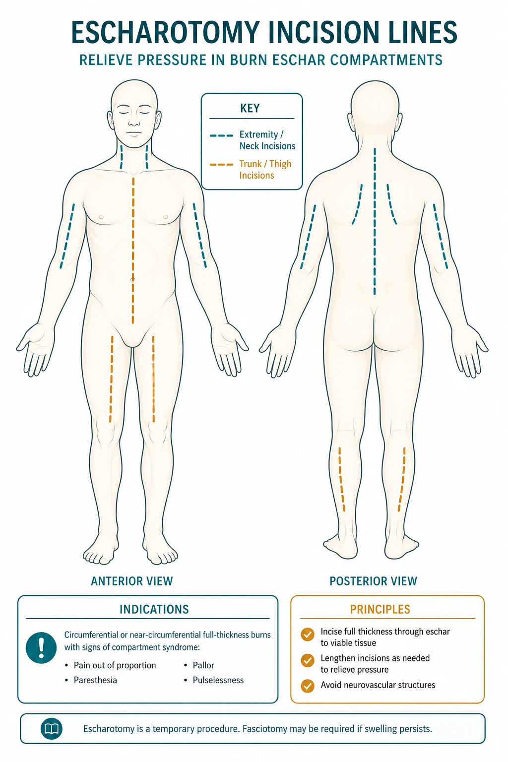
The escharotomy sites — chest wall and limbs
Chest wall
Anterior axillary lines — bilateral
- Circumferential full-thickness chest burn → rigid eschar encases the thorax like a cast → restricted chest wall expansion
- Clinical: rising peak airway pressure (volume control), falling tidal volume (pressure control), falling SpO2, increasing work of breathing
- Incision lines: bilateral, along the mid-axillary OR anterior axillary lines, from the clavicle to the costal margin; may connect subcostally across the upper abdomen
- Effect: immediate fall in peak airway pressure, restoration of tidal volume, improved chest wall compliance
- Often performed PROPHYLACTICALLY before intubation in the major burn with the circumferential chest eschar
Upper limb
Midlateral / mid-medial lines
- Circumferential full-thickness burn → the 6 Ps (see below) → the arterial compromise
- Arm: midlateral line (lateral bicipital groove) — avoids the radial nerve in the spiral groove and the ulnar nerve at the elbow
- Forearm: midlateral and mid-medial (volar-radial and ulnar) lines — release both the flexor and extensor compartments
- Hand/fingers: midlateral line of the digits (the non-contact surface) to spare the neurovascular bundles; the dorsum of the hand may need release
- Extend the FULL length of the eschar — from the proximal unburned skin to the distal unburned skin; a short incision is an inadequate release
Lower limb
Midlateral lines — bilateral
- Thigh: midlateral line; the anterolateral and the posteromedial if bilateral release needed
- Leg: midlateral line along the lateral border of the tibia (anterior) AND the posteromedial line — releases the anterior, lateral, and deep/superficial posterior compartments
- CAUTION: the common peroneal nerve at the fibular neck, the posterior tibial neurovascular bundle behind the medial malleolus — avoid
- Foot/toes: midlateral digital release; the dorsum of the foot
- Bilateral release (both sides) is usually required for an adequate circumferential limb decompression
Abdomen / neck / penis
Special sites — specialist
- Abdomen: midline or bilateral subcostal; rarely needed alone — usually continuous with the chest escharotomy
- Neck: midlateral — CAUTION (carotid, internal jugular, vagus, trachea); the specialist
- Penis: dorsal midline / lateral release for the glans ischaemia; the rare
- These sites have critical structures immediately deep to the eschar — the judicious, the experienced operator
The 6 Ps of the limb ischaemia (the classic — the progression)
The circumferential eschar of the limb produces the ischaemic progression — the classic "6 Ps" — but in the burn patient the SEQUENCE and the RELIABILITY differ from the trauma compartment syndrome, because the burn oedema and the resuscitation drive a RAPID progression and the awake burn patient may be distracted by the pain of the burn itself.[1][1]
- Pain — the pain on passive stretch (the earliest); often obscured by the burn pain, the analgesia, and the insensate full-thickness burn overlying the ischaemic muscle.
- Pallor — the cool, the pale, the delayed capillary refill (more than 3 seconds).
- Paraesthesia — the pins-and-needles in the distribution of the compromised nerve (the median, the ulnar, the peroneal).
- Paralysis — the loss of the active movement (the LATE sign — the motor nerve and the muscle ischaemia).
- Poikilothermia — the cool to the touch (the arterial inflow compromise).
- Pulselessness — the LATEST sign — the Doppler signal loss = the arterial occlusion = the imminent irreversible ischaemia. Do NOT wait for this — by the time the pulse is lost, the window for salvage is closing.[1]
The burn-specific caveat: the most RELIABLE early monitor in the burn patient is the continuous pulse oximetry on the involved digit and the hourly handheld Doppler of the peripheral pulses — not the pain (unreliable in the analgosedated patient) and not the pulselessness (too late).[1][1]
The technique — the bedside escharotomy
The escharotomy is performed at the bedside on the ICU or the burns unit. The preparation is rapid. The full-thickness eschar is insensate (the nerve endings in the dermis are destroyed) — anaesthesia of the eschar itself is NOT required. The subcutaneous fat and the deeper tissues ARE sensate, so the patient receives the analgesia (the opioid, the ketamine) and the local anaesthetic infiltration at the proximal and distal unburned margins. The aseptic (not the full sterile) technique is standard.[1][1][1]
The preparation:
- The patient supine; the limb exposed and supported; the electrocautery and the blunt-tipped scissors available; the diathermy pad applied (the electrocautery for the haemostasis — the eschar vessels are largely thrombosed so the bleeding is minimal, but the subcutaneous vessels bleed).[1]
- The analgesia: the IV opioid (the fentanyl, the morphine) ± the low-dose ketamine. The local anaesthetic (the lidocaine 1% with adrenaline) infiltrated at the proximal and distal ends of the planned incision where the unburned skin is sensate.[1]
- The aseptic prep (the povidone-iodine or the chlorhexidine); the sterile drapes around the field.[1]
The incision:
- The scalpel (no. 10 or 15 blade) or the electrocautery; the incision along the chosen line (the midlateral limb, the anterior axillary chest).[1]
- The incision passes through the full thickness of the eschar — the leathery necrotic skin — and INTO the subcutaneous fat. The endpoint of an adequate incision is the visible separation of the eschar edges and the bulging of the subcutaneous fat through the defect (the "mushrooming" — the sign of the release).[1][1]
- The incision extends the FULL length of the circumferential eschar — from the proximal margin of unburned (or partial-thickness) skin to the distal margin — passing across the joints (the elbow, the wrist, the knee, the ankle) as needed. A short incision is an inadequate release; the constriction persists.[1]
- The bilateral incisions (the midlateral AND the mid-medial, OR the anterior AND the posterior) are usually required for the full circumferential release of a limb.[1]
- The chest: the bilateral anterior axillary line incisions from the clavicle to the costal margin, joined subcostally if the abdomen is involved. The effect on the ventilated patient is immediate — the peak airway pressure falls, the tidal volume rises.[1][1]
The haemostasis and the aftercare:
- The bleeding is usually minimal (the eschar vessels thrombosed); the subcutaneous bleeders are controlled with the electrocautery. The profound bleeding suggests the incision too deep (into the muscle) or a coagulopathy.[1]
- The wound is dressed with the non-adherent dressing (the silver sulfadiazine, the nano-crystalline silver, the Vaseline gauze); the limb is elevated to reduce the oedema.[1]
- The post-escharotomy monitoring: the continuous pulse oximetry, the hourly Doppler, the capillary refill, the limb warmth — to confirm the adequate release. If the perfusion does not improve, consider the inadequate release (extend the incision) OR the compartment syndrome beneath (the fasciotomy).[1][1]
The key technical pearls
- Anaesthesia NOT required for the eschar — the full-thickness burn is insensate. But the analgesia for the subcutaneous fat and the patient's overall comfort is humane and standard.[1]
- Incise to the fat, not into the muscle — the escharotomy releases the skin/eschar only. Cutting into the muscle is the fasciotomy (a different operation) and risks the bleeding and the damage to the neurovascular structures.[1]
- Full length, full release — the incision must traverse the entire circumferential eschar. The "split not enough" is the common error; if the perfusion does not improve, extend the incision.[1]
- Beware the nerves at the joints — the ulnar nerve at the elbow (the medial epicondyle), the radial nerve in the spiral groove (the posterolateral arm), the common peroneal nerve at the fibular neck, the posterior tibial nerve behind the medial malleolus. The midaxillary/midlateral lines are designed to AVOID these.[1]
- The escharotomy does NOT release the deep fascial compartments — if the deep muscle oedema (the electrical injury, the profound fluid resuscitation) is the problem, the patient needs the FASCIOTOMY in addition.[1][1]
The chest wall escharotomy — to allow ventilation
The circumferential full-thickness burn of the chest wall is a respiratory emergency in disguise. The rigid eschar encases the thorax like a plaster cast; as the tissue swells from the resuscitation and the patient is placed on the positive-pressure ventilation, the chest wall compliance plummets.[1][1]
The clinical signs in the ventilated patient:
- The rising peak airway pressure (the volume-control mode) — the chest wall cannot expand to accept the set tidal volume.
- The falling tidal volume (the pressure-control mode) — the same mechanism, the same rigid encasement.
- The falling SpO2; the rising PaCO2; the increasing work of breathing in the spontaneously breathing patient.
- The abdominal component (the subcostal extension of the eschar) restricts the diaphragmatic excursion and compounds the problem.[1][1]
The incision: the bilateral incisions along the anterior axillary lines, from the clavicle/second intercostal space down to the costal margin, with a subcostal connecting incision if the abdomen is involved. The release is immediate and dramatic — the peak airway pressure falls, the tidal volume rises, the chest wall "gives."[1][1]
The prophylactic chest escharotomy: in the major burn with the circumferential chest eschar who WILL be intubated (the inhalation injury, the >40% TBSA, the facial burn, the altered mental state), perform the chest escharotomy BEFORE or AT the time of the intubation. The positive-pressure ventilation against an encased thorax is the recipe for the barotrauma, the hypoxaemia, and the cardiovascular compromise — the prophylactic release averts this.[1]
The compartment syndrome — the deep muscle oedema
The deep muscle oedema (from the burn + the fluid resuscitation + especially the electrical injury) increases the pressure within the fascial compartments:[1][1][1]
The escharotomy releases only the overlying skin/eschar. The muscle within the unyielding fascial compartments continues to swell — from the direct thermal injury, the reperfusion, the inflammatory mediators, and (critically) the large-volume crystalloid resuscitation. When the intracompartmental pressure rises above the perfusion pressure (the diastolic pressure minus the compartment pressure gives the perfusion pressure / delta pressure — the threshold for the fasciotomy is a delta pressure of LESS THAN 30 mmHg), the muscle and the nerve within the compartment become ischaemic.[1][1]
- The limb compartment syndrome: the pain on passive stretch (the earliest), the tense/swollen, the paraesthesia, the pulselessness (the late).[1]
- The management: the FASCIOTOMY (not the escharotomy — the fascial release). The escharotomy only cuts the eschar; the fasciotomy releases the deep compartments. The electrical injury especially needs the fasciotomy (the deep muscle oedema).[1][1]
Escharotomy versus fasciotomy — the distinction
Escharotomy
Skin/eschar release
- Pathology: circumferential full-thickness BURN (rigid eschar = tourniquet)
- Structure cut: the full-thickness eschar down to the subcutaneous fat ONLY
- Depth: skin + eschar; the fascia is NOT opened
- Anaesthesia: the eschar is insensate — anaesthesia NOT required (analgesia for the subcutaneous fat is humane)
- Bleeding: minimal (the eschar vessels thrombosed)
- Indication: the circumferential full-thickness burn with the perfusion/ventilation compromise
- Setting: the bedside, the ICU/burns unit
Fasciotomy
Deep fascial compartment release
- Pathology: compartment SYNDROME (the deep muscle oedema within the fascial compartment)
- Structure cut: the skin, the subcutaneous fat, AND the deep fascia overlying the muscle compartment
- Depth: down to and through the fascia — the muscle bulges out
- Anaesthesia: the fascia and muscle ARE sensate — general/regional anaesthesia usually required
- Bleeding: more (the viable subcutaneous and muscle vessels)
- Indication: the compartment syndrome (especially the electrical injury, the fluid resuscitation, the deep burn)
- Setting: usually the operating theatre; may be bedside in the emergency
The burn patient with the circumferential full-thickness burn AND the deep muscle involvement (the electrical injury, the high-voltage contact, the crush) often needs BOTH — the escharotomy to release the skin, the fasciotomy to release the deep compartments. The electrical injury is the classic indication for the early, the generous, the multiple-compartment fasciotomy (the deep muscle oedema outpaces any escharotomy).[1][1]
The compartment syndrome from the fluid resuscitation
The fluid resuscitation (the crystalloid, the Parkland 4 mL/kg/%TBSA) is the iatrogenic driver of the compartment syndrome in the major burn. The crystalloid leaks into the interstitium (the capillary leak, the hypoalbuminaemia); the muscle swells within the fascial compartments; the compartment pressure rises; the perfusion pressure falls; the muscle and the nerve become ischaemic.[2][3]
The prevention — the careful resuscitation:
- The guideline-adherent resuscitation (the Brooke/modified Brooke, the Parkland) — titrated to the urine output (0.5 mL/kg/h adult, 1 mL/kg/h child).[2]
- The early colloid (the albumin at 8–12 hours, the fresh frozen plasma) — reduces the total crystalloid, the "fluid creep."[2]
- The high-volume resuscitation (the greater than 6 mL/kg/%TBSA) is the RED FLAG for the fluid creep — the risk of the abdominal compartment syndrome AND the limb compartment syndrome.[2]
- The monitoring of the bladder pressure (the intra-abdominal pressure surrogate) in the major burns (the greater than 30% TBSA) for the early abdominal compartment syndrome.[4]
The management of the established limb compartment syndrome from the fluid resuscitation: the FASCIOTOMY. The escharotomy alone will not suffice — the deep fascial compartments must be opened. The high index of suspicion in the patient with the rising fluid requirement, the tense limb, the falling urine output (the oliguria from the abdominal compartment syndrome) — the compartment pressure measurement (the delta pressure less than 30 mmHg) — the fasciotomy.[1][1]
The abdominal compartment syndrome
The fluid creep (the over-resuscitation) → the bowel and retroperitoneal oedema → the intra-abdominal pressure (IAP) rises:[2][1][1][4]
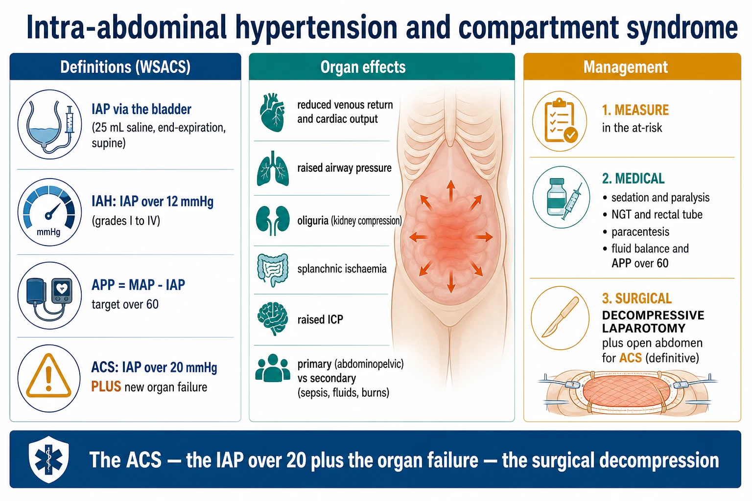
The abdominal compartment syndrome is the sustained intra-abdominal pressure ABOVE 20 mmHg with NEW organ dysfunction (the renal — the oliguria, the rising creatinine; the respiratory — the rising airway pressure, the falling tidal volume, the hypoxaemia from the cephalad diaphragm; the cardiovascular — the reduced venous return, the low cardiac output, the raised CVP that does not reflect the volume status). The WSACS definitions:[4]
- Intra-abdominal hypertension (IAH): the sustained IAP above 12 mmHg (Grade I 12–15, II 16–20, III 21–25, IV above 25 mmHg).[4]
- Abdominal compartment syndrome (ACS): the sustained IAP above 20 mmHg WITH new organ dysfunction (the renal, the respiratory, the cardiovascular, the splanchnic).[4]
- The APP (abdominal perfusion pressure) = MAP − IAP — the target APP above 50–60 mmHg (the analogue of the cerebral perfusion pressure).[4]
The measurement of the IAP: the intravesical (the bladder) pressure — the gold standard, the bedside, the inexpensive. The Foley catheter is clamped, 25 mL of sterile saline instilled, the pressure transduced via the catheter at the symphysis pubis (the end-expiratory, the supine). The serial measurement (every 4–6 hours) in the major burn at risk.[1][4]
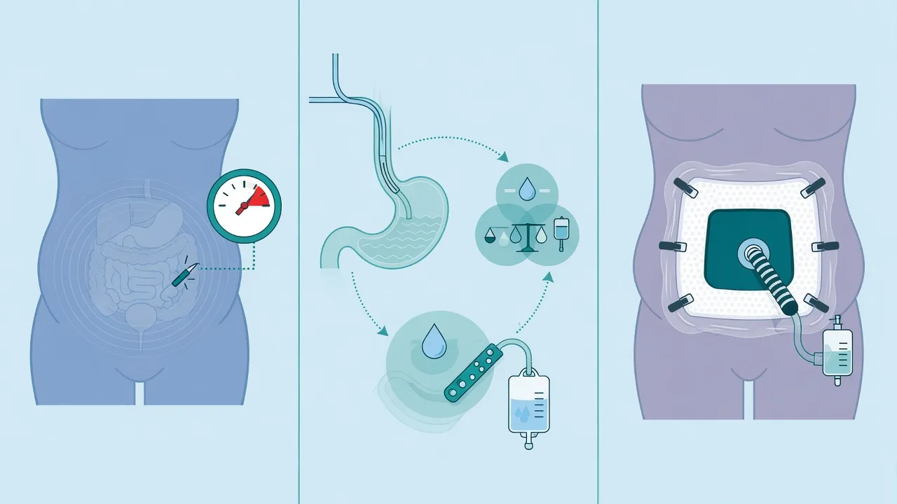
The management of the abdominal compartment syndrome:[2][1][4]
- The reduce the fluid — the colloid (the albumin), the diuretic (the furosemide) if the volume-overloaded; the avoidance of the further crystalloid.
- The decompression — the surgical laparostomy (the open abdomen) if the severe (the IAP above 25, the refractory organ dysfunction), with the temporary abdominal closure (the negative-pressure dressing, the Bogota bag). The surgical decompression is the definitive but the morbid (the open abdomen, the entero-cutaneous fistula, the planned re-closure).
- The supportive — the vasopressor to maintain the APP (the noradrenaline), the renal replacement therapy for the AKI, the lung-protective ventilation adjusted for the reduced chest wall compliance.
The monitoring of the circumferential burn
The circumferential full-thickness burn demands the continuous, the vigilant, the hourly monitoring for the evolving compromise — from the time of the admission until the eschar separates or the escharotomy/fasciotomy is performed. The intensivist who waits for the pulselessness waits too long.[1][1]
- The peripheral Doppler — the hourly handheld Doppler of the radial, ulnar, posterior tibial, dorsalis pedis pulses; the loss of the signal = the urgent.[1]
- The continuous pulse oximetry — on the involved digit/limb; the loss of the trace = the perfusion compromise.[1]
- The capillary refill — the distal nailbed; the delayed (more than 3 seconds) = the venous/arterial compromise.[1]
- The limb temperature and the colour — the cool, the pale, the mottled = the arterial compromise.[1]
- The limb firmness — the tense, the woody, the non-compliant = the compartment syndrome (measure the compartment pressure).[1]
- The ventilated patient with the chest eschar — the continuous airway pressure, the tidal volume, the SpO2; the rising peak pressure / falling tidal volume = the chest escharotomy.[1][1]
- The major burn at risk of the ACS — the serial bladder pressure (every 4–6 hours), the urine output (the oliguria = the early sign), the airway pressure (the rising = the diaphragmatic compression).[4]
Escharotomy & compartment decompression in the burn patient — the ICU workflow
Recognise the risk
On admission, identify EVERY circumferential or near-circumferential full-thickness burn of a limb, the chest, the abdomen, the penis, the neck. Document the depth (the leathery, dry, insensate, non-blanching full-thickness), the circumference, and the proximal/distal extent. This patient is at risk of the tourniquet effect — plan the monitoring from the start.
Set up the continuous monitoring
Continuous pulse oximetry on the involved digit; hourly handheld Doppler of the radial/ulnar/tibial/dorsalis pedis pulses; hourly capillary refill, limb temperature and colour; for the chest burn, the continuous airway pressure and tidal volume (if ventilated). For the major burn (greater than 30% TBSA), the serial bladder pressure every 4–6 hours. The intensivist who waits for the pulselessness waits too long.
Anticipate the swelling
The fluid resuscitation (the Parkland 4 mL/kg/%TBSA, the crystalloid) WILL swell the limb over the first 24 hours. The high-volume resuscitation (greater than 6 mL/kg/%TBSA), the inhalation injury, the delayed resuscitation, the electrical injury = the higher risk of the fluid creep, the compartment syndrome, and the abdominal compartment syndrome. Titrate the fluids to the urine output (0.5 mL/kg/h), consider the early colloid.
Identify the indication for the escharotomy
The clinical trigger: the decreased capillary refill, the cool/pale limb, the slow venous filling, the pulse oximetry loss, the paraesthesia, OR the rising airway pressure / falling tidal volume in the chest burn. The Doppler-pulse loss = the LATE sign = the URGENT — do not wait for this. In the high-risk patient, the prophylactic escharotomy at the time of the resuscitation (the chest BEFORE the intubation).
Perform the escharotomy at the bedside
Analgesia (IV opioid, low-dose ketamine); the local anaesthetic at the sensate margins (the eschar itself is insensate — anaesthesia NOT required). Aseptic prep. The scalpel or the electrocautery along the chosen line (the midlateral limb, the anterior axillary chest). Incise through the full-thickness eschar INTO the subcutaneous fat. The endpoint: the separation of the eschar edges, the bulging of the fat (the mushrooming). The FULL length of the eschar; the bilateral release for the full circumferential limb. The chest: the bilateral anterior axillary lines, clavicle to costal margin.
Confirm the adequate release
The immediate reassessment: the Doppler signal returns, the capillary refill improves, the limb warms, the pulse oximetry trace returns. For the chest: the peak airway pressure falls, the tidal volume rises. If the perfusion does NOT improve, the release is INADEQUATE — extend the incision. If the perfusion still does not improve, suspect the compartment syndrome beneath — measure the compartment pressure (the delta pressure less than 30 mmHg) and proceed to the FASCIOTOMY.
Add the fasciotomy if the deep compartment is involved
The deep muscle oedema (the electrical injury, the high-voltage contact, the profound fluid resuscitation) is NOT relieved by the escharotomy — the deep fascia must be opened. The clinical trigger: the tense/woody limb, the pain on passive stretch, the rising creatine kinase, the myoglobinuria. The fasciotomy: the skin, the subcutaneous fat, AND the deep fascia over each involved compartment; the muscle bulges out; the general/regional anaesthesia (the fascia and muscle ARE sensate). The electrical injury: the early, the generous, the multiple-compartment fasciotomy.
Manage the abdominal compartment syndrome
In the major burn with the rising fluid requirement and the falling urine output, the serial bladder pressure (every 4–6 hours). The IAP above 20 mmHg with new organ dysfunction (the oliguria, the rising airway pressure, the low cardiac output) = the ACS. The management: reduce the fluid (the colloid, the diuretic), the supportive (the vasopressor for the APP above 50–60, the RRT for the AKI), the surgical decompression (the laparostomy) if the severe and refractory.
Aftercare and the dressings
The escharotomy/fasciotomy wound dressed with the non-adherent dressing (the silver sulfadiazine, the nano-crystalline silver); the limb elevated to reduce the oedema; the continued hourly monitoring to confirm the sustained release. The fasciotomy wounds managed for the delayed closure or the skin grafting once the swelling resolves. The physiotherapy to prevent the contracture; the nutritional support for the wound healing.
Prognosis
The prompt escharotomy restores the perfusion and the ventilation. The delay → the ischaemia (the amputation, the nerve damage), the respiratory failure. The compartment → the irreversible muscle/nerve damage if not decompressed within 6 hours.[1][1][1]
The outcome of the escharotomy is determined by the TIMING — the early (the prophylactic or the early clinical signs) restores the perfusion with the minimal tissue loss; the late (after the pulselessness, after the irreversible ischaemia) leaves the amputation, the Volkmann contracture, the nerve palsy. The compartment syndrome decompressed within 4–6 hours recovers; beyond 6–8 hours the irreversible muscle necrosis and the rhabdomyolysis (the myoglobinuria, the AKI). The abdominal compartment syndrome decompressed promptly recovers; the delay → the multiple organ failure, the mortality up to 50–70% in the untreated.[1][1][4]
The burn patient who needs BOTH the escharotomy and the fasciotomy (the electrical injury, the high-voltage contact) has the worst prognosis — the deep muscle involvement, the myoglobinuric AKI, the risk of the amputation. The meticulous monitoring, the early decompression, and the aggressive management of the rhabdomyolysis (the hydration, the alkaline diuresis, the RRT) determine the limb salvage.[1][1]
Evidence and the key references
Burns escharotomy, compartment syndrome, and the abdominal compartment syndrome — the evidence
Ibrahim 2006 (Burns): the decompression (the escharotomy AND the fasciotomy together) in the acute burn — the deep muscle oedema often needs the fasciotomy in addition to the escharotomy; the escharotomy alone is insufficient in the electrical and the deep burns.[1] Kumar 2023 (J Burn Care Res): the adherence to the burn resuscitation guidelines reduces the total fluid volume (the fluid creep) — and by extension the rate of the abdominal compartment syndrome and the limb compartment syndrome. The guideline-adherent resuscitation (the titrated crystalloid, the early colloid) is the primary prevention of the compartment syndromes.[2] Monafo 1996 (NEJM): the classic — the initial treatment of burns; the fluid resuscitation (the Parkland, the Brooke), the recognition of the circumferential burn and the escharotomy, the monitoring of the perfusion. The foundation of the modern burn care.[3] Kirkpatrick 2013 / WSACS (Intensive Care Med): the updated consensus definitions of the intra-abdominal hypertension (the IAP above 12) and the abdominal compartment syndrome (the IAP above 20 with the organ dysfunction); the abdominal perfusion pressure (the MAP minus the IAP, the target above 50–60); the intravesical (the bladder) pressure as the standard measurement; the medical and the surgical management. The reference for the ACS in the burn patient.[4] The principle: the escharotomy and the fasciotomy are the BEDSIDE emergencies — the early (the prophylactic or the early clinical signs) restores the perfusion and the ventilation; the late (after the pulselessness, after the irreversible ischaemia) leaves the amputation, the contracture, the nerve palsy, the mortality.[1][1]
Short answer questions
SAQ — Circumferential chest wall burn with ventilation compromise
10 minutes · 10 marks
A 52-year-old man is admitted to the ICU after a house fire with a 45 per cent TBSA flame burn that is circumferential and full-thickness across the entire anterior and lateral chest and upper abdomen. He was intubated in the ED for inhalation injury and is now ventilated in volume-control mode (set tidal volume 450 mL). Forty minutes after admission, during the initial Parkland resuscitation, the peak airway pressure rises from 28 to 52 cmH2O, the exhaled tidal volume falls to 250 mL, SpO2 drops to 88 per cent, and the BP falls to 84/50 with a CVP of 24 mmHg. The chest is firm, hard and unyielding to palpation. Blood gas: pH 7.18, PaCO2 68, PaO2 56.
SAQ — Compartment syndrome in a high-voltage electrical burn
10 minutes · 10 marks
A 28-year-old electrician is brought to the ED 40 minutes after contacting an 11 000 V overhead AC line with his right hand. He is in VF at the scene, is defibrillated with 200 J and regains a perfusing rhythm. On arrival he is intubated, GCS 8, HR 120, BP 102/64. There is a small charred entry wound on the right palm and a large, irregular exit wound on the right forearm and antecubital fossa. The right forearm is tense, woody and exquisitely tender; capillary refill in the fingers is 6 seconds and the pulse oximetry trace on the right index finger is lost. CK is 22 000 U/L, K+ 6.4 mmol/L, creatinine 142 micromol/L, and the urine is dark red-brown. There is no circumferential full-thickness cutaneous burn on the forearm.
Clinical pearls
Red flags
References
- [1]Ibrahim AE, et al. Decompression not escharotomy in acute burns Burns, 2006.PMID 16527416
- [2]Kumar R, et al. Adherence to Burn Resuscitation Guidelines Reduces Resuscitation Fluids and Mortality J Burn Care Res, 2023.PMID 35709512
- [3]Monafo WW, West MA. Exacerbation of inflammation-associated colonic injury in rat through inhibition of cyclooxygenase-2 J Clin Invest, 1996.PMID 8903327
- [4]Kirkpatrick AW, Roberts DJ, De Waele J, et al. Role of (18)F-FDG PET-CT in head and neck squamous cell carcinoma Acta Otorhinolaryngol Ital, 2013.PMID 23620633