ICU · Burns
Inhalation Injury & Carbon Monoxide in Burns
Also known as Smoke inhalation injury · Airway burn · Thermal airway injury · Chemical pneumonitis smoke · Carbon monoxide poisoning burn · Bronchoscopy inhalation injury · Supraglottic thermal oedema · Subglottic chemical tracheobronchitis · Cyanide toxicity from smoke
Smoke inhalation injury is the leading cause of death at the fire scene and, in the burn unit, DOUBLES the mortality of a given cutaneous burn. It is anatomically and mechanistically THREE distinct injuries layered on one exposure: (1) SUPRAGLOTTIC THERMAL injury — heat is dissipated above the vocal cords so direct thermal damage is confined to the supraglottic airway, where progressive oedema over 24–48 h threatens complete obstruction → the imperative for EARLY INTUBATION before the airway becomes impossible; (2) SUBGLOTTIC CHEMICAL injury — smoke carries water-soluble toxins (hydrogen chloride, ammonia) that deposit in the upper airway and lipid-soluble toxins (phosgene, nitrogen dioxide, acrolein, aldehydes) that reach the alveoli, causing bronchospasm, mucosal sloughing, fibrin cast formation and ARDS; (3) SYSTEMIC TOXICITY — carbon monoxide (CO) and cyanide (CN) generated by incomplete combustion in enclosed spaces; CO produces carboxyhaemoglobin (pulse oximetry is FALSELY NORMAL — use a CO-oximeter), cyanide blocks cytochrome c oxidase (suspect when lactate >10 mmol/L). Diagnosis rests on clinical predictors + bronchoscopy (soot, erythema, oedema, mucosal necrosis, casts) + carboxyhaemoglobin level + lactate. Management: early intubation, 100% oxygen, hydroxocobalamin for cyanide, lung-protective ventilation, bronchodilators + airway toilet (nebulised heparin/NAC), and a 30–40% upward adjustment of fluid resuscitation.
On this page & tools
Your progress
Saved locally on this device.
Target exams
Red flags
Overview & definition
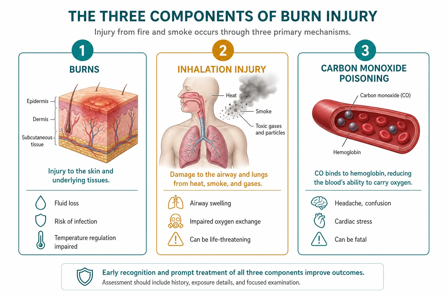
The inhalation the injury in the burns — the three the components: the upper the airway the thermal, the lower the airway the chemical, the systemic the CO / the cyanide. The doubles the burn the mortality. The recognise the early (the soot, the singed the hair, the enclosed-the-space the fire) → the early the intubation.[1][3]
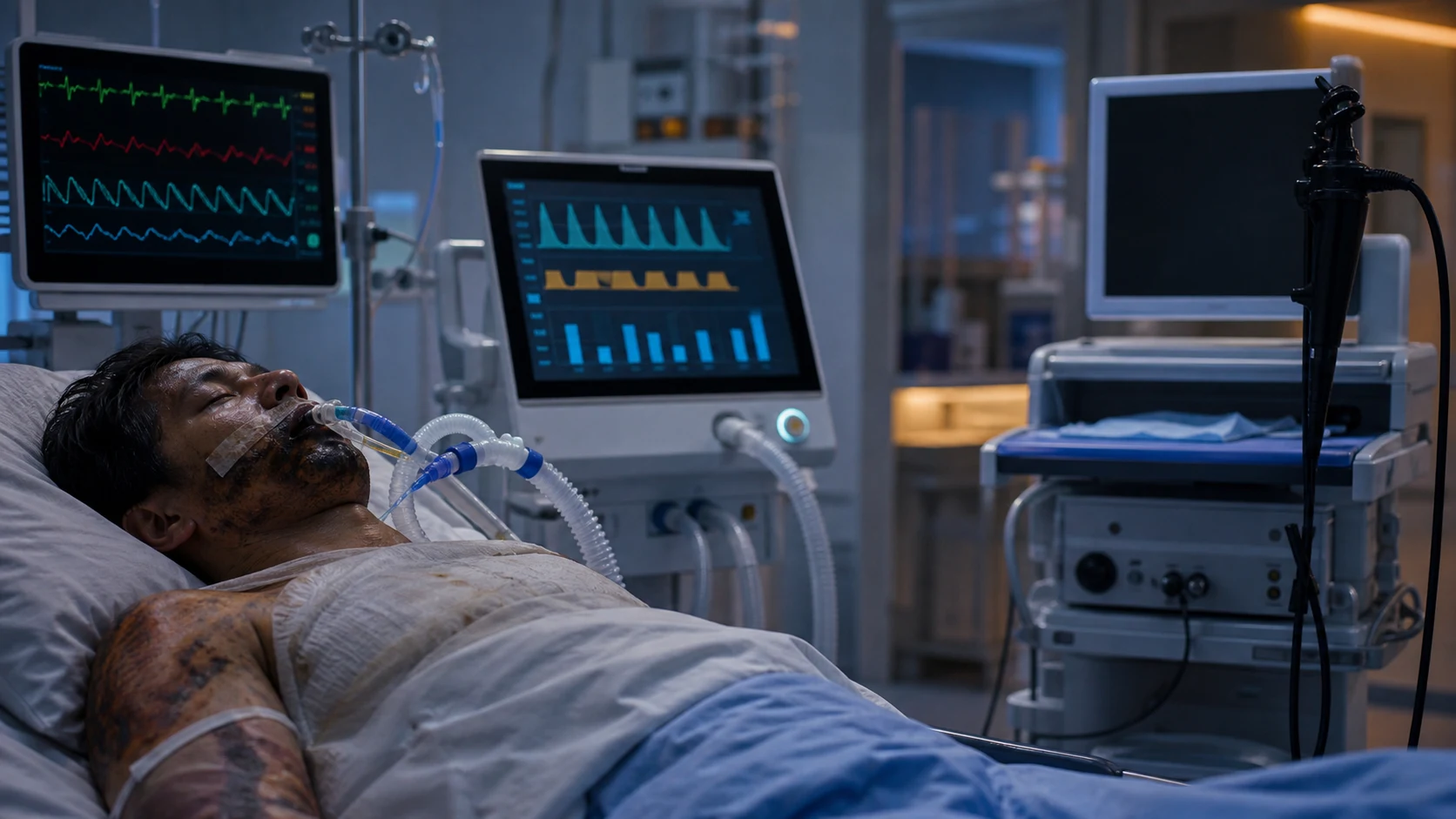
Smoke inhalation is a time-critical, multi-compartment injury that kills more fire victims than the cutaneous burn itself: most fire-related deaths occur at the scene from CO/cyanide and upper-airway obstruction, not from the burn wound. Among patients who reach a burn centre, concomitant inhalation injury doubles the mortality of an equivalent-sized cutaneous burn and adds a 30–40% surcharge to fluid requirements. Three anatomically and mechanistically distinct processes coexist after any significant smoke exposure and each demands a specific intervention: [1]
- Supraglottic thermal injury — heat is a poor penetrant of gas; the upper airway (nasopharynx, oropharynx, supraglottic larynx) acts as a heat exchanger that cools dry inspired gas below the vocal cords. Thermal damage is therefore largely confined above the cords, producing progressive mucosal oedema that peaks at 24–48 h and can occlude the laryngeal inlet → the single most important reason to intubate before the airway becomes impossible.
- Subglottic chemical tracheobronchitis / pneumonitis — combustion generates a toxic aerosol. Water-soluble irritants (HCl, ammonia, sulphur dioxide) deposit in the upper airway causing laryngospasm and bronchospasm; lipid-soluble toxins (phosgene, nitrogen dioxide, acrolein, the aldehydes) penetrate to the alveoli, denaturing surfactant, sloughing mucosa, forming fibrin casts, and triggering ARDS over 24–72 h.
- Systemic asphyxiant toxicity — CO and cyanide — incomplete combustion in an enclosed space generates CO (binds haemoglobin with ~210x the affinity of O2) and cyanide (combustion of wool, silk, polyurethane, plastics — CN blocks cytochrome c oxidase). Pulse oximetry is falsely normal in CO poisoning; a high lactate (>10 mmol/L) is the bedside surrogate for cyanide.[2][1] />[1] />
The three the components
1. The upper the airway the thermal the injury
The thermal the injury the limited to the supraglottic the area (the airway the cools the inspired the gas the below the vocal the cords — the EXCEPT the steam, the which the carries the more the heat).[1][3]
- The progressive the oedema (the 24 to the 48 h) → the airway the obstruction.[1][3]
- The signs (the soot, the singed the hair, the hoarse, the stridor).[3]
- The the early the intubation (the before the difficult).[1][3]
2. The lower the airway the chemical the pneumonitis
The smoke the toxins (the acrolein, the hydrogen the chloride, the aldehydes, the ammonia) → the chemical the pneumonitis.[1][3]
- The bronchospasm, the mucosal the sloughing, the cast the formation, the V/Q the mismatch.[1][3]
- The ARDS (the 24 to the 72 h).[3][4]
- The the bronchoscopy (the diagnostic — the soot, the erythema, the oedema, the mucosal the necrosis, the casts).[1][3]
3. The systemic the CO / the cyanide
The enclosed-the-space the fire → the CO + the cyanide.[2][3]
- The CO: the pulse the ox the the falsely the normal (the use the CO-the oximeter). The 100 per cent the oxygen; the HBO for the severe (the see the Toxicology #10 the topic).[2][3]
- The cyanide: the hydroxocobalamin (the see the Toxicology #10).[3][4]
The three components of smoke inhalation injury — anatomy, mechanism, and signature management
| Feature | Supraglottic thermal injury | Subglottic chemical injury | Systemic CO / cyanide toxicity |
|---|---|---|---|
| Anatomical level | Naso/oropharynx, supraglottic larynx (above the cords) | Tracheobronchial tree + alveoli (below the cords) | Systemic — blood + mitochondria |
| Mechanism | Direct heat (dry gas cooled above cords) | Combustion toxins — water-soluble deposit high, lipid-soluble reach alveoli | CO binds Hb (210x O2 affinity); CN blocks cytochrome c oxidase |
| Onset / time course | Oedema progressive, peaks 24–48 h | Bronchospasm immediate; ARDS 24–72 h | Immediate; COHb half-life drives timeline |
| Hallmark signs | Hoarseness, stridor, drooling, soot in mouth, singed nasal hairs | Wheeze, cough, carbonaceous sputum, hypoxaemia, falling compliance | Cherry-red skin (late/rare), confusion, coma, lactate >10 |
| Diagnostic test | Laryngoscopy/bronchoscopy (supraglottic oedema) | Bronchoscopy (soot, erythema, casts, mucosal necrosis) | CO-oximeter (COHb%); blood lactate; whole-blood cyanide (slow) |
| Pulse oximetry | Normal (until obstruction) | May fall (V/Q mismatch) | Falsely normal (COHb indistinguishable from O2Hb) |
| Signature treatment | EARLY intubation (before oedema) | Lung-protective ventilation; bronchodilators; nebulised heparin/NAC; airway toilet | 100% O2 (CO); hydroxocobalamin (CN); consider HBO |
| Key risk if missed | Can't-intubate / can't-ventilate | ARDS, pneumonia, respiratory failure | Death / anoxic brain injury; delayed neuro sequelae |
Carbon monoxide vs cyanide toxicity in smoke inhalation — differentiating the two asphyxiants
| Feature | Carbon monoxide (CO) | Cyanide (CN) |
|---|---|---|
| Source | Incomplete combustion of any organic material (the dominant toxicant of smoke) | Combustion of wool, silk, polyurethane, plastics, synthetic furnishings |
| Mechanism | Binds haemoglobin → carboxyhaemoglobin (COHb); ~210x the affinity of O2 → functional anaemia + left-shift of dissociation curve | Binds cytochrome a3 (cytochrome c oxidase) → blocks mitochondrial oxidative phosphorylation → histotoxic hypoxia |
| Lactate | Mild–moderate elevation | Marked elevation; lactate >10 mmol/L strongly suggests CN (Baud 1991) |
| Venous O2 | Low (tissue extraction) | High (cells cannot consume O2 — histotoxic block) → narrowed arteriovenous O2 difference |
| Pulse oximetry | Falsely normal (COHb reads as O2Hb) | Normal (or low if CO coexists) |
| CO-oximetry | COHb elevated (diagnostic) | COHb may be normal; direct blood CN assay is definitive but slow |
| Classic (rare) sign | Cherry-red skin; "red venous blood" | Bitter-almond breath (unreliable); same red venous blood from high venous O2 |
| Bedside surrogate | COHb level (CO-oximeter) | Lactate >10 mmol/L + depressed consciousness after enclosed-space fire |
| Definitive treatment | 100% oxygen (↓COHb half-life 320 min → ~80 min); HBO for severe | Hydroxocobalamin (Cyanokit) — binds CN → cyanocobalamin; sodium thiosulphate as co-therapy; 100% O2 supportive |
| Onset | Immediate; symptoms at COHb 15–20%, severe >30–40% | Rapid; coma, seizures, cardiovascular collapse |
| Coexistence | Common — CO and CN almost always co-exist in enclosed-space smoke exposure; treat empirically for CN if lactate >10 or refractory shock | Common — same fire; do not wait for CN level |
Pathophysiology — why smoke is so destructive
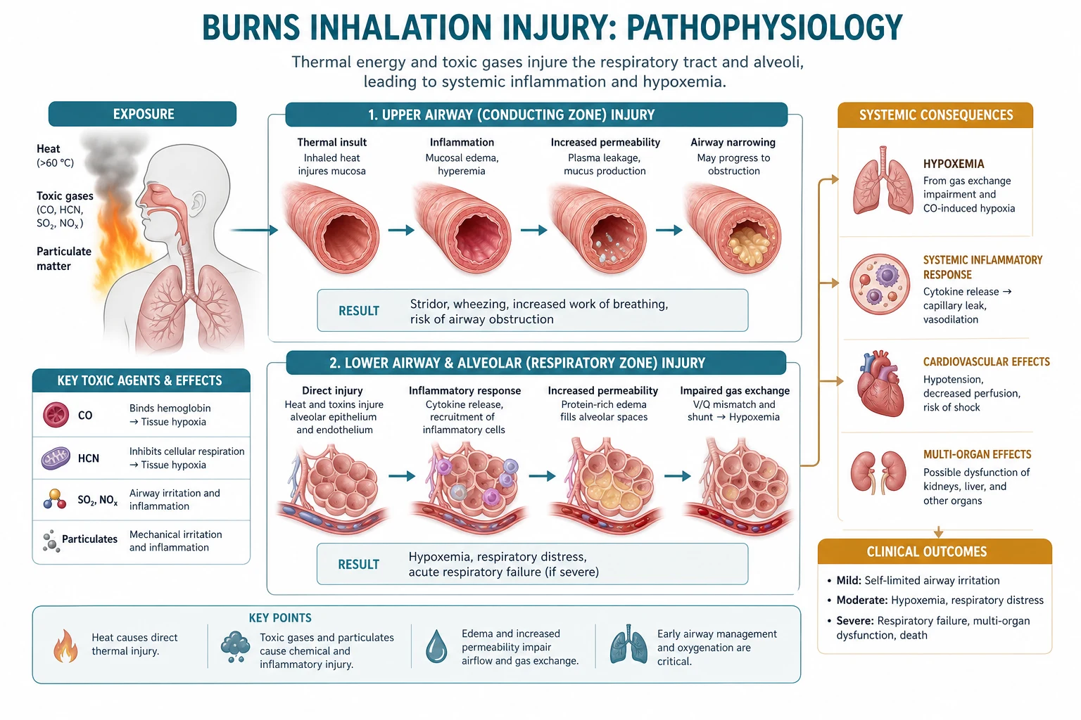
Water-soluble vs lipid-soluble smoke toxins — where they deposit
| Property | Water-soluble irritants (HCl, NH3, SO2, chlorine) | Lipid-soluble toxins (phosgene, NO2, acrolein, aldehydes) |
|---|---|---|
| Site of deposition | Upper airway / proximal tracheobronchial tree | Distal airways + alveoli |
| Solubility behaviour | Dissolve in moist mucosa → high proximal injury | Penetrate deep, dissolve slowly → delayed alveolar injury |
| Symptom onset | Immediate — laryngospasm, bronchospasm, upper-airway irritation | Delayed — dyspnoea, pulmonary oedema, ARDS at 6–72 h |
| Signature | Stridor, wheeze, mucosal oedema above the carina | Hypoxaemia, low compliance, alveolar flooding |
| Classic agent | Phosgene — lipid-soluble, delayed (up to 24 h) pulmonary oedema | Acrolein — ciliatoxic, denatures surfactant |
| Clinical pearl | Water-soluble agents warn the victim early (cough, escape) → often lower exposure | Lipid-soluble agents produce few warning symptoms → deeper inhalation, worse delayed injury |
- Thermal physics of the upper airway. The naso-oropharyngeal mucosa is an efficient heat exchanger: dry gases above ~150°C inspired at the fire are cooled to body temperature by the time they reach the carina, so direct thermal damage below the cords is rare — except with steam. Steam has a heat-carrying capacity ~4000x that of dry air (latent heat of vaporisation released on condensation), so steam inhalation can inflict direct thermal tracheobronchial and even alveolar injury.[1][3]
- Chemical pneumonitis cascade. Smoke toxins denature surfactant → atelectasis and compliance loss; strip the ciliated epithelium and mucus blanket → bacterial overgrowth and pneumonia; produce mucosal sloughing and fibrin casts that physically obstruct small airways (the rationale for nebulised heparin). The result is severe V/Q mismatch, increased shunt, and an ARDS picture peaking at 24–72 h.[1][4]
- CO pathophysiology. CO binds haemoglobin with an affinity ~210x that of oxygen, forming carboxyhaemoglobin (COHb), which (a) cannot carry O2 (functional anaemia) and (b) shifts the remaining O2 dissociation curve leftward (impaired unloading). CO also binds myoglobin (cardiac and skeletal dysfunction) and cytochrome c oxidase (direct cellular hypoxia). The net effect is tissue hypoxia with a normal PaO2 (because PaO2 measures dissolved O2, not saturation) — which is why the ABG PaO2 is misleading and the CO-oximeter is essential.[2][1] />
- Cyanide pathophysiology. CN binds the ferric (Fe3+) iron of cytochrome a3 in complex IV of the electron transport chain, halting oxidative phosphorylation. Cells switch to anaerobic glycolysis → severe lactic acidosis; oxygen cannot be consumed despite being delivered → high venous O2 saturation and a narrow arteriovenous O2 difference (cells "can't use" the oxygen).[1] />
Predictors of inhalation injury (the history + bedside exam)
Inhalation injury is a clinical diagnosis supported by bronchoscopy — there is no single blood test that confirms it at the door. The history and bedside examination drive the decision to look inside the airway (and to intubate). The high-yield predictors:[1][3]
- Enclosed-space fire — the single strongest historical predictor. Combustion in a closed room concentrates CO, cyanide, and smoke; reduced oxygen and the inability to escape lengthen exposure. An open-air flash burn, by contrast, is far less likely to produce significant inhalation injury.
- Loss of consciousness at the scene / found unconscious — implies a high enough asphyxiant (CO/CN) load to depress mentation; also implies prolonged exposure (the victim could not self-extricate).
- Facial / perioral burns, singed nasal hairs, singed eyebrows — markers that the face was in the fire plume; correlate with supraglottic thermal injury.
- Soot in the mouth, nares, oropharynx, or sputum — direct evidence that smoke particles have been inhaled; strongly predicts lower-airway chemical injury on bronchoscopy.
- Hoarse voice / change in voice / throat pain — the cardinal early sign of evolving supraglottic oedema. Any voice change after a fire warrants laryngoscopy and a low threshold to intubate.
- Drooling, dysphagia, stridor — late, dangerous signs of imminent airway loss; by the time stridor appears the airway is already critically narrowed.
- Carbonaceous (black) sputum, cough, wheeze — lower-airway chemical injury.
- Tachypnoea, hypoxaemia, elevated carboxyhaemoglobin — physiological evidence of injury. [1]
Predictors of inhalation injury — strength and what each predicts
| Predictor | Predictive strength | What it most strongly predicts |
|---|---|---|
| Enclosed-space fire | Very high | CO + cyanide toxicity; subglottic chemical injury |
| Loss of consciousness / found down | High | Systemic CO/cyanide; prolonged exposure |
| Soot in mouth / sputum / nares | High | Lower-airway chemical injury (bronchoscopy positive) |
| Facial / perioral burns | High | Supraglottic thermal oedema |
| Singed nasal hairs / eyebrows | Moderate–high | Supraglottic thermal injury |
| Hoarse voice | High (early) | Evolving supraglottic oedema → prepare to intubate |
| Stridor / drooling | Late, dangerous | Imminent airway obstruction — emergency airway |
| COHb elevation | Confirms CO | Systemic CO toxicity (also predicts coexistent CN) |
| Lactate >10 mmol/L | High for CN | Cyanide toxicity (Baud 1991) |
Diagnosis — bronchoscopy, CO-oximetry, lactate, imaging
Bronchoscopy — the diagnostic gold standard
Flexible bronchoscopy confirms and grades inhalation injury and is performed (where possible) after airway protection. Findings range from normal through hyperaemia/erythema, mucosal oedema, blisters, mucosal sloughing/necrosis, carbonaceous (soot) deposits in the airway, and the formation of fibrin casts. Grading (e.g. the 0–4 Abbreviated Injury Score-derived scheme used by Endorf & Gamelli) correlates with survival: grades 2–4 carry significantly worse outcomes than grades 0–1.[1][1] />
- Grade 0 — normal
- Grade 1 — erythema/hyperaemia of the mucosa
- Grade 2 — oedema, blisters, with or without erythema
- Grade 3 — mucosal sloughing, necrosis, carbonaceous deposits
- Grade 4 — fulminant: massive necrosis, casts, airway obstruction [1]
Carboxyhaemoglobin (COHb) — the CO test
A CO-oximeter (multi-wavelength spectrophotometry on an arterial or venous sample) directly measures COHb percentage. Standard pulse oximetry cannot detect COHb — it reads it as oxyhaemoglobin, so the SpO2 is falsely normal. The ABG PaO2 is also normal (it reflects dissolved O2). Correlate COHb level with the clinical picture, but remember the level falls once high-flow oxygen is started (COHb half-life ~320 min on room air, ~80 min on 100% O2, ~20 min at 2.5–3 atm HBO) — so an early COHb is informative and a late one is reassuring but not exculpatory.[2][1] />
COHb levels and the clinical correlation
| COHb (%) | Typical clinical features |
|---|---|
| <10 | Usually asymptomatic; subtle neurocognitive effects possible |
| 10–20 | Headache, dyspnoea on exertion, nausea |
| 20–30 | Throbbing headache, irritability, impaired judgement (may look alcohol-intoxicated) |
| 30–40 | Severe headache, nausea, confusion, syncope |
| 40–50 | Confusion, tachycardia, ECG changes, marked acidosis |
| 50–60 | Coma, seizures, severe acidosis, cardiovascular instability |
| >60 | Lethal in most patients |
Lactate — the cyanide surrogate
Cyanide blood assays are slow and rarely available in time to guide therapy. In fire victims, a plasma lactate >10 mmol/L (in the absence of severe cutaneous burn or shock from another cause) is a sensitive bedside surrogate for clinically significant cyanide toxicity (Baud 1991, NEJM — fire victims with lactate >10 mmol/L had blood cyanide >40 µmol/L). A rising or refractory lactate, a high central/mixed venous saturation (cells unable to extract O2), and refractory metabolic acidosis after enclosed-space smoke exposure all argue for empirical hydroxocobalamin.[1] />
Arterial blood gas
- Normal PaO2 (CO does not lower dissolved O2) — the trap.
- Metabolic acidosis with raised lactate (CN, and severe CO).
- High venous O2 saturation if measured (CN histotoxic block).
Imaging and physiology
- Chest X-ray is frequently normal on admission even with severe inhalation injury; infiltrates/pulmonary oedema develop over 24–72 h. A normal initial CXR does NOT exclude injury.
- Chest CT is more sensitive for early interstitial/alveolar change.
- Falling PaO2/FiO2 ratio and falling respiratory compliance are the physiological signatures of evolving lower-airway injury/ARDS.
- ECG — CO causes myocardial ischaemia, arrhythmia, ST changes; patients with pre-existing coronary disease are vulnerable at lower COHb levels.
The management
1. The airway — the early the intubation.[1][3][4]
- The intubate the before the oedema → the difficult.
- The small-the-bore the ETT (the oedema → the smaller); the experienced the operator.[1][3]
- The 100 per cent the oxygen (the CO the elimination).
- The lung-the-protective (the if the ARDS — the low the TV, the plateau below 30).[4]
- The PEEP (the alveolar the recruitment).[3]
3. The bronchodilators + the airway the toilet.[1][3]
- The beta-2 (the bronchospasm).
- The suction, the saline the lavage, the mucolytic (the N-the acetylcysteine — the nebulised).[1][3]
- The nebulised the heparin (the cast the prevention — the controversial but the used).[1]
4. The CO / the cyanide.[2][3]
- The 100 per cent the oxygen (the CO the t1/2 the 80 min).
- The HBO for the severe (the LOC, the neuro, the pregnancy, the cardiac).[2]
- The hydroxocobalamin for the cyanide (the smoke).[3]
- The inhalation the injury the increases the fluid the requirement by the 30 to the 40 per cent (the greater the capillary the leak).[1]
Management of smoke inhalation injury — airway, oxygen, ventilation, antidote, fluid
- AIRWAY — decide to intubate EARLY (before oedema makes it impossible). (a) Indications to intubate: hoarse voice / change in voice, stridor, drooling/dysphagia, deep facial/perioral burns, pharyngeal blistering/erythema on laryngoscopy, depressed consciousness (CO/CN), respiratory failure, or a large burn requiring transfer where airway loss in transit is a risk. (b) TIMING is the key exam point: supraglottic oedema PROGRESSES and peaks at 24–48 h — once it is severe, laryngoscopy reveals a swollen, distorted, bleeding larynx and the patient becomes the classic can't-intubate/can't-ventilate disaster. Intubate while you still can. (c) TECHNIQUE: use a smaller ETT than usual (oedematous glottis — start at 6.0–6.5 mm cuffed in an adult if needed); have a senior/experienced operator; have a surgical airway / rigid bronchoscope / difficult-airway trolley immediately available; perform an awake fibreoptic intubation if airway oedema is already advanced but the patient is cooperative; do NOT use a long-acting paralytic that will commit you before the tube is secured. (d) Secure the tube meticulously — facial burns make standard tape impossible; use harness ties. (e) Do NOT extubate early — re-assess with a cuff-leak test and repeat laryngoscopy; oedema can persist for several days.[1][3]
- OXYGEN — 100% oxygen for everyone, immediately. (a) Every smoke-exposed patient gets 100% oxygen (FiO2 1.0) via non-rebreather mask or, if intubated, the ventilator. (b) RATIONALE for CO: high alveolar PO2 accelerates dissociation of CO from haemoglobin — COHb half-life falls from ~320 min on room air to ~80 min on 100% normobaric O2. (c) RATIONALE for cyanide: oxygen is supportive (does not displace CN from cytochrome oxidase, but enhances detoxification and limits further injury) and is co-therapy with hydroxocobalamin. (d) Continue 100% O2 until COHb is near-zero AND the patient is neurologically intact AND acidosis has resolved. (e) Remember the pulse-oximetry trap — SpO2 is falsely normal in CO; track the CO-oximeter COHb, not the SpO2.[2][1] />
- CYANIDE ANTIDOTE — hydroxocobalamin when CN is suspected. (a) WHEN: any enclosed-space fire with depressed consciousness, lactate >10 mmol/L, refractory metabolic acidosis or refractory shock — do not wait for a blood cyanide level (it is slow and rarely actionable). (b) DRUG: hydroxocobalamin (Cyanokit) 5 g IV (adult; repeat if severe), preferred over the dicobalt edetate / sodium thiosulphate kit because it is rapid, effective at the bedside, and well tolerated in the smoke-inhalation setting. It binds cyanide to form cyanocobalamin (vitamin B12) which is renally excreted (turns urine/SKIN red-orange — warn the team, this is harmless). (c) CO-THERAPY: 100% O2; consider sodium thiosulphate as a co-administered agent in some protocols (donates sulphur for rhodanese-mediated CN→thiocyanate conversion). (d) AVOID the classical sodium nitrite/amyl nitrite kit in fire victims — it generates methaemoglobin, which worsens the functional anaemia already caused by coexisting COHb.[1] />
- VENTILATION — lung-protective once intubated (and the lower-airway injury is real). (a) Many inhalation-injury patients develop ARDS (24–72 h); ventilate from the outset with a lung-protective strategy: tidal volume 6 mL/kg predicted body weight, plateau pressure <30 cmH2O, PEEP titrated to oxygenation/recruitment, permissive hypercapnia if needed. (b) The tracheobronchial casts and sloughed mucosa raise airway resistance and risk plugging — humidification, frequent suctioning, and periodic saline/bronchoscopic toilet are essential to prevent tube obstruction and atelectasis. (c) In the most severe cases (refractory hypoxaemia) consider prone positioning, inhaled pulmonary vasodilators (nitric oxide), neuromuscular blockade, and ECMO as rescue therapy — the same algorithm as any severe ARDS. (d) Avoid barotrauma — the heat-/chemical-injured lung is stiff and vulnerable.[3][4]
- BRONCHODILATORS + AIRWAY TOILET + ADJUNCTS. (a) Beta-2 agonists (salbutamol) nebulised for bronchospasm — common with chemical pneumonitis. (b) Airway toilet: regular suctioning, saline lavage, and periodic therapeutic bronchoscopy to remove casts and soot. (c) Nebulised N-acetylcysteine — mucolytic that liquefies inspissated secretions and casts. (d) Nebulised heparin (typically 5000–10,000 units every 4–6 h, often alternating with NAC) — anticoagulates the fibrin casts that obstruct small airways; reduces cast formation, atelectasis and reintubation; widely used in burn centres despite being "controversial" by trial-evidence standards. (e) Nebulised bronchodilator + steroid may help refractory bronchospasm. (f) Early bronchoscopy is both diagnostic (grading) and therapeutic (toilet).[1][3]
- FLUID RESUSCITATION — adjust UPWARD by 30–40%. (a) Inhalation injury adds a large systemic inflammatory/capillary-leak burden on top of the cutaneous burn → fluid requirements rise 30–40% above the calculated Parkland (or local) estimate. (b) Use the burn-fluid protocol (see Burns Resuscitation topic) but titrate to end-points (urine output 0.5 mL/kg/hr adult, 1 mL/kg/hr child, MAP, lactate clearance) and expect to exceed the textbook volume. (c) BALANCE: under-resuscitation → shock, AKI, conversion of partial- to full-thickness burn; over-resuscitation ("fluid creep") → pulmonary oedema, abdominal compartment syndrome, worsened airway oedema. (d) P/F ratio on admission may be a better predictor of fluid need than bronchoscopic grade (Endorf & Gamelli 2007).[1][1] />
- HYPERBARIC OXYGEN (HBO) for severe CO — selective. (a) 100% normobaric O2 is standard; HBO is adjunctive and selective, not universal — it accelerates COHb clearance (half-life ~20 min at 2.5–3 atm) and may reduce delayed neurological sequelae. (b) The Weaver 2002 NEJM RCT found HBO (3 treatments within 24 h) reduced cognitive sequelae at 6 weeks (25% vs 46%); subsequent trials are conflicting, so controversy persists. (c) Typical indications: loss of consciousness, neurological signs/symptoms, COHb >25% (some say >40%), cardiac ischaemia/arrhythmia, pregnancy, severe acidosis. (d) Do NOT transfer an unstable, airway-compromised, or oedematous burn patient to a hyperbaric chamber at the cost of airway/resuscitation — airway and resuscitation come first. (e) Practical reality in a major burn: HBO is rarely feasible; 100% normobaric O2 + supportive care is the mainstay.[2][1] />
- SUPPORTIVE + COMPLICATION MANAGEMENT. (a) Pneumonia prophylaxis/treatment — inhalation injury markedly increases pneumonia and sepsis risk; surveillance cultures, pulmonary toilet, early enteral nutrition, head-up position, strict glucose control, and prompt antibiotic therapy for proven infection (avoid prolonged broad empiric cycles). (b) Pain and sedation — burns are agonising; titrate opioids/sedation, recognise delirium. (c) Nutrition — early enteral feeding (within 24–48 h); the hypermetabolic burn + inhalation injury drives enormous caloric/nitrogen demand. (d) Deep vein thrombosis and stress-ulcer prophylaxis per burn protocol. (e) Topical airway care and serial bronchoscopy as needed. (f) Plan for tracheostomy only after the airway oedema has been reassessed (typically after several days); not as the primary airway in the acute phase.[3][4]
Prognosis
The inhalation the injury the doubles the mortality (the compared to the same the TBSA without). The increases the infection risk (the pneumonia, the sepsis), the fluid the requirement, the ARDS.[1][3][4]
Inhalation injury is an independent predictor of death in burns: it roughly doubles the mortality of an equivalent TBSA burn, drives a 30–40% increase in fluid requirements, and substantially raises the incidence of pneumonia, sepsis, ARDS and multi-organ failure. Bronchoscopic grade correlates with outcome — grades 2–4 fare significantly worse than grades 0–1 (Endorf & Gamelli 2007). Among survivors of severe CO poisoning, delayed neurological sequelae (cognitive impairment, parkinsonism, mood disorders) occur in up to a third and may appear days to weeks after apparent recovery; HBO is debated as a means to reduce this.[1] />[1] />[1] />
Evidence, trials, and outcomes in inhalation injury and CO/cyanide poisoning
Baud 1991 (NEJM) — cyanide in smoke inhalation and the lactate surrogate. In 109 fire victims plus controls, blood cyanide was markedly elevated in non-survivors (mean 116.4 µmol/L) vs survivors (21.6) and controls (5.0). A plasma lactate >10 mmol/L was a sensitive bedside indicator of cyanide toxicity (blood CN >40 µmol/L) in fire victims without severe burns — the cornerstone surrogate that allows empirical hydroxocobalamin without waiting for a cyanide assay.[1] /> Weaver 2002 (NEJM) — HBO RCT in acute CO poisoning. Double-blind, sham-controlled RCT of three HBO treatments vs three normobaric sessions within 24 h in 152 patients. Cognitive sequelae at 6 weeks were significantly less frequent with HBO (25.0% vs 46.1%); benefit persisted at 12 months. The most influential trial supporting selective HBO use in significant CO poisoning — though later and external data are conflicting, it frames the standard indications (LOC, neuro signs, high COHb, pregnancy, cardiac ischaemia).[1] /> Weaver 2009 (NEJM) — Clinical Practice review of CO poisoning. The definitive modern review: COHb ~210x O2 affinity; pulse oximetry falsely normal; PaO2 normal; treatment = 100% normobaric O2 (COHb t½ 320 min → ~80 min) ± HBO for severe; delayed neuro sequelae common; HBO role debated. The backbone of contemporary CO management.[1] /> Rose 2017 (AJRCCM) — CO pathogenesis, management and future directions. Comprehensive mechanistic review: CO binds Hb, myoglobin and cytochrome oxidase; oxidative stress and neuroinflammation underpin delayed neurotoxicity; reinforces 100% O2 and selective HBO. [2] Endorf & Gamelli 2007 (J Burn Care Res) — inhalation injury, pulmonary perturbations and fluid resuscitation. Bronchoscopic grade (0–4) correlated with mortality (grades 2–4 worse than 0–1); the bronchoscopic grade did NOT directly predict fluid volume — admission P/F ratio was a better predictor of fluid requirement. Key teaching: grade the injury, but resuscitate to physiology, not to a number.[1] /> Walker 2018 (Ann Plast Surg) — Inhalation injury in the burned patient (overview). Three components (supraglottic thermal, subglottic chemical, systemic CO/cyanide); bronchoscopy diagnostic; early airway; doubles mortality. [1] Mortality summary: inhalation injury adds ~20–30 percentage points to the mortality of a major burn (i.e. roughly doubles it); the addition of pneumonia triples the risk of death. Poor-prognosis features: high bronchoscopic grade, large TBSA, age, comorbidity, need for mechanical ventilation, refractory hypoxaemia/ARDS, severe COHb, profound acidosis, delayed neurotoxicity.[1][3][1] />
Exam practice — SAQs
SAQ — Enclosed-space fire with combined CO and cyanide toxicity
10 minutes · 10 marks
A 48-year-old man is brought to the emergency department after being rescued unconscious from a house fire in a small enclosed room. He has facial burns, singed nasal hairs, and soot in his mouth and sputum. GCS 9 (E2V3M4). SpO2 99 per cent on 15 L oxygen via a non-rebreather mask. HR 124, BP 86/54, RR 28, temp 35.4°C. Arterial blood gas on 15 L: pH 7.20, PaO2 96 mmHg, PaCO2 32 mmHg, bicarbonate 14, lactate 11.8 mmol/L, base excess −12. CO-oximetry on arterial blood: carboxyhaemoglobin 34 per cent. He has a 35 per cent TBSA partial- and full-thickness burn. Central venous saturation 84 per cent.
SAQ — Airway management in inhalation injury with impending obstruction
10 minutes · 10 marks
A 38-year-old woman is brought to the emergency department two hours after being rescued from a house fire. She has deep perioral and facial burns, singed eyebrows and nasal hairs, and soot in the oropharynx. She is alert and anxious but her voice is hoarse. RR 26, SpO2 93 per cent on 10 L oxygen via a non-rebreather mask, with mild inspiratory stridor. She has a 25 per cent TBSA partial-thickness burn. The team plans transfer to a regional burns centre in three hours.
Clinical pearls
Red flags
References
- [1]Walker PF, et al. Inhalation Injury in the Burned Patient Ann Plast Surg, 2018.PMID 29461292
- [2]Rose JJ, et al. Carbon Monoxide Poisoning: Pathogenesis, Management, and Future Directions of Therapy Am J Respir Crit Care Med, 2017.PMID 27753502
- [3]Galeiras R, Seoane-Quiroga L, Pértega-Díaz S Prevalence and prognostic impact of inhalation injury among burn patients: A systematic review and meta-analysis J Trauma Acute Care Surg, 2020.PMID 31688831
- [4]Brusselaers N, Monstrey S, Vogelaers D, Hoste E, Blot S Severe burn injury in Europe: a systematic review of the incidence, etiology, morbidity, and mortality Crit Care, 2010.PMID 20958968