ICU · Burns
Burns — Metabolic & Nutritional Management
Also known as Burn hypermetabolism · Burn nutrition · Burn catabolism · Oxandrolone burn · Propranolol burn · Trace elements burn
The metabolic and nutritional management of the burns — the hypermetabolic response (the metabolic rate 150 to 200 per cent above normal; the massive catabolism, the hyperglycaemia). The early enteral nutrition (within 24 to 48 h). The high protein (1.5 to 2 g/kg/day). The trace elements (the zinc, the copper, the selenium). The anabolic (the oxandrolone, the propranolol, the insulin).
On this page & tools
Your progress
Saved locally on this device.
Target exams
Overview & definition
The burn produces a massive hypermetabolic response — the metabolic rate rises to 150 to 200 per cent above normal in major burns. The massive catabolism (the protein loss up to 150 g per day), the hyperglycaemia (the insulin resistance), the immune suppression. The early enteral nutrition (within 24 to 48 hours) + the targeted pharmacological modulation (the oxandrolone, the propranolol) are the core.[1][1]

Of all the insults encountered in critical care, major burn injury is the most powerful sustained driver of hypermetabolism — more so than severe sepsis, multiple trauma, or major surgery. The magnitude of the response scales with total body surface area (TBSA): it is clinically significant above ~20% TBSA, and reaches its ceiling (resting energy expenditure 150-200% of predicted, sometimes higher) in burns greater than ~40-50% TBSA. Unlike other critically ill patients in whom the stress response peaks and then resolves over days, the burn patient remains hypermetabolic for weeks to months — driven by the open wound itself, which continues to evaporate water, lose heat, and demand substrate until the wound is closed. Nutritional and metabolic management is therefore not an afterthought to resuscitation; in the post-resuscitation phase it becomes the central determinant of survival, infection risk, ICU-acquired weakness, and long-term functional outcome.[1][3]
The hypermetabolic response
The burn triggers a sustained hypermetabolic response driven by catecholamines, cytokines (TNF, IL-1, IL-6), and the inflammatory cascade:[2][1][1]
- The metabolic rate rises to 150 to 200 per cent of normal (the peak at 5 to 10 days; the sustained for weeks).[2]
- The massive catabolism — the protein breakdown up to 150 g per day (3 times normal) → the muscle wasting, the ICU-acquired weakness, the delayed wound healing.[1][1]
- The hyperglycaemia (the insulin resistance from the catecholamines + the cytokines).[1][1]
- The hyperlipidaemia (the lipolysis).[2]
- The immune suppression (the T-cell dysfunction, the low immunoglobulin).[2][1]
- The fever (the hypermetabolic — the not the infectious; the baseline the 38 to 38.5).[1]
The two phases — ebb and flow
The burn metabolic response, like all stress responses, divides into two phases — a concept Cuthbertson first described and that the burn paradigm (Herndon, Wilmore) refined. Understanding which phase the patient is in explains the physiology and guides therapy.[3]
The ebb phase vs the flow phase of burn injury
| Feature | Ebb phase (first 0-48 h) | Flow phase (from ~48 h, for weeks-months) |
|---|---|---|
| Timing | From injury to completion of resuscitation (~0-48 h) | Begins as resuscitation completes; peaks day 5-10; may persist for months |
| Metabolic rate | LOW — reduced oxygen consumption, hypothermia | HIGH — 150-200% of normal REE |
| Cardiac output | Low (hypovolaemia) | High (hyperdynamic, tachycardia) |
| Glucose | High (glycogenolysis + gluconeogenesis, impaired clearance) | High (insulin resistance, persistent gluconeogenesis) |
| Dominant hormones | Catecholamines, cortisol, glucagon (counter-regulatory) | Catecholamines (sustained) + insulin resistance |
| Core problem | Under-resuscitation, shock, hypoperfusion | Hypermetabolism, catabolism, heat/water loss from wound |
| Therapeutic focus | Fluid resuscitation (Parkland/modified Brooke), airway, escharotomy | Nutrition, glucose control, beta-blockade, anabolic agents, wound closure |
| Temperature | Often hypothermic | Febrile (baseline 38-38.5°C, not necessarily infection) |
Mediators of the sustained flow phase
The flow-phase hypermetabolism is driven by three converging inputs that the examiner will expect you to name:[3]
- Catecholamine surge — plasma adrenaline and noradrenaline rise 10-fold and stay elevated. They drive tachycardia, thermogenesis, lipolysis, and hepatic glucose production. The magnitude of the catecholamine response correlates directly with the magnitude of the hypermetabolism — which is precisely why beta-blockade (propranolol) is the single most effective pharmacological brake on the response.
- Inflammatory cytokines — TNF-α, IL-1β and especially IL-6 are released from the burn wound and drive the acute-phase response, fever, and muscle proteolysis (via the ubiquitin-proteasome pathway).
- The wound itself — the open burn wound is a huge evaporative surface. Water evaporating from the wound carries latent heat of vaporisation (~2.4 MJ/L) away from the body; the patient must generate this heat metabolically to defend core temperature. This evaporative heat loss accounts for a substantial fraction of the excess energy expenditure, and is reduced by wound closure and by humidified, warm ambient environments. [1]
Pathophysiological cascade of the burn hypermetabolic response
Wound + neuroendocrine trigger
The burn wound releases cytokines (TNF-α, IL-1β, IL-6) and activates sympathetic outflow. Loss of skin barrier creates a massive evaporative surface for water AND heat loss.
Catecholamine surge
Plasma catecholamines rise ~10-fold and persist for weeks → tachycardia, thermogenesis, lipolysis, gluconeogenesis. This is the dominant driver of the elevated REE.
Catabolism dominates over synthesis
Muscle protein breakdown (ubiquitin-proteasome pathway) outstrips synthesis → up to 150 g protein/day lost → 0.5-1 kg lean mass per week if unopposed. Glutamine and alan released from muscle to fuel gut and liver.
Insulin resistance + hyperglycaemia
Catecholamines, cytokines and counter-regulatory hormones impair insulin receptor signalling → persistent hyperglycaemia with intracellular glucose starvation. Hyperglycaemia itself impairs wound healing and immunity.
Immune suppression
T-cell dysfunction, reduced immunoglobulin, and nutrient depletion (zinc, selenium, vitamin C) combine → high risk of bacteraemia, wound infection, and sepsis — the leading late cause of death.
Consequences: wasting, weakness, delayed healing
Net catabolism → ICU-acquired weakness (CIP/CIM), respiratory muscle failure delaying weaning, delayed wound healing, multiorgan dysfunction. Treatment aims to flatten this cascade at every step.
The time course and the magnitude question
Resting energy expenditure (REE) does not rise immediately. During the ebb phase it is normal or low. It climbs over the first 48-72 hours as resuscitation completes, peaks at 5-10 days at 150-200% of predicted (Harris-Benedict) for burns over 40% TBSA, and then plateaus — remaining elevated for as long as the wound remains open. Once the wound is surgically closed (excision and grafting), REE falls progressively toward normal, but a measurable elevation can persist for up to 12 months — which is why propranolol and oxandrolone are often continued into the rehabilitation phase. A useful exam number: in an ungrafted 50% TBSA burn, REE is roughly 1.5-2.0 times baseline; aggressive excision and closure bring this toward 1.2-1.3 times baseline.[3]
Nutritional requirements — energy expenditure
Calculating how many calories to deliver is one of the highest-yield exam topics in burns nutrition, because there are several named formulae and a clear gold standard (indirect calorimetry) that examiners love to contrast.[1][1]
The formulae
Caloric-requirement formulae in burn nutrition
| Formula | Calculation | Comment |
|---|---|---|
| Curreri (1974) | 25 kcal/kg + (40 kcal × %TBSA burn) | The classic; the most frequently examined. Tends to overestimate energy needs in modern practice (leads to overfeeding). Reassess daily as the wound closes. |
| Harris-Benedict + stress factor | Basal metabolic rate (from sex/weight/height/age) × stress factor (~1.5-2.0 for major burn) | Underlying BMR is accurate; the stress factor is the weak, subjective step. |
| Toronto (Allard) | BMR × 1.25 + (fractional change in %3rd-sp burn × 0.5) + ... (a multivariable equation) | Derived empirically in burn patients; more accurate than Curreri but complex and less used bedside. |
| Schofield / Mifflin-St Jeor | Weight/age/sex-based BMR × stress factor | Used in many ICUs; the burn stress factor is again the variable step. |
| Simplified weight-based | 25-30 kcal/kg/day | Simplest; reasonable for most ICU patients but may underestimate the major burn. |
| Indirect calorimetry (gold standard) | Measured REE from VO₂ and VCO₂ | The reference standard — measures the actual metabolic rate rather than estimating it. Targets measured REE × 1.0-1.2. Use wherever available; reassess as clinical state changes. |
Exam point. The Curreri formula — 25 kcal/kg + 40 kcal per %TBSA — is the single formula you must be able to reproduce. Know that it tends to over-feed (with modern early excision and grafting, the true requirement is lower than the original Curreri estimate), and that over-feeding causes its own harms: hyperglycaemia, hepatic steatosis, increased CO₂ production (weaning difficulty), and fluid overload. For this reason the modern consensus (ASPEN/SCCM, EBA) favours indirect calorimetry where available, falling back to a simplified weight-based or Harris-Benedict estimate with a stress factor of ~1.3-1.5.[1][1]
Macronutrient distribution in the burn patient
| Substrate | Target | Rationale / cautions |
|---|---|---|
| Carbohydrate | 60-70% of non-protein calories (~5-7 mg/kg/min glucose) | Preferred fuel for the burn wound, brain, and the obligate glucose-users (leucocytes, fibroblasts). Protein-sparing. Limit glucose infusion to avoid hyperglycaemia and over-feeding CO₂ load. |
| Protein | 1.5-2.0 g/kg/day (up to 2.5 in major burns); ~20-25% of calories | The single most important target. Replaces massive catabolic losses and provides substrate for wound healing and acute-phase proteins. |
| Fat | 20-30% of non-protein calories; favour omega-3 (anti-inflammatory) | Fat is calorically dense but excessive long-chain triglyceride can impair immune function and promote hepatic steatosis. Use mixed medium- and long-chain; consider added eicosapentaenoic/γ-linolenic acid. |
Protein — the keystone macronutrient
Protein intake is the nutritional variable most tightly linked to survival and wound healing in major burns. The target is 1.5-2.0 g/kg/day, with some authorities advocating up to 2.5 g/kg/day for very major burns; in children, 3 g/kg/day is sometimes used. The aim is to achieve net positive nitrogen balance — measured as nitrogen balance (intake minus urinary urea nitrogen plus a wound-loss estimate), targeting +2 to +5 g/day. Additional points examiners probe:[1][1]
- Wound nitrogen loss is large and independent of intake — the burn wound weeps protein-rich exudate continuously. Add ~0.2 g nitrogen × %TBSA to measured urinary losses.
- Branched-chain amino acids (BCAAs: leucine, isoleucine, valine) are preferentially oxidised by skeletal muscle and may reduce catabolism; many burn-specific enteral feeds are enriched with BCAAs.
- Glutamine becomes conditionally essential in burns — the gut, immune cells and fibroblasts consume it avidly. Supplementation (often enteral, ~0.3-0.5 g/kg/day) is supported by reduced infection in some trials, though the REDOXS trial in general ICU patients tempered enthusiasm for high-dose parenteral glutamine.
- Arginine supports T-cell function and collagen synthesis; over-supplementation may be harmful in uncontrolled sepsis. [1]
The nutritional management
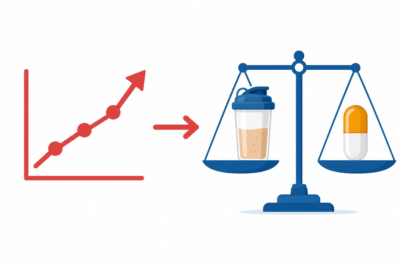
1. Early enteral nutrition (within 24 to 48 hours).[1][1]
- The reduces the gut mucosal atrophy, the bacterial translocation, the sepsis, the mortality.[1]
- The nasogastric / the nasojejunal tube; the start the low-rate the day 1; the advance the daily.[1]
- The 1.5 to 2 g/kg/day (the some the recommend the even the higher for the major the burns).[1]
- The carbohydrate (the 60 to 70 per cent of the non-protein the calories — the glucose for the brain, the wound; the protein-sparing).[1]
- The fat (the moderate — the 20 to 30 per cent; the omega-3 the anti-inflammatory).[1]
- Various formulas (the Curreri — 25 kcal/kg + 40 x %TBSA; the Harris-the-Benedict with the stress the factors; the simpler 25 to 30 kcal/kg/day).[2]
4. Trace elements + the vitamins.[1][1]
- The zinc, the copper, the selenium (the depleted in the burns — the wound healing, the immune function).[1]
- The vitamin C, the vitamin E (the antioxidant — the massive the free-the-radical the production).[1]
- The glutamine (the conditionally the essential — the gut, the immune).[1]
Early enteral nutrition — why, when, and how
Early enteral nutrition (within 24 hours, certainly within 48 h) is one of the few interventions in burns nutrition with consistent outcome benefit. The mechanisms are worth knowing in detail because they recur in every exam.[1][3]
Why early enteral nutrition works in burns
Maintains gut mucosal integrity
The enterocyte is fuelled principally by luminal glutamine and short-chain fatty acids. Luminal substrate maintains villous height and tight-junction integrity; starvation causes rapid mucosal atrophy.
Prevents bacterial translocation
Mucosal atrophy + splanchnic hypoperfusion (shock) allow enteric bacteria and endotoxin to cross into the portal and systemic circulation — a putative driver of sepsis and multiorgan failure. Early feed attenuates this.
Supports the gut-associated lymphoid tissue (GALT)
Enteral feed stimulates IgA and mucosal immunity, which protect against respiratory and wound infection. Parenteral nutrition does not do this.
Blunts the hypermetabolic/catecholamine response
Early feed is associated with a lower peak REE and a less severe catabolic phase — the gut appears to signal ("gut talk") to modulate the systemic stress response.
Improves outcomes
Consistent association with fewer infections, shorter ICU stay, lower mortality, and better wound healing. The benefit is lost when feed is delayed beyond ~48 h.
Enteral vs parenteral nutrition in major burn
| Feature | Enteral nutrition (preferred) | Parenteral nutrition |
|---|---|---|
| First-line? | YES — always preferred | NO — reserve for intolerance, prolonged ileus, or inability to meet targets enterally |
| Gut mucosal integrity | Maintained (trophic effect of luminal substrate) | Mucosal atrophy occurs despite full nutrition |
| Infection risk | Lower | Higher (line sepsis, bacterial translocation) |
| Hyperglycaemia | Less pronounced | More pronounced (IV glucose) |
| Cost / complexity | Lower | Higher (central line, sterile compounding) |
| When to use PN | — | Genuine enteral failure (prolonged ileus, mesenteric ischaemia, high-output fistula), or as supplemental PN when enteral alone cannot meet targets after 5-7 days |
| Exam mantra | "Use the gut whenever it works" | "PN does not replace the trophic effect of feed on the gut" |
Practical points of enteral feeding in burns
- Route: nasogastric first; consider nasojejunal/post-pyloric if high gastric residuals or if feeding during surgical procedures/prone ventilation. A feeding jejunostomy is placed at laparotomy if needed.
- Timing: start at a trophic rate (10-20 mL/h) within 24 h even if ileus is present; advance toward the target over 3-5 days as tolerated. Do NOT wait for ileus to "fully resolve" — the gut tolerates trophic feed even in low-flow states, and the feed itself helps resolve ileus.
- Gastric residual volumes: a permissive approach (re-feed even at residuals up to 250-500 mL in the absence of distension/vomiting) is now favoured, with prokinetics (metoclopramide, erythromycin) if needed.
- Continuous vs bolus: continuous is often better tolerated initially in the ICU; cyclical or bolus feeding can be introduced as the patient recovers. [1]
Glucose control — target 6-10, never tight
Hyperglycaemia is near-universal after major burn, driven by catecholamine- and cytokine-mediated insulin resistance plus unrestrained hepatic gluconeogenesis. It is not benign: hyperglycaemia impairs neutrophil function, wound healing, and is associated with infection and mortality. But tight glycaemic control (4.4-6.1 mmol/L), as trialled in the Leuven protocol, was shown in the burn population (Jeschke, Herndon) to cause an unacceptable rate of hypoglycaemia — itself an independent predictor of death.[4][5]
The target and the agents
- Target blood glucose: 6-10 mmol/L (some units accept up to 11). Avoid hypoglycaemia absolutely — it is more dangerous than moderate hyperglycaemia in this population.[4]
- Insulin is first-line. It lowers glucose AND is anabolic (promotes muscle protein synthesis, inhibits proteolysis). The burn patient is profoundly insulin-resistant, so doses are high (often 5-20+ units/hour). Use a titrated infusion protocol.
- Metformin is an attractive alternative investigated by Jeschke's group: it lowers glucose without the hypoglycaemia risk (it does not stimulate insulin secretion directly), and addresses insulin resistance at its root (hepatic gluconeogenesis inhibition). In the phase II trial it achieved comparable glucose control to insulin with fewer hypoglycaemic events.[5] Limitations: lactic acidosis risk in shock/renal failure, cannot be given to the unstable patient.
- Avoid "tight" (4.4-6.1) control. The hypoglycaemia risk is 4-5× higher in burn patients than in general ICU patients, and severe hypoglycaemia independently predicts mortality.[4]
Glucose-control strategies in major burn
| Strategy | Mechanism | Glucose target | Hypoglycaemia risk | Role in burns |
|---|---|---|---|---|
| Moderate insulin infusion | Exogenous insulin — lowers glucose, anabolic | 6-10 mmol/L | Moderate (titrate carefully) | First-line; anabolic bonus |
| Tight insulin infusion (Leuven) | Exogenous insulin | 4.4-6.1 mmol/L | High — NOT recommended | Avoid — excessive hypoglycaemia |
| Metformin | Hepatic gluconeogenesis inhibition; insulin sensitiser | 6-10 mmol/L | Low | Investigated in phase II; useful where tolerated (not in shock/AKI) |
| Other (GLP-1 agonists, SGLT2-i) | Vary | — | Vary | Not established in burns |
The pharmacological modulation
1. The propranolol (the beta-blocker — the reduces the hypermetabolic the drive, the catecholamine the surge, the heart rate, the catabolism). The titrate to the heart rate.[2][1]
2. The oxandrolone (the testosterone the analogue — the anabolic; the reduces the catabolism, the increases the lean the body the mass).[1][1]
3. The insulin (the glycaemic the control — the moderate; the target the 6 to 10; the NOT the tight). The anabolic (the protein the synthesis).[1][1]
Pharmacological modulation in depth
The three agents above, plus the trace elements below, constitute the evidence-supported pharmacological arm of burn metabolic care. Each targets a different limb of the hypermetabolic cascade, and they are synergistic when combined.[3]
Pharmacological modulation of burn hypermetabolism — the four pillars
| Agent | Class / mechanism | Effect on hypermetabolism | Key evidence / dose | Cautions |
|---|---|---|---|---|
| Propranolol | Non-selective β-blocker — blocks catecholamine drive | ↓ REE, ↓ heart rate (titrate to ~15-20% reduction), ↓ cardiac work, ↓ muscle catabolism, preserves lean mass, ↓ fatty infiltration of liver | Standard of care in many burn centres; titrated to HR reduction. Start once resuscitation complete and haemodynamics stable. | Avoid in shock/bradycardia; can mask hypoglycaemia (β2 symptoms). Monitor for bronchospasm (inhalation injury). |
| Oxandrolone | Synthetic testosterone analogue (anabolic, low androgenic effect) | Net anabolic — ↑ muscle protein synthesis, ↑ lean body mass, ↓ weight loss, ↓ length of stay | RCTs (Wolf et al.; Demling & DeSanti): faster lean-mass regain, shorter hospital stay. Dose ~10-20 mg/day (adult). | Hepatotoxicity — monitor LFTs. Avoid in prostate/breast cancer, pregnancy. |
| Insulin | Anabolic + hypoglycaemic | ↓ glucose, ↑ muscle protein synthesis | Moderate target 6-10 mmol/L | Hypoglycaemia (the dangerous end). High doses required (insulin resistance). |
| Metformin (investigational) | Biguanide — insulin sensitiser | ↓ glucose without hypoglycaemia | Phase II RCT (Jeschke 2016) — comparable control to insulin, fewer hypoglycaemic events | Lactic acidosis in shock/renal failure/HE — NOT for the unstable patient. |
Why propranolol is the cornerstone. Of all the agents, propranolol most directly attacks the cause of the hypermetabolism — the catecholamine surge. By titrating to a 15-20% reduction in heart rate it lowers REE, reduces cardiac work (the hyperdynamic burn heart is working near its limit), reduces skeletal-muscle catabolism, and reduces the fatty infiltration of the liver seen in prolonged hypermetabolism. The Galveston (Herndon) group has shown benefit even when continued for up to a year in children, including preservation of bone mineral density. The non-selective (β1+β2) action is deliberate — β2-blockade is what blunts the thermogenic/metabolic effect.[3]
Why oxandrolone. Testosterone analogues restore the anabolic drive that burns abolish. Oxandrolone is preferred because it is orally bioavailable and has a high anabolic-to-androgenic ratio (fewer virilising effects). The burn-specific RCT evidence (Wolf, Demling & DeSanti) shows reduced lean-mass loss, faster weight regain in rehabilitation, and shorter hospital stay. The main caution is hepatotoxicity — monitor transaminases.[7]
Trace elements, vitamins, and the antioxidant deficit
Major burns produce a massive, rapid depletion of trace elements and antioxidant vitamins — through wound exudate (which is rich in zinc, copper, selenium and iron), through increased urinary losses, and through the consumption of the body's antioxidant defences by the huge free-radical load of the inflamed wound. Repletion is not a nicety: it is associated with reduced infection and improved wound healing.[6]
Trace elements and vitamins in burns — what, why, how much
| Micronutrient | Role in burns | Consequence of depletion | Supplementation |
|---|---|---|---|
| Zinc | Co-factor for >300 enzymes; collagen synthesis, wound healing, T-cell and neutrophil function | Delayed wound healing, alopecia, dermatitis, immunosuppression, impaired taste | Enteral/parenteral repletion; doses above standard trace-element mixes in major burn |
| Copper | Co-factor for cytochrome oxidase (energy), lysyl oxidase (collagen cross-linking), superoxide dismutase (antioxidant) | Anaemia, neutropenia, impaired collagen cross-linking → weak scar, osteoporosis | IV copper in major burn (Berger protocol) |
| Selenium | Co-factor for glutathione peroxidase — the principal intracellular antioxidant | Loss of antioxidant defence → oxidative damage; impaired immunity | IV selenium in major burn; associated with ↓ infection in RCTs |
| Iron | Haemoglobin, myoglobin, cytochromes | Anaemia (also lost through repeated blood sampling + wound) | Replace as indicated; beware over-supplementation (infection, oxidative stress) |
| Vitamin C | Antioxidant; co-factor for collagen synthesis (prolyl hydroxylase) | Scurvy-like: poor wound healing, capillary fragility | High-dose vitamin C sometimes used (also as a resuscitation adjuvant — see burns-resuscitation) |
| Vitamin E | Lipid-soluble antioxidant (protects cell membranes from lipid peroxidation) | Membrane damage, haemolysis | Replete |
| Vitamin A | Epithelialisation and immune function | Delayed epithelialisation | Replete |
| B vitamins (incl. thiamine) | Carbohydrate metabolism | Re-feeding syndrome (critical — see below) | Give before/at commencement of feed in the malnourished |
The Berger protocol and the trial evidence
The seminal work here is Berger's (Lausanne) series of RCTs. The 2007 trial showed that IV trace-element supplementation (copper, selenium, zinc) in major burns raised tissue concentrations, improved antioxidant status, reduced infectious episodes (especially pulmonary), and reduced the need for regrafting.[6] Subsequent meta-analytic work confirms a reduction in infectious complications. The practical message for the exam: in the major burn, standard enteral multivitamin/trace-element preparations are insufficient — additional IV selenium, zinc and copper are given, particularly in the early exudative phase.
Refeeding syndrome — the trap of starting feed
When feed is commenced (or escalated) in a patient who has been starved, malnourished, or chronically unwell, refeeding syndrome can occur. Burns patients are at risk because of the combination of pre-injury under-nutrition, the prolonged catabolic state, and the sudden delivery of a carbohydrate load. The mechanism: carbohydrate → insulin surge → intracellular shift of phosphate, potassium and magnesium → precipitous falls in serum levels; thiamine depletion as it is consumed for carbohydrate metabolism. Untreated, refeeding syndrome causes arrhythmia, heart failure, respiratory failure, seizures, death.[1]
Prevention: identify high-risk patients (low BMI, prolonged reduced intake, electrolyte abnormalities, alcoholism), check and correct phosphate/potassium/magnesium BEFORE feeding, give thiamine before and during the first week, and start feed at low rate (10-20 kcal/kg/day) and advance slowly over a week, monitoring electrolytes daily. This must be balanced against the imperative of early enteral nutrition in burns — the compromise is trophic-low-rate feed from day 1 with cautious advancement and electrolyte vigilance. [1]
Monitoring the burn patient's metabolic and nutritional state
Nutrition in burns is a moving target. The wound closes, sepsis comes and goes, the patient is operated on. Reassess continuously:[1][1]
- Indirect calorimetry — the gold standard for energy expenditure; repeat when clinical state changes (new sepsis, grafting, weaning).
- Nitrogen balance — urinary urea nitrogen (+ estimated wound loss) vs protein intake; target +2 to +5 g/day.
- Blood glucose — frequently (Q1-4 hourly on insulin infusion); target 6-10.
- Serum electrolytes — Na, K, Mg, PO₄ (refeeding), daily.
- Liver function — to detect fatty infiltration (overfeeding) and oxandrolone hepatotoxicity.
- Weight — difficult (fluid shifts, dressings) but trend where possible.
- Feed tolerance — gastric residuals, abdominal exam, stool output.
- Wound healing / infection — the ultimate functional read-out of nutritional adequacy. [1]
Prognosis
The adequate the nutrition + the pharmacological the modulation → the reduces the catabolism, the infection, the mortality, the ICU-the-acquired the weakness, the length of the stay. The long-the-term the rehabilitation (the scarring, the contracture, the psychological).[1][1][1]
Adequately nourished, beta-blocked, anabolic-supported and trace-element-repleted patients lose less lean mass, heal their wounds faster, develop fewer infections, wean earlier, and have measurably better long-term functional outcomes. The hypermetabolic response can persist for up to a year, which is why propranolol and oxandrolone are often continued into the rehabilitation phase. Conversely, the under-fed or over-fed patient faces ICU-acquired weakness, respiratory failure, sepsis, and delayed graft take — all of which lengthen stay and increase mortality. Metabolic and nutritional management is therefore not "supportive care" bolted onto resuscitation; in the post-resuscitation phase it is the central, outcome-determining therapy.[3][7]
The structured exam answer (the long form). The burn hypermetabolic response has two phases: an early ebb phase (0-48 h, low metabolic rate, shock, focus on resuscitation) and a prolonged flow phase (peaks day 5-10 at 150-200% of normal REE, sustained for weeks-months until the wound is closed). It is driven by the catecholamine surge, inflammatory cytokines (TNF, IL-1, IL-6), and evaporative heat loss from the wound. The consequences — massive catabolism (protein loss up to 150 g/day), hyperglycaemia from insulin resistance, immune suppression, ICU-acquired weakness — are mitigated by (1) early enteral nutrition within 24 h (maintains gut mucosa, reduces bacterial translocation, supports GALT); (2) high protein 1.5-2 g/kg/day; (3) caloric targeting by indirect calorimetry (or Curreri 25 kcal/kg + 40 × %TBSA, knowing it over-estimates); (4) glucose control to 6-10 mmol/L (never tight — hypoglycaemia kills); (5) pharmacological modulation with propranolol (the β-blocker that blunts the catecholamine drive), oxandrolone (anabolic), and insulin; and (6) trace-element and antioxidant repletion (zinc, copper, selenium, vitamins C and E — losses through the wound are large). Beware refeeding syndrome when starting feed in the malnourished. Net effect: reduced catabolism, infection, ICU-acquired weakness, length of stay, and mortality.[1][1][3][6]
Key trials and evidence
Herndon & Tompkins — The metabolic response to burn injury (PMID 15183630)
Source
Lancet 2004 — landmark review from the Galveston/Shriners group
Type
Authoritative narrative review synthesising decades of burn-physiology research
Key concepts
Defines the modern understanding of the burn hypermetabolic response: the ebb/flow phases, catecholamine-driven REE of 150-200%, sustained catabolism, insulin resistance
Clinical bottom line
Established the rationale for the current therapeutic pillars: early excision/grafting, early enteral nutrition, beta-blockade (propranolol), anabolic agents (oxandrolone), glucose control, and trace-element repletion
Jeschke et al. — Intensive insulin therapy in severely burned children (PMID 20395554)
Study design
Prospective randomised trial — severely burned paediatric patients
Intervention
Intensive (tight) insulin therapy vs conventional glucose management
Key finding
Tight insulin therapy improved glucose control and several metabolic/infectious endpoints BUT at the cost of a markedly increased rate of hypoglycaemia
Clinical bottom line
Tight glycaemic control in burns is unsafe because of severe hypoglycaemia — the modern target is moderate (6-10 mmol/L). Hypoglycaemia is an independent predictor of mortality in burn patients
Jeschke et al. — Metformin for glucose control in severe burns (PMID 27355267)
Study design
Phase II randomised controlled trial — metformin vs insulin in severely burned patients
Intervention
Metformin (insulin sensitiser) vs insulin infusion for glucose control
Key finding
Metformin achieved comparable glucose control with significantly fewer hypoglycaemic events, and may provide additional anti-inflammatory and insulin-sensitising benefit
Clinical bottom line
Metformin is a promising alternative to insulin for glucose control in stable burn patients; not suitable for the shocked or renally impaired patient (lactic acidosis risk)
Berger et al. — Trace element supplementation after major burns (PMID 17490965)
Study design
Randomised, placebo-controlled trial — major burn patients
Intervention
IV copper, selenium and zinc supplementation vs placebo, in addition to standard nutrition
Primary outcome
Improved antioxidant status and tissue trace-element concentrations
Key finding
Supplementation reduced infectious episodes (especially pulmonary infections) and reduced the need for regrafting — consistent with improved wound healing and immune function
Clinical bottom line
Major burns deplete trace elements through wound exudate; routine IV repletion of selenium, copper and zinc (above standard trace-element mixes) reduces infection and improves healing
Demling & DeSanti — Oxandrolone in the rehabilitation phase of burn care (PMID 14636753)
Study design
Clinical study of oxandrolone in the rehabilitation phase of burn recovery
Intervention
Oxandrolone (oral anabolic agent) vs nutrition alone
Key finding
Patients receiving oxandrolone regained weight and lean mass 2-3 times faster than those on nutrition alone, with gains maintained for months after discontinuation
Clinical bottom line
Oxandrolone is an effective anabolic adjunct that reduces catabolism and accelerates lean-mass recovery in burn rehabilitation; monitor liver function
Short answer questions
SAQ — Pharmacological modulation of the burn hypermetabolic response
10 minutes · 10 marks
A 42-year-old man sustained a 50 per cent TBSA flame burn (30 per cent full-thickness) in a house fire, with inhalation injury requiring intubation. It is now day 7. He is tachycardic at 128/min in sinus rhythm, temperature 38.6 deg C, blood glucose 14 mmol/L despite an insulin infusion at 8 units/h, and he has lost 6 kg since admission. Indirect calorimetry shows a resting energy expenditure of 1.8 times the Harris-Benedict prediction. The wound is only 40 per cent excised and grafted.
SAQ — Nutritional plan for a major burn at risk of refeeding
10 minutes · 10 marks
A 55-kg, 28-year-old woman has a 45 per cent TBSA scald burn (35 per cent full-thickness). She was intubated for inhalation injury and resuscitated with the modified Brooke regimen. It is now 18 hours post-injury; she has bowel sounds and a soft abdomen. Her BMI is 18.5 and she describes a chronically poor oral intake. Phosphate is 0.55 mmol/L, magnesium 0.5 mmol/L, potassium 3.1 mmol/L.
Clinical pearls
Red flags
Summary — the ten things to take to the exam
- The burn is the most powerful sustained hypermetabolic insult in medicine — REE 150-200% of normal, peaking day 5-10, sustained for weeks-months until wound closure.
- Two phases: ebb (0-48 h, low metabolic rate, focus on resuscitation) → flow (peak hypermetabolism, focus on nutrition/metabolic care).
- Three drivers: catecholamine surge, inflammatory cytokines (TNF, IL-1, IL-6), evaporative heat/water loss from the wound.
- Early enteral nutrition within 24 h — maintains gut mucosa, reduces bacterial translocation, supports GALT, improves outcomes. Enteral preferred over parenteral.
- High protein 1.5-2 g/kg/day — the keystone macronutrient; target positive nitrogen balance.
- Calories by indirect calorimetry (gold standard) or Curreri
25 kcal/kg + 40 × %TBSA(know it over-estimates). - Glucose 6-10 mmol/L — never tight control (hypoglycaemia is an independent mortality predictor).
- Pharmacological modulation: propranolol (catecholamine brake), oxandrolone (anabolic), insulin (glucose + anabolic), ± metformin.
- Trace elements + antioxidants: IV zinc, copper, selenium above standard mixes (Berger protocol); vitamins C and E; glutamine.
- Watch for refeeding syndrome and do not over-feed — accurate is better than generous.[1][1][3][6]
References
- [1]Porro LJ, et al. Nutrition in Pediatric Burns Semin Plast Surg, 2024.PMID 38746694
- [2]Demling RH, et al. Thermal injury Crit Care Clin, 1999.PMID 10331132
- [3]Herndon DN, Tompkins RG Support of the metabolic response to burn injury Lancet, 2004.PMID 15183630
- [4]Jeschke MG, et al. Intensive insulin therapy in severely burned pediatric patients: a prospective randomized trial Am J Respir Crit Care Med, 2010.PMID 20395554
- [5]Jeschke MG, et al. Glucose Control in Severely Burned Patients Using Metformin: An Interim Safety and Efficacy Analysis of a Phase II Randomized Controlled Trial Ann Surg, 2016.PMID 27355267
- [6]Berger MM, et al. Trace element supplementation after major burns modulates antioxidant status and clinical course by way of increased tissue trace element concentrations Am J Clin Nutr, 2007.PMID 17490965
- [7]Demling RH, DeSanti L Oxandrolone induced lean mass gain during recovery from severe burns is maintained after discontinuation of the anabolic steroid Burns, 2003.PMID 14636753