ICU · Burns
Burn Resuscitation Fluids (Parkland)
Also known as Burn fluid resuscitation · Parkland formula · Modified Brooke formula · Fluid creep · Burn shock · Rule of 10s · Goal-directed burn resuscitation · Brooke formula · Evans formula · Consensus formula · Albumin in burns · Abdominal compartment syndrome in burns · Burn oedema · Capillary leak in burns
The burn the resuscitation — the massive the capillary the leak → the burn the shock. The Parkland the formula (the 4 mL/kg/%TBSA — the half the 8 h from the burn, the rest the 16 h). The titration the to the urine the output. The the fluid the creep (the over-the-resuscitation → the pulmonary the oedema, the abdominal the compartment, the extremity). The lactated the Ringer's the preferred. The colloid the after the 24 h. The goal-the-directed. Major burn resuscitation replaces the massive plasma volume lost through the post-burn capillary leak. The Parkland formula (Baxter, 4 mL/kg/%TBSA, half in the first 8 h from the TIME of the burn) remains the world standard starting point, but actual volumes given are typically 50% higher than calculated (fluid creep). Titrate to urine output 0.5 mL/kg/h (adult) or 1 mL/kg/h (child). Modified Brooke (2 mL/kg/%TBSA) is a conservative alternative. Albumin is added after 8-24 h once capillary integrity is restored (SAFE trial showed equivalence overall; cautious burn-subgroup signal). Over-resuscitation causes abdominal compartment syndrome, pulmonary oedema, and orbital/extremity compartment syndromes. Children need dextrose-containing maintenance fluid IN ADDITION to resuscitation because of limited glycogen stores.
On this page & tools
Your progress
Saved locally on this device.
Target exams
Red flags
Overview & definition
The burn the resuscitation — the burn the produces the massive the systemic the capillary the leak → the hypovolaemic the shock. The formula-the-based the fluid the resuscitation the prevents the burn the shock; the titration the prevents the fluid the creep. The the Parkland the formula the standard.[1][1]
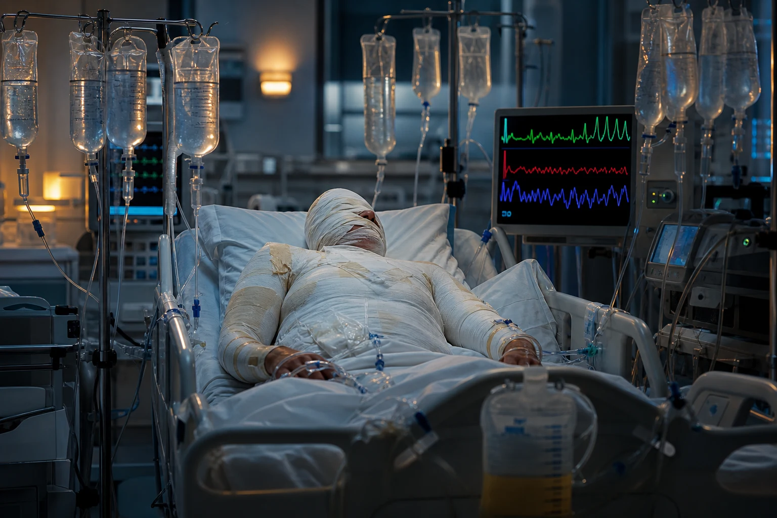
Burn resuscitation is one of the most examined and most poorly executed areas in critical care. A major burn (>20% TBSA in adults, >10% in children) produces a systemic inflammatory response that converts the entire capillary bed — not just the burnt skin — into a leaky sieve. Plasma water AND albumin pour into the interstitium, producing the combination of hypovolaemic/burn shock (low intravascular volume) and massive whole-body oedema (high interstitial volume) simultaneously. The art of burn resuscitation is to give enough fluid to prevent shock and renal failure while giving little enough to avoid pulmonary oedema, abdominal compartment syndrome, and extremity/orbital compartment syndromes. Every formula is only a starting estimate — the real resuscitation is titrated, hour by hour, to urine output and perfusion.[1][1][6]
The pathophysiology — the burn the shock
The burn the triggers the systemic the inflammatory the response → the massive the capillary the leak (the fluid + the protein the into the interstitium) → the hypovolaemia, the oedema, the hypoalbuminaemia. The first the 24 h. The then the colloid the (the after the 12 to 24 h).[1][1][1]
The three phases of burn shock — exam depth
The capillary leak is not constant; it follows a predictable time course that dictates WHY colloid is withheld early and added late. Understanding the phases explains the formula and the timing of every intervention. [1]
The three temporal phases of the burn capillary leak
| Phase | Time window | Capillary permeability | Dominant fluid movement | Clinical / therapeutic implication |
|---|---|---|---|---|
| Phase 1 — fluid loss & accumulation | 0-8 h (peak leak) | Maximum — both water and albumin escape | Plasma volume plummets; interstitial oedema accumulates fastest | Crystalloid only (lactated Ringer's). Colloid given here simply leaks out and worsens oedema. Give the first half of the Parkland volume. |
| Phase 2 — ongoing leak / "flow phase" | 8-24 h | Still leaky but beginning to seal | Continued plasma loss; oedema peaks ~24 h | Continue crystalloid, titrated to UO. Albumin may be added from 12 h in many protocols once leak is slowing. |
| Phase 3 — reabsorption / "diuretic phase" | After 24-48 h | Capillary integrity restored | Interstitial fluid returns to intravascular space; spontaneous diuresis; oedema resolves | Reduce/stop resuscitation fluid. Switch to maintenance + colloid. Watch for fluid overload as oedema mobilises. |
The leak is driven by a burn toxin / mediator cascade: complement, histamine, prostaglandins, leukotrienes, thromboxane A2, bradykinin, cytokines (IL-1, IL-6, TNF-alpha), and oxygen free radicals. The result is endothelial cell gap formation, loss of the glycocalyx, and a fall in plasma oncotic pressure (hypoalbuminaemia) that compounds the hydrostatic leak. Cardiac output falls early (a circulating "myocardial depressant factor" + hypovolaemia), then rises as resuscitation proceeds. Systemic vascular resistance is high initially (sympathetic response) then falls. Smoke inhalation injury dramatically increases fluid requirements by 30-50% through direct alveolar-capillary injury and systemic inflammation.[1][11]
The Parkland the formula
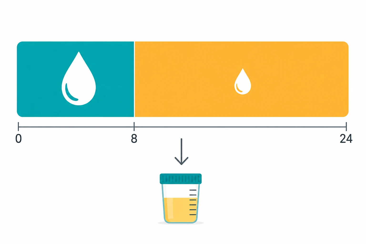
The the 4 mL/kg/%TBSA the lactated the Ringer's.[1][1]
- The the half the first the 8 h from the TIME OF THE BURN (the NOT the admission).
- The the rest the next the 16 h.[1]
- The endpoint the urine the output the 0.5 mL/kg/h the adult; the 1 mL/kg/h the child. The titrate (the NOT the rigid the formula).[1][1]
The Parkland formula in detail — Baxter's contribution
The Parkland (Baxter) formula was developed by Charles Baxter at Parkland Memorial Hospital in the 1960s from dog experiments and then validated in humans. The original derivation: the volume of lactated Ringer's required to restore and maintain a normal cardiac output and extracellular volume after a burn was 3.8-4.2 mL per kg per %TBSA — rounded to 4 mL/kg/%TBSA for the first 24 h.[1][1]
The formula is: [1]
First 24 h fluid (mL) = 4 × weight (kg) × %TBSA burn (2nd degree + 3rd degree only)
Worked example — the exam arithmetic
Parkland worked example — 70 kg adult, 40% TBSA, presents 2 h after the burn
Calculate the 24-h total
4 mL × 70 kg × 40% = 11,200 mL (11.2 L) of lactated Ringer's over 24 h.
Split into halves
Half (5,600 mL) in the first 8 h from the TIME OF BURN; half (5,600 mL) in the next 16 h.
Correct for the delay
The patient presents 2 h post-burn, so only 6 h remain for the first half. First-half rate = 5,600 mL / 6 h = ~933 mL/h. Second-half rate = 5,600 mL / 16 h = 350 mL/h.
Titrate hourly
Aim for urine output 0.5 mL/kg/h = 35 mL/h. If UO <0.3 mL/kg/h increase the rate by ~25-33%; if UO >1.0 mL/kg/h reduce it. Do NOT chase a high UO with more fluid — that is the route to fluid creep.
Count ONLY 2nd and 3rd degree burns
Superficial (1st degree / erythema) burns are EXCLUDED from %TBSA. Count partial- and full-thickness only.
Reassess at 24 h
Switch from crystalloid resuscitation to colloid (albumin) + maintenance + enteral nutrition. The "diuretic phase" begins — expect spontaneous diuresis and REDUCE fluids.
Who needs formula-based resuscitation?
Not every burn. Formal resuscitation is indicated for: [1]
- Adults: burns >20% TBSA (partial + full thickness)
- Children: burns >10-15% TBSA
- Smaller burns are managed with oral fluids or maintenance IV fluids — over-treating minor burns is a common error that causes iatrogenic morbidity. [1]
The alternative the formulas
- The modified the Brooke (the 2 mL/kg/%TBSA) — the more the conservative; the growing the favour (the fluid-the-creep the concern).[2][1]
- The Rule of the 10s — the 10 × %TBSA = the initial the hourly the rate (the first the 8 h).[1][1]
- The hypertonic the saline — the reduced the fluid the volume but the risk the hypernatraemia; the experimental.[1]
The historical and modern formula landscape — a comparison
Burn resuscitation formulas compared — what the exams ask
| Formula | First-24h crystalloid volume | Origin / rationale | Resurgence / current status |
|---|---|---|---|
| Evans (1952) | 1 mL/kg/%TBSA crystalloid + 1 mL/kg/%TBSA colloid + 2000 mL glucose-water | First formula; over-weighted colloid early | Obsolete — colloid in first 8 h worsens oedema |
| Brooke (1953) | 1.5 mL/kg/%TBSA crystalloid + 0.5 mL/kg/%TBSA colloid + 2000 mL glucose | US Army Burn Unit, Korean War | Obsolete as a primary formula |
| Parkland / Baxter (1968) | 4 mL/kg/%TBSA crystalloid (LR), no colloid first 24 h | Crystalloid-only, capillary-leak aware | World standard starting point; titrated to UO |
| Modified Brooke (1969/1970s) | 2 mL/kg/%TBSA crystalloid (LR) | Re-analysis showed less was adequate | Growing favour — antidote to fluid creep; Pruitt advocated |
| Consensus / "Uribe" (Cape Town, 2008, modified 2024) | 2-4 mL/kg/%TBSA LR; colloid after 8-12 h if >20% TBSA | Acknowledges creep; builds in albumin early | Used in South Africa, parts of Europe |
| Rule of 10s (US Army ISR, 2009) | Initial rate = %TBSA × 10 mL/h (titrate from there); add 100 mL/h for inhalation injury | Designed for combat — calculator-free, weight-agnostic | US military standard; excellent for mass casualty / austere |
| Shriners / Salt Lake City (albumin-based) | LR 4 mL/kg/%TBSA + 5% albumin after 12 h + dextrose maintenance in children | Paediatric-focused; leverages early colloid once leak slows | Used in major paediatric burn centres |
| Hypertonic saline | Na+ 250 mEq/L; volume reduced ~1/3 | Draw fluid from interstitium to intravascular space | Largely abandoned — hypernatraemia, renal failure, no outcome benefit |
The Rule of 10s in detail (Chung 2009):[8] designed for combat casualty care where scales and calculators are impractical. Estimate %TBSA to the nearest 10; multiply by 10 to get the INITIAL hourly rate in mL/h (e.g., 40% → 400 mL/h). Add 100 mL/h if there is inhalation injury. This is the starting rate only — titrate thereafter to urine output, exactly as with Parkland. It is now the US Army Institute of Surgical Research standard and is increasingly used in civilian mass-casualty planning.
The fluid the creep
The the fluid the creep — the over-the-resuscitation (the formula the over-the-estimates the → the fluid the overload).[2][3][1]
Fluid creep — the defining problem of modern burn resuscitation
Coined by Saffle in 2007, fluid creep is the steady rise in resuscitation volumes over and above what the formula predicts, culminating in over-resuscitation and its complications.[3] The data are stark: across published series, the median first-24 h volume rose from ~3.3 mL/kg/%TBSA in the 1970s-80s (close to Parkland) to ~5.5-6.5 mL/kg/%TBSA in the 2000s — roughly 50% more than calculated.[2]
- The the pulmonary the oedema.
- The the abdominal the compartment the syndrome (the IAP the above the 20 → the renal, the respiratory, the cardiac the compromise).
- The the extremity the compartment the syndrome (the escharotomy).
- The the orbital the compartment (the vision).[1]
Causes of fluid creep — why we keep giving too much
| Driver | Mechanism | Frequency |
|---|---|---|
| Slavish adherence to formula | Calculating Parkland and delivering it uncritically without titrating DOWN when UO is high | Most common |
| Fear of under-resuscitation | Defensive medicine — "more is safer" | Very common |
| Inhalation injury (unrecognised) | Adds 30-50% to requirements; if not identified the team chases a higher UO target | Common |
| Delayed presentation / electrical injury | Underestimation of true tissue injury (electrical, deep 4th-degree) inflates apparent "formula failure" | Common in electrical burns |
| Opiate sedation causing hypotension | Treating drug-induced hypotension with fluid rather than reducing sedation | Common |
| Wrong UO endpoint | Aiming for 1 mL/kg/h in an adult, or accepting transient oliguria without context | Common |
| Albumin withheld too long | Persistent capillary leak after 24 h with crystalloid-only regimen | Less common |
Predictors of higher-than-calculated requirements (Cancio 2004):[9] patients likely to need >4 mL/kg/%TBSA can be identified early — inhalation injury, full-thickness burns >20%, electrical injury, delayed resuscitation (>2 h), and high base deficit/lactate on arrival. Recognising these lets you start higher AND, critically, lets you add albumin earlier.
The prevention — the titrate the to the urine the output the (the 0.5 the adult); the NOT the rigid the formula; the colloid the early the if the crystalloid the high.[1][2][1]
Preventing fluid creep — the disciplined resuscitation protocol
START with a formula, not a feeling
Calculate Parkland (or Rule of 10s). Set the initial rate. Document the time of burn.
TITRATE HOURLY to urine output
Adult 0.5 mL/kg/h, child 1.0 mL/kg/h. UO <0.3 → increase rate 25-33%; UO 0.5-1.0 → keep rate; UO >1.0 → DECREASE rate 25-33%. Never chase a high UO.
ADD ALBUMIN when crystalloid exceeds ~150% of calculated
Once the leak is sealing (12-24 h), switch part of the regimen to 5% albumin to maintain oncotic pressure and pull water back intravascularly. Lawrence 2010 (retrospective) found routine supplemental albumin was NOT beneficial — so use it selectively, not universally.
CHECK inhalation injury / adequacy of analgesia
If requirements climb, look for inhalation injury (bronchoscopy, carboxyhaemoglobin), untreated pain, or deep/electrical injury rather than just escalating fluid.
MONITOR intra-abdominal pressure if fluid >6 mL/kg/%TBSA
Bladder pressure every 4-6 h once you cross into "creep" territory. ACS is preventable if caught early.
STOP at 24 h and switch regimen
Resuscitation fluid is a 24-h event. After 24 h, cap crystalloid at maintenance, add colloid, and let the diuretic phase occur.
Crystalloid vs colloid — the central debate (albumin and the SAFE trial)
This is the single most contested question in burn fluid therapy and a guaranteed exam topic. The debate centres on what to give (crystalloid vs colloid), when (early vs late), and whether colloid reduces total volume or improves outcome. [1]
The physiological case for colloid (albumin)
After the first 8-12 h, as the capillary leak begins to seal, colloid (albumin) becomes physiologically rational: it stays intravascular, raises plasma oncotic pressure, and pulls water back from the interstitium. Albumin at 5% (iso-oncotic) or 20% (hyperoncotic) reduces the total crystalloid volume needed and limits ongoing oedema. The Shriners (Galveston) and Salt Lake City protocols add 5% albumin after the first 12 h precisely for this reason, with excellent results in paediatric series.[6][12]
The evidence — SAFE trial and the burn subgroup
SAFE — Saline vs Albumin Fluid Evaluation (Finfer 2004)
Design
Multicentre, randomised, double-blind — 6,997 ICU patients (incl. 66 burn patients)
Intervention
4% albumin vs 0.9% saline for all fluid resuscitation for 28 days
Primary outcome
28-day mortality: 20.9% albumin vs 21.1% saline (RR 0.99, 95% CI 0.91-1.09) — NO difference
Burn subgroup (n=66)
Trend to HIGHER mortality with albumin (RR 2.40, 95% CI 0.79-7.31) — NOT statistically significant, but a cautionary signal
Bottom line
Albumin and saline are equivalent for general ICU resuscitation; the burn subgroup signal was insufficient to condemn albumin but argues against routine early use
How to reconcile: the SAFE burn subgroup is small and its signal is not definitive. Most burn centres interpret the totality of evidence as: albumin is acceptable and often beneficial when added AFTER the capillary leak has sealed (8-24 h), but should NOT be the primary first-8-h fluid. Lawrence 2010 (single-centre retrospective, n≈400)[10] found routine supplemental albumin did NOT reduce fluid volumes or improve outcomes, so albumin is used selectively — typically when crystalloid requirements exceed ~150% of calculated, or in the second 24 h.
Crystalloid vs colloid in burns — the synthesis
| Question | Answer | Evidence |
|---|---|---|
| First 8 h — crystalloid or colloid? | CRYSTALLOID (lactated Ringer's) ONLY. Colloid leaks out and worsens oedema. | Physiological dogma; universal practice[5] |
| 8-24 h — add colloid? | Reasonable, selective. Albumin once leak is sealing; reduces total volume. | Shriners/Salt Lake protocols[6][12] |
| After 24 h — crystalloid or colloid? | Colloid (albumin) preferred. Capillary integrity restored; crystalloid causes overload. | Standard; Oh's, Marino[1][1] |
| Does albumin improve survival? | No proven survival benefit. SAFE neutral; Lawrence 2010 neutral. | SAFE, Lawrence[4][10] |
| Starch / HES in burns? | NEVER. Increased AKI, coagulopathy, pruritus. | CHEST, 6S (sepsis); extrapolated |
| Hypertonic saline? | No — abandoned. Hypernatraemia, renal failure, no outcome benefit. | Historic trials; obsolete[5] |
Oral resuscitation — the "Wong" / Brooke approach
For conscious adults with burns 20-30% TBSA and no shock, oral rehydration solution is increasingly accepted as an alternative to IV fluids, mirroring the cholera-treatment philosophy. The WHO ORS is hypotonic; a carbohydrate-electrolyte solution (e.g., CeraLyte) is preferred. Volumes are titrated to thirst and urine output. Contraindicated in shock, vomiting, altered consciousness, or burns >30%.[6]
The post-the-24-hour the fluid
After the 24 h (the capillary the leak the resolves), the colloid (the albumin) + the maintenance. The children: the dextrose-containing the maintenance (the hypoglycaemia).[1][1]
Second-24-h and beyond — the "diuretic phase"
From 24-48 h the capillary seal is restored and interstitial fluid begins to return to the intravascular space. The patient enters the diuretic (mobilisation) phase: urine output rises spontaneously, blood pressure stabilises, and oedema begins to resolve. The resuscitation requirement falls dramatically. Typical second-24-h regimen: [1]
- Albumin at 0.3-0.5 mL/kg/%TBSA of 5% albumin (provides colloid + oncotic pull)
- Maintenance crystalloid (dextrose-containing in children)
- Enteral nutrition started early (within 24-48 h) — the gut is the most metabolically affected organ and must be fed
- Reduce/stop the resuscitation crystalloid — giving Parkland-style volumes beyond 24 h is a classic error causing fluid overload [1]
The hypermetabolic phase now dominates: resting energy expenditure rises to 1.5-2× baseline; nutritional targets are high (see the metabolic-nutrition topic).[1][1]
Paediatric burn resuscitation — maintenance in addition to resuscitation
Children are NOT small adults in burn care. Two physiological differences dictate a fundamentally different approach: (1) a higher surface-area-to-mass ratio (larger %TBSA for the same burn) and (2) limited glycogen stores that are exhausted within hours, producing hypoglycaemia if only lactated Ringer's is given.[1][1]
The cardinal rule — give maintenance IN ADDITION to resuscitation
A child resuscitated with Parkland volumes of lactated Ringer's alone will become hypoglycaemic within hours. The solution is to run a separate dextrose-containing maintenance line alongside the resuscitation: [1]
Resuscitation (Parkland): 3 mL/kg/%TBSA lactated Ringer's (slightly less than adult — see below)
target UO 1.0 mL/kg/h
Maintenance (SEPARATE): dextrose-containing fluid (e.g., 5% dextrose + 0.45% saline, or
D5-1/4NS per local protocol) per Holliday-Segar:
100 mL/kg for first 10 kg + 50 mL/kg for next 10 kg + 20 mL/kg above 20 kg
``` <Cite id="1" />
The maintenance volume is NOT part of the Parkland calculation — it is additional. Failure to provide it is a common and serious error.<Cite id="1" /><Cite id="1" />
<Compare title="Adult vs paediatric burn resuscitation — the key differences">
| Parameter | Adult | Child (<30 kg, or <14 y) |
|---|---|---|
| **%TBSA threshold for IV resuscitation** | >20% TBSA | >10% TBSA |
| **Resuscitation formula** | 4 mL/kg/%TBSA LR (Parkland) | 3 mL/kg/%TBSA LR (Shriners/modified Parkland) |
| **Urine output target** | **0.5 mL/kg/h** | **1.0 mL/kg/h** (higher — reflects higher baseline metabolic rate) |
| **Maintenance fluid** | Usually not needed separately | **Dextrose-containing maintenance IN ADDITION** to resuscitation |
| **Glucose monitoring** | Routine | **Hourly** — hypoglycaemia common and dangerous |
| **Albumin timing** | After 12-24 h | Often after 8-12 h (Shriners uses albumin early) |
| **Risk of fluid creep** | High | Very high — small total body water amplifies errors |
</Compare><Cite id="1" /><Cite id="1" /><Cite id="12" />
### Why children need dextrose — the physiology
A 10 kg child has ~40 g of glycogen (4 g/kg) versus an adult's ~400 g. A major burn doubles glucose turnover immediately and exhausts glycogen within 4-6 h. Without exogenous dextrose, the child develops **hypoglycaemia, ketosis, and (paradoxically) hyperosmolar dehydration** as the kidney struggles to conserve glucose. Dextrose-containing maintenance fluid prevents this. Monitor capillary glucose hourly; target 4-8 mmol/L.<Cite id="1" />
## The goal-the-directed the approach
The modern the trend — the NOT the formula; the titrate the to the urine the output, the lactate, the haemodynamics, the mental the state. The permissive the hypovolaemia the (the conservative the fluid).<Cite id="1" /><Cite id="2" />
### Goal-directed / "smart" resuscitation — the modern standard
The dogma of "give the formula" is dying. The consensus (ABA, ISBI, ANZBA) is that a formula provides only the **starting rate**; the actual resuscitation is titrated continuously to multiple endpoints.<Cite id="12" /><Cite id="13" />
<Compare title="Resuscitation endpoints — what to monitor">
| Endpoint | Target | Comment |
|---|---|---|
| **Urine output** | **Adult 0.5 mL/kg/h; child 1.0 mL/kg/h** | The PRIMARY endpoint; titrate hourly. Do NOT accept oliguria without investigating. |
| **Mean arterial pressure** | MAP >65 mmHg (age-appropriate in children) | Adequate perfusion pressure; respect chronic hypertension |
| **Heart rate** | Trending down | Tachycardia is early shock; persistent tachycardia may be pain or under-resuscitation |
| **Base deficit / lactate** | Normalising | Base deficit >-6 or lactate >2 = under-resuscitation; trend over hours |
| **Mental state** | Alert, oriented | Confusion/agitation = hypoperfusion or hypoxia |
| **Capillary refill** | <2 s | Simple bedside microcirculation marker |
| **Central venous pressure / ScvO2** | Optional | Not routinely needed; useful in refractory shock |
| **Intra-abdominal pressure** | <12 mmHg | Monitor if fluid volumes high or >6 mL/kg/%TBSA |
</Compare><Cite id="1" /><Cite id="1" />
**Permissive hypovolaemia / conservative fluid:** the modern trend (echoing Pruitt's "push the pendulum back"<Cite id="5" />) is to accept a **slightly lower-than-"normal" urine output (0.3-0.5 mL/kg/h)** and modest haemodynamic under-filling in exchange for avoiding fluid creep and its complications — provided lactate is clearing and perfusion is maintained. This is the burn equivalent of the sepsis "restrictive fluid" movement.<Cite id="2" />
<RedFlag title="Permissive hypovolaemia does NOT mean 'no fluids'">
"Permissive hypovolaemia" means titrating to the LOW end of acceptable urine output (0.3-0.5 mL/kg/h in an adult) with clearing lactate — NOT abandoning resuscitation. A patient with rising lactate, base deficit, or oliguria <0.3 mL/kg/h is UNDER-resuscitated and needs more fluid, not less. The strategy is conservative, not negligent.<Cite id="2" /><Cite id="5" />
</RedFlag>
## Abdominal compartment syndrome from over-resuscitation — deep dive
Abdominal compartment syndrome (ACS) is the most feared complication of fluid creep in burns. Ivy (2000) first defined it in this population: bladder pressure >25 mmHg with new organ failure (oliguria unresponsive to fluid, rising airway pressures, falling cardiac output).<Cite id="7" /> In burn patients the incidence correlates directly with the volume of resuscitation given — typically appearing at **>6 mL/kg/%TBSA** or a positive fluid balance >250 mL/kg in the first 24 h.<Cite id="7" />
### Pathophysiology — why burn patients are uniquely at risk
Major burns cause **generalised oedema of the bowel wall** (the gut is the most oedema-prone organ in shock). Aggressive crystalloid resuscitation compounds this: the oedematous bowel swells within the non-compliant abdominal cavity, raising intra-abdominal pressure (IAP). Once IAP exceeds ~12 mmHg (intra-abdominal hypertension) the cycle accelerates — reduced venous return, reduced renal perfusion, splinting of the diaphragm — each worsening the next.<Cite id="7" />
<Compare title="WSACS grading of intra-abdominal hypertension and ACS">
| Grade | Bladder pressure (mmHg) | Clinical significance | Action |
|---|---|---|---|
| I | 12-15 | Intra-abdominal hypertension (IAH) — early | Reduce fluid; reassess |
| II | 16-20 | IAH — moderate | Position head of bed flat; evacuate bowel (NG, rectal tube); reduce fluid |
| III | 21-25 | Severe IAH | Above + consider sedation/paralysis, neuromuscular blockade; consider decompression |
| **IV** | **>25 with NEW organ failure** | **Abdominal compartment syndrome (ACS)** | **Surgical decompression (laparostomy) — emergency** |
</Compare> <Cite id="1" />
The diagnosis of ACS requires BOTH a sustained IAP >20-25 mmHg AND new organ dysfunction (the WSACS definition). The signs: <Cite id="1" />
- **Oliguria/anuria** unresponsive to further fluid (the classic first sign — the kidney is compressed)
- **Rising peak airway pressures** on the ventilator (diaphragmatic splinting)
- **Falling cardiac output / worsening shock** (reduced venous return, increased afterload)
- **Tense, distended abdomen** (late)
- **Metabolic acidosis** from gut hypoperfusion <Cite id="1" />
<RedFlag title="Oliguria in a burn patient on big fluids = think ACS, NOT 'give more fluid'">
The single most dangerous reflex in burns is to respond to falling urine output by escalating the fluid rate. If a patient who is already on high-rate resuscitation becomes oliguric, **the first action is to measure bladder pressure**, not to increase fluids. Giving more fluid to a patient with intra-abdominal hypertension accelerates the spiral to ACS, renal failure, and death. The differential of oliguria in burns: under-resuscitation (falling lactate, low CVP → give fluid), ACS (high IAP → decompress), or established AKI (high IAP, no response → RRT).<Cite id="7" />
</RedFlag>
### Management of ACS in burns
<FlowSteps title="Managing intra-abdominal hypertension / ACS in burns — escalation" steps={[
{ title: 'MEASURE bladder pressure', detail: 'Instill 25 mL sterile saline into the Foley catheter, clamp, transduce. Measure every 4 h if on >4 mL/kg/%TBSA. Pressure >12 = IAH; >25 with organ failure = ACS.' },
{ title: 'REDUCE / STOP fluid creep', detail: 'Switch part of the regimen to colloid (albumin); reduce crystalloid to maintenance; treat the cause of high requirements (pain, inhalation injury).' },
{ title: 'EVACUATE bowel contents', detail: 'NG tube decompression, rectal tube, prokinetics (metoclopramide, erythromycin). Bowel distension raises IAP.' },
{ title: 'POSITION and PARALYSE', detail: 'Head of bed flat (sitting raises IAP). Consider neuromuscular blockade (cisatracurium) to relax abdominal wall tone.' },
{ title: 'SURGICAL DECOMPRESSION if ACS (grade IV)', detail: 'Laparostomy / decompressive laparotomy. Indicated for IAP >25 with organ failure not responding to medical measures. Temporising with percutaneous drainage if localised fluid collection.' },
{ title: 'PREVENT recurrence', detail: 'Manage the open abdomen (VAC dressing); planned re-closure once IAP normal and oedema resolves (often days later). Maintain colloid-based, volume-restricted regimen.' },
]} /><Cite id="7" /><Cite id="1" />
## The other complications of fluid creep — orbital and extremity
Beyond the abdomen, over-resuscitation threatens two other compartments: <Cite id="1" />
- **Extremity compartment syndrome** — circumferential full-thickness limb burns form an inelastic eschar; the oedematous muscle beneath swells against it, producing ischaemia. **Escharotomy** (incision through the eschar to fascia, along the long axis of the limb) relieves it. Indicated with signs of compromised perfusion (cool, pulseless, tense, painful — though the burn patient is often sedated). Prophylactic escharotomy for circumferential full-thickness burns of the limbs or chest (the latter restricting ventilation).
- **Orbital compartment syndrome** — facial burns with massive periorbital oedema can compress the optic nerve and retina, causing irreversible blindness within hours. **Lateral canthotomy** decompresses the orbit. Urgent if there is vision loss, afferent pupillary defect, or extreme proptosis.<Cite id="1" />
<RedFlag title="Circumferential full-thickness chest burn = prophylactic escharotomy">
A circumferential full-thickness burn of the chest wall forms a rigid eschar that splints the chest, causing progressive respiratory failure as oedema accumulates beneath it. **Prophylactic escharotomy** (mid-axillary and mid-clavicular lines) restores chest wall compliance. Do not wait for ventilatory failure — the rise in airway pressures may be abrupt and fatal.<Cite id="1" /><Cite id="1" />
</RedFlag>
## Prognosis
The adequate the fluid the resuscitation the prevents the burn the shock + the acute the kidney the injury. The fluid the creep the → the complications (the pulmonary the oedema, the compartment).<Cite id="1" /><Cite id="2" /><Cite id="1" />
Adequate resuscitation converts a uniformly fatal injury (untreated major burns >40% TBSA were 100% lethal before 1950) into a survivable one — modern mortality for isolated burns >40% TBSA in adults is <20% in good centres. The LA50 (burn size lethal to 50% of patients) has risen from ~40% TBSA in the 1950s to >80% TBSA today. Death, when it occurs, is usually from **sepsis / multi-organ failure** days to weeks later, not from burn shock. The keys to survival: **early adequate resuscitation, prevention of fluid creep, early excision and grafting, infection control, and nutritional support.**<Cite id="1" /><Cite id="1" />
<AnswerCard title="The one-paragraph exam answer">
The burn the resuscitation — the massive the capillary the leak → the burn the shock. The **Parkland the formula** (the 4 mL/kg/%TBSA the lactated the Ringer's — the half the first the 8 h from the TIME OF THE BURN, the rest the 16 h). The endpoint the urine the output (the 0.5 mL/kg/h the adult; the 1 mL/kg/h the child). The **the fluid the creep** (the over-the-resuscitation → the pulmonary the oedema, the abdominal the compartment, the extremity). The modified the Brooke (the 2 mL/kg/%TBSA) the more the conservative. The colloid (the albumin) the after the 24 h. The goal-the-directed the (the titrate the to the urine the output; the NOT the rigid).<Cite id="1" /><Cite id="2" /><Cite id="1" />
</AnswerCard>
<AnswerCard title="The expanded exam answer — for the long-form / viva question">
Major burn resuscitation replaces the plasma lost through the post-burn systemic capillary leak. The **Parkland formula** (Baxter, 1968) — **4 mL/kg/%TBSA of lactated Ringer's over the first 24 h, half in the first 8 h from the TIME of the burn** — is the standard starting estimate; it is titrated hourly to **urine output 0.5 mL/kg/h (adult), 1.0 mL/kg/h (child)**, MAP, and clearing lactate. **Fluid creep** — giving 50%+ more than calculated, driven by formula-worship, unrecognised inhalation injury, and fear of under-resuscitation — causes **abdominal compartment syndrome** (bladder pressure >25 mmHg with oliguria and rising airway pressures), pulmonary oedema, and extremity/orbital compartment syndromes; prevent it by titrating down when UO is high, adding **albumin after 8-24 h** once the leak seals (SAFE trial showed albumin and saline equivalent overall — use albumin selectively, not as first-8-h fluid), and switching to maintenance + colloid at 24 h. The **modified Brooke (2 mL/kg/%TBSA)** is a conservative alternative. Children need **dextrose-containing maintenance fluid IN ADDITION** to resuscitation because of limited glycogen stores. Lactated Ringer's is preferred over 0.9% saline (hyperchloraemic acidosis). The modern trend is **goal-directed, permissive-hypovolaemic** resuscitation — a formula to start, endpoints to finish.<Cite id="1" /><Cite id="2" /><Cite id="1" /><Cite id="4" /><Cite id="7" />
</AnswerCard>
## Red flags
<RedFlag title="The half the FIRST 8 h from the TIME OF THE BURN (the NOT the admission)">
The Parkland — the half the first the 8 h from the TIME OF THE BURN (the NOT the admission). The if the delayed (the 2 h) → the remaining the 6 h. The NOT the 8 h the from the admission.<Cite id="1" />
</RedFlag>
<RedFlag title="The fluid the creep — the abdominal the compartment the syndrome">
The fluid the creep (the over-the-resuscitation) → the abdominal the compartment the syndrome (the IAP the above the 20 → the renal, the respiratory, the cardiac the compromise). The monitor the IAP; the decompression if the severe. The titrate the to the urine the output (the 0.5 the adult).<Cite id="2" /><Cite id="3" />
</RedFlag>
<RedFlag title="The lactated the Ringer's the preferred the over the saline (the hyperchloraemic the acidosis)">
The saline (the 0.9 per cent) → the hyperchloraemic the acidosis (the high the chloride → the renal the vasoconstriction → the AKI). The lactated the Ringer's the preferred.<Cite id="1" /><Cite id="1" />
</RedFlag>
<RedFlag title="The children — the dextrose the maintenance (the hypoglycaemia)">
The children the limited the glycogen → the hypoglycaemia. The dextrose-containing the maintenance the fluid. The urine the output the 1 mL/kg/h (the higher the than the adult).<Cite id="1" /><Cite id="1" />
</RedFlag>
<RedFlag title="Albumin in the FIRST 8 h is futile — it leaks out and worsens oedema">
During peak capillary leak (first 8-12 h) the capillary is permeable to albumin. Giving colloid here simply relocates albumin to the interstitium, raises interstitial oncotic pressure, and worsens oedema. Reserve albumin for after 8-24 h when the leak is sealing. Pruitt called early albumin "an exercise in futility."<Cite id="5" /><Cite id="6" />
</RedFlag>
<RedFlag title="A rising UO target does not mean 'push fluids' — and a falling UO does not always mean 'give more'">
Two opposite errors kill burn patients: (1) chasing a urine output above target with escalating fluids (the engine of fluid creep), and (2) responding to oliguria in an over-resuscitated patient with more fluid (the engine of ACS). If UO is ABOVE target, REDUCE the rate. If UO falls AND the patient has had big fluids, MEASURE BLADDER PRESSURE before escalating.<Cite id="2" /><Cite id="7" />
</RedFlag>
<RedFlag title="Electrical burns and 4th-degree burns underestimate TBSA — the formula will under-resuscitate">
Surface %TBSA underestimates true tissue injury in electrical burns (deep muscle injury invisible from the skin) and deep 4th-degree burns. These patients routinely need 50-100% more than calculated and may need surgical fasciotomy for compartment syndrome of unburned limbs. Monitor for myoglobinuria (dark urine) and rhabdomyolysis.<Cite id="9" />
</RedFlag>
## Short answer questions
<SaqBlock
title="SAQ — Parkland formula in a 40 per cent TBSA flame burn with delayed presentation"
stem="A 52-year-old, 75 kg man is brought to the emergency department after being rescued from a house fire at 10:30; he arrives in the resuscitation bay at 13:30. He has partial- and full-thickness burns involving 40 per cent TBSA (anterior chest, abdomen, both arms). He is intubated for suspected inhalation injury (soot in the oropharynx, carboxyhaemoglobin 18 per cent). HR 124, BP 98/60, SpO2 96 per cent on FiO2 1.0, urine output has not yet been charted. Lactate 3.4 mmol/L."
duration={10}
totalMarks={10}
parts={[
{ question: 'Calculate his first 24-hour fluid resuscitation, giving the fluid, the total volume, and the hourly rates. Justify your choice of crystalloid and your timing of colloid. (5 marks)', marks: 5, answer: 'Calculate the 24-hour total using the Parkland (Baxter) formula: 4 mL/kg/%TBSA of lactated Ringer\'s (Hartmann\'s) = 4 x 75 x 40 = 12,000 mL over 24 h. Split into halves: the first 6,000 mL goes in the first 8 h counted from the TIME OF THE BURN (not from admission); the second 6,000 mL over the next 16 h. The burn occurred at 10:30 and it is now 13:30, so 3 h have elapsed and only 5 h remain for the first half. First-half rate = 6,000 / 5 = 1,200 mL/h. Second-half rate = 6,000 / 16 = 375 mL/h. Use lactated Ringer\'s, NOT 0.9 per cent saline: large-volume saline causes hyperchloraemic metabolic acidosis, renal vasoconstriction and AKI (consistent with the SMART and SALT-ED balanced-crystalloid trials). Count only partial- and full-thickness burns toward the 40 per cent; superficial erythema is excluded. With suspected inhalation injury (which Cancio 2004 identified as a predictor of high requirements, adding 30-50 per cent to fluid needs) expect to titrate upward from this starting estimate. Do NOT give colloid in the first 8-12 h: during peak capillary leak the endothelium is permeable to albumin, so colloid translocates to the interstitium, raises interstitial oncotic pressure and worsens oedema — Pruitt called early albumin "an exercise in futility." Reserve 5 per cent albumin for after 8-24 h once the leak begins to seal.' },
{ question: 'Outline the endpoints you will use to titrate this resuscitation hour by hour. (3 marks)', marks: 3, answer: 'The PRIMARY endpoint is urine output: 0.5 mL/kg/h in an adult (= ~37-38 mL/h for this patient), or 1.0 mL/kg/h in a child under 30 kg. Titrate hourly: if UO is under 0.3 mL/kg/h increase the rate by 25-33 per cent; if 0.5-1.0 mL/kg/h maintain the rate; if above 1.0 mL/kg/h REDUCE the rate by 25-33 per cent. Never chase a high urine output with more fluid — that is the engine of fluid creep. Secondary endpoints: MAP above 65 mmHg, heart rate trending down, base deficit clearing (above -6) and lactate falling below 2 mmol/L, capillary refill under 2 s, and normal mentation (limited here by sedation). Measure intra-abdominal (bladder) pressure every 4-6 h if fluid exceeds ~6 mL/kg/%TBSA. A formula is a STARTING estimate only — the actual resuscitation is goal-directed.' },
{ question: 'How does the regimen change at 24 hours, and why? (2 marks)', marks: 2, answer: 'By 24 h the capillary leak has largely sealed and the patient enters the mobilisation (diuretic) phase — interstitial fluid returns to the intravascular space and urine output rises spontaneously. STOP the crystalloid resuscitation: cap it at maintenance and switch part of the regimen to colloid (5 per cent albumin at ~0.3-0.5 mL/kg/%TBSA) to maintain oncotic pressure and pull water back from the interstitium. Begin enteral nutrition early, as the hypermetabolic phase raises resting energy expenditure to 1.5-2x baseline. Continuing Parkland crystalloid volumes beyond 24 h is a classic error that causes fluid overload as oedema mobilises.' },
]}
/> <Cite id="1" />
<SaqBlock
title="SAQ — Fluid creep with abdominal compartment syndrome in a major burn"
stem="An 80 kg, 45-year-old man with a 50 per cent TBSA flame burn is now 18 h into resuscitation. He has received 18 L of lactated Ringer\'s (the calculated 24-h Parkland is 16 L, of which ~12 L was due by 18 h). Over the last 3 h his urine output has fallen from 50 to 10 mL/h despite the rate being escalated to 1,400 mL/h. Peak airway pressure has risen from 28 to 44 cmH2O on the same ventilator settings, the abdomen is tense, and he is becoming hypotensive (MAP 58). Lactate has risen from 2.1 to 4.0 mmol/L."
duration={10}
totalMarks={10}
parts={[
{ question: 'What is fluid creep? Explain the phenomenon with the supporting data, the factors that drive it in this patient, and how you would have prevented it. (5 marks)', marks: 5, answer: 'Fluid creep, coined by Saffle in 2007, is the steady escalation of resuscitation volumes above what the Parkland formula calculates, culminating in over-resuscitation. The data are stark: the Faraklas quantitative review showed the median first-24-h volume rose from ~3.3 mL/kg/%TBSA in the 1970s-80s (close to Parkland) to ~5.5-6.5 mL/kg/%TBSA in the 2000s — roughly 50 per cent more than calculated — and Saffle showed his own centre median rise from 3.3 to 6.0 mL/kg/%TBSA over 25 years. This patient has received 18 L against an expected ~12 L by 18 h — clear creep. Drivers: (1) slavish adherence to the formula without titrating DOWN when UO is high; (2) fear of under-resuscitation ("more is safer"); (3) unrecognised inhalation injury adding 30-50 per cent to requirements; (4) treating opiate-induced hypotension with fluid rather than reducing sedation; (5) aiming for an inappropriately high UO target; (6) withholding albumin too long after the leak has sealed. Cancio (2004) identified predictors of high requirements — inhalation injury, full-thickness burns over 20 per cent, electrical or deep injury, delayed resuscitation, and high base deficit — recognising these lets you start higher AND plan albumin earlier. Prevention: start with a formula or the Modified Brooke (2 mL/kg/%TBSA, the conservative antidote advocated by Pruitt), titrate hourly to UO and REDUCE when high, add albumin selectively once crystalloid exceeds ~150 per cent of calculated, monitor bladder pressure once over 6 mL/kg/%TBSA, and stop resuscitation crystalloid at 24 h. Note Lawrence 2010 showed routine supplemental albumin is NOT beneficial, so use it selectively.' },
{ question: 'Give the differential diagnosis for his falling urine output, state the first investigation you will perform, and the diagnostic criteria for abdominal compartment syndrome. (3 marks)', marks: 3, answer: 'Three differentials: (1) under-resuscitation (lactate would be falling, filling pressures low — give more fluid); (2) abdominal compartment syndrome compressing the kidneys (high intra-abdominal pressure — the most likely diagnosis here, given the rising airway pressures, tense abdomen and hypotension in an over-resuscitated patient); (3) established AKI (will need RRT). The FIRST action is to MEASURE BLADDER PRESSURE, NOT to escalate fluids — giving more fluid to a patient with intra-abdominal hypertension accelerates the spiral to ACS, renal failure and death. Technique: instil 25 mL sterile saline into the Foley catheter, clamp, and transduce at the iliac crest in the supine position at end-expiration. Diagnostic criteria (WSACS): intra-abdominal hypertension is a sustained pressure over 12 mmHg (graded I-IV); abdominal compartment syndrome is a sustained pressure over 20 mmHg (grade IV over 25) WITH new organ failure — here the oliguria, rising airway pressures and hypotension. This patient meets criteria.' },
{ question: 'Outline the management of his intra-abdominal hypertension / abdominal compartment syndrome. (2 marks)', marks: 2, answer: 'Escalate in steps. (1) Reduce and stop the crystalloid creep — switch part of the regimen to colloid (5 per cent albumin) to maintain oncotic pressure and cut total volume; treat the cause of high requirements (analgesia, inhalation injury). (2) Evacuate the bowel — NG tube, rectal tube, prokinetics (metoclopramide or erythromycin). (3) Position head of bed flat (sitting raises IAP) and consider neuromuscular blockade (cisatracurium) to reduce abdominal wall tone. (4) If grade IV ACS (IAP over 25 with organ failure) does not respond to medical measures, proceed to SURGICAL DECOMPRESSION (laparostomy) — this is life-saving. Manage the open abdomen with a negative-pressure (VAC) dressing and plan delayed re-closure once oedema resolves and IAP normalises.' },
]}
/> <Cite id="1" />
## Clinical pearls — the exam-tested nuggets
<ClinicalPearl title="Burn resuscitation pearls for the CICM / FFICM / EDIC exam">
1. **Parkland = 4 mL/kg/%TBSA lactated Ringer's, half in first 8 h from TIME OF BURN.** Time it from the burn, not the admission. Count only 2nd- and 3rd-degree burns.<Cite id="1" />
2. **Modified Brooke = 2 mL/kg/%TBSA** — the conservative antidote to fluid creep; advocated by Pruitt ("push the pendulum back").<Cite id="2" /><Cite id="5" />
3. **Fluid creep = actual fluid given ~50% MORE than calculated** (Saffle 2007: median rose 3.3 → 6.0 mL/kg/%TBSA over 25 years). Each extra mL/kg/%TBSA raises the odds of abdominal compartment syndrome.<Cite id="2" /><Cite id="3" />
4. **Urine output targets: 0.5 mL/kg/h adult, 1.0 mL/kg/h child (<30 kg).** Titrate hourly; reduce the rate if UO >1 mL/kg/h in an adult.<Cite id="1" />
5. **Lactated Ringer's > 0.9% saline.** Large-volume saline causes hyperchloraemic metabolic acidosis, renal vasoconstriction, and AKI (mirrors the SMART/SALT-ED sepsis data).<Cite id="1" /><Cite id="1" />
6. **Colloid (albumin) only AFTER 8-24 h.** During peak leak the capillary is permeable to albumin — early colloid worsens oedema. SAFE trial showed albumin and saline equivalent overall; use albumin selectively.<Cite id="4" /><Cite id="5" />
7. **Children need dextrose maintenance IN ADDITION to resuscitation.** A child on LR-only will become hypoglycaemic. Use 3 mL/kg/%TBSA LR (less than adult) + a separate dextrose maintenance line. Monitor glucose hourly.<Cite id="1" /><Cite id="1" />
8. **Abdominal compartment syndrome = bladder pressure >25 mmHg + new organ failure.** First sign is oliguria unresponsive to fluid in an over-resuscitated patient. Measure IAP, do NOT just give more fluid. Surgical decompression if grade IV.<Cite id="7" />
9. **Inhalation injury adds 30-50% to fluid requirements.** Diagnose by bronchoscopy, carboxyhaemoglobin, soot in sputum. If requirements climb unexpectedly, look for it rather than escalating fluids blindly.<Cite id="9" /><Cite id="11" />
10. **Rule of 10s (Chung 2009, US military): %TBSA × 10 = initial hourly rate in mL/h; +100 mL/h if inhalation injury.** Calculator-free, weight-agnostic — ideal for mass casualty.<Cite id="8" />
11. **After 24 h, STOP resuscitation crystalloid; switch to albumin + maintenance + enteral nutrition.** Continuing Parkland volumes beyond 24 h is a classic error causing fluid overload in the diuretic phase.<Cite id="1" /><Cite id="1" />
12. **Oescharotomy for circumferential full-thickness burns** of the chest (prophylactic — restores chest wall compliance) or limbs (therapeutic — relieves compartment syndrome). Lateral canthotomy for orbital compartment syndrome from facial burns.<Cite id="1" /><Cite id="1" />
13. **Starch / HES is NEVER used in burns** — increased AKI, coagulopathy, pruritus (CHEST, 6S). Hypertonic saline is abandoned (hypernatraemia, renal failure, no benefit).<Cite id="5" />
14. **Titrate DOWN when UO is high.** The reflex to "give more" is the engine of fluid creep. If UO >1 mL/kg/h in an adult, reduce the rate by 25-33%.<Cite id="2" />
15. **Predict high requirements early (Cancio 2004): inhalation injury, full-thickness >20%, electrical, delayed resuscitation, high base deficit.** Start higher AND plan to add albumin earlier.<Cite id="9" />
16. **Permissive hypovolaemia** — accept UO 0.3-0.5 mL/kg/h in an adult if lactate is clearing and MAP is adequate; the burn equivalent of restrictive sepsis resuscitation. Conservative, not negligent.<Cite id="2" /><Cite id="5" />
17. **Electrical burns underestimate injury** — surface TBSA misses deep muscle damage; expect 50-100% more fluid, watch for myoglobinuria, rhabdomyolysis, and compartment syndrome in unburned limbs.<Cite id="9" />
18. **Oral resuscitation is valid** for conscious adults with 20-30% TBSA burns and no shock (carbohydrate-electrolyte solution, titrated to thirst/UO). Contraindicated in shock, vomiting, altered consciousness, or >30% TBSA.<Cite id="6" />
19. **The LA50 (lethal burn area for 50%) is now >80% TBSA** — up from ~40% in the 1950s. Death when it occurs is usually late sepsis/MOF, not burn shock. Survival hinges on early adequate resuscitation + preventing creep + early excision.<Cite id="1" /><Cite id="1" />
</ClinicalPearl>
## Exam-style cross-questions — what they will probe
<Compare title="Common viva questions and the model answers">
| Question | Model answer |
|---|---|
| **"Calculate the resuscitation for a 70 kg adult with 40% TBSA burn presenting 2 h after injury."** | 4 × 70 × 40 = 11,200 mL LR over 24 h. First half (5,600 mL) over the remaining 6 h of the first 8 h = 933 mL/h. Second half over 16 h = 350 mL/h. Titrate to UO 0.5 mL/kg/h = 35 mL/h. |
| **"Why not give albumin in the first 8 h?"** | The capillary is leaky to albumin for the first 8-12 h; colloid leaks into the interstitium, raises interstitial oncotic pressure, and worsens oedema (Pruitt — "exercise in futility"). Reserve albumin for after 8-24 h. |
| **"Your burn patient's urine output has dropped despite escalating fluids. What do you do?"** | STOP escalating. Measure bladder pressure (ACS — the most likely diagnosis in an over-resuscitated patient). Check for under-resuscitation (lactate, base deficit) vs AKI vs ACS. If ACS, reduce fluid, add colloid, consider decompression. |
| **"What is fluid creep and how do you prevent it?"** | Giving 50%+ more than the formula calculates, from formula-worship, fear, and unrecognised inhalation injury. Prevent by titrating hourly to UO (reduce when high), recognising inhalation injury, adding albumin selectively after 12-24 h, and stopping resuscitation at 24 h. |
| **"How does paediatric resuscitation differ?"** | 3 mL/kg/%TBSA LR (not 4); UO target 1.0 mL/kg/h; PLUS separate dextrose-containing maintenance (children run out of glycogen); monitor glucose hourly; albumin often started earlier. |
| **"Modified Brooke vs Parkland?"** | Modified Brooke (2 mL/kg/%TBSA) is more conservative, growing favour as the antidote to fluid creep; Pruitt advocated it. Parkland (4 mL/kg/%TBSA) remains the world standard starting point; both are titrated to UO. |
| **"What does the SAFE trial tell us about albumin in burns?"** | SAFE (Finfer 2004, n=6,997) found albumin and saline equivalent for general ICU mortality (RR 0.99). The small burn subgroup (n=66) showed a non-significant trend to HIGHER mortality with albumin (RR 2.40) — insufficient to condemn albumin but arguing against routine early use. |
</Compare> <Cite id="1" />
## Key takeaways — the one-page summary
- **Parkland:** 4 mL/kg/%TBSA LR, half in first 8 h from TIME OF BURN, titrate to UO 0.5/1.0 mL/kg/h.<Cite id="1" />
- **Modified Brooke:** 2 mL/kg/%TBSA — conservative alternative; both titrated to UO.<Cite id="2" />
- **Fluid creep:** actual ~50% more than calculated — the modern problem; titrate down, add albumin selectively.<Cite id="3" />
- **Crystalloid vs colloid:** crystalloid first 8-24 h; albumin after the leak seals; SAFE neutral overall.<Cite id="4" />
- **Children:** dextrose maintenance IN ADDITION; 3 mL/kg/%TBSA; UO 1.0 mL/kg/h; glucose hourly.<Cite id="1" />
- **ACS:** bladder pressure >25 + organ failure; measure IAP before escalating fluids; decompress if grade IV.<Cite id="7" />
- **LR not saline; never HES; never hypertonic saline.**<Cite id="5" />
- **Goal-directed, permissive-hypovolaemic** resuscitation is the modern standard.<Cite id="2" /><Cite id="13" />
References
- [1]Pham TN, Cancio LC, Gibran NS. Burn resuscitation Burns, 2009.PMID 18539396
- [2]Faraklas I, et al. Fluid volumes infused during burn resuscitation 1980-2015: A quantitative review Burns, 2020.PMID 31862276
- [3]Saffle JI. The phenomenon of fluid creep in acute burn resuscitation J Burn Care Res, 2007.PMID 17438489
- [4]Finfer S, Bellomo R, Boyce N, French J, Myburgh J, Norton R; SAFE Study Investigators. Forcible medication for courtroom competence--the case of Charles Sell N Engl J Med, 2004.PMID 15163782
- [5]Pruitt BA Jr. Requirement for p38alpha in erythropoietin expression: a role for stress kinases in erythropoiesis Cell, 2000.PMID 10943842
- [6]Cartotto R. Biogas as a resource-efficient vehicle fuel Trends Biotechnol, 2008.PMID 18036686
- [7]Ivy ME, Atweh NA, Palmer J, Possenti PP, Pineau M, D'Andrea J, Fidler PE. Atrial pacing for the prevention of postoperative atrial fibrillation: how and where to pace? J Am Coll Cardiol, 2000.PMID 10807442
- [8]Chung KK, Salinas J, Renz EM, Alvarado RA, King BT, Barillo DJ, Cancio LC, Blackbourne LH, Wolf SE. [Resistance by hypermutable Pseudomonas aeruginosa and beta-lactamases production] Zhong Nan Da Xue Xue Bao Yi Xue Ban, 2009.PMID 19197128
- [9]Cancio LC, Chavez S, Alvarado-Ortega M, Barillo DJ, Walker SC, McManus AT, Goodwin CW. An improved mathematical model of the ovulatory cycle of the laying hen Br Poult Sci, 2003.PMID 14965098
- [10]Lawrence A, Faraklas I, Watkins H, Allen A, Cochran A, Morris SE, Saffle JR. Expanding the scope of metal-free catalytic hydrogenation through frustrated Lewis pair design Angew Chem Int Ed Engl, 2010.PMID 20549755
- [11]Enkhbaatar P, Pruitt BA Jr, Suman O, Mlcak R, Wolf SE, Sakurai H, Herndon DN. Maximising HIV prevention by balancing the opportunities of today with the promises of tomorrow: a modelling study Lancet HIV, 2016.PMID 27365203
- [12]Greenhalgh DG. Developmental expression and serotonergic regulation of relaxin 3/INSL7 in the nucleus incertus of rat brain Regul Pept, 2008.PMID 17870193
- [13]ISBI Practice Guidelines Committee; Steering Subcommittee; Advisory Committee of ISBI. Severe Corneal Disorders Developed After Brimonidine Tartrate Ophthalmic Solution Use Cornea, 2017.PMID 28938378