ICU · Burns
Burn Wound Care & Infection
Also known as Burn wound management · Burn infection · Burn wound sepsis · Silver sulfadiazine · Early excision and grafting · Acticoat · Biobrane · Integra · ABA burn sepsis criteria
Burn wound care and infection — cleaning/debridement, topical antimicrobials (silver sulfadiazine, mafenide, nanocrystalline silver Acticoat), dressings (Biobrane, Integra, synthetic), early excision and grafting. Infection (colonisation to invasion; Gram positive early, Gram negative and fungal late). SIRS is BASELINE in major burns — sepsis must be diagnosed with modified ABA criteria (a CHANGE from baseline, not an absolute threshold). Burn wound sepsis the leading cause of death after 48 hours. Prophylactic systemic antibiotics NOT recommended.
On this page & tools
Your progress
Saved locally on this device.
Target exams
Overview & definition
Burn wound care and infection — the wound management (cleaning, debridement, topical antimicrobials, dressings, early excision and grafting) and the infection (the leading cause of death after the first 48 hours). The burn wound is initially sterile but becomes colonised, then invaded. The prevention of infection and the prompt treatment are central to survival.[1][1]
A major burn (>20% TBSA in adults, >10% in children/elderly) destroys the skin barrier AND induces a systemic inflammatory response so intense that SIRS is the baseline state — tachycardia, fever, leukocytosis, and a hypermetabolic physiology that persist for weeks. This single fact makes the bedside diagnosis of sepsis in a burn patient uniquely difficult: every patient "looks septic" all the time. The American Burn Association (ABA) consensus criteria (Greenhalgh 2007) were developed precisely to solve this problem — sepsis in burns is defined by a change from the patient's own baseline, not by an absolute threshold.[3][6]

The wound care
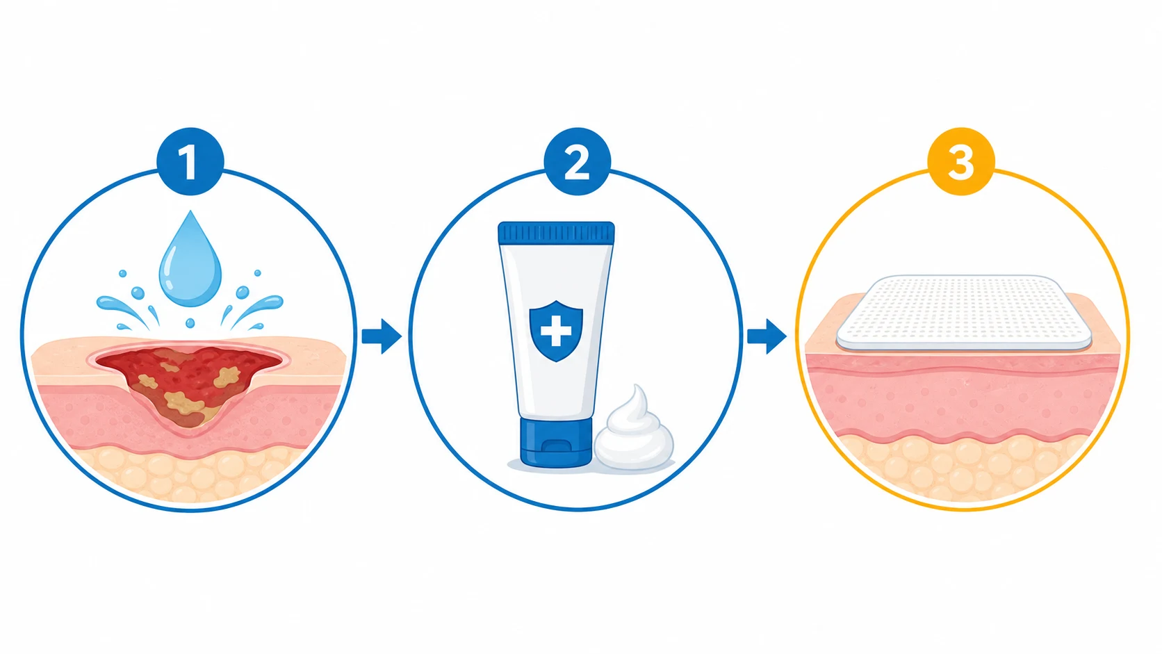
1. Cleaning and debridement. The daily wound cleaning (saline, mild soap). The debridement of the necrotic tissue (the eschar — the surgical tangential excision).[2][1]
2. Topical antimicrobials.[1]
- Silver sulfadiazine (the commonest — broad-spectrum; the side effects: leucopenia, argyria, the sulfa allergy).
- Mafenide acetate (penetrates the eschar — the deeper; the painful; the carbonic anhydrase inhibitor → metabolic acidosis).
- Nanocrystalline silver dressings (Acticoat — the sustained release; the less frequent changing).
- Honey (the osmotic, the antibacterial; the less established).[1]
3. Dressings. The biological (Biobrane — the temporary), the synthetic (the hydrocolloid, the foam), the skin substitutes (Integra — the dermal regeneration template).[1]
4. Early excision and grafting. The modern standard — the tangential excision of the eschar (within 3 to 7 days) + the autograft (the split-thickness skin graft). The reduces the infection, the mortality, the length of stay. The allograft (the cadaveric — the temporary) and the xenograft (the porcine) for the extensive burns (the not enough the autograft).[1][1]
Topical antimicrobials — the agent-by-agent detail
The topical agent is the cornerstone of burn wound infection prevention: it keeps the bacterial load on the wound surface below the <10^5 colony-forming units (CFU) per gram of tissue threshold that defines invasion. Every agent is a trade-off between antimicrobial spectrum, eschar penetration, pain, and systemic toxicity. No single agent is ideal for every wound.[4][5]
Topical antimicrobial agents for the burn wound — the agent, the spectrum, the penetration, the catch
| Agent | Spectrum | Eschar penetration | Key adverse effects / cautions | Dressing change frequency |
|---|---|---|---|---|
| Silver sulfadiazine (SSD) 1% cream | Broad (Gram-positive, Gram-negative, some Candida) | Moderate — does NOT penetrate eschar well | Leucopenia (transient, ~5-15%, recover on cessation); argyria; sulfa allergy; stains; may retard epithelialisation; painful removal | Once or twice daily |
| Mafenide acetate 11% cream / 5% solution | Broad including Pseudomonas | Excellent — penetrates eschar & cartilage (the only one) | Painful on application; carbonic anhydrase inhibition → metabolic acidosis (hyperventilation compensatory); allergic rash; haemolysis in G6PD | Twice daily (q12h) |
| Silver nitrate 0.5% solution | Broad incl. some fungi | Poor — surface only | Hyponatraemia, hypokalaemia, hypocalcaemia, hypomagnesaemia, methaemoglobinaemia (leaches electrolytes); stains everything black/brown; messy (soaked dressings) | Soaked dressings rewetted q4-6h, changed daily |
| Nanocrystalline silver (Acticoat / Aquacel Ag) | Broad (sustained Ag+ release; effective vs MRSA, VRE, Pseudomonas) | Good — sustained release over days | Stains; rare argyria; can dry out (keep moist); cost | Every 3 days (Acticoat) or up to 7 days (Aquacel Ag) — the major advantage |
| Bacitracin / polymyxin B / mupirocin ointments | Gram-positive (Staph, Strep); mupirocin for MRSA nasal/skin | Good on superficial wounds | Contact dermatitis; mupirocin resistance with overuse | Daily |
| Povidone-iodine | Broad | Poor | Cytotoxic to keratinocytes/fibroblasts (impairs healing); metabolic acidosis; thyroid dysfunction with large areas; electrolyte disturbance | Daily — AVOID on large wounds |
| Honey / medical-grade (Medihoney) | Broad (osmotic, low pH, H2O2) | Moderate | Sticky; rare allergy; less evidence than silver | Daily to alternate-day |
Why nanocrystalline silver (Acticoat) displaced silver sulfadiazine as the workhorse in many burn units
| Feature | Silver sulfadiazine 1% cream | Nanocrystalline silver (Acticoat) |
|---|---|---|
| Mechanism | Ag+ + sulfonamide; one-time release per application | Sustained nanocrystalline Ag+ release over 24-72 h (or 7 d for Aquacel Ag) |
| Antimicrobial potency | Good, but requires daily reapplication | More potent per unit silver; effective vs MRSA, VRE, P. aeruginosa, Candida |
| Dressing change frequency | Once-twice daily — painful, labour-intensive, costly | Every 3 days (Acticoat) to 7 days (Aquacel Ag) — major analgesic, nursing-time, and cost benefit |
| Pain | Moderate on application/removal | Low once applied; kept moist |
| Effect on healing | May retard epithelialisation | Neutral or mildly favourable |
| Leucopenia | Yes (transient, 5-15%) | No |
| Evidence | Decades of use; the historical standard | Tredget 1998 RCT: fewer infections, less pain, lower costs vs SSD/nitrate — drove adoption< Cite id="10" /> |
Dressings — biological, synthetic, and skin substitutes
The dressing has three jobs: (1) protect the wound from the environment, (2) deliver a topical antimicrobial, and (3) — when it is a skin substitute — replace lost dermis/epidermis. The choice depends on the wound depth, the TBSA (is there enough donor skin?), and the goal (temporary cover vs permanent closure).[5][1]
Wound cover options for the burn — biological, synthetic, and engineered
| Dressing / substitute | What it is | Indication | Advantages | Limitations |
|---|---|---|---|---|
| Biobrane | Biosynthetic: nylon fabric mesh bonded to silicone film + porcine collagen peptides | Temporary cover for clean superficial-to-intermediate partial-thickness burns; donor sites; clean excised wounds | Adheres to the wound within 24-48 h; reduces pain; allows inspection (semi-transparent); dressing change every 3-5 d; peels off as the wound re-epithelialises | MUST be removed if infection develops beneath (traps bacteria); not for full-thickness or contaminated/infected wounds; acts as a nidus |
| Allograft (cadaveric split-thickness skin) | Human cadaver skin | Temporary biological cover over large excised wounds ("test graft"); bridges to autograft | Adheres, vascularises temporarily (then rejected ~2-4 wks); excellent cover; conforms; reduces pain, heat/water loss | Limited supply; cost; risk of disease transmission (screened); rejection → must replace before autograft |
| Xenograft (porcine skin) | Pig skin | Temporary cover; partial-thickness; paediatric | Cheap, available; reduces pain; good for partial-thickness | Rejected faster than allograft; not vascularised |
| Amniotic membrane | Placental amnion | Temporary cover; paediatric partial-thickness; eye/facial burns | Cheap; anti-inflammatory, anti-scarring, pain relief; promotes epithelialisation | Variable quality; infection-screening required; not durable for large wounds |
| Hydrocolloid / foam / film dressings | Synthetic occlusive/semi-occlusive | Donor sites; small partial-thickness; as an outer layer | Comfortable; maintain moist environment; reduce pain | Not antimicrobial alone (some silver-impregnated); maceration if too occlusive |
| Integra (bilayer dermal regeneration template) | Outer silicone "epidermis" + inner collagen/chondroitin-6-sulfate "dermis" that vascularises in ~2-3 wks → silicone peeled off → thin auto-epidermal graft | Full-thickness and large deep burns where donor skin is scarce; reconstructive burns; scalp/face | Provides neodermis → better pliability, cosmesis, and less contracture than meshed grafts alone; expands the usable cover | Two-stage (vascularise then graft); infection beneath the silicone must be watched; cost; requires a trained team |
| Alloderm (acellular dermal matrix) | Human cadaver dermis, decellularised | Combined with thin autograft for full-thickness | Provides dermal scaffold; single-stage with autograft | Variable take; cost |
| Cultured epithelial autografts (CEA) | Patient's keratinocytes grown in vitro (3-4 wks) | Massive burns (>80% TBSA) with no donor sites | Only option when donor skin is exhausted | Fragile; takes 3-4 wks to grow; poor take on colonised wounds; very expensive; contracture-prone |
The decision tree — choosing the wound cover for a given burn
- Superficial partial-thickness, clean (no infection), out to 48 h — consider Biobrane or a biosynthetic. Biobrane adheres to a clean partial-thickness wound within 24-48 h, dramatically reduces pain, and peels off as the wound heals in 7-14 days. Contraindicated if the wound is infected or heavily colonised — it then traps bacteria and becomes a nidus.[5]
- Deep partial- or full-thickness, <48-72 h, will be excised — plan EARLY EXCISION AND GRAFTING (see below). The definitive cover is the autologous split-thickness skin graft (STSG).
- Extensive burn — not enough donor skin to cover at once — use temporary biological cover (allograft or Biobrane) over the excised wound to prevent infection and protein loss while donor sites re-epithelialise and can be re-harvested every 10-14 days.[1]
- Massive burn (>60-80%) — donor sites exhausted — staged Integra (neodermis) then thin epidermal graft, or cultured epithelial autografts as the last resort.[1]
- Any wound that shows signs of infection beneath a Biobrane/Integra/allograft — REMOVE IT. A trapped infection under a biologic/biosynthetic is worse than an open wound. Convert to a topical antimicrobial cream (SSD or mafenide) and re-cover once controlled.[4]
Early excision and grafting — the modern standard
Early tangential excision and autografting — the single biggest mortality reduction in modern burn care
- THE PRINCIPLE. The burn eschar is dead, devascularised, protein-rich tissue — an ideal culture medium that the host defences and antibiotics cannot reach. Leaving it in place guarantees colonisation → invasion → sepsis. Tangential excision slices the necrotic tissue off in thin layers down to viable (bleeding) tissue; the wound is then closed with an autograft, eliminating the culture medium entirely.[1][1]
- TIMING — within 3-7 days, ideally staged. Excision of the full-thickness and deep partial-thickness wounds within the first week (often starting day 3-5 once the patient is resuscitated and stable). For very large burns the excision is staged (e.g., 15-20% TBSA per session) to limit blood loss and the operative insult.
- BLOOD LOSS — the operative challenge. Burn excision is bloody: historically up to 0.5-1 mL/cm² excised, or 3-5% of blood volume per 1% TBSA. Reduce loss with tourniquets (limbs), sub-eschar adrenaline (epinephrine) infiltration, topical thrombin/spray, and cell-salvage. Plan cross-match; have massive transfusion protocol ready for large excisions.[1]
- GRAFT CHOICE. (a) Autograft — split-thickness skin graft (STSG) is the definitive cover (meshed 1:1 to 1:4 to expand, or sheet for hands/face/cosmetic). (b) Allograft (cadaveric) as temporary cover when autograft is insufficient, or as a "test graft" to confirm the bed will take. (c) Xenograft (porcine) — cheap temporary cover. (d) Integra / cultured epithelium for massive burns (see table above).[5][1]
- WHY IT WORKS — the outcomes. Early excision + grafting reduces infection/sepsis, mortality, length of stay, and hypertrophic scarring, and improves metabolic control (closes the wound → reduces the hypermetabolic drive). This is the single most important advance in modern burn surgery and a high-yield exam point.[1][6]
- CONTRAINDICATIONS / CAUTIONS. Haemodynamic instability (shock not yet resuscitated — fix the patient first); inhalation injury with severe ARDS (limit the operative insult); inability to obtain donor skin; non-viable bed (necrotic fascia/muscle — excise further first); and extensive burns needing staged sessions. The patient must be fit for the operative blood loss and the temperature/fluid challenge of a large excision.[1]
The infection — colonisation, invasion, and sepsis
The burn wound infection progresses from colonisation to invasion:[1][1]
- Colonisation (the first few days) — the Gram positive (Staphylococcus aureus, the Strep). The normally the present on the skin.
- Invasion (the days to weeks) — the Gram negative (Pseudomonas aeruginosa — the classic, the green pigment; Klebsiella, Acinetobacter, E. coli). The more the invasive, the more the resistant.
- Fungal (the late, the broad-spectrum antibiotic exposure) — Candida, Aspergillus, Mucor (the angioinvasive — the rapidly fatal).[1][1]
The temporal pattern of organisms — the exam favourite
The microbial flora of the burn wound changes in a predictable, time-ordered sequence that the fellowship candidate must be able to recite. The pattern reflects the source of the organism (endogenous skin/gut vs hospital environment), the selective pressure of topical/systemic antimicrobials, and the progressive breakdown of the patient's defences.[4][6]
The temporal pattern of burn wound colonisation and invasion
| Time after burn | Predominant flora | Typical organisms | Clinical relevance |
|---|---|---|---|
| Days 0-5 (early) | Gram-positive (endogenous skin flora) | Staphylococcus aureus (incl. MRSA), coagulase-negative Staph, Streptococcus pyogenes, Enterococcus | Wound colonisation; Strep pyogenes can cause early invasive cellulitis/sepsis; cover with Gram-positive agents if invasive |
| Days 5-14 (middle) | Gram-negative (hospital/environmental; gut translocation) | Pseudomonas aeruginosa (the classic), Klebsiella, Enterobacter, E. coli, Acinetobacter baumannii, Proteus, Serratia | The invasive phase; Pseudomonas ecthyma gangrenosum, septicaemia; the major driver of burn sepsis mortality |
| Day 14 onward (late) | Fungal (broad-spectrum antibiotic selection pressure) | Candida spp. (commonest fungal coloniser), Aspergillus, Mucorales (Rhizopus, Mucor) — angioinvasive, Fusarium | Fungal invasion is late, ominous, often angioinvasive (Mucor → necrosis, fatal); requires tissue biopsy (culture insensitive) |
| Late (any time, reactivation) | Viral | Herpes simplex virus (HSV) reactivation in partial-thickness facial/digital burns; CMV | HSV can mimic bacterial infection (delayed re-epithelialisation, vesicles); diagnose with PCR/PCR of vesicle; treat with aciclovir |
Diagnosing the burn wound infection
The diagnosis of the burn wound infection.[1]
- The changing wound appearance (the rapid eschar separation, the dark/brown/black discolouration, the haemorrhagic, the conversion of partial to full thickness).
- The systemic signs (the fever, the tachycardia, the hypotension, the altered mental state).
- The wound biopsy (the quantitative — the bacterial count above 10^5 per gram of tissue = the invasive).
- The blood cultures, the wound swab.[1][1]
Diagnosing burn wound infection — clinical, microbiological, and histological
- SUSPECT on CLINICAL grounds — the changing wound is the key. Look for: (a) rapid or premature eschar separation; (b) dark/brown/black or violaceous discolouration of the wound or surrounding skin; (c) haemorrhagic areas (sub-eschar bleeding without trauma); (d) conversion of a partial-thickness wound to full-thickness (the wound deepens); (e) ecthyma gangrenosum-like lesions (Pseudomonas); (f) a focal violaceous/black edge (Mucor — angioinvasion). None of these are normal — investigate.[4]
- Distinguish COLONISATION from INVASION — the quantitative biopsy is the gold standard. A surface swab only tells you what is colonising the surface (almost always positive and often unhelpful). A wound biopsy for quantitative culture: >10^5 colony-forming units (CFU) per gram of tissue = invasion; <10^4 is colonisation. Histopathology of the biopsy is even more specific: micro-organisms invading viable (non-necrotic) tissue = invasive infection.[4]
- Send BLOOD CULTURES before antibiotics (at least two sets), plus wound biopsy/tissue culture AND a surface swab for surveillance (track resistance patterns). In the stable patient, pause antibiotics to obtain cultures; in septic shock, do NOT delay antibiotics for >45 min.[3]
- Look for SYSTEMIC signs of sepsis using the ABA criteria (see below) — a CHANGE from baseline, not an absolute value. Add a lactate, and consider procalcitonin (trend more useful than single value).[3]
- DIFFERENTIAL of a "failing" wound. Always ask: is this infection, ischaemia/inadequate excision (necrotic bed under the graft), hypoxia (inhalation injury, ARDS, low cardiac output), malnutrition/hypoalbuminaemia, hypothermia, or drug effect (immunosuppression)? Treat the real cause, not just the surface.[1]
Sepsis in burns — SIRS is the baseline, so use the ABA criteria
The burn wound sepsis — the leading cause of death after the first 48 hours. The hypermetabolic state mimics sepsis (tachycardia, fever, high WBC, high CRP — all baseline elevated in burns) → hard to distinguish. The American Burn Association (ABA) criteria for the burn sepsis.[1][1]
A major burn produces a systemic inflammatory response so intense that the conventional Sepsis-2/SIRS and Sepsis-3 definitions do not work — a patient with a 50% TBSA burn will have a heart rate of 120, a temperature of 38.5°C, a WBC of 15, and a respiratory rate of 24 as their resting baseline, every day, for weeks, with no infection at all. Applying "2 of 4 SIRS criteria" would label every major burn septic from day 1. The ABA 2007 consensus solves this by defining burn sepsis as a change from the patient's own established baseline, combined with evidence of infection.[3]
The ABA 2007 consensus criteria for burn sepsis and septic shock
- THE PRINCIPLE — sepsis in a burn = a CHANGE from the patient's baseline + evidence of infection. The absolute values of heart rate, temperature, WBC etc. are unhelpful in isolation; what matters is the departure from that individual patient's stable (post-resuscitation) baseline. Document each patient's baseline HR, RR, temperature, glucose, and platelet count during the stable phase so a change is recognisable.[3]
- BURN SEPSIS = at least 3 of the following 6 criteria + evidence of infection. The six ABA parameters (any 3 of 6):
- (a) Temperature — >39°C OR <36.5°C (a swing up or down from baseline).
- (b) Progressive tachycardia — >110 bpm (or a sustained rise >20 bpm above baseline), not explained by pain/agitation.
- (c) Progressive tachypnoea — >25/min spontaneous, or minute ventilation >12 L/min if ventilated (or a rise > baseline).
- (d) Thrombocytopenia — platelet count <100 × 10^9/L (and it should be RISING in the second week after the initial post-burn fall — a fall or failure to rise is significant; <100 is a late but specific sign).
- (e) Hyperglycaemia — a rising requirement for insulin (in the known diabetic) OR a glucose >12 mmol/L (200 mg/dL) despite no change in intake/insulin (insulin resistance reflects the inflammatory surge of sepsis).
- (f) Altered mental state — new confusion, agitation, or depressed sensorium (once pain, hypoxia, sedation, and delirium are excluded).
- EVIDENCE OF INFECTION is required — at least one of: (i) a documented infection by culture/histology; (ii) a changing burn wound appearance (see diagnosis section); (iii) haemodynamic/organ-dysfunction change attributable to infection. Without infection, the criteria describe the hypermetabolic/inflammatory baseline, not sepsis.[3]
- BURN SEPTIC SHOCK = burn sepsis + requirement for vasopressors to maintain MAP ≥65 mmHg (after adequate fluid resuscitation) OR serum lactate >2 mmol/L (persisting) — the parallel with Sepsis-3's shock definition. The need for inotropes/vasopressors in an adequately resuscitated burn patient is a red flag for septic shock.[3]
- WHAT THE ABA CRITERIA DO NOT USE — and why. Note that procalcitonin, CRP, SOFA score, and the qSOFA are NOT part of the ABA definition (procalcitonin is elevated at baseline in major burns and trends more than absolute thresholds; CRP is chronically elevated; SOFA/qSOFA have not been validated in burns). Use them as adjuncts only. The changing burn wound remains the single most important clue.[3]
The antibiotics — prophylaxis is NOT recommended
Prophylactic systemic antibiotics: NOT recommended.[1]
- The routine systemic prophylaxis does NOT reduce the infection and INCREASES the resistance. The topical antimicrobials + the early excision are the prevention.[1]
- The systemic antibiotics for the DOCUMENTED infection (the invasive, the sepsis) — the culture-guided, the broad initially.[1][1]
Infection prevention in the burn patient — the bundle that works
- TOPICAL ANTIMICROBIALS on the wound, daily to thrice-weekly (silver sulfadiazine, mafenide, or nanocrystalline silver — see table). This is the primary defence — it keeps the wound-surface bacterial load below the 10^5 CFU/g invasion threshold without selecting for resistance the way systemic antibiotics do.[4]
- EARLY EXCISION AND GRAFTING (within 3-7 days) — removes the necrotic culture medium (the eschar) and closes the wound. The single biggest reduction in burn infection and mortality in modern care.[1][1]
- ENVIRONMENTAL AND INFECTION-CONTROL MEASURES. (a) Plenum or laminar-flow ventilation, room temperature 30-32°C (limit heat loss from the open wound), and humidity control. (b) Strict hand hygiene (WHO 5 moments) and contact precautions (gown, gloves) for every wound contact. (c) Dedicated, non-shared equipment (stethoscope, BP cuff, thermometer). (d) Single-room isolation or cohorting of multiply-colonised patients (MRSA, MDR-Gram-negatives). (e) Aseptic technique for dressing changes and line insertion; remove central lines as soon as possible. (f) Routine surveillance cultures (wound, sputum, urine) to track the unit's flora and resistance — guides empiric therapy if sepsis develops.[4]
- NUTRITION — enteral, early, high-protein, high-calorie. The hypermetabolic burn patient runs a 1.5-2x resting energy expenditure; malnutrition impairs wound healing and immunity and predicts infection and mortality. Start enteral feeding within 24-48 h (trophic then target).[1]
- GLYCAEMIC CONTROL — moderate targets (e.g., glucose 8-10 mmol/L) to avoid both hyperglycaemia (impairs neutrophil function, drives infection) and hypoglycaemia. Tight control (<6.1) is NOT recommended (hypoglycaemia risk).[1]
- STRESS-ULCER AND VTE PROPHYLAXIS as for any critically ill patient (PPI/H2RA; LMWH — note doses may need escalation in burns due to hypermetabolic clearance).[1]
- WHAT DOES NOT WORK — NO routine systemic antibiotic prophylaxis. Multiple RCTs and meta-analyses confirm that systemic prophylaxis (peri-operative, wound, or continuous) does NOT reduce burn wound infection and SELECTS for resistant organisms (MRSA, ESBL, VRE, Candida). Reserve systemic antibiotics for documented infection, given culture-guided and broad initially.[4]
When to give systemic antibiotics in a burn patient — and what to give
- DOCUMENTED invasive infection or sepsis (ABA criteria + infection). This is the indication. Send cultures FIRST (blood, wound biopsy, urine, sputum), then start broad empiric therapy within 1 hour, then narrow to sensitivities at 48-72 h (de-escalate).[3]
- EMPIRIC BROAD COVER (culture pending) — base on the time after burn (temporal pattern) and the unit antibiogram:
- Week 1: Gram-positive cover — flucloxacillin/vancomycin (add vancomycin/linezolid if MRSA prevalent or known colonised).
- Week 2 onward: add Gram-negative / anti-Pseudomonal cover — piperacillin-tazobactam, ceftazidime, cefepime, or meropenem ± aminoglycoside (gentamicin/tobramycin/amikacin — once-daily, monitor levels) for severe sepsis or known MDR.
- Late, broad-spectrum antibiotic exposure, suspected fungal invasion: add antifungal — echinocandin (caspofungin/micafungin) for Candida; liposomal amphotericin B if Mucorales suspected (plus urgent debridement).[4]
- SOURCE CONTROL is paramount — antibiotics alone will not control an infected eschar. Excise the infected/necrotic tissue, drain any abscess, remove infected lines, and re-cover with a clean dressing/graft. An infected Biobrane/Integra/allograft must be REMOVED.[4][1]
- DURATION — guided by clinical response and the focus: 7-14 days for most bacteraemias with source control; stop once the patient meets ABA criteria for resolution (baseline parameters return, wound improved, cultures negative). Prolonged courses select for resistance and fungal superinfection.[3]
- ANTIBIOTIC STEWARDSHIP — "start smart, then focus." Review at 48-72 h, de-escalate to the narrowest effective agent, use the shortest effective duration. Never give continuous prophylactic antibiotics to "prevent" infection in a colonised wound — that is exactly the practice proven to cause resistance.[4]
Prognosis
The infection is the leading cause of death after the first 48 hours. The prevention (the topical antimicrobials, the early excision, the environmental controls) + the prompt treatment (the culture-guided systemic antibiotics) are the core.[1][1][1]
After the first 48 hours — once shock has been survived — sepsis and multi-organ failure are the leading causes of death in the burn ICU, followed by respiratory failure (inhalation injury/ARDS) and, in children, over-whelming Gram-negative septicaemia. The mortality of an infected burn rises steeply once invasion occurs: invasive Pseudomonas or Mucor sepsis carries mortality of 40-80% despite optimal therapy, which is why prevention (excision + topical antimicrobials) is so much more effective than treatment. Long-term survivors face hypertrophic scarring, contracture, and the metabolic/neurological consequences of prolonged critical illness.[6][1]
Exam practice — SAQs
SAQ — Wound dressing selection in a 35 per cent mixed-depth flame burn
10 minutes · 10 marks
A 42-year-old man (80 kg) sustained a 35 per cent TBSA mixed-depth flame burn to the torso and both arms in a house fire 18 hours ago. He has been resuscitated per Parkland and is now stable: HR 108, BP 118/72, urine output 0.8 mL/kg/h. Wound inspection shows: (i) clean, moist, blistered, exquisitely tender red patches over the anterior chest (superficial partial-thickness, about 8 per cent); (ii) dry, pale, mottled, insensate areas over the anterior abdomen and right forearm (deep partial- to full-thickness, about 22 per cent); (iii) a circumferential full-thickness burn on the left forearm. The burns unit asks you to plan the wound cover.
SAQ — Day-6 burn wound infection: diagnosis by ABA criteria, microbiology, and the prevention bundle
10 minutes · 10 marks
A 50-year-old man (75 kg) is on day 6 of admission for a 45 per cent TBSA flame burn. He was extubated on day 3, is enterally fed, and had tangential excision with split-thickness skin grafting to the anterior torso on day 4. His documented baseline since day 3 is HR 104, RR 22, temperature 38.2°C, platelets 320 (and rising), glucose 8.5 mmol/L on a sliding scale. This morning: HR 122, RR 28, temperature 39.4°C, platelets 95, glucose 14 mmol/L despite unchanged insulin, and he is mildly confused. The grafted abdomen shows a 4 cm patch of dark, haemorrhagic discolouration with premature separation at the graft edge. Two sets of blood cultures have been drawn.
Clinical pearls
Key trials and evidence
ABA 2007 Consensus Conference — defining sepsis and infection in burns (Greenhalgh, J Burn Care Res 2007, PMID 17211294)
Source
American Burn Association Multicenter Trials Group consensus
Problem
Conventional SIRS/Sepsis criteria over-diagnose sepsis in burns (SIRS is the baseline hypermetabolic state)
Definition
Burn sepsis = at least 3 of 6 parameters (temperature, tachycardia, tachypnoea, platelet, glucose, mental state) showing a CHANGE FROM BASELINE + evidence of infection; septic shock = burn sepsis + vasopressor need or lactate >2 mmol/L
Clinical bottom line
The standard definition of sepsis in burns — use these criteria, not absolute SIRS thresholds, in the burn ICU. Know all 6 parameters.
Tredget 1998 — Acticoat (nanocrystalline silver) vs silver sulfadiazine/nitrate (PMID 9820837)
Design
Matched-pair randomised study of burn patients
Intervention
Acticoat nanocrystalline silver dressing vs silver sulfadiazine / silver nitrate
Key findings
Acticoat: fewer wound infections, less pain, fewer dressing changes, lower overall cost (despite higher per-dressing cost); comparable safety
Clinical bottom line
Established nanocrystalline silver as a workhorse topical — driven by the 3-day (vs daily) change interval, lower pain, and avoidance of SSD's leucopenia. Now first-line in many units.
Church 2006 — Burn wound infections review (Clin Microbiol Rev, PMID 16614254)
Source
Definitive review of burn wound microbiology, prevention, and treatment
Key teaching
Temporal colonisation pattern (Gram-positive → Gram-negative → fungal); quantitative biopsy >10^5 CFU/g defines invasion; topical antimicrobials + early excision are prevention; systemic prophylaxis NOT recommended (drives resistance)
Clinical bottom line
The canonical reference for the microbiology and prevention of burn infection — cite for the temporal pattern, the biopsy threshold, and the no-prophylaxis principle.
Williams 2009 — Leading causes of death after burn injury (Crit Care, PMID 19995436)
Design
Single-centre review of deaths after major burn (paediatric burn centre)
Key finding
After the first 48 h, sepsis/multi-organ failure is the leading cause of death (~50-70% of deaths), followed by respiratory failure (inhalation injury/ARDS) and, in children, overwhelming Gram-negative septicaemia
Clinical bottom line
Frames the central place of infection prevention in burn critical care — survival past resuscitation depends on preventing and treating sepsis.
Burn excision and grafting evidence — outcomes of early tangential excision
Intervention
Early (within 3-7 days) tangential excision + autograft vs conservative (expectant) eschar separation
Outcomes
Early excision reduces infection/sepsis, mortality, length of stay, and hypertrophic scarring; improves metabolic control (closes the wound → reduces hypermetabolic drive)
Operative challenge
Blood loss historically 0.5-1 mL/cm² excised (~3-5% blood volume per 1% TBSA); mitigate with tourniquets, sub-eschar adrenaline, topical thrombin, cell salvage; staged for large burns (15-20% TBSA per session)
Clinical bottom line
The single biggest mortality and infection reduction in modern burn surgery — the standard of care for full-thickness and deep partial-thickness burns. A guaranteed exam point.
Red flags
References
- [1]Wang PH, et al. Recent advances in molecular mechanisms of skin wound healing and its treatments Front Immunol, 2024.PMID 38835782
- [2]Granick MS, et al. Management of Acute Wounds-Expert Panel Consensus Statement Adv Wound Care (New Rochelle), 2024.PMID 38618741
- [3]Greenhalgh DG, Saffle JR, Holmes JH 4th, et al. Risk factors for development of critical limb ischemia -- a survey of diabetic vs. nondiabetic population Prilozi, 2006.PMID 17211294
- [4]Church D, Elsayed S, Reid O, Winston B, Lindsay R. Clinically relevant chromosomally encoded multidrug resistance efflux pumps in bacteria Clin Microbiol Rev, 2006.PMID 16614254
- [5]Rowan MP, Cancio LC, Elster EA, et al. Disentangling direct and growth-mediated influences on early survival: a mechanistic approach J Anim Ecol, 2015.PMID 25882771
- [6]Williams FN, Herndon DN, Hawkins HK, et al. Cytosolic N-terminal arginine-based signals together with a luminal signal target a type II membrane protein to the plant ER BMC Plant Biol, 2009.PMID 19995436
- [7]Tredget EE, Shankowsky HA, Groeneveld A, Burrell R. A guide to oral anticoagulant therapy. Italian Federation of Anticoagulation Clinics Haemostasis, 1998.PMID 9820837