ICU · Cardiovascular / aortic
Acute Aortic Syndromes — Dissection
Also known as Acute aortic syndrome · Aortic dissection · Stanford classification · Type A dissection · Type B dissection · Intimal flap · TEVAR · Malperfusion
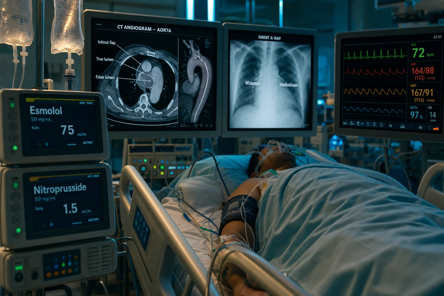
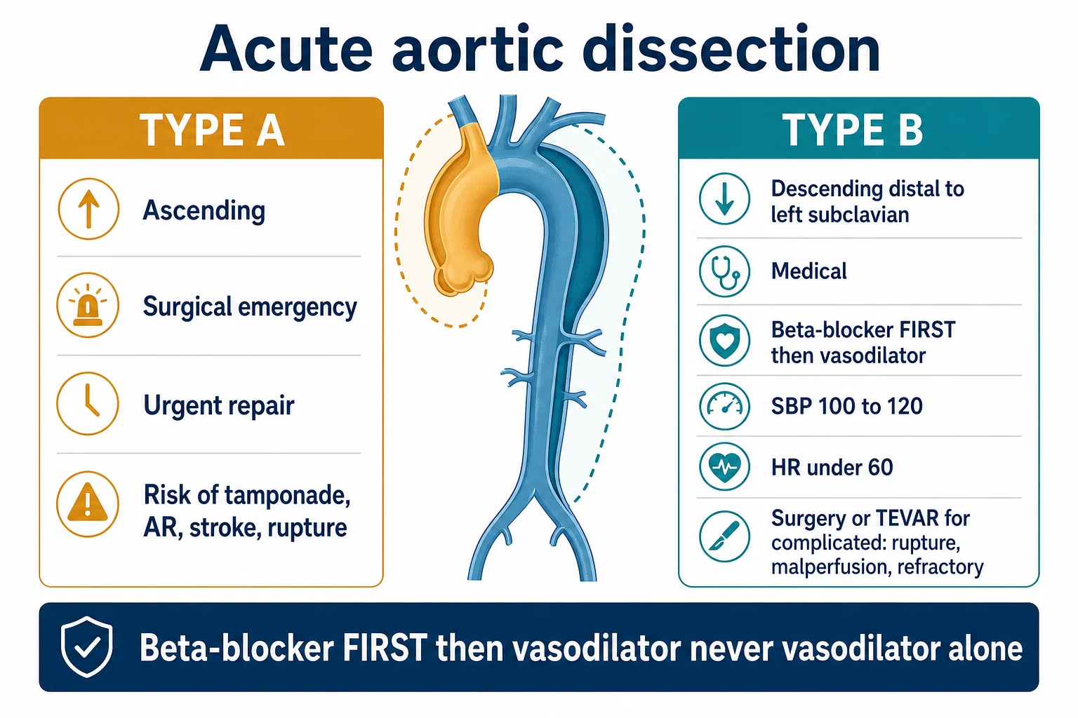
Acute aortic dissection is classified by the Stanford system: Type A (involving the ascending aorta — a surgical emergency with a high risk of rupture, tamponade, aortic regurgitation, and stroke) and Type B (descending aorta, distal to the left subclavian — managed medically with aggressive BP control). The classic presentation is a tearing chest or back pain, maximal at onset, with a pulse deficit or a new aortic regurgitation murmur. The diagnosis is by CT angiography (the intimal flap, the true and the false lumen). Type A requires urgent surgery; Type B is managed with a beta-blocker FIRST (to control the heart rate and the sheer stress), then a vasodilator (to lower the BP to SBP 100-120). Never give a vasodilator without a beta-blocker first — the reflex tachycardia worsens the dissection.
On this page & tools
Your progress
Saved locally on this device.
Target exams
Overview & definition
Acute aortic dissection is a tear in the aortic intima that allows blood to enter the aortic media, creating a false lumen that propagates along the aorta, potentially obstructing branch vessels and rupturing. It is classified by the Stanford system, which determines the management:[1]
- Type A — involves the ascending aorta (regardless of the origin). A surgical emergency — the risk of rupture, tamponade, aortic regurgitation, and stroke is high; the mortality without surgery is 1-2 per cent per hour.
- Type B — involves the descending aorta (distal to the left subclavian artery), without the ascending. Managed medically (BP control); surgery or endovascular repair only for complications.[1]
Clinical presentation
The classic presentation:[1]
- Tearing or ripping chest or back pain — sudden onset, maximal at onset (unlike the gradual onset of ACS pain); may migrate as the dissection extends distally.
- A pulse deficit — a blood pressure difference between the arms (over 20 mmHg) or an absent pulse.
- A new aortic regurgitation murmur (the dissection flap disrupts the aortic valve).
- Neurological symptoms — a stroke or syncope (from carotid involvement).
- Hypotension or shock (from tamponade, rupture, or severe AR).
- Mesenteric or renal ischaemia (abdominal pain, AKI — from visceral artery involvement).
Risk factors: hypertension (the commonest), connective tissue disease (Marfan, Ehlers-Danlos, Loeys-Dietz), bicuspid aortic valve, aortic coarctation, pregnancy, trauma, age over 60.[1]
Imaging
- CT angiography — the gold standard; shows the intimal flap, the true and the false lumen, the extent, and the branch vessel involvement. Rapid and widely available.[1]
- Transoesophageal echo (TOE) — for the unstable patient who cannot go to CT; also assesses the AR and the pericardial effusion.[1]
- A chest X-ray — a widened mediastinum (nonspecific; not reliable for the diagnosis, but may raise the suspicion).[1]
Management
Type A — urgent surgery
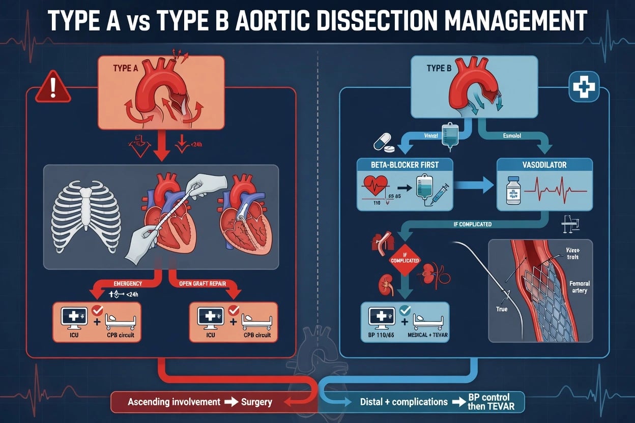
Type A dissection is a surgical emergency. The ascending aorta is repaired or replaced (with or without the arch, depending on the extent), the aortic valve may be resuspended or replaced, and the false lumen is obliterated. The mortality without surgery is very high (1-2 per cent per hour from rupture, tamponade, or malperfusion).[1]
While preparing for surgery, start the medical BP control (the same as Type B — a beta-blocker first, then a vasodilator).[1]
Type B — medical management
Type B dissection is managed medically with aggressive BP and heart-rate control:[1]
- A beta-blocker FIRST — esmolol (an ultra-short-acting beta-blocker, IV infusion) or labetalol (alpha + beta). The goal: heart rate under 60 beats per minute. This reduces the sheer stress on the dissection flap (the force of each cardiac contraction drives the dissection distally).
- Then a vasodilator — nitroprusside or nicardipine (IV infusion). The goal: SBP 100-120 mmHg. This reduces the wall stress.
- Pain control — morphine (reduces the sympathetic drive from the pain).
- Monitor for complications.
The critical rule: always give the beta-blocker first, then add the vasodilator. Giving a vasodilator without a beta-blocker causes a reflex tachycardia and increased contractility, which increases the sheer stress on the dissection flap and worsens the dissection.[1]
Complicated Type B — surgery or TEVAR
Surgery or endovascular repair for Type B dissection is indicated for:[1]
- Rupture or an impending rupture.
- Malperfusion — mesenteric ischaemia, renal failure, limb ischaemia, spinal cord ischaemia (paraplegia).
- Rapid expansion or a large aneurysm.
- Refractory pain or hypertension (not controllable medically).
- The preferred intervention is thoracic endovascular aortic repair (TEVAR) — a covered stent placed over the entry tear, decompressing the false lumen and restoring the true-lumen flow.[1]
Complications
- Rupture — into the pericardium (tamponade), the pleural space (a haemothorax), or the mediastinum. Fatal.
- Malperfusion — a stroke (carotid), mesenteric ischaemia (visceral arteries), an AKI (renal arteries), a limb ischaemia (iliac/subclavian), or a spinal cord ischaemia (paraplegia).
- Aortic regurgitation — from the dissection disrupting the valve.
- Tamponade — from the dissection rupturing into the pericardium.[1]
Red flags
Stanford classification in detail
The Stanford system is the operationally decisive classification because the ascending aorta is the surgical watershed — involvement of the ascending segment places the coronary ostia, the aortic valve, the cerebral vessels and the pericardium in immediate jeopardy, and that combination of threats is what makes Type A a surgical emergency.[1]
Stanford Type A vs Type B aortic dissection
| Feature | Type A (ascending aorta) | Type B (descending — distal to the left subclavian) |
|---|---|---|
| Incidence | ~65 per cent of dissections | ~35 per cent of dissections |
| Anatomy | Any dissection involving the ascending aorta, regardless of where the intimal tear originates or how far distally the flap propagates | Dissection confined to the descending aorta, distal to the left subclavian artery take-off |
| Untreated mortality | 1–2 per cent per hour (≈50 per cent dead within 48 hours) | ~10 per cent at 30 days (uncomplicated) |
| Definitive treatment | Emergency surgery — open repair of the ascending aorta ± arch ± valve | Medical (beta-blocker first, then vasodilator); TEVAR/surgery only if complicated |
| Pre-op BP target | SBP 100–120 mmHg, HR under 60 bpm | SBP 100–120 mmHg, HR under 60 bpm |
| Mechanism of death | Tamponade (retrograde rupture into pericardium — ~80 per cent of fatal Type A), free rupture, acute aortic regurgitation, coronary occlusion (usually RCA), stroke | Rupture, malperfusion (mesenteric, renal, spinal, limb), refractory pain/hypertension |
| Bedside herald sign | New diastolic murmur of aortic regurgitation, widened mediastinum, pericardial effusion | BP differential between arms, pulse deficit, refractory hypertension, abdominal pain |
| DeBakey equivalent | DeBakey I (arch + ascending) and DeBakey II (ascending only) | DeBakey III (descending only) |
The older DeBakey classification subdivides by extent (I = whole aorta, II = ascending only, III = descending only) but offers no management advantage over Stanford in the acute setting and is now largely an exam curiosity.[2]
The acute aortic syndrome spectrum
Acute aortic syndrome (AAS) is the umbrella term for the three non-traumatic, life-threatening aortic wall pathologies that share presentation (sudden tearing chest/back pain), mechanism (medial disruption with haemorrhage), and an analogous Stanford-based management logic. Recognising the variant matters because intramural haematoma and penetrating ulcer are easily missed on a single arterial-phase CT.[6]
Classic dissection
Intimal tear + false lumen
- Frank tear in the intima admits pulsatile blood into the aortic media, splitting it longitudinally into a true and a false lumen.
- The false lumen is typically larger and slower-flowing; it propagates distally (or, in Type A, retrogradely into the pericardium), shearing branch vessels off as it advances.
- The commonest AAS variant and the prototype against which the others are defined.
- Risk factors: hypertension (the dominant one), age over 60, male sex, Marfan / Ehlers-Danlos / Loeys-Dietz, bicuspid aortic valve, pregnancy, cocaine.
Intramural haematoma (IMH)
Bleeding within the media, no intimal tear
- Rupture of the vasa vasorum within the aortic media produces a circumferential high-attenuation wall thickening with no intimal flap and no flowing false lumen.
- Best seen on the non-contrast phase of the CT (a high-density crescent in the wall) — easy to miss on arterial phase alone.
- May regress spontaneously or evolve into a frank dissection/rupture; ascending IMH behaves aggressively and is managed like Type A.
- Account for 5–20 per cent of AAS; more common in Asian cohorts and slightly older patients.
Penetrating atherosclerotic ulcer (PAU)
Ulcer through the intima into the media
- An atherosclerotic plaque erodes through the internal elastic lamina into the media, forming a focal crater that can progress to a pseudoaneurysm, an IMH, or a localised dissection.
- Typically an elderly patient with heavy atherosclerotic burden; most often in the descending aorta.
- Asymptomatic PAU is managed medically with surveillance imaging; symptomatic, expanding, or ruptured PAU is treated with TEVAR.
Traumatic aortic injury
Deceleration / blunt chest trauma
- A distinct entity — blunt deceleration injury at the aortic isthmus (the tethered ligamentum arteriosum) producing a transection or a periaortic haematoma.
- Managed with TEVAR in almost all cases; urgent when there is a contrast extravasation, a large pseudoaneurysm, or haemodynamic compromise.
- Always co-managed with the trauma team — the patient often has competing life-threats (brain, solid organ, pelvic bleeding) that re-prioritise the aortic repair.
Diagnosis in detail
CT angiography (CTA)
Gold standard — first-line
- Sensitivity and specificity greater than 98 per cent; fast, widely available, and reproducible.
- Demonstrates the intimal flap (pathognomonic), the true and the false lumen, the entry/exit tears, the extent, and branch vessel involvement.
- Identifies complications: tamponade (a pericardial effusion with contrast), contained or free rupture (contrast extravasation), and malperfusion.
- A three-phase protocol is ideal: non-contrast (for intramural haematoma), arterial phase (CTA — the flap), and delayed phase (for false-lumen flow and end-organ perfusion).
Transoesophageal echo (TOE)
For the unstable patient
- Performed at the bedside or in theatre; ideal when the patient is too unstable for CT or while the operating room is being prepared.
- Excellent for the ascending aorta, the aortic valve (severity of AR), the presence of a pericardial effusion, and the intimal tear of Type A.
- Has blind spots (the distal descending aorta behind the trachea, and the upper abdominal aorta), is operator-dependent, and is semi-invasive (oesophageal probe in an unintubated patient).
- In the shocked patient with a suspected Type A, a bedside TOE confirming the flap and an effusion is enough to mobilise the surgical team.
D-dimer
A rule-OUT, never a rule-IN
- A D-dimer below 500 ng/mL in a patient with a low pre-test probability effectively rules out dissection (sensitivity around 97 per cent).
- Useful to spare a low-risk patient an unnecessary CTA; the ADAPT trial algorithm reduced CT use by roughly a third without missing a dissection.
- Non-specific — elevated in PE, sepsis, trauma, malignancy, pregnancy, and after surgery. A high D-dimer never confirms dissection.
- Do NOT delay imaging in a high-risk patient to await the result.
Chest X-ray
Cheap, fast, nonspecific
- A widened mediastinum (over 8 cm at the aortic knob), an obliterated aortic knob, a left apical cap, a depressed left main bronchus, or a left pleural effusion are all supportive but not diagnostic.
- Present in only ~60–90 per cent of dissections, and absent in many Type B presentations — a normal CXR does not exclude dissection.
- Its real role is to exclude alternative diagnoses (pneumothorax) and to raise the index of suspicion in the undifferentiated patient.
The ADD-Risk score (Aortic Dissection Detection Risk) sharpens the pre-test probability before imaging:[1]
- High-risk condition (1 point each, max 2): Marfan / connective tissue disease, aortic valve disease, recent aortic manipulation, family history of aortic disease, known thoracic aneurysm.
- High-risk pain feature (1 point): chest, back, or abdominal pain that is sudden, severe, and tearing/ripping.
- High-risk examination feature (1 point): pulse deficit, systolic BP differential over 20 mmHg between arms, focal neurological deficit with pain, or a new murmur of aortic regurgitation.
A score of 2 or 3 places the patient in a high-risk group in whom imaging must not be deferred — the aortic-dissection mnemonic is "the pain is maximal at onset, tearing, and migrates".[6]
Type A management — the surgical pathway
Type A dissection — the hour that matters
Recognise and resuscitate in parallel
Two large-bore IV cannulae, oxygen, continuous ECG, and a BP cuff on BOTH arms (a differential greater than 20 mmHg is itself a red flag). Take blood for group-and-save, cross-match (at least 8 units), coagulation, and a D-dimer. Analgesia with IV morphine — uncontrolled pain drives sympathetic surge and worsens the dissection. ECG to exclude a simultaneous inferior STEMI (the right coronary ostium is the one usually sheared off).
Confirm with CTA — but do not delay theatre
CT angiography is the default. In the crashing patient (shock, suspected tamponade), a bedside TOE is sufficient to commit to theatre — never send an unstable Type A across the hospital to CT.
Begin medical BP control WHILE mobilising surgery
IV esmolol (500 mcg/kg load over 1 min, then 50–200 mcg/kg/min) or labetalol (20 mg bolus, then 1–2 mg/min infusion). Target HR below 60 bpm FIRST, then SBP 100–120 mmHg. Add nitroprusside or nicardipine only after beta-blockade. The cardiothoracic surgeon and the on-call anaesthetic/perfusion team are activated the moment Type A is suspected — not after the CT report.
Definitive surgery
Median sternotomy, cardiopulmonary bypass, and deep hypothermic circulatory arrest for arch work. Resection of the torn ascending segment with an interposition Dacron graft. The aortic valve is resuspended if salvageable, or replaced (mechanical or bioprosthetic) if the root is destroyed. The coronary ostia are reimplanted (a Bentall if the root is replaced en bloc with the valve). The arch is repaired if it is involved.
Manage the life-threatening complications
Tamponade — operate, do not tap: pericardiocentesis is reserved for the patient arresting in the resus bay, because decompression can release the tamponade that was containing the retrograde rupture. Coronary occlusion (usually RCA) — restore flow at surgery, do not stent in isolation. Acute aortic regurgitation — valve repair or replacement. Stroke — individualised, often haemorrhagic on reversal of heparin at the end of the case.
Post-operative ICU care
Invasive BP monitoring (an arterial line), strict SBP 100–120 mmHg, beta-blockade continued, hourly neurological and peripheral pulse checks, watch for spinal cord ischaemia (paraplegia) especially after long-segment arch/descending repair, and serial renal function (CPB + contrast + renal malperfusion). A surveillance CTA is performed before discharge to confirm false-lumen thrombosis and to plan lifelong imaging.
Type B management — the medical pathway
Type B dissection — uncomplicated medical pathway
Beta-blocker FIRST (reduce dP/dt)
IV esmolol (500 mcg/kg loading over 1 min, then 50–200 mcg/kg/min infusion) or labetalol (20 mg bolus, then 20–80 mg every 10 min, or 1–2 mg/min infusion). Titrate to a HEART RATE under 60 bpm first. Reducing the rate of pressure rise (dP/dt) — the velocity of the systolic pressure wave — directly reduces the shear stress that propagates the flap distally.
THEN add a vasodilator
Once the heart rate is controlled, add IV nicardipine (5–15 mg/h), sodium nitroprusside (0.3–3 mcg/kg/min — beware cyanide toxicity above 10 mcg/kg/min or in renal failure), or nitroglycerin. Target SBP 100–120 mmHg. The vasodilator must ALWAYS follow the beta-blocker — never precede it.
Analgesia and the sympathetic axis
IV morphine or fentanyl for pain; pain itself drives the hypertension and tachycardia that extend the dissection. A calm, pain-controlled, beta-blocked patient is a stable dissection.
Monitor for conversion to "complicated"
ICU/HDU admission. Hourly peripheral pulses and abdominal examination; serial lactate, renal function, and urine output; serial neurological checks for paraplegia. A repeat CTA at 24–48 hours documents extent, expansion, and any evolving malperfusion. Any deterioration converts an uncomplicated Type B to a complicated one — and to TEVAR.
Transition to oral and plan lifelong care
Convert IV to oral beta-blocker (bisoprolol, metoprolol, or labetalol) plus an ACE-inhibitor or ARB once the patient is stable. Target an outpatient BP below 130/80 mmHg. Lifelong beta-blockade reduces the rate of late expansion and redissection. Schedule surveillance CT or MRI (MRI preferred in younger patients and in Marfan to spare radiation) at 1, 3, 6, and 12 months, then annually.
Complicated Type B — TEVAR
Complicated Type B — TEVAR pathway
Define the complication
A Type B becomes "complicated" with ANY of: rupture or impending rupture (a contained haematoma, a haemothorax, haemodynamic compromise); malperfusion syndrome (mesenteric, renal, spinal, or limb ischaemia); rapid expansion on serial imaging; an aneurysmal degeneration to over 55–60 mm; or refractory pain/hypertension despite maximal medical therapy. Each of these is an independent indication for intervention.
Choose the access and the landing zones
TEVAR is delivered via a femoral artery cut-down or percutaneous access. The proximal landing zone is a healthy segment of aorta (usually 2 cm of non-dissected wall proximal to the entry tear), and the distal landing zone is the healthy aorta below the tear. Coverage of the left subclavian may be required — pre-stage with a carotid-subclavian bypass if the vertebral or the internal mammary (a future LIMA graft) is dominant.
Deploy the stent graft
A covered stent is advanced over a stiff wire and deployed across the entry tear. The graft excludes the false lumen from systemic pressure, restores true-lumen flow, decompresses the false lumen, and relieves branch vessel malperfusion. Technical success exceeds 90 per cent in experienced centres.
Address malperfusion explicitly
If a branch vessel remains under-perfused after the entry tear is covered, perform percutaneous fenestration (a window cut in the flap to equalise true/false lumen pressure) and/or a branch-vessel stent (SMA, renal, iliac). Mesenteric malperfusion with rising lactate mandates a laparotomy for bowel assessment — a revascularised but necrotic bowel still kills the patient.
Watch for spinal cord ischaemia
Coverage of intercostal arteries that feed the artery of Adamkiewicz produces paraplegia in 2–5 per cent of TEVAR cases. Prophylaxis is a lumbar CSF drain (target CSF pressure 8–10 mmHg) and an augmented mean arterial pressure. Any new leg weakness post-TEVAR is spinal cord ischaemia until proven otherwise — drop the CSF pressure, raise the MAP, and call vascular.
Long-term surveillance
A residual patent false lumen is the substrate for late aneurysmal degeneration and rupture. Surveillance CTA at 1, 6, and 12 months, then annually, looking for endoleak (a type I or III is a re-intervention), false-lumen expansion, or a new distal tear.
Malperfusion syndrome — the hidden killer
Malperfusion is the single most important determinant of survival in both Type A and Type B. The mechanism is one of two: dynamic obstruction (the dissection flap prolapses across the branch ostium with each systole, collapsing the true lumen) or static obstruction (the flap is fixed across the ostium, thrombosing the origin). Both can coexist.[6]
Coronary
Usually the right coronary
- The right coronary ostium is sheared off by the dissection flap far more often than the left — presenting as an inferior STEMI in a hypertensive patient with a widened mediastinum.
- Always consider dissection before stenting an inferior STEMI in an atypical patient — stenting the coronary without repairing the dissection is futile and dangerous.
- Management is surgical repair of the dissection, which restores coronary flow by re-establishing the root and the true lumen.
Mesenteric
Bowel ischaemia
- Dissection flap covers the SMA ostium; presents as severe abdominal pain out of proportion to the examination, a rising lactate, and a metabolic acidosis.
- The mortality of mesenteric malperfusion with bowel necrosis exceeds 50 per cent — the highest of any single complication.
- Management: urgent TEVAR/fenestration to restore SMA flow, then a laparotomy for a second-look assessment of bowel viability.
Renal
AKI and refractory hypertension
- The flap covers a renal ostium, producing an AKI and a renin-mediated refractory hypertension from the ischaemic kidney.
- A unilateral renal malperfusion can be silent (the contralateral kidney compensates); a bilateral malperfusion produces a sudden, oliguric, dialysis-requiring AKI.
- Management: TEVAR with renal artery stenting, or a surgical bypass.
Spinal cord
Paraplegia
- Occlusion of the intercostal/lumbar arteries feeding the artery of Adamkiewicz produces a devastating anterior spinal artery syndrome — a flaccid paraplegia with a sensory level.
- Management: lumbar CSF drainage to raise the spinal cord perfusion pressure (MAP minus CSF pressure), an augmented MAP with noradrenaline, and avoidance of any further hypotension.
Limb
Acute limb ischaemia
- The dissection extends into the iliac or the subclavian, producing a pulseless, painful, cold, paraesthetic limb (the 6 Ps).
- Often the most visible sign of an otherwise occult Type B — a pulse deficit IS the dissection until proven otherwise.
- Management: TEVAR with branch-vessel stenting, or a surgical bypass; fasciotomy if the ischaemia has been prolonged.
Cerebral
Stroke or syncope
- Involvement of the arch vessels (innominate, left common carotid) produces a hemispheric stroke; a transient occlusion produces syncope.
- Syncope at the onset of "chest pain" in a hypertensive patient is a dissection until proven otherwise — the transient cerebral hypoperfusion from a flap covering the carotid.
- The stroke may be haemorrhagic — the dissection patient is often anticoagulated on the bypass circuit, and the post-arrest CT brain must be reviewed before any antithrombotic is given.
Complications and prognosis
Aortic dissection outcomes
Evidence and trials
Key trials and guidelines in aortic dissection
2022 ACC/AHA Guideline (Isselbacher, Circulation, PMID 36322642)[1] — the contemporary standard. Type A is surgery; Type B uncomplicated is medical with the beta-blocker-first dogma; complicated Type B is TEVAR. Adds the ADD-Risk score for pre-imaging stratification and refines the long-term surveillance imaging schedule.
2014 ESC Guideline (Erbel, European Heart Journal, PMID 25173340)[2] — the European parallel. Same Stanford-based management logic; emphasises intramural haematoma and PAU as part of the AAS spectrum and codifies the role of TOE in the unstable patient.
INSTEAD-XL (Nienaber, Circulation, 2013, PMID 23922146)[3] — randomised TEVAR plus optimal medical therapy vs medical therapy alone in UNCOMPLICATED Type B. At 5 years, the TEVAR arm had better false-lumen thrombosis (aortic remodelling) and a trend to fewer late aortic interventions; no early survival difference. It established TEVAR as a reasonable option in carefully selected uncomplicated Type B, but did not mandate it as first-line.
ADSORB (Brunkwall, EJVES, 2014, PMID 24962744)[4] — randomised TEVAR vs medical in acute uncomplicated Type B. At 1 year, TEVAR promoted favourable aortic remodelling (false-lumen thrombosis) — consistent with INSTEAD-XL. Mortality was low in both arms, confirming that uncomplicated Type B is safe on medical therapy in the short term.
Pape, JACC Historical Breakthroughs (PMID 33004136)[5] — the IRAD-derived five-decade overview of Type A. Surgical mortality has fallen from over 30 per cent to roughly 10–20 per cent; the dominant drivers of the improvement are earlier diagnosis (CTA), faster transfer to theatre, and better perfusion and cerebral-protection strategies.
Golledge & Eagle, Lancet, 2008 (PMID 18603160)[6] — the canonical Lancet review of acute aortic dissection. The source for the clinical presentation triad (sudden tearing pain, pulse deficit, neurological deficit), the malperfusion phenotype table, and the framing of AAS as a spectrum.
Exam practice
SAQ — Type B dissection with mesenteric malperfusion
10 minutes · 10 marks
A 66-year-old hypertensive man presents with sudden severe tearing back pain radiating to the abdomen. BP 184/102 (right arm), 152/94 (left arm). HR 108 sinus. CXR shows a widened mediastinum. CT angiography confirms a dissection originating distal to the left subclavian artery, extending to the aortic bifurcation. The superior mesenteric artery arises from the false lumen. Creatinine 168 (baseline 92). Lactate 4.6. Severe, disproportionate abdominal pain.
Differential diagnosis — the four killers
Aortic dissection
Tearing, migrating
- Sudden onset, maximal at onset, tearing/ripping quality, may migrate from chest to back.
- BP differential over 20 mmHg, a pulse deficit, a new AR murmur, a focal neurological deficit.
- A widened mediastinum on CXR; an intimal flap on CTA is diagnostic.
Acute coronary syndrome
Crushing, gradual crescendo
- Builds to a crescendo (not maximal at onset), crushing/pressure, radiates to arm or jaw.
- ECG changes (ST elevation or depression, T-wave inversion); troponin rise (timing-dependent).
- No pulse deficit; equal BPs in both arms.
Pulmonary embolism
Pleuritic, dyspnoea
- Sudden-onset pleuritic chest pain, dyspnoea, hypoxia; risk factors for VTE.
- Right-heart strain on echo (RV dilation, McConnell sign); a filling defect on CTPA.
- ECG may show sinus tachycardia, S1Q3T3 (nonspecific); a D-dimer is also elevated (does NOT distinguish from dissection).
Tension pneumothorax / Boerhaave
Sudden, pleuritic or post-emetic
- Pneumothorax: sudden pleuritic pain, dyspnoea, hyperresonance and absent breath sounds, tracheal deviation.
- Boerhaave (oesophageal rupture): severe retching/vomiting followed by excruciating chest pain, subcutaneous emphysema, a left pleural effusion; diagnosis by contrast oesophagram/CT.
Special circumstances
Pregnancy (especially Marfan)
Third trimester / postpartum
- Hormonal (relaxin softens the aortic wall) and haemodynamic (increased volume and stroke volume) changes raise the dissection risk, especially in Marfan with an aortic root over 40 mm.
- Pre-pregnancy counselling is essential — a Marfan root over 40 mm warrants prophylactic root replacement BEFORE conception. Beta-blocker throughout pregnancy; serial echocardiography of the root.
- Type A in pregnancy: emergency surgery (with caesarean at the same anaesthetic if viable). Type B: medical, with early delivery if the fetus is mature.
Iatrogenic / catheter-induced
Post-cardiac catheter / guidewire
- A guidewire or a catheter tip that intusses the intima can produce a localised dissection — usually in the descending aorta, often retrograde into the ascending.
- Suspect it in any patient with new chest pain, a neurological deficit, or a haemodynamic change after a catheter procedure.
- Most small catheter-related dissections are managed medically; ascending involvement is managed as Type A.
Cocaine / sympathomimetic
Severe transient hypertension
- Cocaine produces a surging hypertension and tachycardia — a massive acute shear load on a (often already abnormal) aorta.
- A young hypertensive male with tearing chest pain after cocaine use is dissection until proven otherwise; the same is true of amphetamine and MDMA toxicity.
- Avoid pure beta-blockers in acute cocaine toxicity (unopposed alpha) — favour a combined alpha-beta blocker (labetalol) plus a benzodiazepine, then titrate the vasodilator.
Connective tissue disease
Marfan, Ehlers-Danlos, Loeys-Dietz
- Marfan (fibrillin-1) — aortic root dilation, the highest dissection risk; beta-blockade and elective root replacement above 50 mm (40 mm in Loeys-Dietz, which is more aggressive).
- Vascular Ehlers-Danlos (type III collagen) — fragile arteries and bowel; diagnosis often only at complication. Avoid angiography if possible (groin/femoral rupture).
- Loeys-Dietz (TGF-beta receptor) — aggressive, early, widely metastatic arterial aneurysms; intervene at smaller diameters.
Clinical pearls [1]
Additional red flags
References
- [1]Isselbacher EM, Preventza O, Hamilton Black Iii J, et al. 2022 ACC/AHA Guideline for the Diagnosis and Management of Aortic Disease: A Report of the American Heart Association/American College of Cardiology Joint Committee on Clinical Practice Guidelines Circulation, 2022.PMID 36322642
- [2]Erbel R, Aboyans V, Boileau C, et al. 2014 ESC Guidelines on the diagnosis and treatment of aortic diseases: Document covering acute and chronic aortic diseases of the thoracic and abdominal aorta of the adult. The Task Force for the Diagnosis and Treatment of Aortic Diseases of the European Society of Cardiology (ESC) Eur Heart J, 2014.PMID 25173340
- [3]Nienaber CA, Kische S, Rousseau H, et al. Endovascular repair of type B aortic dissection: long-term results of the randomized investigation of stent grafts in aortic dissection trial Circ Cardiovasc Interv, 2013.PMID 23922146
- [4]Brunkwall J, Kasprzak P, Verhoeven E, et al. Endovascular repair of acute uncomplicated aortic type B dissection promotes aortic remodelling: 1 year results of the ADSORB trial Eur J Vasc Endovasc Surg, 2014.PMID 24962744
- [5]Pape LA, Awais M, Woznicki EM, et al. Type A Aortic Dissection-Experience Over 5 Decades: JACC Historical Breakthroughs in Perspective J Am Coll Cardiol, 2020.PMID 33004136
- [6]Golledge J, Eagle KA. Acute aortic dissection Lancet, 2008.PMID 18603160