ICU · Cardiovascular / ACS
Acute Coronary Syndromes (STEMI/NSTEMI) in the ICU
Also known as Acute coronary syndrome · ACS · STEMI · NSTEMI · Unstable angina · Primary PCI · Dual antiplatelet therapy · DAPT · Ticagrelor · PLATO trial · SHOCK trial · GRACE score
Acute coronary syndromes (ACS) span the spectrum of acute myocardial ischaemia — unstable angina (UA), NSTEMI and STEMI — now unified under the Fourth Universal Definition of Myocardial Infarction (Thygesen 2018, Circulation), which classifies MI into five pathophysiological types (spontaneous, supply-demand, sudden death, PCI-related and CABG-related). STEMI (complete coronary occlusion) is treated with primary PCI within 90 minutes or thrombolysis within 30 minutes; NSTEMI is risk-stratified by the GRACE score for early invasive PCI within 24 hours. All ACS receives aspirin plus a P2Y12 inhibitor (ticagrelor preferred per PLATO, NEJM 2009) plus parenteral anticoagulation (heparin or bivalirudin per HORIZONS-AMI). In MI complicated by cardiogenic shock, early revascularisation is the standard (SHOCK, NEJM 1999); routine IABP confers no mortality benefit (IABP-SHOCK II, NEJM 2012), culprit-only PCI beats multivessel PCI in shock (CULPRIT-SHOCK, NEJM 2017), and early routine VA-ECMO did not improve outcomes (ECLS-SHOCK, NEJM 2023). The mechanical complications of MI — VSD, papillary muscle rupture and free wall rupture — classically occur at days 2 to 7 and present with sudden haemodynamic collapse.
On this page & tools
Your progress
Saved locally on this device.
Target exams
Red flags
Overview & definition
Acute coronary syndrome (ACS) encompasses the spectrum of acute myocardial ischaemia: STEMI (ST-elevation — a complete coronary artery occlusion requiring immediate reperfusion), NSTEMI (non-ST-elevation with a troponin rise — a partial or subtotal occlusion), and unstable angina (ischaemia without a troponin rise). The ICU manages the complications — cardiogenic shock, the mechanical complications of MI, the life-threatening arrhythmias, and the post-cardiac-arrest ACS patient.[1][2][1]

STEMI management
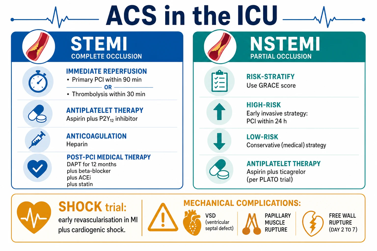
Immediate (the MONA-B plus reperfusion):[2][1]
- Aspirin 300 mg loading.
- A P2Y12 inhibitor — ticagrelor 180 mg loading (preferred over clopidogrel per the PLATO trial, NEJM 2009, which showed ticagrelor reduced cardiovascular death and all-cause mortality versus clopidogrel in ACS without an increase in major bleeding)[2] — or prasugrel 60 mg, or clopidogrel 600 mg.
- Anticoagulation — unfractionated heparin or bivalirudin.
- Reperfusion:
- Primary PCI (preferred) — door-to-balloon within 90 minutes.
- Thrombolysis (if PCI unavailable within 120 minutes) — tenecteplase or alteplase; then transfer for PCI (pharmacoinvasive).
Post-reperfusion (secondary prevention):[1]
- Dual antiplatelet therapy (DAPT) for 12 months (aspirin plus the P2Y12 inhibitor).
- Beta-blocker (reduces mortality; caution in acute heart failure and hypotension).
- ACE inhibitor / ARB (especially if LV dysfunction, diabetes, or hypertension).
- High-intensity statin (atorvastatin 80 mg).
- Aldosterone antagonist (eplerenone) if LVEF under 40 per cent with heart failure.[1]
NSTEMI management
- Risk stratification with the GRACE score (Global Registry of Acute Coronary Events) — predicts the in-hospital and the 6-month mortality.[1]
- High-risk (refractory ischaemia, haemodynamic instability, dynamic ECG changes, a rising troponin, a GRACE score above 140): early invasive PCI within 24 hours.[1]
- Low-risk: conservative management with a delayed or outpatient angiography.
- Antiplatelet: aspirin plus ticagrelor (PLATO — ticagrelor superior to clopidogrel).[2]
- Anticoagulant: heparin, fondaparinux, or bivalirudin.[1]
ACS with cardiogenic shock — the SHOCK trial
The SHOCK trial (Hochman, NEJM 1999) randomised patients with MI complicated by cardiogenic shock to early revascularisation (PCI or CABG within 24 hours) versus initial medical stabilisation. Early revascularisation reduced the 6-month and the long-term mortality (at 6 months 50 vs 63 per cent; at 6 years 47 vs 56 per cent), even though the 30-day mortality was not significantly different.[1] The principle: in MI with cardiogenic shock, early revascularisation is the standard.[1]
The mechanical complications of MI
These occur at days 2 to 7 post-MI (the necrotic myocardium weakens and ruptures):[1]
- Ventricular septal rupture (VSR/VSD) — a new pansystolic murmur at the lower left sternal border; sudden haemodynamic collapse with a left-to-right shunt; echo diagnostic; surgical or percutaneous closure.
- Papillary muscle rupture — acute severe mitral regurgitation; the murmur may be soft (low forward flow in shock); pulmonary oedema; echo; urgent MV repair or replacement.
- Free wall rupture — cardiac tamponade with sudden death or electromechanical dissociation; echo (a pericardial effusion with RA/RV collapse); surgical repair.[1]
Arrhythmias complicating ACS
- VT/VF — amiodarone, defibrillation; beta-blockade (reduces the arrhythmia); correct the potassium and magnesium.
- Atrial fibrillation — rate control (beta-blocker, amiodarone); anticoagulate.
- Complete heart block — especially in inferior MI (the RCA supplies the AV node); a temporary pacemaker.[1]
Red flags
The Fourth Universal Definition of Myocardial Infarction (five types)
The 2018 Fourth Universal Definition of MI (Thygesen, Circulation 2018) standardises how an MI is diagnosed and classified worldwide.[9] A diagnosis of acute MI requires a rise and/or fall of cardiac troponin (with at least one value above the 99th percentile upper reference limit) together with at least one of: symptoms of myocardial ischaemia; new ischaemic ECG changes; development of pathological Q waves; imaging evidence of new loss of viable myocardium or new regional wall motion abnormality; or identification of a coronary thrombus by angiography or autopsy.[9][1]
The definition then stratifies MI into five pathophysiological types, each with fundamentally different management implications — the type determines whether the patient needs a catheter or whether the underlying trigger is the real target:[9]
Type 1
Spontaneous MI (plaque rupture)
- Atherosclerotic plaque rupture, ulceration, erosion or dissection with intraluminal thrombus
- The "classic" STEMI / NSTEMI
- Management: immediate reperfusion, DAPT, anticoagulation, statin
- Troponin rise/fall with ischaemic symptoms or ECG changes
Type 2
Supply-demand mismatch
- Imbalance between myocardial O2 supply and demand WITHOUT plaque rupture
- Triggers: tachy-/brady-arrhythmia, hypotension/shock, anaemia, hypoxia, hypertension with LVH, sepsis, severe respiratory failure
- Common in ICU — often mislabelled as Type 1
- Management: treat the TRIGGER, not routine DAPT/catheter
Type 3
MI causing sudden death
- Sudden cardiac death with symptoms/signs of ischaemia and new STE / new LBBB, but biomarkers unavailable (death before samples drawn)
- Intracoronary thrombus at angiography or autopsy
- Recognised by the definition so these patients are counted
Type 4
PCI-related (4a / 4b / 4c)
- 4a: MI related to PCI (cTn >5x 99th percentile within 48 h)
- 4b: stent/scaffold thrombosis (documented by angiography/autopsy)
- 4c: restenosis from in-stent neointimal hyperplasia or progressive native atherosclerosis
Type 5
CABG-related
- MI within 48 h of CABG (cTn >10x 99th percentile)
- Graft occlusion, side-branch loss, or native vessel compromise
- Distinguished from Type 4a by the surgical context
ACS classification — STEMI vs NSTEMI vs unstable angina
The three clinical phenotypes of ACS share a common pathology (an unstable ruptured or eroded atherosclerotic plaque with varying degrees of overlying thrombus) and are separated by the ECG and the troponin:[1][15]
STEMI
ST-elevation MI
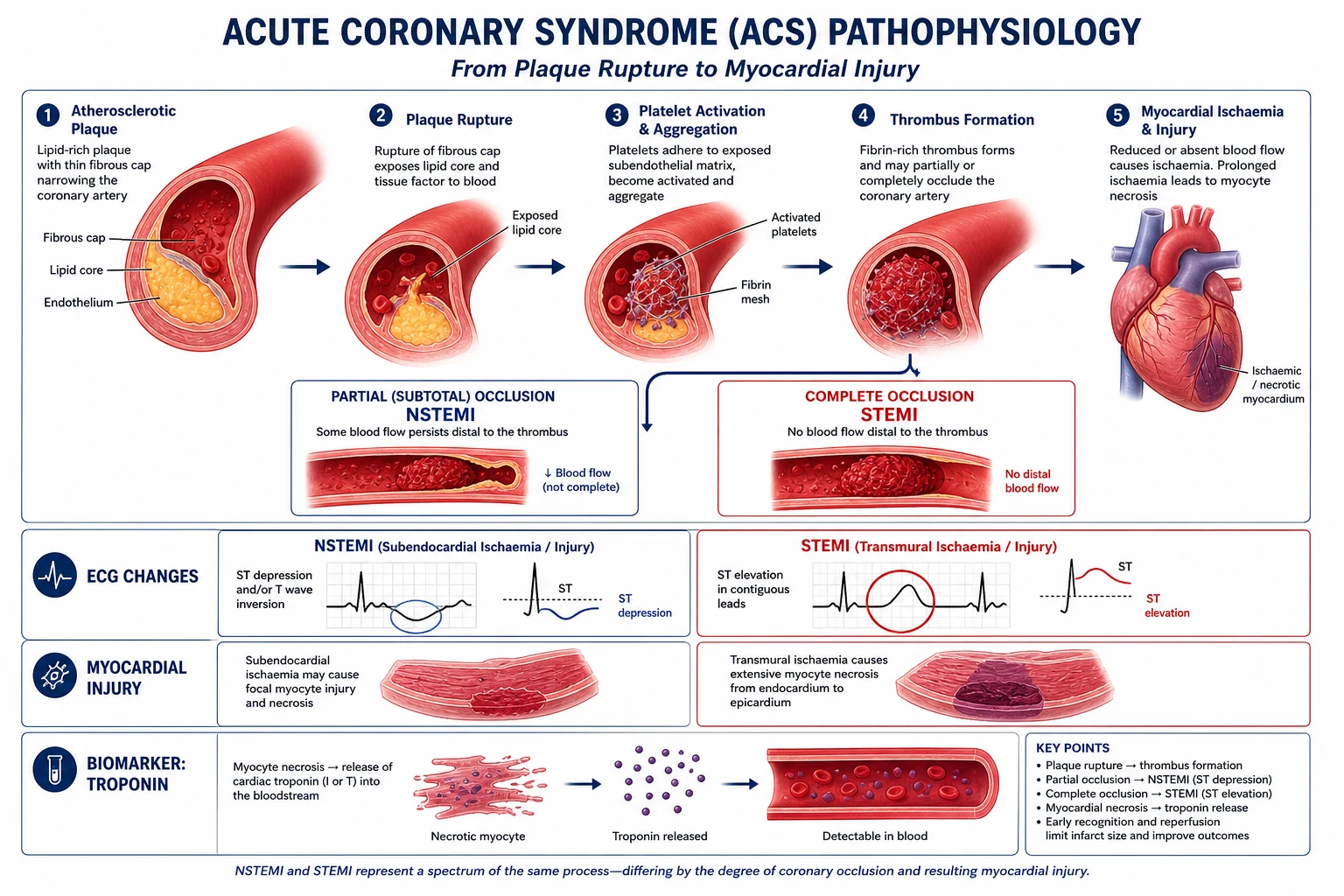
- Complete / sustained coronary occlusion → transmural ischaemia
- ECG: ST elevation ≥1 mm in 2 contiguous limb leads OR ≥2 mm in 2 contiguous precordial leads, or new LBBB (or true posterior MI — V1-V2 ST depression / tall R waves)
- Troponin: rises (may be normal at presentation)
- Reperfusion: PRIMARY PCI within 90 min, or thrombolysis within 30 min
- Pathology: fibrin-rich "red" clot over ruptured plaque
NSTEMI
Non-ST-elevation MI
- Subtotal / transient occlusion → subendocardial ischaemia
- ECG: ST depression, T-wave inversion, or transient changes — may be normal
- Troponin: RISES above the 99th percentile (the defining feature)
- Reperfusion: risk-stratified (GRACE) — high-risk → early invasive PCI within 24 h
- Pathology: platelet-rich "white" clot, subtotal occlusion
UA
Unstable angina
- Rest / crescendo / new-onset angina with NO troponin rise
- ECG: may show ST depression or T inversion
- Troponin: NEGATIVE (by definition — otherwise it is NSTEMI)
- Management: as for NSTEMI (DAPT, anticoagulant, risk-stratified invasive strategy)
- Now uncommon with high-sensitivity troponin assays
Pathophysiology — the unstable plaque
ACS begins with atherosclerotic plaque disruption (rupture of a thin-cap fibroatheroma, or superficial erosion). Circulating platelets adhere to exposed collagen and von Willebrand factor, become activated (via the ADP/P2Y12 and thromboxane A2 pathways), and aggregate. The coagulation cascade is simultaneously activated (tissue factor → extrinsic pathway), generating thrombin that converts fibrinogen to fibrin and further amplifies platelet activation. The result is a thrombus that partially (NSTEMI/UA) or completely (STEMI) occludes the lumen.[1] This dual pathway — platelets (white clot) and coagulation (red clot) — is the rationale for the two pillars of ACS pharmacotherapy: antiplatelets (aspirin + P2Y12 inhibitor) and anticoagulants (heparin or bivalirudin).
Antiplatelet therapy in ACS
Antiplatelet therapy targets the platelet arm of the thrombus. Dual antiplatelet therapy (DAPT) — aspirin plus a P2Y12 inhibitor — is the foundation of ACS management and is given to every patient unless contraindicated.[2][14]
Aspirin
- Mechanism: irreversibly acetylates and inhibits cyclo-oxygenase-1 (COX-1), blocking thromboxane A2 production for the lifespan of the platelet (7-10 days).
- Dose: 150-300 mg loading (chewed for fastest absorption), then 75-100 mg daily lifelong.
- Adverse: gastrointestinal bleeding, hypersensitivity, allergy. [1]
P2Y12 inhibitors
The P2Y12 (ADP) receptor is the dominant amplifier of platelet activation — its blockade is the key second antiplatelet. Three oral agents are available:[2][6][8]
Ticagrelor
Preferred for ACS (PLATO)
- Reversible, direct-acting cyclopentyl-triazolo-pyrimidine P2Y12 inhibitor
- Load 180 mg, then 90 mg BD
- Onset: fast (reversible — does not need hepatic activation)
- PLATO: ↓ CV death and all-cause mortality vs clopidogrel, no ↑ in major bleeding
- Adverse: dyspnoea (adenosine re-uptake), bradyarrhythmia, ↑ uric acid
- Avoid: prior intracranial haemorrhage, active bleeding
Prasugrel
Potent thienopyridine
- Irreversible thienopyridine prodrug — needs hepatic activation (one-step)
- Load 60 mg, then 10 mg daily (5 mg if <60 kg)
- TRITON-TIMI 38: ↓ ischaemic events vs clopidogrel, but ↑ major (including fatal) bleeding
- CONTRAINDICATED: prior stroke / TIA
- Caution: age >75 (bleeding) and weight <60 kg (use 5 mg)
- Best for STEMI going to primary PCI (where no pretreatment delay)
Clopidogrel
Thienopyridine (reference)
- Irreversible thienopyridine prodrug — needs two-step hepatic activation (variable, CYP2C19-dependent)
- Load 300-600 mg, then 75 mg daily
- Slower onset, weaker inhibition, genetic variability (CYP2C19 loss-of-function)
- Use when ticagrelor/prasugrel contraindicated or unavailable, or for oral anticoagulation (OAC + clopidogrel preferred)
- Best established safety in elderly and with OAC
PLATO
NEJM 2009
18,624 pts with ACS — ticagrelor vs clopidogrel (both + aspirin)
Key finding
CV death/MI/stroke: 9.8% vs 11.7% (HR 0.84, p<0.001). All-cause mortality ↓ (4.5% vs 5.9%). No ↑ in major bleeding.
Practice change
Ticagrelor preferred P2Y12 inhibitor in ACS (unless contraindicated)
TRITON-TIMI 38
NEJM 2007
13,608 pts with ACS going to PCI — prasugrel vs clopidogrel
Key finding
CV death/MI/stroke: 9.9% vs 12.1% (HR 0.81). BUT major bleeding ↑ (2.4% vs 1.8%) incl fatal bleeding, esp in prior stroke/TIA, age >75, <60 kg
Practice change
Prasugrel more potent but with higher bleeding — avoid in prior stroke/TIA
ISAR-REACT 5
NEJM 2019
4018 pts with ACS — ticagrelor vs prasugrel (investigator-selected strategy)
Key finding
1-yr death/MI/stroke: 9.3% prasugrel vs 5.9% ticagrelor in NSTE-ACS — prasugrel superior overall (driven by lower events, no ↑ bleeding)
Practice change
Prasugrel preferred over ticagrelor in NSTE-ACS in some European centres
Glycoprotein IIb/IIIa inhibitors (GPI)
The GPIs (abciximab, tirofiban, eptifibatide) block the final common pathway of platelet aggregation (fibrinogen cross-linking via GPIIb/IIIa). They are not used routinely in the era of potent oral P2Y12 inhibitors — reserve for bailout during PCI (large thrombus burden, slow/no reflow, massive thrombus) or in patients not yet loaded with a P2Y12 inhibitor.[1]
Duration of DAPT
- Default: 12 months for all ACS (the index ischaemic risk is highest in the first year).
- Ticagrelor beyond 12 months: the PEGASUS-TIMI 54 trial showed that extending ticagrelor (60 mg BD) beyond 12 months in patients with prior MI and additional high-risk features reduced MACE (but increased major bleeding).[13] Consider in patients with high ischaemic and low bleeding risk.
- Dual pathway inhibition: low-dose rivaroxaban 2.5 mg BD + aspirin (ATLAS ACS 2-TIMI 51, then COMPASS) reduces CV events post-ACS / in stable CAD, at a cost of more bleeding.[12] Useful in selected stable post-MI patients.
- Patients on oral anticoagulation (AF): OAC + aspirin (≤1 week) + clopidogrel then OAC + clopidogrel (triple→dual therapy) to minimise bleeding — NOT ticagrelor/prasugrel routinely.
Anticoagulation in ACS
Anticoagulation targets the thrombin/fibrin arm of the thrombus. Parenteral anticoagulation is given at presentation and continued through PCI (then stopped after successful stenting, as DAPT takes over long-term).[1][7]
UFH (heparin)
Default — flexible, reversible
- Mechanism: potentiates antithrombin III → inactivates thrombin (IIa) and factor Xa
- Dose: 60-70 IU/kg bolus (max 4000 IU) then 12 IU/kg/h infusion (PCI); or weight-based bolus only for primary PCI
- Monitored by ACT (PCI) or aPTT (infusion)
- Advantage: complete reversal with protamine; short half-life; cheap
- Risk: heparin-induced thrombocytopenia (HIT), bleeding
Bivalirudin
Direct thrombin inhibitor
- Mechanism: direct, reversible thrombin inhibitor — inhibits clot-bound AND free thrombin
- Dose: 0.75 mg/kg bolus, then 1.75 mg/kg/h infusion (periprocedural)
- HORIZONS-AMI: ↓ major bleeding and ↓ mortality vs heparin + GPI in primary PCI (early results)
- Advantage: no HIT, more predictable in renal/hepatic disease (partial renal clearance)
- Risk: higher early (acute) stent thrombosis than heparin
Fondaparinux
Indirect factor Xa inhibitor
- Mechanism: indirect, antithrombin-mediated selective Xa inhibitor
- Dose: 2.5 mg SC daily
- OASIS-5: non-inferior to enoxaparin with LESS major bleeding and ↓ mortality in NSTE-ACS
- NOT suitable as sole anticoagulant in primary PCI (catheter thrombosis) — add UFH
- No reversal agent; renal clearance — avoid if eGFR <20
LMWH (enoxaparin)
Alternative in conservatively-managed NSTEMI
- Mechanism: preferential Xa inhibition via antithrombin (also some IIa)
- Dose: 1 mg/kg SC BD (age >75 or renal impairment → reduce)
- Convenient (no monitoring) but partial reversibility with protamine
- Risk: HIT (much less than UFH), accumulation in renal failure
HORIZONS-AMI
NEJM 2008
3602 pts with STEMI undergoing primary PCI — bivalirudin vs heparin + GPI
Key finding
↓ Major bleeding at 30 d (4.9% vs 8.3%) and ↓ 1-yr mortality (3.5% vs 4.8%). BUT ↑ acute stent thrombosis within 24 h
Practice change
Bivalirudin accepted as alternative anticoagulant in primary PCI
Reperfusion strategy
STEMI — the time-critical reperfusion
In STEMI, every minute of coronary occlusion means more myocardium lost ("time is muscle"). The two reperfusion strategies are primary PCI (preferred) and fibrinolysis (when PCI cannot be delivered in time).[15]
STEMI reperfusion — first 60 minutes (door-to-needle / door-to-balloon)
Recognise and confirm
12-lead ECG within 10 minutes of arrival. Confirm ST elevation criteria (≥1 mm limb, ≥2 mm precordial in 2 contiguous leads) or new LBBB / true posterior pattern. Notify the catheter lab immediately ("STEMI activation").
Loading medications (simultaneously)
Aspirin 300 mg PO (chewed), P2Y12 inhibitor load (ticagrelor 180 mg OR prasugrel 60 mg OR clopidogrel 600 mg), atorvastatin 80 mg. Parenteral anticoagulation (UFH or bivalirudin). Avoid GPI routinely (bailout only).
Decision: PCI vs lysis
PRIMARY PCI if door-to-balloon ≤90 minutes (the global standard). If PCI cannot be delivered within 120 minutes of first medical contact → FIBRINOLYSIS (tenecteplase, single bolus) within 30 minutes (door-to-needle), then transfer for PCI (pharmacoinvasive strategy — routine angiography 3-24 h after successful lysis).
Analgesia and oxygen
Morphine/fentanyl for pain (caution — delays oral P2Y12 absorption). Oxygen ONLY if SpO2 <90% (avoid routine O2 — AVOID trial showed ↑ infarct size). Sublingual or IV nitrate if SBP >90 and no RV infarct/sildenafil.
During primary PCI
Drug-eluting stent to culprit lesion. Anticoagulation (UFH ACT 250-300 or bivalirudin). Thrombus aspiration (selective — routine aspiration NOT beneficial per TASTE/TOTAL). GPI only for bailout (large thrombus, slow flow).
Post-PCI ICU admission
Monitor for re-infarction, arrhythmia, heart failure, bleeding (access site). Continue DAPT. Assess LVEF by echo. Plan complete revascularisation if multivessel disease.
Complete vs culprit-only revascularisation
~50% of STEMI patients have multivessel disease. The strategy for the non-culprit lesions has evolved:[4][10][11]
- In stable STEMI (no shock): complete revascularisation (staging the non-culprit lesions, either during the index procedure or a staged PCI) reduces the composite of CV death/MI/ischaemia-driven revascularisation versus culprit-only — supported by COMPLETE (NEJM 2019) and DANAMI-3-PRIMULTI (Lancet 2015).[11][10]
- In cardiogenic shock: culprit-only PCI is preferred — CULPRIT-SHOCK (NEJM 2017) showed culprit-only PCI reduced 30-day death or severe renal failure versus immediate multivessel PCI.[4]
STEMI, no shock
Multivessel disease
- COMPLETE: complete revasc ↓ CV death/MI (HR 0.74)
- DANAMI-3-PRIMULTI: ↓ MACE with FFR-guided complete revasc
- Staged non-culprit PCI (during index or staged)
- FFR-guidance if uncertainty about significance
STEMI + shock
Multivessel disease
- CULPRIT-SHOCK: culprit-only PCI superior to immediate multivessel
- 30-day death/severe renal failure: 45.9% culprit vs 55.4% multivessel
- Stage non-culprit lesions after stabilisation
- Excessive contrast in shock worsens AKI
COMPLETE
NEJM 2019
4012 pts with STEMI + multivessel disease (no shock) — complete vs culprit-only revasc
Key finding
CV death/MI: 7.8% complete vs 10.5% culprit-only (HR 0.74, p<0.001). No difference in moderate bleeding.
Practice change
Complete revascularisation is standard in stable STEMI with multivessel disease
DANAMI-3-PRIMULTI
Lancet 2015
627 pts with STEMI + multivessel disease — FFR-guided complete vs culprit-only
Key finding
Composite of death/MI/ischaemia-driven revasc: 17% complete vs 27% culprit-only (HR 0.56)
Practice change
FFR-guided complete revascularisation reduces MACE in STEMI
ACS complicated by cardiogenic shock — the mechanical circulatory support ladder
Cardiogenic shock complicates ~6-10% of STEMI and is the leading cause of in-hospital death. The defining principle (SHOCK trial) is that early revascularisation (PCI or CABG within 24 hours) reduces long-term mortality — do not delay the catheter for medical stabilisation.[1] When shock persists despite revascularisation, vasoactive drugs (noradrenaline first-line; dobutamine/milrinone as inotropes) are added, and if these fail, mechanical circulatory support (MCS) escalates from IABP → Impella → VA-ECMO.[1][5]
Escalation of support in ACS-cardiogenic shock
Revascularise early (the non-negotiable)
Urgent coronary angiography + culprit-only PCI (CULPRIT-SHOCK). SHOCK trial: early revasc (≤24 h) ↓ long-term mortality vs medical stabilisation. The catheter is the treatment.
Secure airway + monitoring
Intubate/ventilate if shock-induced respiratory failure. Arterial line + central venous catheter. Lung-protective ventilation (Vt 6 mL/kg PBW, PEEP 5-8, plateau <30).
Pharmacological support
Noradrenaline 0.05-1.0 mcg/kg/min to MAP ≥65 (first-line — SOAP-2: fewer arrhythmias than dopamine). Add dobutamine 2.5-20 mcg/kg/min (or milrinone if beta-blocked) for low cardiac output.
Consider IABP selectively
IABP-SHOCK II: NO mortality benefit of routine IABP. Use selectively (e.g., as a bridge, or for mechanical complications like acute VSD/MR, RV infarct). NOT contraindicated in shock per se, but not routine.
Escalate to active LV support / Impella
If escalating inotrope/vasopressor demand with end-organ dysfunction, consider an Impella (2.5-5 L/min active LV unloading) — particularly if the LV is failing and dilating, or for high-risk PCI.
VA-ECMO for biventricular / refractory failure
For SCAI Stage D/E shock, refractory arrest, or severe biventricular failure, VA-ECMO (3-5 L/min full cardiopulmonary bypass). ECLS-SHOCK: NO benefit of EARLY ROUTINE VA-ECMO — use individualised, not protocolised. Remember VA-ECMO does NOT unload the LV — add an Impella (ECPELLA) if the LV dilates.
Define the goal and exit
Every MCS patient needs a defined endpoint: bridge to recovery, bridge to decision, bridge to transplant, or bridge to durable LVAD. MCS without a defined goal accumulates complications (bleeding, infection, limb ischaemia, thrombosis).
IABP
0.5–1.0 L/min · counterpulsation
- Diastolic balloon inflation ↑ coronary perfusion; systolic deflation ↓ afterload
- IABP-SHOCK II: NO routine mortality benefit in ACS-CS
- Use selectively: bridge, mechanical complications, RV infarct
- Contraindicated: aortic regurgitation, aortic dissection, severe PVD
Impella
2.5–5.0 L/min · active LV unloading
- Axial-flow pump across the aortic valve — drains LV to descending aorta
- Actively reduces LV preload, volume and wall stress
- Indicated: severe LV failure, high-risk PCI, LV distension on VA-ECMO (ECPELLA)
- Risks: haemolysis (↑ LDH), limb ischaemia, device migration
VA-ECMO
3–5 L/min · full cardiopulmonary bypass
- Provides systemic flow AND oxygenation (biventricular + lung support)
- Does NOT unload LV — retrograde flow ↑ afterload → LV distension
- ECLS-SHOCK: NO benefit of early ROUTINE VA-ECMO (use individualised)
- Risks: limb ischaemia (~25%, use distal perfusion cannula), bleeding, stroke, North-South syndrome
SHOCK
NEJM 1999
302 pts with AMI + cardiogenic shock — early revascularisation vs initial medical stabilisation
Key finding
6-month mortality: 50% early revasc vs 63% delayed (p=0.027). Benefit persists at 6 years.
Practice change
Early revascularisation became the standard in AMI-cardiogenic shock
IABP-SHOCK II
NEJM 2012
600 pts with AMI + cardiogenic shock — IABP vs no IABP
Key finding
30-day mortality: 39.7% vs 41.3% (p=0.69) — no difference. No benefit across all subgroups.
Practice change
Routine IABP no longer recommended in ACS cardiogenic shock
CULPRIT-SHOCK
NEJM 2017
1075 pts with AMI + cardiogenic shock — culprit-only vs immediate multivessel PCI
Key finding
30-day death/severe renal failure: 45.9% culprit vs 55.4% multivessel (RR 0.83, p=0.01)
Practice change
Culprit-only PCI preferred in AMI-cardiogenic shock
ECLS-SHOCK
NEJM 2023
420 pts with AMI + cardiogenic shock — early VA-ECMO vs no VA-ECMO
Key finding
30-day composite death/ECMO dependency: 63.5% vs 61.0% (not significant). High crossover (32%) limits interpretation.
Practice change
Early routine VA-ECMO not recommended — MCS use individualised
Mechanical complications of MI — the day 2-7 catastrophe
The mechanical complications result from myocardial necrosis weakening the wall, which then ruptures or dysfunction. They classically occur at days 2 to 7 post-MI (peak around days 3-5), and present as a sudden haemodynamic collapse in a patient who was previously improving.[1] A sudden deterioration in this window is a mechanical complication until proven otherwise — the immediate action is an urgent echocardiogram.[1]
Ventricular septal rupture (VSR/VSD)
Day 2-7 · large anterior or inferior MI
- A new harsh pansystolic murmur at the lower left sternal edge ± thrill
- Acute left-to-right shunt → RV volume overload → biventricular failure
- Echo: drop-out of the septum with left-to-right flow on colour Doppler
- Management: IABP/MCS bridge, urgent surgical or percutaneous closure
- Mortality 30-50% despite repair
Papillary muscle rupture
Day 2-7 · inferior MI (posteromedial papillary muscle)
- Acute severe mitral regurgitation → flash pulmonary oedema
- Murmur may be SOFT or absent in low-output shock (low forward flow = no murmur)
- The posteromedial papillary muscle has a single blood supply (PDA/RCA) — more vulnerable than the anterolateral (dual LAD/LCx supply)
- Echo: flail leaflet, eccentric MR jet, pulmonary oedema
- Management: MV repair/replacement (urgent), MCS bridge
Free wall rupture
Day 1-5 · usually first MI, elderly, female, hypertension
- Sudden cardiac death, electromechanical dissociation (EMD) arrest, or tamponade
- May be preceded by sharp pleuritic/positional chest pain and pericardial rub
- Echo: pericardial effusion with RA/RV diastolic collapse (tamponade), echo-dense space (clot)
- Management: emergent surgical repair, pericardiocentesis as a bridge. Survival low.
LV aneurysm
Weeks to months · large anterior MI
- Dyskinetic bulge of the LV wall (fibrotic, thin) — persistent ST elevation even in the chronic phase
- Complications: LV thrombus (systemic embolism), heart failure, ventricular arrhythmias
- Echo: dyskinetic outpouching; anticoagulate if thrombus present
- Management: ACEi/beta-blocker/GDMT, anticoagulation for thrombus, surgical/CABG if refractory
Pseudoaneurysm (contained rupture)
Day to weeks · contained free-wall rupture
- Myocardial wall rupture contained by pericardium and clot → false aneurysm with a narrow neck
- High risk of delayed complete rupture — surgical repair indicated even if asymptomatic
- Echo/MRI: outpouching with a narrow neck (vs true aneurysm with a wide neck)
Pericarditis (early & Dressler)
Days to weeks
- Early peri-infarct pericarditis (days 1-4): pleuritic chest pain, pericardial rub, diffuse ST elevation
- Dressler syndrome (weeks-months): autoimmune post-MI pericarditis, fever, pleuritic pain, effusion
- Management: high-dose aspirin (avoid NSAIDs/steroids in early MI — impair healing, ↑ rupture risk)
Right ventricular infarction — a special case
RV infarction complicates ~30-50% of inferior STEMI (the RCA supplies the RV). It presents with hypotension, clear lung fields and raised JVP (the triad) in the setting of an inferior MI. The management is counterintuitive and frequently tested:[1]
- Maintain RV preload — give fluid boluses (250 mL aliquots); AVOID nitrates, diuretics, and preload-reducing agents (they collapse the RV and worsen hypotension).
- Restore AV synchrony — atrial contribution to RV filling is critical; atrial fibrillation or heart block is poorly tolerated — cardiovert / pace.
- Inotropic support — dobutamine if hypotension persists after volume.
- Reperfuse the RCA — primary PCI restores RV function (often recovers better than the LV — the RV is more tolerant of ischaemia).
- ECG clues: ST elevation in right-sided leads V3R-V4R, and ST elevation in V1 with ST depression in V2 (posterior involvement). [1]
Post-MI secondary prevention — the four pillars plus device therapy
After reperfusion, the goals are to prevent recurrent ischaemia, limit adverse LV remodelling, and reduce mortality. The evidence-based regimen is the "four pillars" of post-MI therapy.[1][14]
Post-MI secondary prevention bundle
1. Antiplatelet — DAPT
Aspirin 75-100 mg lifelong + P2Y12 inhibitor for 12 months (ticagrelor preferred per PLATO). Extend ticagrelor to 36 months if high ischaemic risk (PEGASUS). In AF on OAC: OAC + clopidogrel (avoid ticagrelor/prasugrel routinely).
2. Beta-blocker
Started within 24 h if no contraindication (heart failure, hypotension, severe asthma, advanced heart block, cocaine-induced vasospasm). Reduces mortality and arrhythmia. Continue indefinitely if LVEF <40% or heart failure; reassess at 12 months if LVEF normal.
3. ACE inhibitor / ARB
Especially if LVEF <40%, heart failure, diabetes, hypertension, or anterior MI. Reduces adverse remodelling. ARNI preferred in HFrEF. Start low, titrate to target dose.
4. High-intensity statin
Atorvastatin 80 mg or rosuvastatin 20-40 mg started on day 1 regardless of baseline LDL. Target LDL <1.4 mmol/L (and <1.0 in recurrent events). Lipid-lowering is proportional to benefit.
+ Aldosterone antagonist
Eplerenone (or spironolactone) if LVEF <40% with heart failure or diabetes (EPHESUS). Monitor K+ and renal function.
+ Device therapy (ICD)
Primary prevention ICD if LVEF ≤35% at ≥40 days post-MI and ≥90 days optimal GDMT (NYHA II-III). Wearable cardioverter-defibrillator as a bridge if LVEF <35% early post-MI. Cardiac resynchronisation therapy if LVEF ≤35% + LBBB.
+ Lifestyle & risk factors
Smoking cessation (single most effective secondary prevention), cardiac rehabilitation, BP <130/80, HbA1c control, diabetes with SGLT2 inhibitor (↓ CV events, EMPA-REG, DECLARE), weight, diet, exercise.
PEGASUS-TIMI 54
NEJM 2015
21,162 pts with prior MI (1-3 yr) + high-risk features — ticagrelor 60/90 mg BD vs placebo (on aspirin)
Key finding
CV death/MI/stroke: 7.85% vs 9.04% (HR 0.84). Major bleeding ↑ (especially with 90 mg).
Practice change
Extended ticagrelor 60 mg BD considered in high ischaemic-risk post-MI patients
ATLAS ACS 2-TIMI 51
NEJM 2012
15,526 pts with recent ACS — rivaroxaban 2.5/5 mg BD vs placebo (on aspirin ± clopidogrel)
Key finding
CV death/MI/stroke: ↓ with rivaroxaban (HR 0.84). 2.5 mg dose ↓ CV and all-cause death. Major bleeding ↑ (incl non-fatal ICH).
Practice change
Low-dose rivaroxaban (+ aspirin ± P2Y12) considered in selected stable post-ACS patients
The ECG in ACS — high-yield patterns
- Hyperacute T waves: the earliest sign of STEMI — broad, tall, asymmetric T waves in the territory of occlusion, often within minutes of onset (before ST elevation develops).
- De Winter T waves: upsloping ST depression at the J point in V1-V3 with tall, symmetrical T waves — an LAD occlusion equivalent; treat as a STEMI.[1]
- Wellens syndrome: deep biphasic (or deeply inverted) T waves in V2-V3 with minimal ST elevation, in a pain-free patient with a history of recent chest pain — critical stenosis of the proximal LAD; urgent (not emergent) angiography. Do NOT stress-test.
- True posterior MI: ST depression in V1-V3 with tall R waves (the mirror image of posterior ST elevation) — confirmed by ST elevation in V7-V9 (posterior leads). Treat as STEMI. Often with RV infarction.
- Left main / triple-vessel ischaemia: diffuse ST depression with ST elevation in aVR (≥1 mm) — critical left main or proximal LAD disease; high risk.
- Right ventricular infarction: ST elevation in V3R-V4R in the setting of an inferior STEMI; haemodynamically significant.
- New LBBB: in the right clinical context, treat as a STEMI (Sgarbossa criteria for diagnosing MI in LBBB/paced rhythm — concordant ST elevation ≥1 mm, discordant ST depression in V1-V3, or concordant ST elevation ≥5 mm).[1]
Arrhythmias complicating ACS — ICU priorities
Arrhythmias in ACS arise from ischaemia, autonomic imbalance, electrolyte disturbance, and reperfusion injury:[1]
- Ventricular fibrillation (VF) / polymorphic VT: defibrillate (200 J biphasic), amiodarone 300 mg IV, correct K+ (≥4.0 mmol/L) and Mg²⁺ (≥0.9 mmol/L). Beta-blockade reduces recurrence. Ischaemia-driven VF requires revascularisation.
- Sustained monomorphic VT: if pulseless → defibrillate; if stable → amiodarone 300 mg IV over 20-60 min. Search for ongoing ischaemia and electrolyte disturbance.
- Atrial fibrillation: common in LV dysfunction and heart failure. Rate-control (beta-blocker, diltiazem, digoxin if heart failure); amiodarone for rhythm control. Anticoagulate (assess CHA₂DS₂-VASc and HAS-BLED). Avoid IV non-dihydropyridine CCBs in heart failure with reduced EF.
- Bradycardia and AV block: inferior MI — usually the AV node (RCA supply); narrow-complex escape; often transient; atropine, and a temporary pacemaker if symptomatic. Anterior MI — infranodal (septal necrosis); wide-complex escape; high-grade block; permanent pacemaker often needed; poor prognosis.[1]
- Idiopathic ventricular arrhythmia (reperfusion): brief runs of VT/AIVR within minutes of restoring flow are common and usually self-terminating; do not over-treat.
Bedside echocardiography in the ACS patient
Bedside echo answers the critical ICU questions in ACS:[1]
- Is there regional wall motion abnormality, and in which territory? (regional hypokinesis/akinesis = ischaemia)
- What is the LVEF? (<40% → start GDMT, plan ICD if persistent)
- Is there a mechanical complication? (VSD with left-to-right shunt on colour Doppler; flail leaflet with MR jet; pericardial effusion with tamponade)
- Is there RV involvement? (RV dysfunction in inferior MI — affects management)
- Is there LV thrombus? (in large anterior MI / aneurysm — needs anticoagulation)
- Volume status (IVC for fluid responsiveness — the failing LV is on a flat Frank-Starling curve) [1]
Landmark trials — the ACS evidence base
PLATO
NEJM 2009
18,624 ACS — ticagrelor vs clopidogrel
Key finding
CV death/MI/stroke 9.8% vs 11.7% (HR 0.84). ↓ all-cause mortality. No ↑ in major bleeding.
Practice change
Ticagrelor preferred P2Y12 in ACS
TRITON-TIMI 38
NEJM 2007
13,608 ACS-PCI — prasugrel vs clopidogrel
Key finding
↓ MACE (HR 0.81) but ↑ major/fatal bleeding. Avoid in prior stroke/TIA, >75, <60 kg.
Practice change
Prasugrel more potent but higher bleeding
ISAR-REACT 5
NEJM 2019
4018 ACS — prasugrel vs ticagrelor
Key finding
Prasugrel superior overall (driven by NSTE-ACS); no ↑ in bleeding.
Practice change
Prasugrel preferred over ticagrelor in NSTE-ACS in some centres
HORIZONS-AMI
NEJM 2008
3602 STEMI-PCI — bivalirudin vs heparin+GPI
Key finding
↓ major bleeding and ↓ mortality; ↑ acute stent thrombosis.
Practice change
Bivalirudin accepted in primary PCI
SHOCK
NEJM 1999
302 AMI-shock — early revasc vs medical
Key finding
↓ 6-month mortality (50% vs 63%); benefit persists at 6 years.
Practice change
Early revasc standard in AMI-shock
IABP-SHOCK II
NEJM 2012
600 AMI-shock — IABP vs no IABP
Key finding
30-day mortality 39.7% vs 41.3% (NS). No benefit.
Practice change
Routine IABP not recommended
CULPRIT-SHOCK
NEJM 2017
1075 AMI-shock — culprit-only vs multivessel PCI
Key finding
↓ 30-day death/severe renal failure (45.9% vs 55.4%).
Practice change
Culprit-only PCI in AMI-shock
ECLS-SHOCK
NEJM 2023
420 AMI-shock — early VA-ECMO vs no VA-ECMO
Key finding
No difference in death/ECMO-dependency (high crossover).
Practice change
Routine early VA-ECMO not recommended
COMPLETE
NEJM 2019
4012 STEMI-multivessel (no shock) — complete vs culprit-only
Key finding
↓ CV death/MI (7.8% vs 10.5%, HR 0.74).
Practice change
Complete revasc in stable STEMI
DANAMI-3-PRIMULTI
Lancet 2015
627 STEMI-multivessel — FFR-guided complete vs culprit-only
Key finding
↓ MACE (17% vs 27%, HR 0.56).
Practice change
FFR-guided complete revasc in STEMI
PEGASUS-TIMI 54
NEJM 2015
21,162 prior MI — extended ticagrelor vs placebo
Key finding
↓ MACE (HR 0.84); ↑ major bleeding.
Practice change
Extended ticagrelor in high-risk post-MI
ATLAS ACS 2-TIMI 51
NEJM 2012
15,526 post-ACS — rivaroxaban 2.5/5 mg BD vs placebo
Key finding
↓ CV death/MI/stroke; ↑ bleeding (incl ICH).
Practice change
Low-dose rivaroxaban in selected post-ACS
Clinical pearls
Additional red flags
Exam practice
SAQ — STEMI complicated by cardiogenic shock
10 minutes · 10 marks
A 68-year-old man presents with 2 hours of crushing central chest pain and diaphoresis. ECG: ST elevation in V1-V4 (anterior STEMI). BP 84/50 (MAP 61), HR 118 (sinus), SpO2 94% on room air. Lactate 3.8 mmol/L. Urine output 20 mL/hr. Coronary angiography shows 100% occlusion of the proximal LAD and a 70% lesion in the RCA. Echo: LVEF 28%, no mechanical complication.
Sample Viva 1 — Risk stratification in NSTEMI
Examiner: "A 72-year-old woman presents with rest chest pain. ECG shows T-wave inversion in the lateral leads. High-sensitivity troponin is elevated. How do you risk-stratify her, and what determines her management?" [1]
Expected response: "This is an NSTEMI — symptoms, troponin elevation, and ischaemic ECG changes. I would risk-stratify her using the GRACE score (Global Registry of Acute Coronary Events), which incorporates age, heart rate, blood pressure, creatinine, Killip class, cardiac arrest at admission, ST-segment deviation, and elevated biomarkers to estimate in-hospital and 6-month mortality. A GRACE score above 140 indicates high risk and mandates an early invasive strategy — coronary angiography within 24 hours. Very-high-risk features (refractory ischaemia, haemodynamic instability, mechanical complication, dynamic ST changes, life-threatening arrhythmia, or cardiac arrest) warrant immediate (≤2 hour) invasive angiography. She should receive aspirin plus ticagrelor (PLATO — ticagrelor preferred over clopidogrel) and parenteral anticoagulation (UFH or bivalirudin). At angiography, revascularisation by PCI or CABG is guided by the anatomy." [1]
Follow-up: "If her angiogram shows multivessel disease and she is in cardiogenic shock, what is your PCI strategy?" [1]
Response: "Culprit-only PCI — the CULPRIT-SHOCK trial (NEJM 2017) showed that treating only the culprit lesion reduced 30-day death and severe renal failure compared with immediate multivessel PCI, primarily because the larger contrast load and longer procedure of multivessel PCI worsened acute kidney injury. The non-culprit lesions are staged after the patient stabilises. This is the opposite of the strategy in stable STEMI, where COMPLETE showed complete revascularisation is superior." [1]
Sample Viva 2 — The troponin in the septic patient
Examiner: "A 65-year-old man is in the ICU with septic shock from a urinary source. His troponin is 800 ng/L (99th percentile 40). He has no chest pain and the ECG shows sinus tachycardia. The cardiology registrar wants to load him with ticagrelor and take him to the catheter lab. What is your response?" [1]
Expected response: "This is almost certainly a Type 2 MI — supply-demand mismatch from the sepsis, tachycardia, hypotension and hypoxia, with no evidence of a spontaneous plaque rupture. The Fourth Universal Definition (2018) requires, for a Type 1 MI, evidence of acute myocardial ischaemia — symptoms, dynamic ST/T changes, a new regional wall motion abnormality, or a coronary thrombus — none of which are present here. Treating him with ticagrelor and anticoagulation exposes him to bleeding without addressing the underlying problem, and a catheter trip is risky in septic shock. My response would be to treat the trigger — optimise oxygenation and ventilation, restore the blood pressure and perfusion, control the heart rate, transfuse if anaemic, and treat the sepsis with antibiotics and source control. I would order a bedside echocardiogram to look for a regional wall motion abnormality, and serial troponins — if they fall as the shock resolves, this confirms Type 2. I would only escalate to a coronary angiogram if there were objective evidence of ongoing ischaemia — new chest pain, dynamic ST changes, or a wall motion abnormality on echo." [1]
Key takeaways
- ACS spans STEMI (immediate reperfusion), NSTEMI (risk-stratified by GRACE) and UA (no troponin rise), unified by the Fourth Universal Definition of MI's five types.[9]
- All ACS receives aspirin + a P2Y12 inhibitor (ticagrelor preferred per PLATO) + parenteral anticoagulation (heparin or bivalirudin).[2][7]
- In MI with cardiogenic shock, early revascularisation is the standard (SHOCK); culprit-only PCI beats multivessel (CULPRIT-SHOCK); routine IABP (IABP-SHOCK II) and routine VA-ECMO (ECLS-SHOCK) confer no mortality benefit.[1][3][4][5]
- The mechanical complications (VSD, papillary muscle rupture, free wall rupture) occur at days 2-7 and present with sudden haemodynamic collapse — a mechanical complication until proven otherwise → urgent echo.[1]
- Post-MI secondary prevention: DAPT 12 months + beta-blocker + ACEi/ARB + high-intensity statin + MRA if LVEF <40% with heart failure; consider ICD if LVEF ≤35% at ≥40 days on optimal GDMT.[13][14]
References
- [1]Hochman JS, Sleeper LA, Webb JG, et al.; SHOCK Investigators. Early revascularization in acute myocardial infarction complicated by cardiogenic shock. SHOCK Investigators. Should We Emergently Revascularize Occluded Coronaries for Cardiogenic Shock N Engl J Med, 1999.PMID 10460813
- [2]Wallentin L, Becker RC, Budaj A, et al.; PLATO Investigators. Ticagrelor versus clopidogrel in patients with acute coronary syndromes N Engl J Med, 2009.PMID 19717846
- [3]Thiele H, Zeymer U, Neumann FJ, et al.; IABP-SHOCK II Trial Investigators. Intraaortic balloon support for myocardial infarction with cardiogenic shock N Engl J Med, 2012.PMID 22920912
- [4]Thiele H, Akin I, Sandri M, et al.; CULPRIT-SHOCK Investigators. PCI Strategies in Patients with Acute Myocardial Infarction and Cardiogenic Shock N Engl J Med, 2017.PMID 29083953
- [5]Thiele H, Zeymer U, Neumann FJ, et al.; ECLS-SHOCK Investigators. Extracorporeal Life Support in Infarct-Related Cardiogenic Shock N Engl J Med, 2023.PMID 37634145
- [6]Wiviott SD, Braunwald E, McCabe CH, et al.; TRITON-TIMI 38 Investigators. Prasugrel versus clopidogrel in patients with acute coronary syndromes N Engl J Med, 2007.PMID 17982182
- [7]Stone GW, Witzenbichler B, Guagliumi G, et al.; HORIZONS-AMI Trial Investigators. Bivalirudin during primary PCI in acute myocardial infarction N Engl J Med, 2008.PMID 18499566
- [8]Schupke S, Neumann FJ, Menichelli M, et al.; ISAR-REACT 5 Trial Investigators. Ticagrelor or Prasugrel in Patients with Acute Coronary Syndromes N Engl J Med, 2019.PMID 31475799
- [9]Thygesen K, Alpert JS, Jaffe AS, et al.; Executive Group on behalf of the Joint European Society of Cardiology / American College of Cardiology Foundation / American Heart Association / World Heart Federation Task Force for the Universal Definition of Myocardial Infarction. Fourth Universal Definition of Myocardial Infarction (2018) Circulation, 2018.PMID 30571511
- [10]Engstrom T, Kelbaek H, Helqvist S, et al.; DANAMI-3-PRIMULTI Investigators. Complete revascularisation versus treatment of the culprit lesion only in patients with ST-segment elevation myocardial infarction and multivessel disease (DANAMI-3—PRIMULTI): an open-label, randomised controlled trial Lancet, 2015.PMID 26347918
- [11]Mehta SR, Wood DA, Storey RF, et al.; COMPLETE Trial Steering Committee and Investigators. Complete Revascularization with Multivessel PCI for Myocardial Infarction N Engl J Med, 2019.PMID 31475795
- [12]Mega JL, Braunwald E, Wiviott SD, et al.; ATLAS ACS 2-TIMI 51 Investigators. Rivaroxaban in patients with a recent acute coronary syndrome N Engl J Med, 2012.PMID 22077192
- [13]Bonaca MP, Bhatt DL, Cohen M, et al.; PEGASUS-TIMI 54 Steering Committee and Investigators. Long-term use of ticagrelor in patients with prior myocardial infarction N Engl J Med, 2015.PMID 25773268
- [14]Collet JP, Thiele H, Barbato E, et al.; ESC Scientific Document Group. 2020 ESC Guidelines for the management of acute coronary syndromes in patients presenting without persistent ST-segment elevation Eur Heart J, 2021.PMID 32860058
- [15]Ibanez B, James S, Agewall S, et al.; ESC Scientific Document Group. 2017 ESC Guidelines for the management of acute myocardial infarction in patients presenting with ST-segment elevation: The Task Force for the management of acute myocardial infarction in patients presenting with ST-segment elevation of the European Society of Cardiology (ESC) Eur Heart J, 2018.PMID 28886621