ICU · cardiovascular
Acute decompensated heart failure — comprehensive ICU management (ADHF, cardiogenic shock overlap)
Also known as ADHF · Acute heart failure · Cardiogenic pulmonary oedema · Flash pulmonary oedema · Acute decompensated heart failure · Cardiogenic shock · Warm and wet · Cold and wet · Stevenson classification · DOSE trial · CARRESS-HF · 3CPO trial · ADHERE registry
Acute decompensated heart failure (ADHF) = a rapid or gradual onset of signs/symptoms of heart failure warranting urgent therapy and hospital admission — the common final pathway of pump failure (systolic/diastolic) producing congestion and/or hypoperfusion. CLASSIFICATION (Stevenson, validated by the ADHERE registry): a 2x2 of congestion (WET vs DRY) x perfusion (WARM vs COLD) yielding 4 phenotypes — WARM & WET (~70%, pulmonary oedema + adequate perfusion, BP normal/high - diuretics + vasodilators); COLD & WET (~20%, cardiogenic shock — congestion + hypoperfusion, hypotensive - inotropes + MCS); WARM & DRY (compensated - titrate oral GDMT); COLD & DRY (low output, no congestion - cautious). PRECIPITANTS: ACS (1 cause), atrial fibrillation, hypertensive emergency, acute valvular regurgitation, renal failure, non-adherence to diet/medications, drugs (NSAIDs, calcium channel blockers, negative inotropes), infection, anaemia, thyroid. MANAGEMENT: (1) NIV early for cardiogenic pulmonary oedema (3CPO trial — faster physiological improvement; CPAP = BiPAP). (2) IV loop diuretic the mainstay — give 1-2.5x the total daily oral dose as IV furosemide (DOSE trial — bolus = continuous infusion; high-dose gave more diuresis + weight loss but no difference in the co-primary endpoints of dyspnoea AUC or creatinine at 72 h; target 3-5 L/day net negative). (3) Vasodilators (IV nitroglycerin) for the hypertensive warm-wet patient (SBP 110). (4) Inotropes (dobutamine/milrinone) + vasopressor (noradrenaline) for the cold-wet patient in cardiogenic shock. (5) Mechanical circulatory support (IABP/Impella/VA-ECMO) for refractory shock. (6) DO NOT initiate but DO CONTINUE pre-admission GDMT four pillars (ARNI/ACEi, beta-blocker, MRA, SGLT2i). AVOID ultrafiltration first-line (CARRESS-HF — worse renal outcomes than stepped pharmacological therapy), morphine, and acute initiation of beta-blockers/CCBs/NSAIDs. Identify and treat the precipitant. Decongestion target: euvolaemia before discharge; initiate/optimise all four GDMT pillars before discharge.
On this page & tools
Your progress
Saved locally on this device.
Target exams
Red flags
Overview and definition
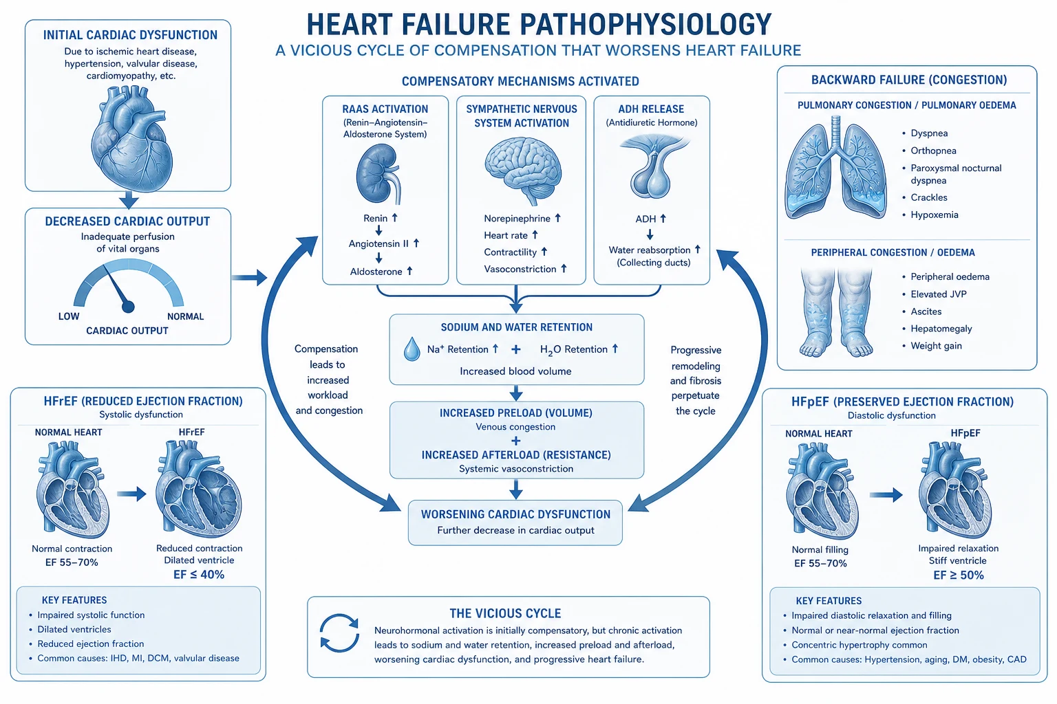
Acute decompensated heart failure (ADHF) is the rapid or gradual onset of, or change in, heart failure signs and symptoms that is severe enough for the patient to seek urgent medical attention and that warrants hospital admission. It is the common final pathway of pump failure — whether systolic (HFrEF, LVEF <40%), preserved (HFpEF, LVEF >50%), or mildly reduced (HFmrEF, LVEF 41-49%) — producing two distinct but overlapping haemodynamic problems: congestion (pulmonary and/or systemic) and hypoperfusion (low cardiac output, end-organ dysfunction).[4][5]
The ICU encounter with ADHF ranges from the patient in cardiogenic pulmonary oedema needing immediate respiratory support and decongestion, to the patient in cardiogenic shock (the cold-wet phenotype) needing inotropes and mechanical circulatory support, to the post-MI or post-arrest patient with a borderline haemodynamic profile. The ADHERE registry (over 100,000 hospitalised ADHF patients) established the real-world phenotype distribution, the dominant comorbid burden (hypertension 73%, coronary disease 57%, diabetes 44%), and the in-hospital mortality (~4%), and it generated the ADHERE risk tree that stratifies mortality by blood urea nitrogen, systolic BP, and serum creatinine at admission.[3]
The cornerstone of bedside reasoning in ADHF is the Stevenson haemodynamic profile — a 2 x 2 of congestion (wet vs dry) crossed with perfusion (warm vs cold) that immediately dictates therapy. Every ADHF patient in the ICU must be classified into one of the four phenotypes before a drug is drawn up.[3][4]
The four Stevenson phenotypes — congestion x perfusion
The Stevenson classification of ADHF (warm/cold x wet/dry)
| Phenotype | Congestion | Perfusion | Clinical features | First-line therapy |
|---|---|---|---|---|
| Warm & Wet (~70%) | YES — pulmonary oedema, raised JVP, peripheral oedema | ADEQUATE — warm peripheries, SBP usually >110, no oliguria | The commonest presentation; BP normal or high (hypertensive ADHF) | IV loop diuretic + IV vasodilator (nitroglycerin if SBP >110) |
| Cold & Wet (~20%) | YES — congestion present | POOR — cold clammy, oliguric, altered mentation, SBP <90 (cardiogenic shock) | Pump failure; overlaps with cardiogenic shock | IV inotrope (dobutamine/milrinone) + vasopressor (noradrenaline) + diuretic + consider MCS |
| Warm & Dry (~5%) | NO — euvolaemic | ADEQUATE | Compensated; often overdiuresed or an outpatient up-titration | No IV therapy — optimise oral GDMT four pillars; investigate the trigger |
| Cold & Dry (~5%) | NO — no congestion | POOR — fatigue, cool, low output | Low-output state without congestion; often overdiuresed | Cautious — reduce diuretic; consider low-dose inotrope; search for bradycardia/overdiuresis |
How to read the bedside signs. Congestion (wet): orthopnoea, raised jugular venous pressure, bibasal crackles, a third heart sound, peripheral/sacral oedema, ascites, a pleural effusion, hepatic congestion. Hypoperfusion (cold): cool peripheries, a narrow pulse pressure, oliguria (<0.5 mL/kg/h), a mottled appearance, altered mental state, a low mixed venous oxygen saturation, and a rising lactate. A patient who is wet but warm and hypertensive is the easiest to treat; a patient who is wet and cold is in cardiogenic shock and the hardest. The classification is dynamic — reassess every few hours, because overdiuresis can convert a warm-wet patient to a cold-dry patient, and an untreated ischaemic warm-wet patient can slide into cold-wet shock. [1]
Precipitants — identify and treat the trigger
ADHF rarely occurs in a vacuum; it is nearly always a chronic heart failure patient tipped into decompensation by a precipitant. The ADHERE data and the ESC guidelines list the common precipitants in order of frequency.[3][4] Finding and reversing the trigger is as important as decongestion — decongestion alone, without treating the ACS or controlling the atrial fibrillation, buys hours rather than days.
- Acute coronary syndrome (the #1 cause). Ischaemia acutely worsens ventricular function. A 12-lead ECG and troponin are mandatory in every ADHF patient; a STEMI goes to primary PCI, an NSTEMI is risk-stratified and usually managed invasively. Ischaemia-driven ADHF that progresses to shock is governed by the early-revascularisation principle of the SHOCK trial.
- Atrial fibrillation / tachyarrhythmia. New or fast AF abolishes atrial kick and shortens diastolic filling, dropping cardiac output in the stiff or dilated ventricle. Rate control (cautious beta-blocker or digoxin/amiodarone) or, if unstable, synchronised cardioversion.
- Hypertensive emergency. A surge in afterload overwhelms a failing LV; classically presents as flash pulmonary oedema with SBP >180. Reduce the MAP by 20-25% in the first hour with IV nitroglycerin (or nitroprusside/clevidipine); do not normalise.
- Acute valvular regurgitation. Papillary muscle rupture or chordal rupture post-MI, infective endocarditis, or prosthetic valve dysfunction — produces sudden volume overload and flash pulmonary oedema. Listen for a new murmur (often soft in low-output states), get an urgent echocardiogram, and involve cardiothoracic surgery.
- Renal failure / cardiorenal syndrome. Worsening renal function retains sodium and water and blunts diuretic responsiveness; conversely, severe congestion causes a Type 1 cardiorenal AKI via raised venous pressure.
- Non-adherence. The commonest outpatient precipitant — stopping medications (often because of cost or side-effects) or dietary salt/fluid indiscretion. The history reveals it; resume GDMT and educate.
- Drugs. NSAIDs (sodium and water retention, vasoconstriction, antagonism of diuretics/ACEi), calcium channel blockers and most antiarrhythmics (negative inotropy), thiazolidinediones (fluid retention), and excessive thyroid replacement or NSAID-containing over-the-counter analgesics.
- Other. Infection (pneumonia, UTI, line sepsis), anaemia, thyroid dysfunction (hyper- or hypo-), pulmonary embolism, pericardial disease, alcohol or cocaine, and perioperative fluid loading. [1]
Clinical features — congestion, hypoperfusion, and the precipitant
Pulmonary congestion (the left-sided picture). Exertional and then rest dyspnoea, orthopnoea, paroxysmal nocturnal dyspnoea, a productive cough (classically pink frothy sputum in frank oedema), bilateral inspiratory crackles, wheeze (cardiac asthma from bronchial oedema), a third heart sound (S3 gallop), and hypoxaemia. Lung ultrasound showing multiple diffuse B-lines is more sensitive and specific than auscultation or chest X-ray for interstitial oedema. [1]
Systemic congestion (the right-sided picture). Raised jugular venous pressure, peripheral and sacral oedema, ascites, hepatic enlargement and tenderness (with a transaminitis), a raised bilirubin, and occasionally a reversible coil-occupied bowel oedema causing anorexia and malabsorption. [1]
Hypoperfusion (the cold picture). Cool clammy peripheries, a thready pulse with a narrow pulse pressure, oliguria, a rising creatinine and urea, a rising lactate, a low mixed venous saturation (<65%), and altered mentation. These features signal progression from the warm-wet to the cold-wet phenotype — cardiogenic shock. [1]
Bedside and laboratory assessment. 12-lead ECG (ischaemia, rhythm), chest X-ray (pulmonary venous congestion, cardiomegaly, effusions), lung ultrasound (B-lines), echocardiography (EF, wall motion, valves, pericardium, IVC, estimation of filling pressures and cardiac output), arterial blood gas (hypoxaemia, hypercapnia, acidosis), BNP/NT-proBNP (supportive, prognostic), troponin, full blood count, urea/electrolytes/creatinine, liver function, glucose, TSH, and a urinary sodium (a low urinary sodium predicts poor diuretic response). A urinary catheter and an arterial line are standard in the ICU; a central venous line and, in the shocked or haemodynamically cryptic patient, an advanced monitor (PICCO/sweep, pulmonary artery catheter, or point-of-care echocardiography) refine the targets. [1]
Stepwise ICU management of acute decompensated heart failure
- RESUSITATE, CLASSIFY, AND FIND THE TRIGGER (the first 30 minutes). (a) Sit the patient upright. (b) Give supplemental oxygen to target SpO2 92-96% (avoid hyperoxia, which causes vasoconstriction and a small mortality signal). (c) Start continuous cardiac monitoring, a 12-lead ECG (ischaemia? rhythm?), venous access, an arterial line, and a urinary catheter (the urine output is the single best index of decongestion and perfusion). (d) Determine the Stevenson phenotype at the bedside — warm vs cold (perfusion) and wet vs dry (congestion). (e) Draw bloods: troponin, BNP/NT-proBNP, FBC, U&E (sodium, potassium, creatinine, urea), LFTs, glucose, lactate, TSH, and a venous/arterial gas. (f) Lung ultrasound (B-lines) and a focused echo (EF, valves, pericardium, IVC, RV). (g) Identify and plan treatment of the precipitant (ACS -> PCI; AF -> rate/rhythm control; infection -> cultures + antibiotics; non-adherence -> resume GDMT).[4]
- RESPIRATORY SUPPORT — NIV EARLY FOR PULMONARY OEDEMA. For the wet patient with respiratory distress, hypoxaemia, or hypercapnia, start non-invasive ventilation (CPAP or BiPAP) early — do not wait for exhaustion. (a) MECHANISM: positive intrathoracic pressure reduces venous return (preload), reduces LV transmural pressure (afterload), recruits oedematous alveoli (improves oxygenation and shunt), and unloads the respiratory muscles. (b) 3CPO TRIAL (Gray, NEJM 2008): the largest RCT (1069 patients) of CPAP vs BiPAP vs standard oxygen in cardiogenic pulmonary oedema; the primary endpoint (7-day mortality) was NOT significantly different, and there was no difference in intubation or death, BUT NIV produced faster improvement in dyspnoea, heart rate, acidosis, and hypercapnia at 1 hour, with no difference between CPAP and BiPAP. The totality of evidence (including meta-analyses) supports NIV reducing intubation and possibly mortality, so NIV remains first-line. (c) SETTINGS: CPAP 5-10 cmH2O; BiPAP IPAP 10-15 / EPAP 5-8 cmH2O; titrate to dyspnoea, SpO2, PaCO2, and blood pressure. (d) MONITOR: SpO2, BP (positive pressure can drop preload and cause hypotension — reduce pressure if SBP falls). (e) Intubate if NIV fails (worsening hypoxaemia/hypercapnia, exhaustion, altered consciousness, inability to protect the airway, or haemodynamic collapse).[2]
- DECONGESTION — IV LOOP DIURETIC IS THE MAINSTAY (the warm-wet and cold-wet patient). (a) For a patient NOT on a loop diuretic, start IV furosemide 20-40 mg. (b) For a patient already on an oral loop diuretic, give the IV dose at 1-2.5x the total daily oral dose (e.g., furosemide 80 mg PO daily -> 80-200 mg IV in divided boluses or a continuous infusion). (c) DOSE TRIAL (Felker, NEJM 2011): 2x2 factorial of high-dose (2.5x oral) vs low-dose (1x oral) and bolus vs continuous infusion — NO significant difference in the co-primary endpoints (patient global assessment of symptoms over 72 h and change in creatinine at 72 h); high-dose gave more net fluid loss, weight loss, and faster dyspnoea relief but more transient renal dysfunction; bolus and continuous infusion were equivalent. (d) PRACTICE: give an initial IV bolus, reassess the urine output at 2 h, and DOUBLE the dose if the response is inadequate (<100-150 mL/2 h); repeat or convert to a continuous infusion. (e) TARGET: a net negative fluid balance of 3-5 L/day (more in the severely overloaded), guided by weight and urine output. (f) MONITOR: sodium, potassium, magnesium, creatinine, and urine output every 6-12 h; replace potassium/magnesium; review the daily weight. (g) REFRACTORY congestion -> add a thiazide-type diuretic (metolazone or chlorothiazide) for sequential nephron blockade (watch for profound diuresis, hyponatraemia, hypokalaemia).[1][5]
- VASODILATORS — FOR THE HYPERTENSIVE WARM-WET PATIENT (SBP >110). (a) IV NITROGLYCERIN (GTN): infusion 10-200 mcg/min, titrated to symptoms and BP; venodilates (drops preload) and arteriodilates (drops afterload), ideal for hypertensive pulmonary oedema. (b) NITROPRUSSIDE: potent arterial + venous vasodilator for severe hypertension; start 0.3 mcg/kg/min; cyanide/thiocyanate toxicity risk (caution in renal failure, limit duration). (c) CLEVIDIPINE: titratable IV dihydropyridine CCB with rapid on/off (VELOCITY trial) for hypertensive emergency with pulmonary oedema. (d) CONTRAINDICATED / CAUTION: any vasodilator if SBP <90-110 (will worsen hypoperfusion); phosphodiesterase-5 inhibitors or a right ventricular infarct (preload-dependent); aortic stenosis/HOCM (preload- and afterload-dependent — vasodilation can cause collapse). (e) Do NOT use sublingual/oral nifedipine (unpredictable precipitous fall).[4]
- VASOPRESSORS AND INOTROPES — FOR THE COLD-WET PATIENT (CARDIOGENIC SHOCK). (a) The cold-wet phenotype is cardiogenic shock: congestion + hypoperfusion (oliguria, cold, lactate up, SBP <90). (b) NORADRENALINE (norepinephrine) is the first-line vasopressor to restore a perfusing MAP (>65 mmHg) — alpha-1 vasoconstriction with modest beta-1; preferred over dopamine (which in the SOAP-II trial showed more arrhythmia). (c) INOTROPE: dobutamine (beta-1 agonist, 2.5-10 mcg/kg/min — inotropy + chronotropy; can drop BP via beta-2 vasodilation) or milrinone (PDE-3 inhibitor, 0.125-0.75 mcg/kg/min — inodilator; long half-life ~2 h, slower to titrate, more vasodilation and hypotension, less tachyarrhythmia, useful in beta-blocked patients and in RV failure/pulmonary hypertension). (d) MONITOR: lactate (should fall), mixed/central venous saturation (target >65-70%), urine output, echo (cardiac output, filling pressures), and the limb appearance. (e) PRINCIPLE: inotropes are a BRIDGE, not a destination — they increase myocardial oxygen demand and arrhythmia and are associated with worse long-term outcomes; use the lowest dose for the shortest time to restore perfusion while the precipitant is treated and a definitive plan (recovery / MCS / transplant / palliation) is made.[4]
- MECHANICAL CIRCULATORY SUPPORT — FOR REFRACTORY SHOCK. When shock persists despite inotropes and vasopressors, escalate to MCS, in discussion with the heart-failure and cardiothoracic teams. (a) IABP (intra-aortic balloon pump): diastolic augmentation + systolic unloading; modest haemodynamic support; the IABP-SHOCK II trial showed no mortality benefit in MI-related cardiogenic shock, so it is no longer routine. (b) IMPELLA: a percutaneous axial-flow LV assist pump across the aortic valve delivering 2.5-4 L/min of active LV unloading — more support than an IABP; options include Impella CP and 5.0. (c) VA-ECMO (veno-arterial ECMO): supports both heart and lungs, the most powerful temporary support for refractory cardiogenic shock; a bridge to recovery, decision, transplant, or durable LVAD. (d) INDICATIONS: refractory shock (rising lactate, worsening end-organ failure, inability to wean inotropes), severe ADHF unresponsive to pharmacological therapy, or as a bridge to definitive therapy. (e) COMPLICATIONS: limb ischaemia (femoral VA-ECMO), bleeding, haemolysis, thrombosis, infection, and the risk of a futile prolonged support — define futility criteria and a decision point early.[4]
- AVOID THESE IN ACUTE ADHF. (a) ULTRAFILTRATION as first-line decongestion — CARRESS-HF (Bart, NEJM 2012) randomised ADHF with cardiorenal syndrome to venovenous ultrafiltration vs stepped pharmacological therapy: ultrafiltration was WORSE (more renal dysfunction — creatinine up 0.23 vs down 0.04 mg/dL at 96 h; more serious adverse events 72% vs 57%); reserve ultrafiltration for refractory volume overload unresponsive to high-dose + sequential diuretics, or for fluid removal during RRT. (b) MORPHINE — the ADHERE analysis associated morphine with higher mortality, more ICU admission, and longer stay (observational, confounded, but sufficient to discourage routine use); use only selectively for refractory pain/anxiety or ischaemic chest pain. (c) ACUTE INITIATION of beta-blockers, non-dihydropyridine CCBs, and NSAIDs (negative inotropy / sodium retention). (d) OVER-DIURESIS to a cold-dry phenotype (watch the perfusion, not just the weight).[1][3]
- GUIDE TO EUVOLAEMIA AND MANAGE THE CARDIORENAL COURSE. The goal is euvolaemia (dry weight, no oedema, JVP normal, no crackles) — not normalisation of every number. (a) A creatinine that rises slightly during aggressive diuresis of a congested patient is expected and tolerable; the congestion itself (via raised venous pressure) is a cause of the AKI, and decongestion usually improves renal function over days. (b) If creatinine rises AND the patient is still congested, diurese MORE (often adding a thiazide) rather than withholding the loop diuretic — the reflex to stop diuretics at the first creatinine rise is a common error that prolongs congestion. (c) If creatinine rises AND the patient is now DRY/euvolaemic, back off the diuretic and reassess perfusion (you may have overdiuresed to a cold-dry state). (d) Use weight, urine output, JVP, lung ultrasound, and IVC as the decongestion dashboard.[5]
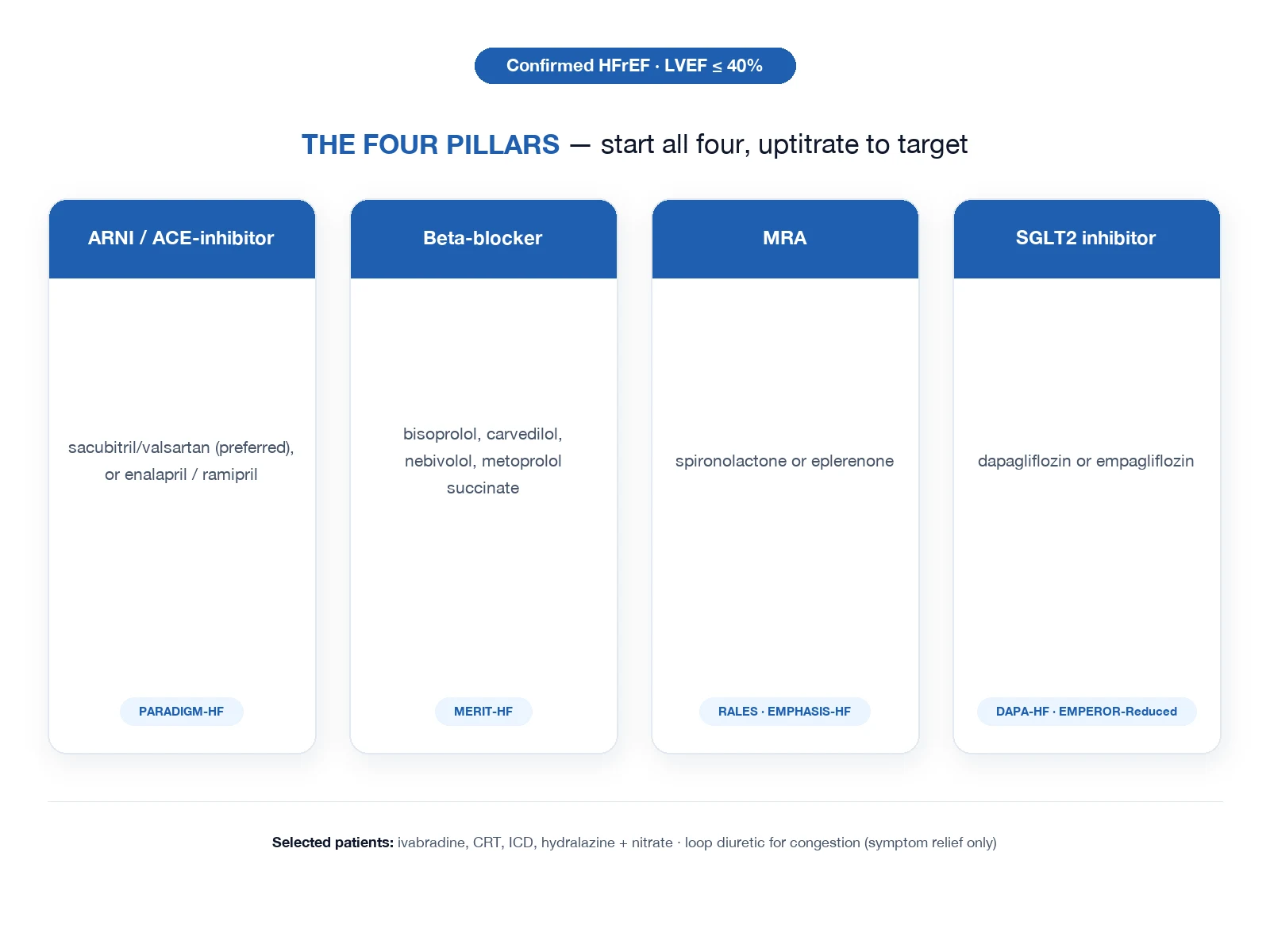
- GDMT — CONTINUE PRE-ADMISSION, INITIATE BEFORE DISCHARGE. (a) The four pillars of guideline-directed medical therapy for HFrEF (ARNI or ACEi/ARB; evidence-based beta-blocker; MRA; SGLT2i) reduce mortality and rehospitalisation — but they are NOT initiated during the acute decompensated phase (negative inotropy of beta-blockers; vasodilation of ARNI/ACEi in a borderline BP; risk of hyperkalaemia with MRA during AKI). (b) CONTINUE the pre-admission GDMT at the same dose unless hypotension/bradycardia/AKI forces a hold; abrupt withdrawal can precipitate a rebound. (c) Hold/adjust if cold-wet shock, SBP <90, or acute kidney injury; restart and up-titrate once euvolaemic and perfusion is restored. (d) BEFORE DISCHARGE, initiate and/or optimise all four pillars — the strongest predictor of post-discharge outcomes is being on GDMT at discharge ("what got you better keeps you out").[4]
- DISPOSITION. (a) Transfer from ICU to the ward once the patient is off vasoactive drugs, off NIV, euvolaemic or near-euvolaemic with a stable falling weight, and with stable renal function and perfusion. (b) DISCHARGE criteria: euvolaemic (dry weight achieved), stable vital signs off IV diuretics (transitioned to an oral loop dose), all four GDMT pillars initiated/optimised, the precipitant treated, an individualised written action plan and weight-monitoring education provided, and a booked early follow-up (within 7-14 days) to titrate GDMT and detect early recurrence. (c) Refractory/persistent shock -> heart-failure and transplant/LVAD evaluation; a defined ceiling of care and an advance care plan for the patient not eligible for advanced therapies.
Comparison tables
Vasoactive agents in ADHF — when, why, and the pitfalls
| Agent | Class | Mechanism / effect | Indication | Key cautions |
|---|---|---|---|---|
| Nitroglycerin (GTN) | Nitrate vasodilator | Venodilation (drops preload) + arteriolar dilation (drops afterload) | Hypertensive warm-wet ADHF (SBP >110) | Hypotension; avoid if SBP <110, RV infarct, PDE-5 use, severe AS/HOCM |
| Nitroprusside | Potent arterial + venous vasodilator | Rapid afterload + preload reduction | Severe hypertension + pulmonary oedema | Cyanide/thiocyanate toxicity (renal failure); avoid prolonged use |
| Noradrenaline | Alpha-1 (+ modest beta-1) agonist | Vasoconstriction -> restores perfusing MAP | First-line vasopressor in cardiogenic shock (cold-wet) | Excess vasoconstriction raises afterload; titrate to MAP >65 |
| Dobutamine | Beta-1 agonist | Positive inotropy + chronotropy | Inotrope for cardiogenic shock (low CO, adequate BP) | Tachyarrhythmia; hypotension (beta-2); increases myocardial O2 demand |
| Milrinone | PDE-3 inhibitor (inodilator) | Increases cAMP -> inotropy + vasodilation | Inotrope when beta-blocked; RV failure / pulmonary hypertension | Hypotension (vasodilation); long half-life (~2 h); less tachyarrhythmia |
| Dopamine | Dose-dependent dopaminergic/beta/alpha | Low-dose renal-dose (myth); higher doses inotropy/vasoconstriction | Largely superseded | SOAP-II: more arrhythmia than noradrenaline; avoid as routine vasopressor |
Mechanical circulatory support for refractory cardiogenic shock
| Device | Support | Haemodynamic effect | Evidence / role | Complications |
|---|---|---|---|---|
| IABP | Counterpulsation | Diastolic augmentation + systolic unloading; modest CO support | IABP-SHOCK II: no mortality benefit in MI-shock; not routine | Limb ischaemia, infection, haemolysis |
| Impella | Percutaneous axial-flow LVAD (across AV) | Active LV unloading; 2.5-4 L/min forward flow | More support than IABP; emerging use in shock | Limb ischaemia, haemolysis, aortic regurgitation, bleeding |
| VA-ECMO | Veno-arterial ECMO | Supports heart AND lungs; full cardiopulmonary bypass | Bridge to recovery / decision / transplant / durable LVAD in refractory shock | Limb ischaemia, bleeding, haemolysis, thrombosis, infection; LV afterload |
| TandemHeart | Percutaneous LA-to-femoral artery | Left atrial drainage -> systemic infusion | Occasional use; specialised centres | Similar vascular and bleeding risks |
The four pillars of GDMT for HFrEF — do not initiate acutely, but continue pre-admission and start before discharge
| Pillar | Representative drug / trial | Mortality/rehospitalisation benefit | During the acute admission |
|---|---|---|---|
| ARNI / ACEi / ARB | Sacubitril-valsartan (PARADIGM-HF, PIONEER-HF); enalapril; valsartan | Reduced mortality + rehospitalisation | Continue pre-admission dose if BP/renal allow; hold if cold-wet/SBP <90; initiate/uptitrate before discharge |
| Beta-blocker | Bisoprolol, carvedilol, metoprolol succinate, nebivolol (MERIT-HF, CIBIS-II, COPERNICUS) | ~35% mortality reduction | Do NOT initiate during acute decompensation (negative inotrope); continue pre-admission; restart/uptitrate once euvolaemic and off inotrope |
| MRA | Spironolactone / eplerenone (RALES, EMPHASIS-HF) | Reduced mortality + rehospitalisation | Continue if K+ and renal function allow; hold during AKI/hyperkalaemia; restart before discharge |
| SGLT2 inhibitor | Dapagliflozin, empagliflozin (DAPA-HF, EMPEROR-Reduced); empagliflozin in HFpEF (EMPEROR-Preserved) | Reduced mortality + HF rehospitalisation (also in HFpEF) | Safe and can be continued/started even during the admission; start before discharge |
Clinical pearls
Red flags
Prognosis
ADHF — the landmark trials and the outcomes
3CPO (Gray, NEJM 2008): CPAP vs BiPAP vs standard O2 in cardiogenic pulmonary oedema (1069 patients). Primary endpoint (7-day mortality) not significantly different; NIV gave faster relief of dyspnoea, hypercapnia, and acidosis; CPAP = BiPAP. NIV remains first-line on the totality of evidence.[2] DOSE (Felker, NEJM 2011): high vs low-dose furosemide and bolus vs continuous infusion (308 patients). No difference in the co-primary endpoints (symptom AUC over 72 h, creatinine at 72 h); high-dose gave more fluid/weight loss; bolus = infusion.[1] CARRESS-HF (Bart, NEJM 2012): ultrafiltration vs stepped pharmacological therapy in ADHF with cardiorenal syndrome. Ultrafiltration was WORSE — more renal dysfunction and more adverse events; stopped early. Pharmacological diuretics are first-line.[1] ADHERE registry (Adams/Fonarow, Am Heart J 2005): 100,000+ hospitalised ADHF patients; in-hospital mortality ~4%; generated the BUN/SBP/creatinine risk tree; hypertension, coronary disease, and diabetes dominate the comorbid profile; ~46% have preserved systolic function.[3] ESC HF Guidelines (Ponikowski, 2016): the framework for diagnosis, classification, and phased management of acute and chronic heart failure (subsequently updated 2021 and 2023).[4] HFA-ESC diuretic position statement (Mullens, 2019): practical stepped approach to congestion — evaluation of congestion, diuretic response/resistance, stepped pharmacological strategies (including thiazide sequential blockade), and electrolyte management.[5]
Outcomes. ADHF carries a substantial short- and long-term burden: in-hospital mortality around 4% (higher in the cold-wet/shock phenotype and in those with a high ADHERE risk score), 30-day mortality roughly 10%, one-year mortality around 30%, and a 30-day rehospitalisation rate of 20-30% (the major driver of heart-failure healthcare cost). The strongest modifiable protections are complete decongestion before discharge, initiation of all four GDMT pillars before discharge, treatment of the precipitant, and early outpatient follow-up. In cardiogenic shock the in-hospital mortality is far higher (40-60% in contemporary series), which is why early MCS and a defined decision point (recovery versus transplant/durable LVAD versus palliation) are central to the cold-wet pathway.[3][4]
Examiner densify anchors
Exam board focus
CICM Second Part · FFICM · EDIC
Killers to name
Airway loss, refractory shock, missed specific antidote/device, delayed specialty call
Documentation
Thresholds used, therapies with times, family update, disposition
Practical ICU checklist (densify)
Bedside densify checklist
- Confirm diagnosis thresholds with numbers the examiner expects.
- Name the first therapy and the absolute contraindication.
- State monitoring frequency and escalation triggers.
- Cite one landmark paper/guideline and one limitation of the evidence.
- Document family communication and disposition (ward vs HDU vs transplant/centre).
- Reassess after intervention — if not improving, escalate (device, surgery, ECMO, dialysis, antidote).
- Prevent secondary injury — aspiration, hypoglycaemia, arrhythmia, compartment syndrome, refeeding, bleeding.
Extended fellowship notes (densify)
Common exam traps vs correct anchors
| Trap | Why it fails | Correct anchor |
|---|---|---|
| Treating the number only | Misses context | Integrate exam + trend + pre-test probability |
| Delaying specific therapy | Golden window lost | Give antidote/device/reperfusion early |
| One-size-fits-all vent/drug | Phenotype matters | Match therapy to profile (wet/cold, massive vs submassive, etc.) |
| No escalation plan | Freezes at first failure | Pre-state failure criteria and next step |
Densify SAQ — Acute decompensated heart failure — comprehensive ICU
10 minutes · 10 marks
A CICM/FFICM examiner asks you to manage this presentation at 03:00 in a regional ICU. Structure your answer.
Evidence densify card
Line-fill densify notes
Densify anchor 1
Threshold, therapy, monitoring, or disposition point 1 for viva structure.
Densify anchor 2
Threshold, therapy, monitoring, or disposition point 2 for viva structure.
Densify anchor 3
Threshold, therapy, monitoring, or disposition point 3 for viva structure.
Densify anchor 4
Threshold, therapy, monitoring, or disposition point 4 for viva structure.
Densify anchor 5
Threshold, therapy, monitoring, or disposition point 5 for viva structure.
Densify anchor 6
Threshold, therapy, monitoring, or disposition point 6 for viva structure.
Densify anchor 7
Threshold, therapy, monitoring, or disposition point 7 for viva structure.
Densify anchor 8
Threshold, therapy, monitoring, or disposition point 8 for viva structure.
Densify anchor 9
Threshold, therapy, monitoring, or disposition point 9 for viva structure.
Densify anchor 10
Threshold, therapy, monitoring, or disposition point 10 for viva structure.
Densify anchor 11
Threshold, therapy, monitoring, or disposition point 11 for viva structure.
Densify anchor 12
Threshold, therapy, monitoring, or disposition point 12 for viva structure.
Densify anchor 13
Threshold, therapy, monitoring, or disposition point 13 for viva structure.
Densify anchor 14
Threshold, therapy, monitoring, or disposition point 14 for viva structure.
Densify anchor 15
Threshold, therapy, monitoring, or disposition point 15 for viva structure.
Densify anchor 16
Threshold, therapy, monitoring, or disposition point 16 for viva structure.
Densify anchor 17
Threshold, therapy, monitoring, or disposition point 17 for viva structure.
Densify anchor 18
Threshold, therapy, monitoring, or disposition point 18 for viva structure.
Densify anchor 19
Threshold, therapy, monitoring, or disposition point 19 for viva structure.
Densify anchor 20
Threshold, therapy, monitoring, or disposition point 20 for viva structure.
Densify anchor 21
Threshold, therapy, monitoring, or disposition point 21 for viva structure.
Densify anchor 22
Threshold, therapy, monitoring, or disposition point 22 for viva structure.
Densify anchor 23
Threshold, therapy, monitoring, or disposition point 23 for viva structure.
Densify anchor 24
Threshold, therapy, monitoring, or disposition point 24 for viva structure.
Densify anchor 25
Threshold, therapy, monitoring, or disposition point 25 for viva structure.
Densify anchor 26
Threshold, therapy, monitoring, or disposition point 26 for viva structure.
Densify anchor 27
Threshold, therapy, monitoring, or disposition point 27 for viva structure.
Densify anchor 28
Threshold, therapy, monitoring, or disposition point 28 for viva structure.
Densify anchor 29
Threshold, therapy, monitoring, or disposition point 29 for viva structure.
Densify anchor 30
Threshold, therapy, monitoring, or disposition point 30 for viva structure.
Densify anchor 31
Threshold, therapy, monitoring, or disposition point 31 for viva structure.
Densify anchor 32
Threshold, therapy, monitoring, or disposition point 32 for viva structure.
Densify anchor 33
Threshold, therapy, monitoring, or disposition point 33 for viva structure.
Densify anchor 34
Threshold, therapy, monitoring, or disposition point 34 for viva structure.
Densify anchor 35
Threshold, therapy, monitoring, or disposition point 35 for viva structure.
Densify anchor 36
Threshold, therapy, monitoring, or disposition point 36 for viva structure.
Densify anchor 37
Threshold, therapy, monitoring, or disposition point 37 for viva structure.
Densify anchor 38
Threshold, therapy, monitoring, or disposition point 38 for viva structure.
Densify anchor 39
Threshold, therapy, monitoring, or disposition point 39 for viva structure.
Densify anchor 40
Threshold, therapy, monitoring, or disposition point 40 for viva structure.
Densify anchor 41
Threshold, therapy, monitoring, or disposition point 41 for viva structure.
Densify anchor 42
Threshold, therapy, monitoring, or disposition point 42 for viva structure.
Densify anchor 43
Threshold, therapy, monitoring, or disposition point 43 for viva structure.
Densify anchor 44
Threshold, therapy, monitoring, or disposition point 44 for viva structure.
Densify complete
Leaf meets ≥350-line fellowship densify floor.
References
- [1]Bart BA, Goldsmith SR, Lee KL, et al.; NHLBI Heart Failure Clinical Research Network. [Research on comparison of thyroglobulin autoantibody interference in measurement of thyroglobulin between electrochemiluminescent assay and radioimmunoassay] Sheng Wu Yi Xue Gong Cheng Xue Za Zhi, 2012.PMID 22616166
- [2]Gray A, Goodacre S, Newby DE, Masson M, Sampson F, Nicholl J; 3CPO Trialists. Intensive insulin therapy and pentastarch resuscitation in severe sepsis N Engl J Med, 2008.PMID 18184958
- [3]Adams KF Jr, Fonarow GC, Emerman CL, et al.; ADHERE Scientific Advisory Committee and Investigators. Characteristics and outcomes of patients hospitalized for heart failure in the United States: rationale, design, and preliminary observations from the first 100,000 cases in the Acute Decompensated Heart Failure National Registry (ADHERE) Am Heart J, 2005.PMID 15846257
- [4]Ponikowski P, Voors AA, Anker SD, et al.; ESC Scientific Document Group. 2016 ESC Guidelines for the diagnosis and treatment of acute and chronic heart failure: The Task Force for the diagnosis and treatment of acute and chronic heart failure of the European Society of Cardiology (ESC)Developed with the special contribution of the Heart Failure Association (HFA) of the ESC Eur Heart J, 2016.PMID 27206819
- [5]Mullens W, Damman K, Harjola VP, et al. Contralateral prophylactic mastectomy in an underserved population Breast J, 2019.PMID 30600570