ICU · Cardiovascular
Acute decompensated heart failure and cardiogenic pulmonary oedema
Also known as Acute decompensated heart failure (ADHF) · Cardiogenic pulmonary oedema · Flash pulmonary oedema · Acute heart failure (AHF)
Acute decompensated heart failure (ADHF) is a life-threatening syndrome of pulmonary and/or systemic congestion from cardiac dysfunction. Presentations: cardiogenic pulmonary oedema (acute breathlessness, orthopnoea, frothy sputum, bilateral crackles, hypoxia), cardiogenic shock (hypoperfusion — cold extremities, oliguria, altered mental status), or isolated right heart failure (peripheral oedema, raised JVP, hepatomegaly). Management: sit upright, high-flow oxygen, IV furosemide (1-2.5x usual oral dose), vasodilator (nitroglycerin if SBP 110), non-invasive ventilation (CPAP/BiPAP — reduces intubation and mortality). Identify and treat precipitant (ACS, arrhythmia, infection, non-adherence). Avoid beta-blockers acutely (negative inotropy).
On this page & tools
Your progress
Saved locally on this device.
Target exams
Red flags
Epidemiology and pathophysiology
Acute heart failure (AHF) is responsible for >1 million hospitalisations annually in the United States and is the leading cause of hospital admission in patients over 65 in the developed world. In-hospital mortality ranges from 4% (warm-wet presentations) to >40% (cardiogenic shock), and 30-day readmission rates approach 25%. Half of all heart failure patients die within 5 years of diagnosis — a prognosis comparable to many metastatic cancers.[1]
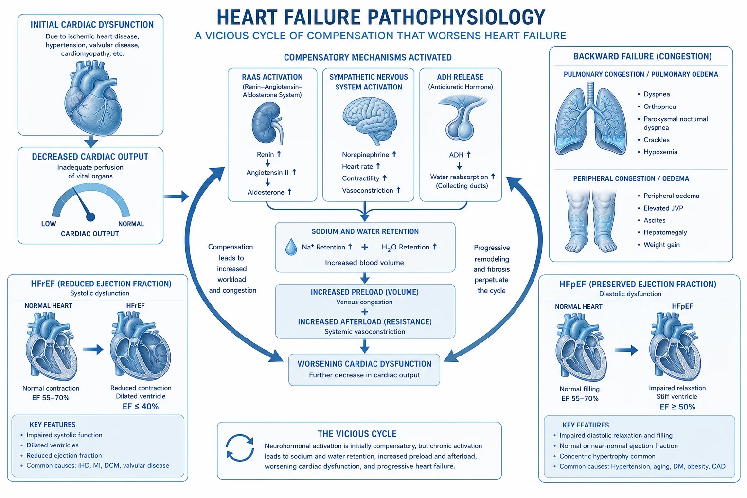
The pathophysiology is a vicious cycle of decompensation: [1]
- Index insult (myocardial ischaemia, arrhythmia, volume load) reduces stroke volume.
- Fall in cardiac output activates the sympathetic nervous system and renin-angiotensin-aldosterone system (RAAS) in a compensatory attempt to maintain perfusion pressure.
- Maladaptive neurohormonal activation drives sodium/water retention (aldosterone, ADH), vasoconstriction (angiotensin II, noradrenaline, endothelin), tachycardia, and direct cardiotoxicity.
- Increased preload and afterload raise ventricular wall stress → further reduction in stroke volume, mitral/tricuspid regurgitation, and myocardial injury (troponin leak from subendocardial ischaemia due to elevated end-diastolic pressure).
- Venous congestion (raised left-sided filling pressures) transudates fluid into the pulmonary interstitium and alveoli → pulmonary oedema and hypoxaemia, which worsens myocardial ischaemia.
- Right heart distension (ventricular interdependence) impairs LV filling and coronary perfusion.
- Cardiorenal syndrome develops as venous congestion (not low output per se) reduces glomerular filtration, creating a self-perpetuating cycle of worsening renal function, more diuretic resistance, and escalating congestion. [1]
The central therapeutic insight is that congestion (raised filling pressures), not low cardiac output in isolation, drives symptoms and most organ injury in the typical warm-wet patient. Decongestion — not augmentation of contractility — is therefore the primary target for the majority of presentations.[1][3]
Clinical classification — Stevenson hemodynamic profiles
The Stevenson (Nohria-Stevenson) classification stratifies AHF at the bedside by two axes — perfusion (warm vs cold, reflecting cardiac output) and congestion (wet vs dry, reflecting filling pressures). It is the dominant clinical framework taught in CICM/FFICM because it is determined without invasive monitoring and directly guides therapy.[5]
| Profile | Congestion | Perfusion | Clinical findings | Treatment | Mortality signal |
|---|---|---|---|---|---|
| Warm-wet | Wet (JVP raised, pulmonary crackles, oedema) | Warm (well perfused, SBP >90, normal lactate) | Most common presentation (~70%). Pulmonary oedema ± peripheral oedema, normal extremities, adequate urine output | IV loop diuretics (1-2.5x oral dose) ± vasodilator (GTN if SBP >110) ± CPAP/BiPAP | Moderate |
| Warm-dry | Dry (no JVP rise, clear chest) | Warm | Euvolaemic, well perfused — the target state after treatment, or a non-congested decompensation (e.g. isolated high-output state) | Continue/optimise oral GDMT; identify non-volume precipitant | Low |
| Cold-wet | Wet | Cold (clammy, mottled, oliguric, confused, SBP <90 or lactate >2) | Cardiogenic shock with congestion. SBP often <90, cold extremities, oliguria, altered mentation | Inotrope (dobutamine/milrinone/levosimendan) ± vasopressor (noradrenaline to MAP ≥65) + decongest; consider mechanical support (Impella/VA-ECMO) early | High |
| Cold-dry | Dry | Cold | Low output without congestion — over-diuresed, or pure forward failure. Often pre-load dependent | Cautious fluid challenge (250 mL crystalloid) ± inotrope; look for occult bleeding/sepsis | Moderate-high |
Warm-wet (most common)
~70% of AHF
- Adequate perfusion preserved (SBP >90, warm extremities, lactate <2 mmol/L)
- Congestion dominates symptoms: orthopnoea, crackles, raised JVP, peripheral oedema
- Therapy target: decongest with IV loop diuretic ± vasodilator; CPAP if pulmonary oedema
- Avoid inotropes (no hypoperfusion — they add arrhythmia/ischaemia risk without benefit)
- Beta-blocker continuation: continue if chronic stable; hold if decompensated
Cold-wet (cardiogenic shock)
~5-8% of AHF
- Hypoperfusion + congestion: SBP <90, cold/clammy, oliguria <0.5 mL/kg/h, lactate >2
- Different pathway: do NOT rely on diuretics alone — they fail in low-output state
- Inotrope first-line: dobutamine, milrinone or levosimendan to augment cardiac output
- Vasopressor (noradrenaline) if MAP <65 despite inotrope
- Escalate EARLY to MCS (Impella/VA-ECMO) — late escalation has poor outcomes
- Revascularise if ACS (CULPRIT-SHOCK: culprit-only PCI)
Cold-dry (over-diuresed / forward failure)
Rare
- Hypoperfusion without congestion: tachycardia, narrow pulse pressure, oliguria
- Often iatrogenic (over-diuresis) or occult hypovolaemia (bleeding, sepsis)
- Cautious fluid bolus 250 mL crystalloid, reassess
- Bedside echo: small hypercontractile LV with kissing septum suggests underfilled
- Inotrope only if confirmed low output not corrected by volume
Warm-dry (target / compensated)
Discharge-ready state
- No congestion, normal perfusion — goal of therapy
- May represent non-congested decompensation: anaemia, thyrotoxicosis, tachyarrhythmia
- Transition to oral GDMT (4 pillars: BB + MRA + ARNI/ACEi + SGLT2i)
- Discharge planning begins here
Forrester classification (invasive hemodynamic)
The earlier Forrester classification (1976) uses invasive Swan-Ganz catheter data — cardiac index (CI) and pulmonary capillary wedge pressure (PCWP) — and is largely of historical and exam interest now that echocardiography and clinical assessment dominate. It is conceptually identical to Stevenson but uses measured rather than inferred values.[1]
Forrester classification — ADHF by cardiac index and PCWP
CI <2.2, PCWP <18 (cold + dry)
Low output without congestion. Hypovolaemic or over-diuresed. Cautious fluid challenge.
Clinical presentation
Pulmonary oedema (left-sided)
Acute backward failure
- Acute breathlessness, orthopnoea, paroxysmal nocturnal dyspnoea
- Pink frothy sputum, bilateral coarse crackles, wheeze ("cardiac asthma")
- Tachypnoea (RR >25), tachycardia, accessory muscle use
- Hypoxia, often hypertensive initially (sympathetic surge)
- CXR: bat-wing perihilar infiltrates, Kerley B lines, upper lobe blood diversion, pleural effusions
Cardiogenic shock (forward failure)
Cold-wet or cold-dry
- Hypotension (SBP <90 or >30 below baseline), narrow pulse pressure
- Cold clammy mottled skin, prolonged capillary refill
- Oliguria (<0.5 mL/kg/h), altered mental status
- Lactate >2 mmol/L, metabolic acidosis
- Often complicating large MI, fulminant myocarditis, end-stage cardiomyopathy
Right heart failure
Isolated backward RV failure
- Raised JVP, hepatomegaly, ascites, marked peripheral/sacral oedema
- Relatively clear lung fields (no pulmonary oedema)
- Causes: RV infarct (inferior MI), pulmonary embolism, severe TR/PR, pulmonary hypertension, ARVC
- Kussmaul sign (JVP rises on inspiration) suggests severe RV failure or constrictive pericarditis
Flash pulmonary oedema
Sudden onset, recurrent
- Explosive pulmonary oedema over minutes — bilateral renal artery stenosis (Pickering syndrome) until proven otherwise
- Other causes: severe mitral regurgitation (flail leaflet), bilateral RAS, phaeochromocytoma, renal failure + volume load
- Clue: AKI after starting ACE inhibitor → suspect bilateral RAS
- Often markedly hypertensive on presentation
Identify the precipitant
Every AHF admission has a precipitant that must be actively sought and treated — decongestion alone without addressing the trigger leads to rapid readmission. [1]
Common precipitants
Find and treat
- Acute coronary syndrome (#1 — check ECG, troponin)
- Arrhythmia (especially new AF — rate control)
- Infection (pneumonia, UTI — check cultures, CRP)
- Medication non-adherence (stopped diuretics/ACEi)
- Anaemia (transfuse if Hb <70)
- Thyroid dysfunction (thyroid storm or myxoedema)
- Renal failure (worsening AKI → fluid overload)
- Dietary indiscretion (high salt intake)
- Drugs (NSAIDs, calcium channel blockers, steroids — sodium retention)
Less common / high-yield for exam
Don't miss
- Negative inotropes: diltiazem, verapamil, most antiarrhythmics
- Cardiotoxic drugs: anthracyclines, trastuzumab, tyrosine kinase inhibitors
- Alcohol-induced (holiday heart), cocaine, amphetamines
- Peripartum cardiomyopathy (last month of pregnancy to 5 months postpartum)
- Takotsubo (stress) cardiomyopathy — apical ballooning
- Acute myocarditis (viral — coxsackie, parvovirus; giant cell)
- Pulmonary embolism (sudden RV strain)
- Endocarditis with acute valvular regurgitation
- Tamponade / constrictive pericarditis
- Tachycardia-mediated cardiomyopathy (persistent SVT/AF)
Investigations
ADHF investigations
A full panel includes: ECG (within 10 minutes — ACS, AF, LVH, old Q waves), chest X-ray (pulmonary oedema, cardiomegaly, effusions, alternative diagnosis), bloods — FBC (anaemia, leucocytosis), U&E (renal function, K+), LFTs (hepatic congestion), troponin (ACS vs type 2 from wall stress), BNP/NT-proBNP (rule-out if <300 ng/L; rule-in if >1800 age-adjusted), CRP, D-dimer (if PE suspected), TSH, lactate (perfusion marker), venous blood gas (acid-base, mixed venous saturation), and urinalysis. Echocardiography (early, ideally within 48 hours) is essential — defines HFrEF (EF <40%) vs HFmrEF (41-49%) vs HFpEF (≥50%), identifies regional wall motion abnormalities, valve lesions, pericardial effusion, and estimates filling pressures/right heart function. [1]
Bedside ultrasound and POCUS in AHF
Point-of-care ultrasound accelerates diagnosis and monitoring of decongestion: [1]
- Lung ultrasound: B-lines (comet-tail artefacts) arising from pleural line indicate interstitial/alveolar fluid. Bilateral diffuse B-lines confirm cardiogenic pulmonary oedema; their resolution tracks successful diuresis. A-lines (horizontal reverberation) indicate dry lung.
- Cardiac POCUS: identifies gross LV/RV dysfunction, pericardial effusion, gross valvular pathology, and estimates LV filling (E/e′). A small, hypercontractile, underfilled LV suggests over-diuresis (cold-dry); a dilated hypocontractile LV confirms cardiomyopathy.
- IVC ultrasound: a dilated plethoric IVC (>2.1 cm with <50% collapse) supports high right atrial pressure and congestion; a small collapsible IVC suggests volume responsiveness.
- ** pleural effusion**: quantified and tapped for diagnostic/therapeutic thoracentesis. [1]
Management of cardiogenic pulmonary oedema
Acute pulmonary oedema management
Position + oxygen
Sit UPRIGHT (reduces preload, improves ventilation). High-flow oxygen via non-rebreather mask (15 L/min). Target SpO2 >92% (or >88% in COPD).
Non-invasive ventilation (CPAP/BiPAP) — EARLY
CPAP 5-10 cmH2O or BiPAP (IPAP 10, EPAP 5). Reduces work of breathing, improves oxygenation, decreases preload and afterload (positive intrathoracic pressure). Evidence: reduces intubation rates and mortality. Start within 15-30 min of presentation. Continue until patient comfortable, SpO2 >92%, RR <25.<Cite id="2" />
IV furosemide
Give 1-2.5x the patient usual oral dose IV. For new-onset HF: furosemide 40-80 mg IV. For chronic HF on diuretics: give 2-2.5x usual oral dose IV. Mechanism: initially venodilation (within 5-10 min, before diuretic effect), then diuresis. Monitor urine output (target >100 mL/h initially).<Cite id="6" />
Vasodilator (nitroglycerin) — if SBP >110
IV nitroglycerin (start 10 mcg/min, titrate up) or sublingual GTN spray (2 puffs). Reduces preload (venodilation) and afterload (arteriolar dilation). Reduces mitral regurgitation. Do NOT use if SBP <110 (risk of hypotension). Contraindicated if phosphodiesterase inhibitor (sildenafil) within 24h.
Morphine — USE CAUTIOUSLY
Morphine 2.5-5 mg IV (reduces anxiety, preload via venodilation, breathlessness). BUT associated with increased ICU admission and mortality in some studies (observational — confounding likely). Use selectively for severe anxiety/distress. Not routinely recommended.
Do NOT give beta-blockers acutely
Acute beta-blockade has negative inotropic effects → may precipitate/worsen cardiogenic shock. Start beta-blockers only once the patient is stabilised and euvolaemic (usually 24-48h after presentation). Continue pre-existing beta-blocker if patient is stable and not in shock.
Oxygen therapy detail
Indications and targets
Saturate judiciously
- Target SpO2 92-96% in most patients; 88-92% in COPD retainers
- Supplemental O2 only if hypoxaemic — routine O2 in non-hypoxaemic patients is NOT beneficial (and may be harmful via coronary vasoconstriction and ROS)
- High-flow nasal cannula (HFNC) is an alternative to NIV in cooperative patients with moderate hypoxaemia
- Reserve endotracheal intubation for: respiratory failure despite NIV, reduced GCS, inability to protect airway, exhaustion (rising CO2)
NIV / CPAP — the evidence
Mortality benefit
- Mechanism: positive intrathoracic pressure → ↑FRC, ↓work of breathing, ↓LV preload and afterload, redistributes alveolar fluid
- CPAP vs BiPAP: 3CPO trial found no mortality difference; either is acceptable — start with whichever is available
- Cochrane meta-analysis: NIV reduces mortality (NNT ~13) and need for intubation vs standard O2 therapy
- Wean when: SpO2 >92% on FiO2 <0.4, RR <25, comfortable, ABG normalized — typically after 2-6 hours
- Contraindications: cardiac arrest, GCS <8, facial trauma, inability to protect airway, severe agitation, vomiting
3CPO (Gray et al, NEJM 2008)
Multicentre RCT: standard O2 vs CPAP vs NIV (BiPAP)
Population: 1069 patients with severe acute cardiogenic pulmonary oedema
Key finding
No significant difference in 7-day mortality between groups (~9.5% all arms). NIV groups had faster improvement in symptoms, HR, BP, and acidosis. Reduced need for intubation/escalation.
Diuretic therapy — DOSE-AHF and practical strategy
Diuretic strategy in ADHF
Initial IV bolus dose
Total daily IV furosemide = 1-2.5x the patient usual total daily oral dose. New-onset HF: furosemide 40-80 mg IV. Chronic HF on 80 mg PO daily: give 80-200 mg IV. Maximum single bolus ~200 mg furosemide (or 5 mg torsemide/bumetanide).
Assess response at 2-6 hours
Target urine output 100-150 mL/h (3-4 L/24h) for effective decongestion. If inadequate (UO <100 mL/h after 2h), double the dose.
Bolus vs continuous infusion
DOSE-AHF: no difference in symptoms or renal function between q12h bolus and continuous infusion. Either strategy is acceptable; boluses allow titration to response.
High-dose vs low-dose
DOSE-AHF: high-dose (2.5x oral) achieved greater fluid loss and weight reduction vs low-dose (1x oral), with transiently worse renal function but no difference in outcomes. Favour higher doses in chronic HF.
Add thiazide if diuretic resistance
If response inadequate after stepwise doubling, add oral metolazone 2.5-5 mg daily OR IV chlorothiazide 500-1000 mg (sequential nephron blockade). Caution: hypokalaemia, hyponatraemia, severe volume depletion.
Add potassium-sparing agent
Spironolactone 25-50 mg daily (also proven mortality benefit in chronic HFrEF). Monitor K+ closely when combined with ACEi/ARB/ARNI.
DOSE-AHF (Felker et al, NEJM 2011)
2x2 factorial RCT: (a) high-dose (2.5x oral) vs low-dose (1x oral) furosemide; (b) bolus q12h vs continuous infusion
Population: 308 patients with acute decompensated heart failure and ≥1 sign of congestion
Key finding
No difference in primary endpoint (global symptom assessment at 72h) or 60-day death/rehospitalisation between any arms. High-dose produced greater net fluid loss and weight loss but transiently more renal dysfunction.
Vasodilators — when and what
Nitroglycerin (GTN)
First-line if SBP >110
- Mechanism: venodilation (↓ preload) at low dose; arteriolar dilation (↓ afterload) at higher doses
- Dose: sublingual spray 400 mcg (1-2 puffs); IV infusion 10-200 mcg/min titrated
- Indications: AHF with SBP >110 and significant congestion/pulmonary oedema, concomitant ischaemia, or acute MR
- Cautions: avoid if SBP <110, right ventricular infarction, PDE5 inhibitor in last 24-48h, severe aortic stenosis
- Tachyphylaxis after 16-24h — useful for the first 24h of acute decompensation
Nesiritide (BNP)
No mortality benefit
- Recombinant B-type natriuretic peptide — vasodilator, natriuretic, RAAS inhibitor
- ASCEND-HF (OConnor 2011): modest dyspnoea improvement, NO mortality or rehospitalisation benefit, no excess renal harm
- Not routinely used in ANZ/Europe; largely abandoned due to cost and lack of hard outcomes benefit
Sodium nitroprusside
Severe hypertension / valve regurg
- Potent arterial + venous vasodilator; rapid onset/offset
- Indications: hypertensive emergency with severe pulmonary oedema, acute severe mitral/aortic regurgitation
- Dose: 0.3-3 mcg/kg/min IV; requires arterial line monitoring
- Caution: cyanide/thiocyanate toxicity with prolonged use (>24-48h) or renal failure; raises ICP
ACEi / ARB / ARNI
Start AFTER stabilisation
- NOT for acute phase — risk of hypotension and AKI in volume-depleted/hypotensive patient
- Initiate once euvolaemic, SBP stable, renal function stable (usually 24-48h post-admission)
- ARNI (sacubitril/valsartan) preferred over ACEi in HFrEF (PARADIGM-HF mortality benefit)
ASCEND-HF (O'Connor et al, NEJM 2011)
RCT: nesiritide (recombinant BNP) vs placebo
Population: 7141 patients with acute decompensated heart failure
Key finding
Nesiritide did not improve 30-day death/rehospitalisation or mortality. Small, non-clinically-significant improvement in dyspnoea. No excess renal dysfunction or mortality (addressing earlier safety concerns).
Inotropes for the cold/shocked patient
Inotropes are NOT indicated in warm-wet AHF — they add arrhythmia, ischaemia, and tachyphylaxis risk without benefit. Reserve for cold-wet or cold-dry profiles (hypoperfusion) where augmentation of cardiac output is required pending definitive therapy.[20]
Dobutamine
β1 agonist
- Dose: 2-20 mcg/kg/min IV
- ↑ cAMP → ↑ contractility and HR (chronotropy); mild β2 vasodilation
- First-line inotrope in most units; cheap, familiar, titratable
- Side effects: tachyarrhythmia, ↑ myocardial O2 demand, hypotension (β2), tolerance
Milrinone
PDE-3 inhibitor
- Dose: 0.125-0.75 mcg/kg/min IV (loading dose often omitted in AHF)
- ↑ cAMP via PDE-3 inhibition → ↑ contractility + vasodilation (pulmonary and systemic)
- Less tachyarrhythmia than dobutamine; favourable in pulmonary hypertension/RV failure
- Caution: renally cleared — halve dose in CKD; significant hypotension on initiation
- OPTIME-CHF: routine milrinone in ADHF did NOT improve outcomes and increased atrial arrhythmias/hypotension
Levosimendan
Calcium sensitiser
- Binds cardiac troponin C → ↑ myofilament Ca2+ sensitivity without ↑ intracellular Ca2+ (less arrhythmia/ischaemia)
- Also opens ATP-sensitive K+ channels → vasodilation (including coronary)
- SURVIVE: no mortality benefit vs dobutamine in ADHF (subgroup signal in prior β-blocker use)
- Used in some European units; not available in Australia; expensive
Noradrenaline (norepinephrine)
First-line vasopressor
- α1 + β1 agonist → vasoconstriction + modest inotropy
- Use when MAP <65 despite inotrope, or as first-line vasoconstrictor in shock
- Preferred over adrenaline (epinephrine) — less lactataemia and arrhythmia
- Dose: 0.05-1 mcg/kg/min IV titrated to MAP ≥65 mmHg
Adrenaline (epinephrine)
Refractory shock
- α + β agonist; potent inotrope and vasoconstrictor
- Reserved for refractory shock not responding to noradrenaline + inotrope
- Causes lactataemia (β2 glycolysis), arrhythmia, worsening renal/splanchnic perfusion
Dopamine
Largely abandoned
- Dose-dependent: low dose "renal" (1-3 mcg/kg/min) — ROSE-AHF showed NO renal benefit
- Higher doses: tachyarrhythmia excess vs noradrenaline (SOAP-II)
- Not recommended for routine use in AHF
ROSE-AHF (Chen et al, JAMA 2013)
2x2 factorial RCT: low-dose dopamine (2 mcg/kg/min) vs placebo, AND low-dose nesiritide vs placebo
Population: 360 patients with acute heart failure and renal dysfunction (eGFR 15-60)
Key finding
Neither low-dose dopamine nor low-dose nesiritide improved the primary endpoints of 72-hour cumulative urine output or cystatin C change. No renal protection.
SURVIVE (Mebazaa et al, JAMA 2007)
RCT: levosimendan vs dobutamine
Population: 1327 patients with AHF requiring inotropic support
Key finding
No significant difference in primary endpoint (180-day all-cause mortality: 26% vs 28%). Subgroup suggested possible benefit of levosimendan in patients with prior β-blocker therapy.
OPTIME-CHF (Cuffe et al, JAMA 2002)
RCT: 48-72h infusion of milrinone vs placebo (not selected by clinician for inotrope indication)
Population: 949 patients with acute exacerbation of chronic systolic heart failure
Key finding
No improvement in 60-day hospitalisation/mortality. Increased adverse events: atrial arrhythmias (HR 1.7), sustained hypotension (HR 1.6). Subgroup trend to worse outcomes in ischaemic cardiomyopathy.
Mechanical circulatory support (MCS)
For refractory cardiogenic shock despite inotropes/vasopressors, MCS bridges to recovery, decision, durable device, or transplant. [1]
IABP (intra-aortic balloon pump)
Counterpulsation
- Mechanism: balloon inflates in diastole (↑ coronary perfusion), deflates in systole (↓ afterload)
- Indications: historically first-line; IABP-SHOCK II showed NO mortality benefit in MI-shock
- Contraindications: aortic regurgitation, aortic dissection, severe PAD, severe sepsis
- Now reserved for mechanical complications of MI (acute MR, VSD) or as bridge to definitive support
Impella (axial flow micro-pump)
LV / RV support
- Mechanism: transvalvular axial pump aspirates LV blood, ejects into aorta (LV unloading)
- Impella 2.5 (2.5 L/min), CP (4.0 L/min), 5.0 (5.0 L/min); RP for RV support
- Active unloading reduces LV wall stress and improves forward flow more than IABP
- Complications: limb ischaemia, haemolysis, device malposition, bleeding at insertion site
VA-ECMO
Venoarterial ECMO
- Mechanism: drains RA blood, returns oxygenated blood to femoral artery — supports both circulation and gas exchange
- Indications: refractory cardiogenic shock, massive PE, fulminant myocarditis, post-cardiotomy, severe ARDS + shock
- Flows 3-6 L/min; provides complete cardiopulmonary support
- Complications: differential hypoxaemia (Harlequin syndrome — femoral configuration), LV distension (increased afterload — consider Impella or IABP venting), limb ischaemia, bleeding, stroke, haemolysis
- Femoral vs central cannulation: central preferred post-cardiotomy; femoral for rapid percutaneous insertion
TandemHeart
Atrial-to-femoral LA→femoral artery
- Drains left atrium via transseptal puncture, returns to femoral artery
- Flows up to 5 L/min; potent LV unloading
- Requires transseptal expertise — less commonly used than Impella/ECMO
IABP-SHOCK II (Thiele et al, NEJM 2012)
RCT: IABP vs no IABP
Population: 600 patients with acute MI and cardiogenic shock undergoing early revascularisation
Key finding
No difference in 30-day mortality (39.7% vs 41.3%) or 12-month mortality. No improvement in secondary endpoints (bleeding, renal function, inflammatory markers, ICU stay).
CULPRIT-SHOCK (Thiele et al, NEJM 2017)
RCT: culprit-lesion-only PCI vs immediate multivessel PCI
Population: 555 patients with acute MI and multivessel disease presenting in cardiogenic shock
Key finding
Culprit-only PCI reduced primary endpoint (death or severe renal failure requiring RRT at 30 days: 45.9% vs 55.4%) and reduced mortality (43.3% vs 51.6%).
Cardiogenic shock (SBP <90)
See dedicated topic: cardiogenic-shock-mechanical-support. Key points:
- Cold + wet = cardiogenic shock → different pathway
- Inotropes: dobutamine or milrinone (improve cardiac output)
- Vasopressor: noradrenaline (maintain MAP >65)
- Mechanical support: IABP, Impella, VA-ECMO
- Urgent revascularisation if ACS (CULPRIT-SHOCK: culprit-only PCI)[16]
- Diurese cautiously (may worsen hypotension)
The SCAI shock stages provide a granular framework: A (at risk), B (beginning — clinical suspicion), C (classic — hypoperfusion meeting criteria), D (deteriorating — worsening despite support), E (extremis — refractory cardiac arrest). Early MCS escalation at stage C/D (before lactate >10, before cardiac arrest) is associated with markedly better outcomes than late "salvage" escalation at stage E. [1]
Diuretic resistance
Defined as failure to achieve euvolaemia despite adequate diuretic dosing. Common in chronic HF, CKD, hypoalbuminaemia, and high sodium intake. [1]
Approach to diuretic resistance
Confirm adherence and intake
Review medication history (adherence, stopped diuretics, NSAIDs). Restrict sodium to <2 g/day and fluid to 1-1.5 L/day.
Stepwise IV furosemide up-titration
Start with 1-2.5x oral dose; reassess UO at 2h. Double each subsequent dose until UO >100 mL/h or 200 mg single bolus reached.
Switch to continuous infusion
May improve efficacy once high doses are needed; titrate infusion to UO 100-150 mL/h.
Add thiazide-type diuretic (sequential nephron blockade)
Oral metolazone 2.5-10 mg daily, IV chlorothiazide 500-1000 mg, or IV hydrochlorothiazide. Acts at distal tubule; synergistic with loop diuretic. Major risk: hypokalaemia, hyponatraemia, severe volume depletion — monitor 6-hourly.
Add acetazolamide
Carbonic anhydrase inhibitor proximal tubule — synergistic in metabolic alkalosis with diuretic resistance (ADVOR trial 2022 showed improved decongestion).
Consider ultrafiltration (rarely)
Reserved for refractory overload NOT responsive to pharmacological therapy. CARRESS-HF showed pharmacologic therapy superior to ultrafiltration.
Address hypoalbuminaemia / nephrotic
In low-albumin states, diuretics enter tubule poorly; consider albumin-furosemide co-infusion (controversial).
Reassess for occult shock / over-diuresis
A "non-responder" may be cold-dry (underfilled) — over-diuresis causes AKI without relieving symptoms. Bedside echo/IVC assessment guides therapy.
CARRESS-HF (Bart et al, NEJM 2012)
RCT: stepped pharmacological therapy (high-dose IV loop + thiazide + dopamine/nitroglycerin) vs ultrafiltration
Population: 188 patients with ADHF, worsened renal function, and persistent congestion
Key finding
Pharmacological therapy was superior: less weight gain (primary endpoint: -1.7 kg worse in UF arm), fewer serious adverse events (72% in UF vs 57% in pharmacologic), and fewer creatinine rises.
UNLOAD (Costanzo et al, JACC 2007)
RCT: ultrafiltration vs standard IV diuretic therapy
Population: 200 patients hospitalised with ADHF
Key finding
Ultrafiltration produced greater weight loss and net fluid loss at 48h and fewer 90-day rehospitalisations. No difference in symptoms or renal function. Significant industry sponsorship bias.
Failed disease-modifying therapies — a recurring pattern
Multiple promising AHF therapies have failed in large RCTs — exam candidates should know these to avoid reaching for unproven agents. [1]
RELAX-AHF (Teerlink et al, Lancet 2013)
RCT: serelaxin (recombinant human relaxin-2) vs placebo for 48h
Population: 1161 patients with acute heart failure, dyspnoea, mild-moderate renal dysfunction, SBP >125
Key finding
Serelaxin improved dyspnoea (visual analogue scale) and reduced 180-day mortality in the original trial (a secondary endpoint). Subsequent RELAX-AHF-2 (2017) FAILED to confirm the mortality signal.
VERITAS (McMurray et al, JAMA 2007)
RCT: tezosentan (endothelin receptor antagonist) vs placebo
Population: 1435 patients with acute heart failure
Key finding
No improvement in symptoms or 30-day death/heart-failure worsening. Increased adverse events (headache, hypotension).
The recurring lesson: decongestion with diuretics + NIV + addressing the precipitant remains the cornerstone; no acute medical therapy (vasodilator, natriuretic peptide, inodilator, endothelin antagonist, relaxin) has convincingly modified AHF mortality. Disease modification happens through chronic GDMT, not acute interventions. [1]
Guideline-directed medical therapy (GDMT) and the 4 pillars
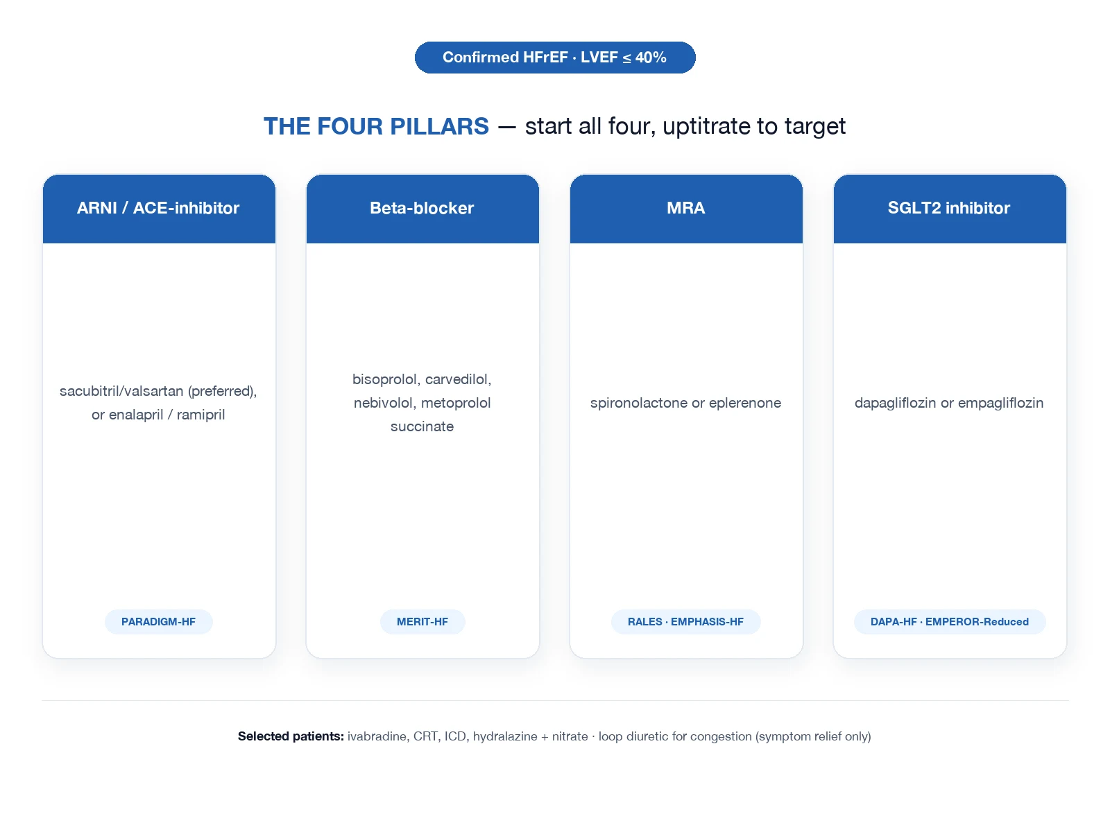
The four pillars of HFrEF therapy, ideally initiated/up-titrated before discharge: [1]
- Beta-blocker (bisoprolol, carvedilol, metoprolol succinate, nebivolol) — start once euvolaemic and stable (NOT in acute decompensation)
- MRA (spironolactone/eplerenone) — K+ <5.0, eGFR >30
- ARNI (sacubitril/valsartan) preferred over ACEi/ARB (PARADIGM-HF); SGLT2 inhibitor (dapagliflozin, empagliflozin) — both HFrEF and HFpEF [1]
STRONG-HF (Mebazaa et al, Lancet 2022)
RCT: intensive (rapid up-titration of GDMT to target doses within 2 weeks pre-discharge and at 2-week follow-up) vs usual care
Population: 1078 patients hospitalised with acute heart failure
Key finding
Intensive up-titration reduced 180-day all-cause death or heart failure readmission (15.2% vs 23.3%, HR 0.64). No excess serious adverse events despite rapid titration.
DAPA-HF (McMurray et al, NEJM 2019)
RCT: dapagliflozin 10 mg daily vs placebo, added to standard GDMT
Population: 4744 patients with HFrEF (EF ≤40%), NYHA II-IV
Key finding
Reduced primary composite (worsening HF or CV death) by 26% (HR 0.74); reduced CV death and HF hospitalisation; reduced all-cause mortality. Benefit across all subgroups including diabetic and non-diabetic.
Discharge criteria
Discharge preparedness is as important as acute stabilisation — premature discharge drives the 25% 30-day readmission rate. The patient should meet ALL of the following: [1]
Clinical — congestion resolved
Required
- Euvolaemic: no orthopnoea, JVP not raised, no new crackles, no ascites
- Stable dry weight for ≥24h (target 2 kg below admission for chronic HF)
- No IV diuretics required — stable on oral diuretic regimen
- Off IV vasoactive drugs for ≥24h; SBP stable on oral medications
- Symptom improvement: NYHA improved by ≥1 class; patient ambulating without dyspnoea
Investigations — stable
Required
- Renal function stable/improving (no ongoing rise in creatinine; K+ 4.0-5.0 mmol/L)
- Haemodynamics stable (HR <100, SBP >100, no orthostatic drop)
- No evidence of ongoing ischaemia (troponin falling, pain-free)
- Echo performed and LV/RV/valve function documented
Medications — GDMT optimised
Required
- On at least low dose of each eligible GDMT pillar (BB + MRA + ARNI/ACEi + SGLT2i)
- Stable oral diuretic dose defined
- NSAIDs, nephrotoxins, and contraindicated drugs stopped
- Vaccinations offered (influenza, pneumococcal, COVID-19)
Education and follow-up
Required
- Patient taught daily weights (call if >2 kg in 3 days or 2.5 kg in a week)
- Sodium (≤2 g/day) and fluid (1.5-2 L/day) restriction counselled
- Medication adherence plan; device therapy (ICD/CRT) referral if eligible
- Cardiology / HF clinic follow-up booked within 7-14 days
- GP follow-up within 1 week
- Cardiac rehabilitation referral
SaqBlocks — fellowship exam practice
SAQ — Acute cardiogenic pulmonary oedema in a warm-wet HFrEF patient
10 minutes · 10 marks
A 72-year-old man with known HFrEF (ejection fraction 30 per cent) on bisoprolol 5 mg, ramipril 5 mg, spironolactone 25 mg and furosemide 80 mg daily presents with two hours of acute severe breathlessness. He is sitting upright, distressed, RR 34, SpO2 84 per cent on room air, BP 168/95, HR 116 (atrial fibrillation), pink frothy sputum, bilateral coarse crackles to the apices, marked accessory-muscle use, JVP raised to the angle of the jaw. ECG shows fast atrial fibrillation with no ischaemic changes. Chest X-ray: bat-wing perihilar infiltrates, Kerley B lines and upper-lobe blood diversion.
SAQ — Cardiogenic shock complicating anterior STEMI
10 minutes · 10 marks
A 58-year-old woman presents with six hours of central crushing chest pain and progressive breathlessness. She is diaphoretic, cold and clammy. BP 78/48 (MAP 58), HR 124 (sinus), RR 32, SpO2 90 per cent on 15 L via non-rebreather, JVP raised, bilateral crackles, urine output 15 mL/h, GCS 14. Lactate 5.2 mmol/L, creatinine 170 (baseline 80). ECG shows 3 mm ST elevation in V1-V4 with reciprocal depression inferiorly. Bedside echo: severe LV dysfunction with anterior and septal akinesis, estimated ejection fraction 25 per cent, no pericardial effusion.
Clinical pearls
[1] [1]Red flags
[1]References
- [1]McDonagh TA, Metra M, Adamo M, et al. 2021 ESC Guidelines for the diagnosis and treatment of acute and chronic heart failure Eur Heart J, 2021.PMID 34447992
- [2]Vital FMR, Ladeira MT, Atallah AN. Non-invasive positive pressure ventilation (CPAP or bilevel NPPV) for cardiogenic pulmonary oedema Cochrane Database Syst Rev, 2013.PMID 23728654
- [3]Chioncel O, Mebazaa A, Harjola VP, et al. Acute heart failure congestion and perfusion status - impact of the clinical classification on in-hospital and long-term outcomes; insights from the ESC-EORP-HFA Heart Failure Long-Term Registry Eur J Heart Fail, 2019.PMID 31127678
- [4]Seferovic PM, Ponikowski P, Anker SD, et al. Clinical practice update on heart failure 2019: pharmacotherapy, procedures, devices and patient management. An expert consensus meeting report of the Heart Failure Association of the European Society of Cardiology Eur J Heart Fail, 2019.PMID 31129923
- [5]Nohria A, Tsang SW, Fang JC, et al. Clinical assessment identifies hemodynamic profiles that predict outcomes in patients admitted with heart failure J Am Coll Cardiol, 2003.PMID 12767667
- [6]Felker GM, Lee KL, Bull DA, et al. Diuretic strategies in patients with acute decompensated heart failure N Engl J Med, 2011.PMID 21366472
- [7]Gray A, Goodacre S, Newby DE, Masson M, Sampson F, Nicholl J (3CPO Trialists). Noninvasive ventilation in acute cardiogenic pulmonary edema N Engl J Med, 2008.PMID 18614781
- [8]Chen HH, Anstrom KJ, Givertz MM, et al. Low-dose dopamine or low-dose nesiritide in acute heart failure with renal dysfunction: the ROSE acute heart failure randomized trial JAMA, 2013.PMID 24247300
- [9]Bart BA, Goldsmith SR, Lee KL, et al. Ultrafiltration in decompensated heart failure with cardiorenal syndrome N Engl J Med, 2012.PMID 23131078
- [10]Teerlink JR, Cotter G, Davison BA, et al. Serelaxin, recombinant human relaxin-2, for treatment of acute heart failure (RELAX-AHF): a randomised, placebo-controlled trial Lancet, 2013.PMID 23141816
- [11]McMurray JJ, Teerlink JR, Cotter G, et al. Effects of tezosentan on symptoms and clinical outcomes in patients with acute heart failure: the VERITAS randomized controlled trials JAMA, 2007.PMID 17986694
- [12]Mebazaa A, Nieminen MS, Packer M, et al. Levosimendan vs dobutamine for patients with acute decompensated heart failure: the SURVIVE Randomized Trial JAMA, 2007.PMID 17473298
- [13]O'Connor CM, Starling RC, Hernandez AF, et al. Effect of nesiritide in patients with acute decompensated heart failure N Engl J Med, 2011.PMID 21732835
- [14]Costanzo MR, Guglin ME, Saltzberg MT, et al. Ultrafiltration versus intravenous diuretics for patients hospitalized for acute decompensated heart failure J Am Coll Cardiol, 2007.PMID 17291932
- [15]Thiele H, Zeymer U, Neumann FJ, et al. Intraaortic balloon support for myocardial infarction with cardiogenic shock N Engl J Med, 2012.PMID 22920912
- [16]Thiele H, Akin I, Sandri M, et al. PCI Strategies in Patients with Acute Myocardial Infarction and Cardiogenic Shock N Engl J Med, 2017.PMID 29083953
- [17]Cuffe MS, Califf RM, Adams KF Jr, et al. Short-term intravenous milrinone for acute exacerbation of chronic heart failure: a randomized controlled trial JAMA, 2002.PMID 11911756
- [18]McMurray JJV, Solomon SD, Inzucchi SE, et al. Dapagliflozin in Patients with Heart Failure and Reduced Ejection Fraction N Engl J Med, 2019.PMID 31535829
- [19]Mebazaa A, Davison B, Chioncel O, et al. Safety, tolerability and efficacy of up-titration of guideline-directed medical therapies for acute heart failure (STRONG-HF): a multinational, open-label, randomised, trial Lancet, 2022.PMID 36356631
- [20]Scheeren TWL, Wicentowski M, Gan TJ, et al. Current use of inotropes in circulatory shock Ann Intensive Care, 2021.PMID 33512597
- [21]Srivastava M, Safi L, Pipil A, et al. Ultrafiltration for acute heart failure Cochrane Database Syst Rev, 2022.PMID 35061249