ICU · Cardiovascular
Acute pericarditis
Also known as Acute pericarditis · Pericardial friction rub · Post-cardiac injury syndrome · Dressler syndrome
Acute pericarditis is inflammation of the pericardium. Causes: viral (1 — coxsackie, echovirus), idiopathic, post-MI (Dressler syndrome — autoimmune, weeks after MI), post-pericardiotomy, autoimmune (SLE, RA, sarcoid), uraemic, malignancy, bacterial (TB), radiation. Presentation: pleuritic chest pain (worse on inspiration, better on sitting forward), pericardial friction rub (three-component: atrial systole, ventricular systole, ventricular diastole), diffuse ST elevation + PR depression on ECG. Treatment: NSAIDs (ibuprofen/indomethacin) + colchicine (reduces recurrence). Steroids for refractory or autoimmune causes (but increase recurrence risk). Most cases resolve within 1-3 weeks.
On this page & tools
Your progress
Saved locally on this device.
Target exams
Red flags
ECG features
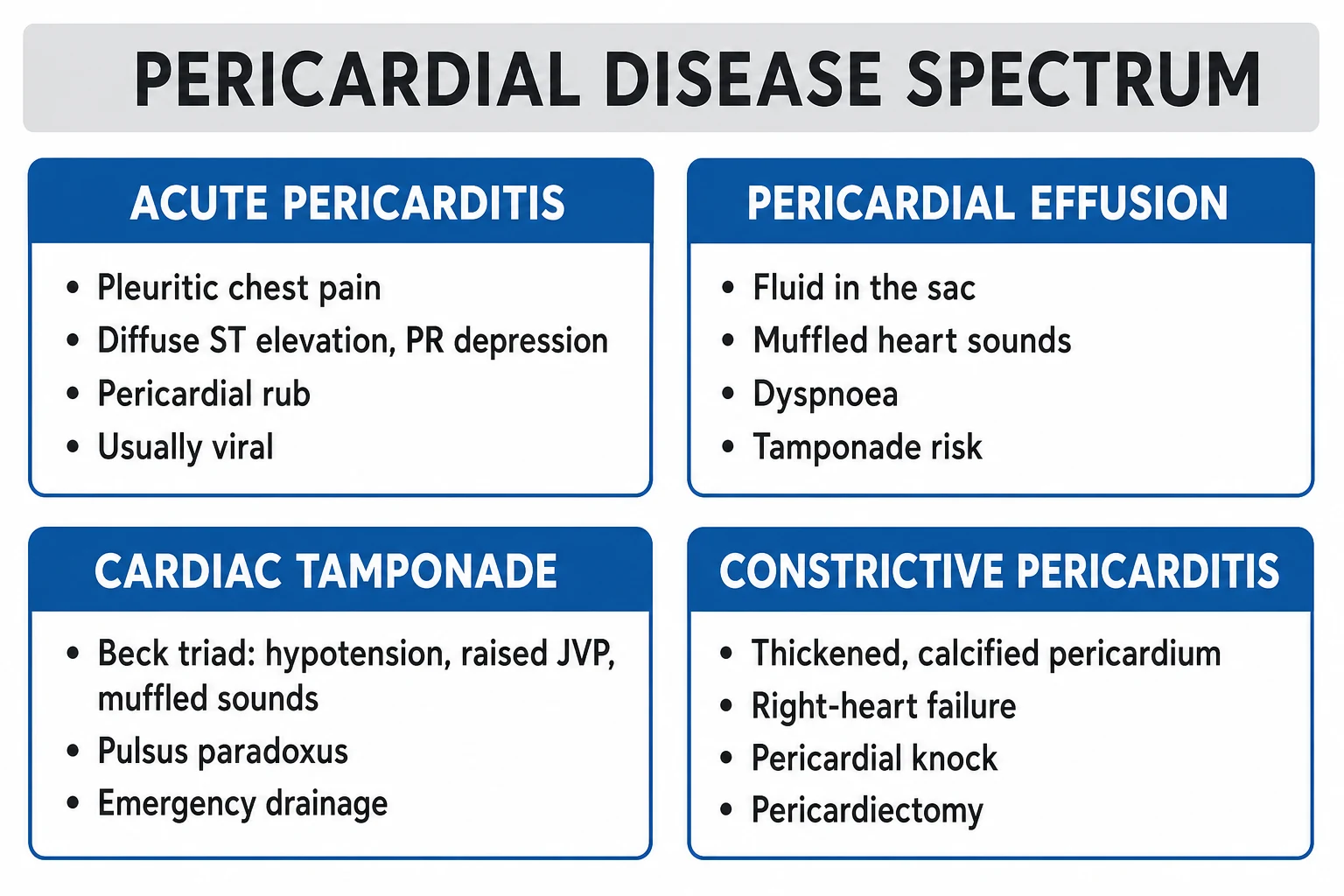
Management
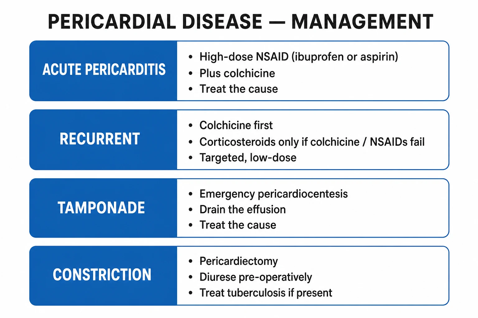
Acute pericarditis treatment
NSAIDs — first-line
Ibuprofen 600 mg PO TID (or aspirin 750-1000 mg PO TID). Duration: 1-2 weeks. Reduces inflammation and pain. Gastroprotection: PPI. Caution: renal impairment, GI bleeding, anticoagulated patients. Alternative: indomethacin 25-50 mg TID.
Colchicine — add to NSAIDs
Colchicine 0.5 mg PO BD (0.5 mg OD if <70 kg). Duration: 3 months. COPPS trial: reduces recurrence rate from ~25% to ~10%. Mechanism: inhibits microtubule polymerisation → inhibits neutrophil function → anti-inflammatory. Side effects: GI (diarrhoea, nausea), bone marrow suppression (rare).
Steroids — ONLY for refractory or specific causes
Prednisolone 0.2-0.5 mg/kg/day (low dose preferred). Indications: NSAID failure/refractory, autoimmune causes (SLE, RA), uraemic pericarditis, pregnancy (NSAIDs contraindicated). CAUTION: steroids increase recurrence rate of pericarditis (paradoxical effect). Use ONLY if NSAIDs fail or contraindicated. Taper slowly.
Treat underlying cause
Viral: supportive (NSAIDs + colchicine). Bacterial: IV antibiotics + drainage if purulent. TB: anti-TB therapy (4 drugs). Uraemic: dialysis. Autoimmune: immunosuppression. Malignancy: treat cancer + pericardial drainage if needed. Dressler: NSAIDs (avoid steroids — high recurrence).
Monitor for complications
Serial echocardiography: assess for pericardial effusion (development or worsening). Watch for tamponade (Beck triad, pulsus paradoxus). Activity restriction: competitive sport avoided during acute illness + until full recovery. Follow-up at 1 week and 1 month.
SAQ — Acute idiopathic/viral pericarditis: diagnosis, ECG discrimination and stepwise management
10 minutes · 10 marks
A 34-year-old previously well man presents with two days of sharp, pleuritic central chest pain that is worse on deep inspiration and lying flat and is relieved by sitting forward. He describes a three-day viral prodrome of coryzal symptoms and myalgia. On examination he is febrile (37.9 degrees C), HR 108 sinus, BP 128/76, SpO2 97 per cent on room air, and a clear three-component friction rub is audible at the left sternal edge with the patient leaning forward in expiration. ECG shows diffuse concave ST elevation in I, II, III, aVL, aVF and V2-V6, with PR depression in the same leads and PR elevation in aVR; there is no reciprocal ST depression and no pathological Q waves. High-sensitivity troponin is mildly raised at 0.08 ng/mL (upper reference 0.04). CRP is 78 mg/L. Bedside echocardiography shows a small 0.5 cm posterior pericardial effusion with no chamber collapse and no regional wall motion abnormality.
SAQ — Recurrent pericarditis in a steroid-dependent patient: escalation to IL-1 blockade
10 minutes · 10 marks
A 42-year-old woman is reviewed in clinic for her third recurrence of pericarditis in 14 months. Each episode has been treated with aspirin plus colchicine, but the last two flares were managed with prednisolone 0.5 mg/kg/day because of NSAID intolerance (chronic kidney disease, eGFR 42). She is currently on prednisolone 12 mg daily and colchicine 0.5 mg daily, with chest pain returning as the steroid is tapered below 15 mg. CRP is 64 mg/L, ECG shows recurrent diffuse PR depression, and echo shows a small effusion with no tamponade. She is distressed by the recurrent admissions and concerned about the cumulative steroid exposure (now Cushingoid, borderline diabetes, osteopenia).
Clinical pearls
Red flags
Aetiology — the full differential and what each cause means
Acute pericarditis is inflammation of the pericardial layers (visceral + parietal). In the developed world the majority of cases are idiopathic/viral — a "diagnosis of exclusion" made after bacterial, neoplastic, and autoimmune causes have been ruled out. The intensivist's job is not simply to label it "viral" but to actively seek and treat the treatable causes (TB, purulent bacterial, uraemic, autoimmune, neoplastic), because the underlying aetiology dictates the management and changes the prognosis.[1][7]
Causes of acute pericarditis — by frequency and clinical importance
| Cause | Relative frequency | Key features / clues | Specific treatment |
|---|---|---|---|
| Idiopathic / viral | ~80-90% (the most common by far) | Often preceded by a viral prodrome (URTI, fever, myalgia). Common viruses: coxsackie B, echovirus, adenovirus, influenza, parvovirus B19, EBV, HIV. By definition, no other cause identified. | NSAIDs + colchicine (the backbone of management). Supportive. |
| Autoimmune / connective tissue disease | ~5-10% | SLE (young woman, malar rash, dsDNA, low complement), rheumatoid arthritis (seropositive, nodules), scleroderma, sarcoidosis, vasculitis (EGPA, granulomatosis with polyangiitis). Often recurrent; multi-system involvement. | Immunosuppression (steroids first, then steroid-sparing — azathioprine, IVIG). Colchicine for the pericarditis component. |
| Post-cardiac injury syndrome | ~5% | Dressler syndrome = 2-10 weeks post-MI (autoimmune response to released cardiac antigens). Post-pericardiotomy syndrome = weeks-months after cardiac surgery/trauma/catheter. Fever + pleuritic pain + effusion + raised inflammatory markers. | NSAIDs + colchicine (preferred). Avoid steroids (high recurrence). |
| Uraemic | Variable (dialysis patients) | End-stage renal failure / under-dialysed. Often haemorrhagic effusion. Does NOT respond to NSAIDs. | Intensify dialysis (the definitive treatment). Drain if tamponade. |
| Neoplastic | ~5-7% | Lung, breast, lymphoma, melanoma most common. Often large, recurrent, haemorrhagic effusion → tamponade. Cytology positive in 50-85% (multiple samples increase yield). | Treat the cancer + pericardial window/sclerotherapy (prevents reaccumulation). |
| Tuberculous | Common in endemic regions | Chronic, low-grade fever, night sweats, weight loss; concomitant pulmonary TB. Effusion often haemorrhagic/exudative, high ADA, lymphocytic. High risk of progression to constrictive pericarditis. | 4-drug anti-TB therapy (RIPE) + adjunctive steroids (reduces constriction risk). |
| Purulent bacterial | Rare but emergency | S. aureus, pneumococcus, meningococcus, Haemophilus. Septic patient, high fever, purulent pericardial fluid. Rapid progression to tamponade; high mortality. | IV antibiotics + immediate surgical/percutaneous drainage. Life-threatening. |
| Radiation | Years after thoracic RT (for Hodgkin, breast) | Late effect — fibrotic, calcific pericardium. Often combined with constrictive physiology + myocardial/coronary damage. | Symptomatic; pericardiectomy (high risk if extensive mediastinal fibrosis). |
| Drug-induced | Rare | Procainamide, hydralazine, isoniazid, phenytoin (drug-induced lupus). Stop the offending drug. | Withdraw the drug + NSAIDs/colchicine. |
| Aortic dissection | Emergency | Type A dissection → haemopericardium → tamponade. This is NOT true pericarditis but mimics it. | Urgent surgery (pericardiocentesis alone is insufficient — bleeding continues). |
Pathophysiology — inflammation of the pericardial layers
The pericardium has two layers: the visceral pericardium (epicardium, adherent to myocardium) and the parietal pericardium (the tough outer fibrous sac). Between them is 15-50 mL of serous fluid. Inflammation causes increased vascular permeability → fibrinous exudate deposition between the layers (the substrate for the friction rub and for adhesions/constriction later) and a variable amount of pericardial effusion (transudate or exudate, sometimes haemorrhagic). [1]
The clinical syndrome arises from three mechanisms:
- Pericardial irritation → the pleuritic, positional chest pain and the friction rub (the roughened, inflamed layers rubbing against each other).
- Superficial epicardial (subepicardial) myocardial involvement → "myopericarditis" — explains the ST elevation (epicardial injury current) and the often-mild troponin elevation. The ST changes of pericarditis are therefore a repolarisation/epicardial-injury phenomenon, not a Q-wave infarct.
- Diffuse atrial and ventricular involvement → explains the diffuse PR depression (diffuse atrial injury current) and diffuse ST elevation across most leads (as opposed to the territorial changes of a coronary occlusion). [1]
This diffuse, subepicardial pattern is the electrophysiological signature that distinguishes pericarditis from STEMI on the ECG.[1]
Clinical presentation — pleuritic, positional pain and the friction rub
The classic triad of acute pericarditis is: (1) pleuritic chest pain, (2) pericardial friction rub, (3) widespread ST elevation / PR depression on ECG. Most patients have at least two; the friction rub is the most easily missed because it is transient and positional.[1][7]
Clinical features of acute pericarditis — mechanism and exam relevance
| Feature | Frequency / detail | Mechanism / notes |
|---|---|---|
| Pleuritic, positional chest pain | ~95% (the cardinal symptom) | Sharp, worse on deep inspiration and coughing (pleural involvement); worse lying flat (the heart's weight presses the inflamed layers against the parietal pericardium/chest wall); better on sitting forward (separates the layers). May radiate to the left trapezius ridge (phrenic nerve C3-C5). Distinguish from the crushing, pressure, non-positional pain of STEMI. |
| Pericardial friction rub | ~30-85% (variable — transient) | Best heard at the left sternal border with the patient leaning forward and breath held in expiration, using the diaphragm. Classic three-component: (1) atrial systole, (2) ventricular systole, (3) rapid ventricular filling (early diastole) — "train wheel" / leather-creak quality. A two- or one-component rub is also valid. Transient — re-examine the patient repeatedly; absence at one moment does not exclude pericarditis. |
| Low-grade fever | Common | Inflammatory; a high swinging fever or rigors should prompt search for purulent bacterial pericarditis. |
| Dyspnoea | Variable | From pain on breathing, or a developing effusion. |
| Tachycardia | Common | Pain, fever, or early tamponade — a rising heart rate with falling BP is a tamponade warning. |
| Cough / dysphagia / hiccoughs | Occasional | From irritation of adjacent structures (phrenic nerve, recurrent laryngeal nerve, oesophagus). |
The ECG in acute pericarditis — four stages and the PR-segment story
The ECG is the single most useful investigation and evolves through four classical stages (Spodick). Roughly half of patients show all four stages; many present in stage 1 and the ECG normalises before stage 4 is captured. Recognising stage 1 (and the PR-segment deviations) is the high-yield exam skill.[1]
The four ECG stages of acute pericarditis (Spodick)
| Stage | Timing | ECG appearance |
|---|---|---|
| Stage 1 (hours to days) | Acute phase | Diffuse concave (upward / "smiley") ST elevation in most leads (I, II, III, aVL, aVF, V2-V6) + diffuse PR depression in the same leads + PR elevation in aVR (and sometimes V1) — the reciprocal mirror. This PR pattern is the most specific early ECG clue. |
| Stage 2 (first few days) | Transition | ST elevation resolves back to baseline; PR depression may persist; T waves begin to flatten. |
| Stage 3 (days to weeks) | Inversion | T-wave inversion develops (in the leads that had ST elevation). No pathological Q waves (distinguishes from evolving STEMI). |
| Stage 4 (weeks to months) | Resolution | T waves return to normal (ECG fully normalises). T-wave inversion may persist indefinitely in some patients without clinical significance. |
Differentiating acute pericarditis from STEMI and early repolarisation
This is a perennial exam favourite and a real-world diagnostic trap: mislabelling pericarditis as STEMI leads to inappropriate antithrombotic/PCI pathways (and bleeding), while missing STEMI is catastrophic. The ECG pattern is usually distinguishable — learn the rules below.[1][7]
Acute pericarditis vs STEMI vs early repolarisation — the ECG rules
| Feature | Acute pericarditis | STEMI | Early repolarisation |
|---|---|---|---|
| ST elevation distribution | Diffuse — multiple vascular territories (I, II, III, aVL, aVF, V2-V6) | Localised / territorial — one coronary territory (e.g., II/III/aVF = inferior) | Diffuse, usually V2-V5, lateral |
| ST morphology | Concave upward ("smiley") | Convex upward ("frowny") — but concave STEMI exists | Concave; prominent J-point notch |
| PR segment | Diffuse PR depression + PR elevation in aVR (key) | PR depression only in the infarcted territory (e.g., atrial infarct) — uncommon | Usually no PR depression; PR elevation may be present |
| Reciprocal ST depression | Absent (the ST change is diffuse, not a single injury vector) | Present — the reciprocal of the ST-elevation territory (e.g., inferior STEMI → anterior ST depression) | Absent |
| Q waves | Never | May develop (pathological) | Never |
| T waves | Inversion after ST returns to baseline (stage 3) — no Q waves | Inversion while ST still elevated — evolutionary | Tall, peaked, symmetrical (benign) |
| ST/T ratio (V6) | > 0.25 favours pericarditis | Variable | < 0.25 favours early repolarisation |
| Serial evolution | Evolves through 4 stages over weeks | Evolves: STE → T inversion → Q waves, over hours-days | Static — unchanged on serial ECGs (benign, stable) |
| Clinical correlate | Pleuritic, positional pain, friction rub, young, viral prodrome | Crushing, pressure, non-positional, radiation, sweating, coronary risk factors | Asymptomatic; incidental on a young male athlete's ECG |
| Troponin | Mildly raised (myopericarditis) or normal | Markedly raised (territorial necrosis) | Normal |
Investigations — confirming the diagnosis and excluding mimics
Acute pericarditis is a clinical diagnosis (typical pain + friction rub + characteristic ECG). Investigation is targeted at (a) confirming pericarditis, (b) excluding STEMI and the specific secondary causes, and (c) detecting complications (effusion/tamponade).[1][7]
- ECG — the cornerstone (see above). Repeat daily during the acute phase to track evolution and exclude evolving STEMI.
- Echocardiogram — mandatory in every case. Looks for: pericardial effusion (size, loculation), tamponade physiology (RA/RV collapse, IVC plethora), wall motion abnormalities (RWMA = STEMI/myocarditis, not pericarditis), and baseline LV function.
- Bloods:
- Inflammatory markers — CRP and ESR are raised; CRP guides treatment duration (treat until CRP normalises).
- Troponin — mildly raised in ~30-50% (myopericarditis); does NOT mean infarction. Markedly raised troponin with RWMA = myocarditis/MI.
- FBC, U&E, LFTs — screen for uraemia, infection, leukaemia.
- Cultures, viral serology, TB (interferon-gamma release assay, AFB), autoimmune screen (ANA, dsDNA, rheumatoid factor, complement), thyroid function — for the secondary causes.
- CXR — usually normal; may show an enlarged cardiac silhouette if effusion >250 mL, a pleural effusion (usually left-sided), or signs of TB/malignancy. A normal CXR with a large effusion is still possible (effusion sits anteriorly).
- Pericardial fluid analysis (if drained): cell count, Gram stain + culture, AFB/TB PCR, cytology (malignancy), glucose (low = bacterial/rheumatoid), protein/LDH (Light's criteria — exudate), ADA (high = TB), triglycerides (chylopericardium), pH.
- CT/MRI — for complex cases: pericardial thickening, loculated/haemorrhagic effusion, contrast uptake of inflamed pericardium (MRI), and to exclude aortic dissection/mass. [1]
Management — the stepwise pharmacological approach
The pillars are (1) anti-inflammatory analgesia (NSAIDs/aspirin), (2) colchicine, and (3) specific treatment of the underlying cause. Steroids are deliberately demoted to second/third line because they increase recurrence. The goal of treatment is symptom control and prevention of recurrence (the most common complication).[1][4][3]
Pharmacology of pericarditis — NSAIDs, colchicine, steroids
| Drug | Dose | Mechanism / rationale | Cautions |
|---|---|---|---|
| Aspirin | 750-1000 mg PO TID for 1-2 weeks, then taper | First-line especially post-MI (antiplatelet + anti-inflammatory). ESC-preferred NSAID in ischaemic contexts. | GI bleed, asthma, anticoagulation. Add PPI. |
| Ibuprofen | 600 mg PO TID for 1-2 weeks | First-line; well tolerated, titratable. | Renal impairment, GI bleed, heart failure. |
| Indomethacin | 25-50 mg PO TID | Alternative NSAID. | Avoid in the elderly and in coronary disease — reduces coronary blood flow. |
| Colchicine | 0.5 mg PO BD (>70 kg) or 0.5 mg PO OD (≤70 kg) for 3 months (first episode) / 6 months (recurrent) | Inhibits microtubule polymerisation → blocks neutrophil chemotaxis/activation → anti-inflammatory. Reduces recurrence by ~50% (ICAP, CORP, COPPS). | GI (diarrhoea, nausea — dose-limiting); CKD (dose-reduce, narrower margin); P-gp/CYP3A4 interactions (clarithromycin, statins → rhabdomyolysis); cytopenias (rare). Avoid loading dose (no benefit, more GI). |
| Prednisolone (steroid) | Low dose 0.2-0.5 mg/kg/day, then taper over weeks-months | Second/third line ONLY. Suppresses inflammation when NSAIDs fail or are contraindicated. | Increases recurrence rate (dose-dependent) — avoid as first-line. Use low dose + slow taper. Osteoporosis, hyperglycaemia, infection prophylaxis. |
Principles of the NSAID course: start at a high anti-inflammatory dose, continue until the patient is pain-free and CRP has normalised, then taper gradually over 1-2 weeks. Stopping too early is a common cause of early recurrence. Add a PPI for gastroprotection. [1]
Landmark trials — the evidence base for colchicine and beyond
The modern management of acute pericarditis rests on a coherent series of Imazio randomised trials (CORE → COPE → COPPS → ICAP → CORP → CORP-2), reinforced by IL-1 blockade (RHAPSODY).[4][3][5][6]
ICAP — colchicine for a first episode of acute pericarditis (Imazio 2013, NEJM; PMID 24088085)
Design
Multicentre, randomised, double-blind, placebo-controlled trial — 240 adults with a first episode of acute pericarditis
Intervention
Colchicine (0.5 mg BD if >70 kg, 0.5 mg OD if ≤70 kg for 3 months) vs placebo, BOTH on top of aspirin/ibuprofen
Primary outcome
Incessant/recurrent pericarditis at 18 months: **16.7% colchicine vs 37.5% placebo** (RRR 0.56; NNT 4)
Secondary outcomes
Higher 1-week remission (85% vs 58%), fewer symptom persistence at 72 h, fewer hospitalisations
Clinical bottom line
Colchicine added to an NSAID is the standard of care for a first episode — it roughly halves recurrence. This is the foundational RCT for colchicine in acute pericarditis.
CORP — colchicine for recurrent pericarditis (Imazio 2011, Ann Intern Med; PMID 21873705)
Design
Multicentre, randomised, double-blind, placebo-controlled trial — 120 patients with a first recurrence of pericarditis
Intervention
Colchicine (1-2 mg day 1 then 0.5-1.0 mg/day for 6 months) vs placebo, on top of conventional anti-inflammatory therapy
Primary outcome
Recurrence at 18 months: **24% colchicine vs 55% placebo** (RRR 0.56; ARR 0.31; NNT 3)
Secondary outcomes
Less symptom persistence at 72 h, higher 1-week remission, fewer recurrences/hospitalisations; no excess side effects
Clinical bottom line
Colchicine is effective for the secondary prevention of recurrent pericarditis — it is now first-line add-on therapy for every recurrence.
COPPS — colchicine to prevent post-pericardiotomy syndrome (Imazio 2010, Eur Heart J; PMID 20843851)
Design
Multicentre, randomised, double-blind, placebo-controlled trial — 360 patients after cardiac surgery
Intervention
Colchicine (1 mg then 0.5 mg BD for 1 month) vs placebo, started post-operatively
Primary outcome
Post-pericardiotomy syndrome at 12 months: **8.9% colchicine vs 21.1% placebo**
Clinical bottom line
Colchicine prevents the post-pericardiotomy syndrome (a post-cardiac-injury pericarditis). Extends the colchicine rationale beyond idiopathic/viral disease. (The pre-operative COPPS-2 design did not improve on this.)
RHAPSODY — rilonacept for recurrent pericarditis (Klein 2021, NEJM; PMID 33175185)
Design
Multicentre trial with a run-in (withdrawal-to-relapse) design — 86 patients with recurrent pericarditis despite NSAIDs/colchicine/steroids
Intervention
Rilonacept (a subcutaneous IL-1α/β 'trap' — interleukin-1 blockade) weekly
Primary outcome
Time to first pericarditis recurrence: dramatically delayed with rilonacept; pain and CRP fell rapidly. Most placebo patients relapsed within weeks of withdrawal
Clinical bottom line
IL-1 blockade is a **steroid-sparing** breakthrough for colchicine-refractory, steroid-dependent recurrent pericarditis. FDA-approved for recurrent pericarditis (2021). Confirms the autoimmune/IL-1-driven nature of refractory disease.
Complications — what pericarditis becomes if it goes wrong
Most episodes of acute viral/idiopathic pericarditis are benign and self-limiting. The complications below are the ones that change management and that examiners probe.[1][7]
Complications of acute pericarditis
| Complication | Frequency | Recognition | Management |
|---|---|---|---|
| Pericardial effusion | Common (variable) | Echo (dark, echo-free space around the heart). Small effusions are expected; size + haemodynamic effect matter, not presence. | Treat the pericarditis. Serial echo. Drain only if large/progressive or tamponade. |
| Cardiac tamponade | ~3% (higher in malignancy, TB, purulent, uraemic) | Hypotension, tachycardia, pulsus paradoxus, distended neck veins, electrical alternans, echo: RA collapse (early diastole, most sensitive) + RV collapse (late diastole, most specific) + IVC plethora. | Echo-guided pericardiocentesis + drain. Avoid PPV/diuretics/vasodilators. Treat the cause. (See the cardiac tamponade topic.) |
| Recurrent pericarditis | ~20-30% after first episode (down to ~10% with colchicine) | Recurrence of typical pain + ECG + raised CRP after a symptom-free interval of >4-6 weeks. | Re-treat with NSAID + colchicine (3-6 months); low-dose steroid only if refractory; anakinra/rilonacept for multiply-recurrent/steroid-dependent. |
| Incessant pericarditis | Subset | Symptoms persisting >4-12 weeks without a symptom-free interval (different from recurrent). | Same drugs; escalate to steroid-sparing IL-1 blockade early; exclude a missed specific cause (TB, autoimmune). |
| Constrictive pericarditis | ~1-2% after first episode (higher after TB, radiation, recurrent) — months to years later | Thickened/calcified pericadium restricts filling: Kussmaul sign, pericardial knock, raised JVP, ascites, peripheral oedema. Echo: septal bounce, restrictive diastolic pattern, plethoric IVC. CT/MRI: pericardial thickening (>3-4 mm), calcification. | Pericardiectomy (surgical stripping) — curative but high morbidity. |
| Purulent pericarditis → tamponade/sepsis | Rare, but an emergency | Septic patient, purulent pericardial fluid, rapid tamponade. | IV antibiotics + immediate drainage (often surgical). High mortality if delayed. |
Recurrent pericarditis — the management ladder
Recurrence is the most common complication and the one that brings patients (repeatedly) to the ICU. Recurrent pericarditis is increasingly understood as an autoimmune / IL-1-driven process (autoreactive inflammation), which is why IL-1 blockade is so effective in refractory cases. The stepwise approach:[1][6][3]
Management ladder for recurrent pericarditis
Step 1 — re-establish full-dose anti-inflammatory therapy
Re-treat with an NSAID/aspirin at full anti-inflammatory dose (e.g., aspirin 750-1000 mg TID or ibuprofen 600 mg TID) until pain-free AND CRP normalises, then taper over 1-2 weeks. The single most common reason for "treatment failure" is an inadequate initial course / premature taper.
Step 2 — colchicine 0.5 mg BD for 6 months
Add (or confirm adherence to) colchicine 0.5 mg BD (>70 kg) / 0.5 mg OD (≤70 kg) for AT LEAST 6 months for recurrence (CORP trial). Continue at least 6 months after the last recurrence. Dose-reduce in CKD.
Step 3 — low-dose steroid, only if NSAIDs fail or are contraindicated
Prednisolone 0.2-0.5 mg/kg/day (LOW dose preferred) — taper VERY slowly over weeks-months. Higher doses and rapid tapering drive more recurrences. Re-institute/titrate the NSAID + colchicine as the steroid is weaned.
Step 4 — IL-1 blockade (steroid-sparing) for refractory / steroid-dependent disease
Anakinra (IL-1 receptor antagonist, daily SC) or rilonacept (IL-1 trap, weekly SC — RHAPSODY trial, FDA-approved). Indicated for colchicine + steroid-refractory or steroid-dependent recurrent pericarditis. Enables steroid weaning and dramatically reduces recurrence.
Step 5 — pericardiectomy (last resort)
Rarely needed; reserved for truly refractory, multiply-recurrent disease failing all medical therapy. Performed in expert centres. High threshold — most patients respond to IL-1 blockade.
Throughout — exclude a missed specific cause
Recurrent disease that does not behave like typical idiopathic recurrence should prompt re-investigation for TB, autoimmune disease (ANA, dsDNA), and malignancy. "Idiopathic" is a diagnosis of exclusion, renewed each time.
Constrictive pericarditis — the chronic cousin
Constrictive pericarditis is the late, fibrotic sequela: the pericardium becomes thick, rigid, and often calcified, encasing the heart in a non-compliant shell that restricts diastolic filling. The physiology mimics tamponade (impaired filling → raised venous pressure → low output) but develops over months-years and, crucially, the treatment is surgical. It most often follows TB, radiation, recurrent idiopathic pericarditis, or prior cardiac surgery.[1]
Constrictive pericarditis vs cardiac tamponade — both impair filling, very different urgency
| Feature | Constrictive pericarditis | Cardiac tamponade |
|---|---|---|
| Pathology | Thickened / calcified rigid pericardium (fibrosis) | Fluid under pressure compressing the heart |
| Onset | Months-years (chronic) | Hours-days (acute/subacute) |
| Kussmaul sign (JVP rises on inspiration) | Present (rigid pericardium cannot accommodate the increased venous return) | Usually absent (free-flowing fluid allows inspiratory venous return) |
| Pericardial knock (early diastolic sound) | Present (abrupt halt to filling by the rigid pericardium) | Absent |
| Pulsus paradoxus | Mild or absent | Marked (>10 mmHg) |
| Echo | Pericardial thickening/calcification, septal bounce, respiratory variation in inflow, plethoric IVC | Effusion + RA/RV collapse + IVC plethora + swinging heart |
| CT/MRI | Pericardial thickening (>3-4 mm), calcification | Effusion (fluid) |
| Treatment | Pericardiectomy (surgical stripping — curative but high morbidity) | Pericardiocentesis (drainage) |
Special situations — when the standard pathway does not apply
Special situations in pericarditis management
| Situation | What changes | Recommended approach |
|---|---|---|
| Pregnancy | NSAIDs contraindicated after 20 weeks (premature closure of the ductus arteriosus, oligohydramnios, fetal renal injury); avoid after 28 weeks. Aspirin also restricted near term. | Paracetamol + colchicine (safe in pregnancy) first; low-dose steroid if needed. Avoid NSAIDs in the 2nd/3rd trimester. |
| Anticoagulated / high bleeding risk | NSAIDs + anticoagulant = major bleeding risk; also raises INR unpredictably. | Prefer colchicine ± paracetamol; if an NSAID is unavoidable, use the shortest course + PPI + close INR monitoring. Steroid if necessary. |
| Early post-MI (<72 h) | NSAIDs/steroids impair scar formation → risk of ventricular free-wall rupture. | High-dose aspirin (750-1000 mg TID) preferred; avoid non-aspirin NSAIDs and steroids in the early healing phase. |
| Dressler / post-cardiac injury | Autoimmune recurrence risk high with steroids. | NSAID + colchicine (preferred). Avoid steroids. |
| Uraemic | Does not respond to NSAIDs/steroids; often haemorrhagic. | Intensify dialysis (the definitive treatment). Drain if tamponade. |
| Tuberculous | High risk of progression to constriction. | 4-drug anti-TB therapy + adjunctive steroids (reduce constrictive evolution). |
| Purulent bacterial | Rapid sepsis + tamponade; emergency. | IV antibiotics + immediate surgical/percutaneous drainage. |
| Neoplastic | Recurrent, often haemorrhagic. | Pericardiocentesis → pericardial window ± sclerotherapy (doxycycline/bleomycin) to prevent reaccumulation + treat the cancer. |
| CKD / elderly | NSAID + colchicine nephrotoxicity; colchicine narrow therapeutic window. | Dose-reduce colchicine; avoid NSAIDs if eGFR <30; consider steroid-sparing IL-1 blockade earlier. |
Additional clinical pearls — exam-exhaustive
Additional red flags
Exam one-liners — the rapid recall set
[1]Comparison with adjacent topics — high-yield cross-references
- Cardiac tamponade: the haemodynamic emergency of pericardial disease — pulsus paradoxus, electrical alternans, RA/RV collapse on echo, urgent pericardiocentesis. Pericarditis is the cause; tamponade is the complication.
- Acute coronary syndromes: the principal ECG and clinical mimic. STEMI = territorial convex ST elevation + reciprocal ST depression + marked troponin + RWMA on echo; pericarditis = diffuse concave ST elevation + PR depression + no RWMA.
- Myocarditis: shares troponin elevation and ST/T changes; myopericarditis is the overlap. Biopsy/MRI distinguishes pure myocarditis. Treat pericarditis component with NSAIDs + colchicine.
- Constrictive pericarditis: the chronic fibrotic sequela — Kussmaul sign, pericardial knock, calcification, septal bounce; treatment is pericardiectomy. [1]
References
- [1]Adler Y, et al. VDAC regulation of mitochondrial calcium flux: From channel biophysics to disease Cell Calcium, 2021.PMID 33529977
- [2]Imazio M, et al. Notum palmitoleoyl-protein carboxylesterase regulates Fas cell surface death receptor-mediated apoptosis via the Wnt signaling pathway in colon adenocarcinoma Bioengineered, 2021.PMID 34402722
- [3]Imazio M, Brucato A, Cemin R, et al. Colchicine for recurrent pericarditis (CORP): a randomized trial Ann Intern Med, 2011.PMID 21873705
- [4]Imazio M, Brucato A, Ciminati G, et al. Motor speech treatment protocol for developmental motor speech disorders Dev Neurorehabil, 2015.PMID 24088085
- [5]Imazio M, Trinchero R, Brucato A, et al. Maternal investment of female mallards is influenced by male carotenoid-based coloration Proc Biol Sci, 2011.PMID 20843851
- [6]Klein AL, Imazio M, Cremer PC, et al. Immunocytochemical assessment of cell differentiation of podoplanin-positive osteoblasts into osteocytes in murine bone Histochem Cell Biol, 2021.PMID 33175185
- [7]Imazio M, Gaido L, LeWinter M, et al. Injury risks of EMS responders: evidence from the National Fire Fighter Near-Miss Reporting System BMJ Open, 2015.PMID 26068510