ICU · cardiovascular
Acute Severe Pulmonary Embolism — Comprehensive ICU Management
Also known as Pulmonary embolism · PE · Massive PE · Submassive PE · Pulmonary embolism with shock · Thrombolysis for PE · Pulmonary embolectomy · PERT
Acute severe pulmonary embolism (PE) — obstruction of the pulmonary arterial tree by thrombus (usually from deep vein thrombosis) causing increased dead space, right ventricular afterload, RV failure, reduced cardiac output, and potentially cardiac arrest. Risk factors: immobility, malignancy, surgery, pregnancy, OCP, inherited thrombophilia (factor V Leiden, prothrombin gene mutation, antithrombin deficiency). Severity classification: massive PE (high-risk — sustained hypotension SBP <90 or drop 40, or cardiac arrest — mortality 25-65%), submassive PE (intermediate-risk — normotensive but RV strain — RV dysfunction on echo + biomarker elevation), low-risk PE (normal BP + no RV strain — mortality <1%). ICU management: massive PE → thrombolysis (alteplase 50-100 mg IV over 2h OR 50 mg bolus if cardiac arrest) OR catheter-directed thrombolysis OR surgical embolectomy; submassive PE → anticoagulation (LMWH then warfarin/DOAC) + monitor for deterioration (consider rescue thrombolysis if decompensates); low-risk → anticoagulation + early discharge. VA-ECMO for refractory cardiac arrest from PE (bridge to definitive therapy). PESI/sPESI risk scores guide disposition. Mortality: massive 25-65%, submassive 5-10%, low-risk <1%.
On this page & tools
Your progress
Saved locally on this device.
Target exams
Red flags
Overview
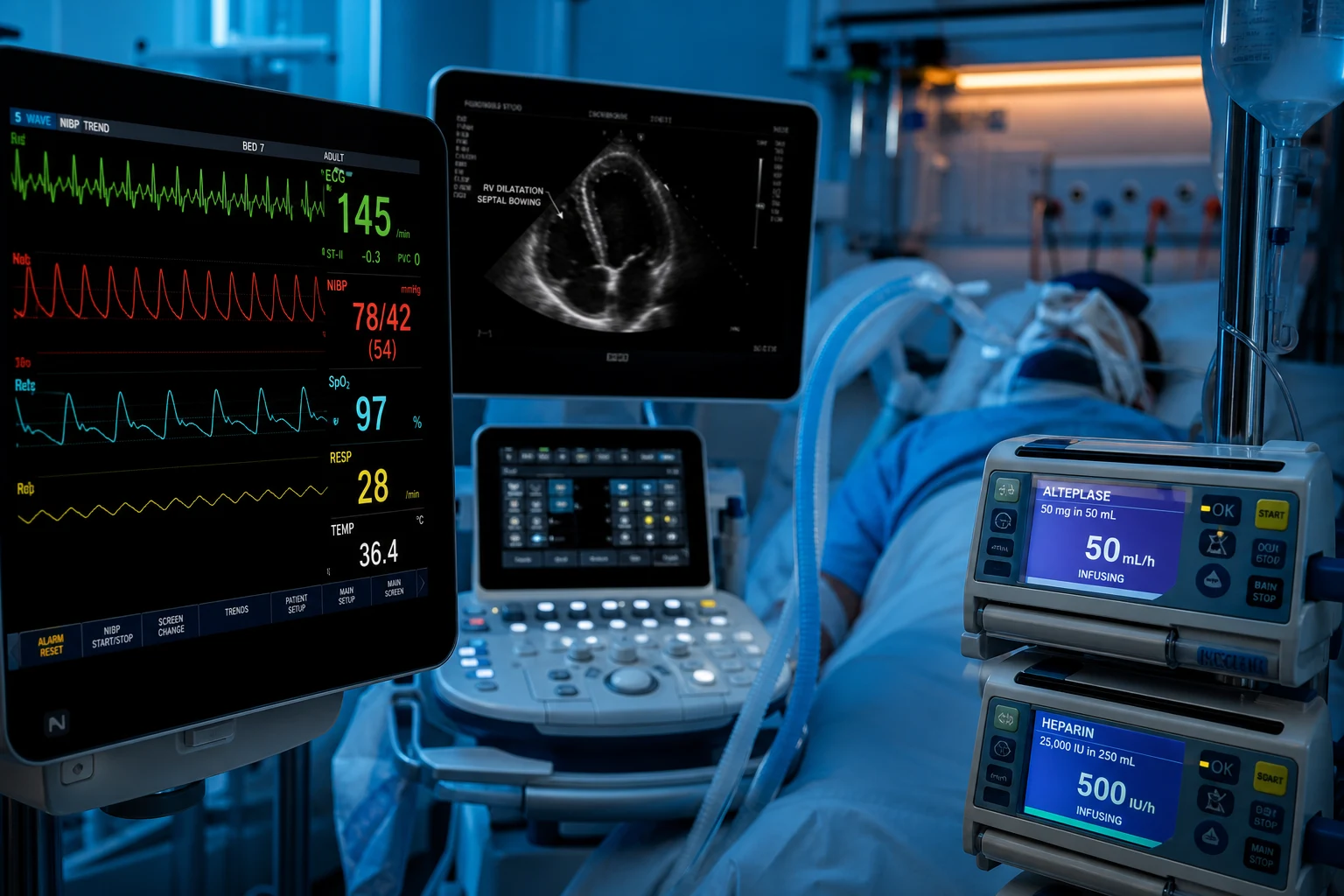
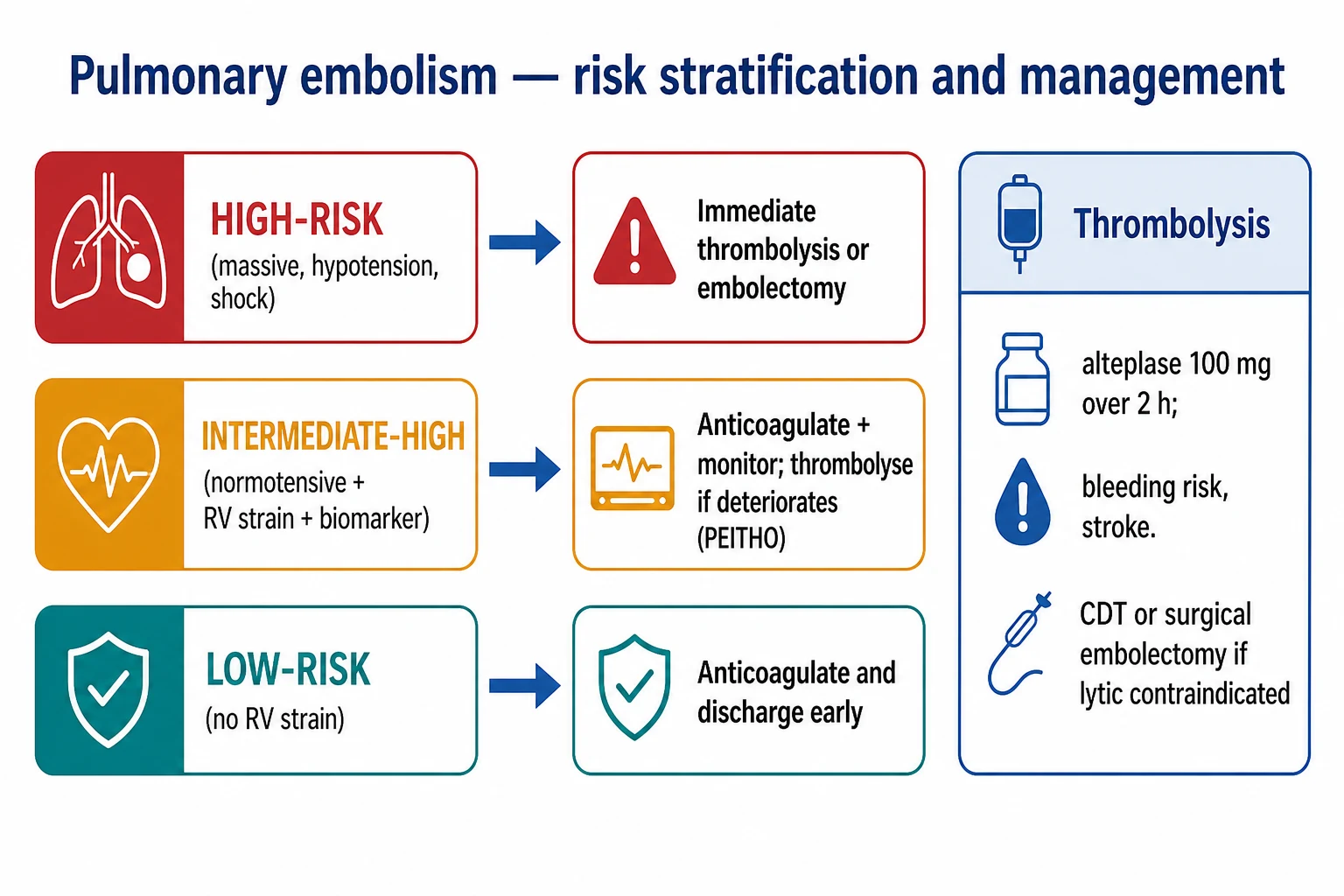
Severity classification — the key triage decision
PE severity classification (ESC 2019) — drives management
| Category | Definition | Haemodynamics | RV function | Biomarkers | Mortality | Management |
|---|---|---|---|---|---|---|
| High-risk (massive) | PE with haemodynamic instability | SBP <90 for >15 min OR drop >40 from baseline OR requiring vasopressors OR cardiac arrest | Usually impaired (but not required for diagnosis) | Usually elevated | 25-65% | Immediate thrombolysis (alteplase 50-100 mg IV over 2h) OR catheter-directed OR surgical embolectomy if thrombolysis contraindicated. Do NOT wait for CTPA if crashing |
| Intermediate-risk (submassive) | Normotensive but evidence of RV strain | SBP >90 (stable) | RV dysfunction (echo: RV/LV ratio >0.9, RV hypokinesis, McConnell sign; CT: RV/LV ratio >0.9) | Myocardial injury (elevated troponin, BNP/NT-proBNP) | 5-10% | Anticoagulation (LMWH). Monitor closely (ICU/HDU). Consider catheter-directed thrombolysis for high-risk features. Rescue systemic thrombolysis if deteriorates |
| Intermediate-risk (low subcategory) | Normotensive, RV strain OR biomarkers (but NOT both) | Stable | One abnormal (either echo OR biomarker — not both) | — | 2-3% | Anticoagulation. Ward monitoring |
| Low-risk | Normotensive, no RV strain, normal biomarkers | Stable | Normal | Normal | <1% | Anticoagulation. Consider early discharge / outpatient (sPESI = 0) |
ICU management of massive PE — the first 60 minutes
- RECOGNISE — sudden onset dyspnoea + hypotension (SBP <90) + hypoxia + risk factors (DVT, malignancy, post-op, immobility, pregnancy, OCP). ECG: sinus tachycardia (most common), S1Q3T3, right axis deviation, RBBB, T inversion V1-V4. Bedside echo: RV dilation, McConnell sign (RV free wall hypokinesis with apical sparing), septal flattening, TR jet >2.6 m/s, IVC dilation (non-collapsible)
- OXYGEN + VASOPRESSORS — high-flow oxygen. Noradrenaline for MAP >65 (alpha-1 vasoconstriction → supports BP AND improves coronary perfusion). AVOID excessive fluid boluses (>500 mL may worsen RV failure — the dilated RV compresses the LV through septal shift → reduced LV filling → worse output — "RV-LV interdependence")
- BEDSIDE ECHO (if available) — confirms RV strain (in the crashing patient, bedside echo is diagnostic — do NOT send to CT). McConnell sign (RV free wall hypokinesis with apical sparing) is 77% sensitive, 94% specific for PE
- GIVE THROMBOLYSIS IMMEDIATELY if clinical suspicion is high and patient is crashing:
- Alteplase (rt-PA) 50-100 mg IV over 2 hours (standard dose for massive PE — 100 mg over 2h per ESC/AHA guidelines, or 50 mg bolus if cardiac arrest)
- For cardiac arrest: alteplase 50 mg IV bolus (can be given during CPR — does NOT require cessation of CPR — continue resuscitation for at least 20-30 minutes after bolus as lysis takes time)
- DO NOT wait for CTPA confirmation if the patient is unstable (CT is dangerous in a crashing patient)
- Relative contraindications to thrombolysis: recent surgery, recent haemorrhagic stroke, active bleeding, intracranial neoplasm. In MASSIVE PE with cardiac arrest, there are NO absolute contraindications — the patient will die without treatment
- ANTICOAGULATION — unfractionated heparin (UFH — 80 IU/kg bolus, then 18 IU/kg/hr infusion, target APTT 1.5-2.5x control). Start UFH (not LMWH) if thrombolysis given or may be needed (UFH is rapidly reversible with protamine — LMWH is not). If thrombolysis NOT given: LMWH (enoxaparin 1 mg/kg BD) preferred over UFH (lower major bleeding and recurrent VTE — meta-analysis)
- CONSIDER ALTERNATIVES if thrombolysis contraindicated:
- Surgical embolectomy — open surgical removal of clot via median sternotomy on cardiopulmonary bypass — for patients with absolute contraindication to thrombolysis (recent intracranial surgery, haemorrhagic stroke) and available cardiothoracic surgery
- Catheter-directed thrombolysis (ultrasound-assisted — EKOS — ULTIMA trial) — lower-dose alteplase (10-20 mg) delivered directly into the clot via catheter — lower systemic bleeding risk — for submassive PE or massive PE with relative contraindications
- Percutaneous thrombectomy (FlowTriever, AngioVac) — mechanical clot removal via catheter — emerging technology — no thrombolytic needed
- VA-ECMO for refractory cardiac arrest or profound shock from massive PE — bridge to definitive therapy (thrombolysis/embolectomy). ECMO supports the failing RV + provides oxygenation while the clot lyses
- MONITOR: continuous BP (arterial line), ECG, SpO2, urine output, lactate (clearance = improving perfusion). Repeat echo at 6-12h (RV recovery). Check bleeding (thrombolysis risk — cerebral, GI, retroperitoneal, access sites)
Key trials and evidence
PEITHO trial — fibrinolysis for intermediate-risk PE (PMID 12502823)
Study design
Randomised, double-blind — 1,006 patients
Population
Normotensive PE with RV strain + biomarker elevation (intermediate-risk)
Intervention
Tenecteplase + heparin vs placebo + heparin
Primary outcome
Death or haemodynamic decompensation at 7 days: 2.6% (tenecteplase) vs 5.6% (placebo) — significant reduction
Key finding
Stroke rate: 2.4% (tenecteplase) vs 0.2% (placebo) — including haemorrhagic stroke (10 patients)
Key finding
Major extracranial bleeding: 6.3% (tenecteplase) vs 1.5% (placebo)
Clinical bottom line
Routine systemic thrombolysis for submassive PE REDUCES haemodynamic deterioration BUT increases major bleeding and stroke. Use SELECTIVELY — not for all submassive PE. Reserve for patients with high-risk features (severe RV dysfunction, worsening biomarkers, clinical deterioration)
ESC 2019 Guidelines — PE management (PMID 22179143)
Source
European Society of Cardiology — clinical practice guidelines
Risk stratification
High-risk (massive): shock/arrest → primary reperfusion (thrombolysis preferred). Intermediate-risk (submassive): RV strain → anticoagulation + monitor. Low-risk: anticoagulation + possible outpatient
Diagnosis
Wells/REVISED Geneva score → D-dimer (if low probability, age-adjusted cutoff) → CTPA (if moderate/high). Bedside echo for unstable patients
Anticoagulation
LMWH/fondaparinux preferred over UFH (unless thrombolysis candidate). DOACs (apixaban, rivarizoxaban) first-line for stable PE
Thrombolysis
Alteplase 100 mg IV over 2h for massive PE. 50 mg bolus for cardiac arrest
Clinical bottom line
The definitive PE management guideline — risk stratification determines treatment intensity
Clinical pearls
Red flags
Prognosis
PE prognosis by severity
| Severity | Mortality | Key prognostic factors |
|---|---|---|
| Massive (high-risk) | 25-65% | Time to thrombolysis, cardiac arrest, comorbidity |
| Submassive (intermediate) | 5-10% | RV dysfunction severity, biomarker level, deterioration |
| Low-risk | <1% | sPESI = 0, normal RV, normal biomarkers |
| CTEPH (long-term) | Variable | 0.4-4% of PE survivors develop CTEPH |
Key trials and evidence (supplementary)
VA-ECMO for massive PE (PMID 30615053)
Source
Retrospective multicentre — ELSO registry analysis
Population
Patients with massive PE on VA-ECMO
Survival
50-70% in selected patients
Key finding
ECMO is a BRIDGE — supports circulation while thrombolysis/embolectomy works — not a standalone treatment
Clinical bottom line
VA-ECMO for refractory massive PE: 50-70% survival — use as bridge to definitive therapy (thrombolysis or surgical embolectomy)
Pathophysiology — the vicious cycle of acute RV failure in PE
Understanding the RV failure cascade is the single most important concept for the PE exam, because every management decision (vasopressor choice, fluid restriction, thrombolysis, ECMO) follows from it. The right ventricle is a volume pump, not a pressure pump: its wall is thin, it operates in a low-pressure, low-resistance circuit (normal PVR ~1 Wood unit), and it has minimal contractile reserve. When a sudden embolic load occludes >30-50% of the pulmonary vascular bed, PVR rises acutely and the RV cannot generate the pressure needed to eject against it. [1]
The cascade — step by step
- Pulmonary arterial obstruction — thrombus (usually embolised from a proximal DVT) lodges in the main/lobar/segmental pulmonary arteries. Mechanical obstruction + reflex vasoconstriction (mediated by serotonin, thromboxane, and hypoxia) raise pulmonary vascular resistance (PVR).
- Increased RV afterload — the RV must now generate higher systolic pressure to push blood across the obstructed bed. Normal RV systolic pressure is ~25 mmHg; in massive PE it must exceed 50-60 mmHg, which exceeds the RV's maximum generated pressure.
- RV dilatation and ischaemia — the RV dilates (volume overload) and becomes hypokinetic. RV wall tension rises (LaPlace's law) → RV myocardial oxygen demand rises while RV coronary perfusion falls (RV perfusion occurs in both systole and diastole, but is compromised when RV diastolic pressure rises and systemic BP falls). The result is RV subendocardial ischaemia (the mechanism of troponin leak and of right-sided chest pain).
- Septal shift (RV-to-LV interdependence) — the dilated, pressurised RV pushes the interventricular septum leftward (paradoxical septal motion) during systole and diastole. This bowing of the septum into the LV cavity reduces LV preload and LV diastolic filling.
- Reduced cardiac output — reduced LV preload + reduced LV compliance → falling stroke volume → falling cardiac output → systemic hypotension. This further worsens RV coronary perfusion (the vicious cycle).
- Cardiogenic shock and arrest — falling coronary perfusion + worsening RV ischaemia → RV pump failure → profound low output → cardiogenic shock → PEA (the classic "narrow-complex PEA" arrest of massive PE). The most common terminal rhythm in PE-induced arrest is PEA with a rate <60/min. [1]
Why this matters at the bedside — the haemodynamic determinants of death
- The RV fails because of ischaemia, not because of pressure alone. Anything that drops systemic diastolic pressure (aortic root pressure) worsens RV coronary perfusion and accelerates the death spiral. This is why noradrenaline (raises systemic vascular resistance and diastolic BP, improving RV coronary perfusion pressure) is the preferred vasopressor, and why avoiding hypotension is critical.
- Fluid can kill. A dilated RV on the steep portion of its Frank-Starling curve does not benefit from preload — extra volume just dilates it further, worsens tricuspid regurgitation, worsens septal shift, and reduces LV filling. Fluid challenges >500 mL are harmful. Give small aliquots (250 mL) only if preload-dependence is demonstrated.
- Tachycardia is the only compensatory lever left. As RV stroke volume falls, the heart rate rises to maintain cardiac output. Bradycardia in massive PE is a pre-arrest sign (loss of compensation). Drugs that cause bradycardia must be avoided.
- Hypoxia worsens PVR. Hypoxic pulmonary vasoconstriction raises PVR further, worsening RV afterload. Correct hypoxia aggressively (high-flow oxygen) to break the cycle. [1]
The pressure-volume loop of the failing RV
In acute PE the RV pressure-volume loop shows: increased end-systolic pressure, increased end-diastolic volume (dilatation), reduced stroke volume (narrow loop width), and a shift to the right. Because the RV is on the flat (decompensated) portion of its Starling curve, increases in preload do not augment stroke volume — confirming that fluid loading is futile and dangerous. The therapeutic goal is to reduce afterload (lyse/remove the clot, lower PVR with oxygen and pulmonary vasodilators), support perfusion pressure (noradrenaline), and unload the RV (consider inodilators such as dobutamine/milrinone with care — they can worsen hypotension). [1]
Dead-space ventilation
Obstructed/under-perfused alveoli become ventilated but not perfused → increased physiological dead space (high V/Q). This manifests as raised PaCO2-ETCO2 gradient (ETCO2 falls because less CO2 is delivered to the lungs) and as hypocapnia from hyperventilation in the awake patient. A sudden drop in ETCO2 in an intubated patient is a red flag for PE. The dead space also explains why these patients are hard to ventilate: increased minute volume is needed to clear CO2, raising intrathoracic pressure and further reducing RV preload. [1]
ECG changes in pulmonary embolism
The ECG in PE is neither sensitive nor specific — the commonest finding is simply sinus tachycardia. The classic "S1Q3T3" pattern is present in only ~10-20% of patients with proven PE. Nevertheless, ECG findings are heavily examined and useful for risk stratification (the degree of RV strain correlates with clot burden) and for differential diagnosis. [1]
ECG findings in PE — frequency and significance
| Finding | Frequency | Significance / mechanism |
|---|---|---|
| Sinus tachycardia | Most common (up to 70-80%) | Compensatory response to reduced cardiac output; almost universal in massive PE. Bradycardia is a pre-arrest sign |
| S1Q3T3 (McGinn-White pattern) | ~10-20% | Deep S wave in lead I, Q wave in lead III, inverted T waves in lead III. Reflects acute RV dilation/strain. NON-specific (also seen in RV infarct, pulmonary hypertension, LBBB) |
| T wave inversion V1-V4 (anteroseptal) | ~20-50% in massive/submassive PE | Reflects severe RV strain and ischaemia. Correlates with clot burden and worse prognosis (Daniel score). Must be distinguished from anterior STEMI (PE has no ST elevation, no reciprocal changes, and RV strain on echo) |
| Right axis deviation (RAD) | ~15-25% | Shift of QRS axis >+90 degrees from RV dilation/strain |
| Right bundle branch block (RBBB) | ~10-25% (often incomplete) | Acute RV dilation stretches/delays the right conduction pathway. May be new and transient |
| Atrial arrhythmia (AF/flutter) | ~5-15% | RV stretch irritates the right atrium; new AF in PE worsens haemodynamics (loss of atrial kick) |
| Low limb-lead voltage / poor R progression | Variable | RV dilation displaces the heart clockwise, attenuating anterior R wave amplitude |
| P pulmonale (peaked P in lead II) | Uncommon | Right atrial enlargement in chronic/subacute cases |
| S1S2S3 pattern / SIQIII | Variable | Right heart overload patterns |
| ST elevation in lead aVR | Up to 20% in massive PE | Reflects diffuse subendocardial ischaemia from low output; correlates with severity and mortality |
The Daniel ECG score
A 21-point scoring system quantifying RV strain on ECG (T-wave inversions, S1Q3T3, RBBB, etc.). A score >10 is rare without PE and correlates with high clot burden and RV dysfunction. Examinable as a semi-quantitative marker of severity. [1]
"ECG in PE vs anterior STEMI" — the critical distinction
T-wave inversion in V1-V4 from PE is frequently mistaken for anterior ischaemia. Clues favouring PE: concurrent sinus tachycardia, S1Q3T3, T inversion also in lead III (not V1-V4 alone), right axis deviation, RBBB, no ST elevation, no reciprocal changes, RV strain on echo, and the clinical context (sudden dyspnoea, DVT risk factors, hypoxia with normal lung fields). Bedside echo is decisive: anterior STEMI shows LV wall motion abnormality, PE shows RV dilation with McConnell sign. [1]
Echocardiographic assessment of pulmonary embolism
Bedside echocardiography is the key diagnostic and prognostic tool in the haemodynamically unstable patient with suspected PE (where CTPA is unsafe), and it stratifies risk in the stable patient. The echo diagnoses RV dysfunction (not the clot itself, except in rare cases of clot-in-transit seen in the right atrium/ventricle or main PA on transthoracic or transoesophageal echo). [1]
Direct signs (clot visualised)
- Clot in transit — serpiginous/mobile echodensity in the right atrium, RV, or proximal PA. Seen in ~4-18% of PE patients. Associated with high mortality (up to 40%) and an indication for aggressive therapy (embolectomy or thrombolysis). May traverse a patent foramen ovale (PFO) and cause paradoxical embolism (stroke).
- Clot in main PA on TOE — TOE can visualise clot in the main/right/left PA (sensitivity limited for left PA). Useful in the intubated unstable patient. [1]
Indirect signs (RV dysfunction / strain) — diagnostic performance
Echocardiographic signs of PE — sensitivity and specificity
| Sign | Finding | Sensitivity | Specificity | Mechanism / significance |
|---|---|---|---|---|
| McConnell sign | RV free-wall hypokinesis with apical sparing (RV apex contracts normally) | ~77-83% | ~94-96% | Pathognomonic-ish: the RV apex is tethered to the hyperdynamic LV apex, which pulls it inward. Highly specific for PE vs other causes of RV dysfunction. Mimic: tako-tsubo cardiomyopathy |
| RV/LV end-diastolic ratio >0.9 | RV dilation relative to LV (normally RV < LV). Ratio >0.9 (some use >1.0) in apical 4-chamber view | ~60-70% | ~75-85% | Reflects acute RV volume overload. Also measurable on CT (RV/LV >0.9 = RV strain). Correlates with clot burden |
| TR jet velocity >2.6 m/s | Tricuspid regurgitation peak velocity elevated (RV systolic pressure = 4v² + RA pressure) | ~55-70% | ~70-80% | Reflects elevated RV systolic pressure from increased afterload. In acute PE usually <3.5 m/s (RV cannot generate very high pressures acutely) |
| IVC plethora | IVC >2.1 cm with <50% collapse on sniff (fixed, dilated, non-collapsible IVC) | ~70-80% | ~70-80% | Reflects elevated RA pressure from RV failure. A non-plethoric, collapsible IVC argues against significant RV failure |
| 60/60 sign | Pulmonary acceleration time <60 ms (on RV outflow Doppler) PLUS mild-moderate TR jet velocity <2.6 m/s | ~25-48% | ~92-96% | The mid-systolic "notch" on RVOT Doppler + sub-maximal TR velocity distinguishes PE (acute, RV cannot generate high pressure) from chronic pulmonary hypertension (high TR velocity). Highly specific when present |
| Flattened/bowed interventricular septum | D-shaped LV on parasternal short axis; septal flattening in systole and/or diastole | ~40-60% | ~85-95% | Reflects RV pressure/volume overload pushing septum leftward → reduces LV compliance/preload (RV-LV interdependence). Diastolic flattening = volume overload; systolic flattening = pressure overload |
| Reduced TAPSE (<1.6 cm) | Tricuspid annular plane systolic excursion reduced | ~40-60% | ~70-80% | Global RV longitudinal systolic dysfunction. Less specific for PE (reduced in any RV failure) |
| Right heart thrombus (in transit) | Mobile mass in RA/RV | — | — | Direct sign; ~4-18% prevalence; high mortality; consider urgent embolectomy/thrombolysis |
The role of echo in risk stratification and the "intermediate-risk" patient
Echo converts a clinically stable patient into intermediate-risk (submassive) PE when RV dysfunction is present. This is why the ESC 2019 algorithm adds RV imaging (echo or CT) AND biomarkers (troponin, NT-proBNP) on top of clinical risk (sPESI): sPESI ≥1, OR sPESI = 0 with RV dysfunction AND biomarker elevation identifies higher-risk patients needing inpatient monitoring. A normal echo in a normotensive PE patient strongly supports low-risk status and suitability for outpatient/ward care. [1]
Serial echo — the haemodynamic monitor
In the ICU, serial focused echoes track RV recovery (reduction in RV/LV ratio, return of apical contraction, resolution of septal flattening, improving TAPSE) and detect deterioration (worsening McConnell sign, rising TR velocity, new pericardial effusion, worsening IVC plethora). Echo guides escalation (rescue thrombolysis) and de-escalation of therapy. [1]
Catheter-directed thrombolysis (CDT) — technique and evidence
Ultrasound-assisted catheter-directed thrombolysis (USAT / EKOS — EkoSonic endovascular system) is the preferred reperfusion strategy when systemic thrombolysis is contraindicated or its bleeding risk is unacceptable, particularly in submassive PE with high-risk features. The principle: deliver a fraction of the systemic thrombolytic dose directly into the pulmonary arterial clot via an infusion catheter, augmented by low-energy (2 MHz) high-frequency ultrasound that disaggregates fibrin and increases drug penetration into the thrombus. [1]
Technique — step by step
- Vascular access — right internal jugular or common femoral vein under ultrasound guidance.
- Pulmonary angiography — a guiding catheter is advanced to the right heart and selective pulmonary angiography is performed from the main PA to localise the clot (filling defects, perfusion cutoffs), measure the pulmonary artery pressure, and calculate the RV/LV ratio.
- Device placement — the EKOS catheter (with ultrasound core and drug-infusion side-holes) is advanced over a wire and positioned across the burden of the thrombus in the affected (usually right or left main/lobar) PA. Bilateral catheters may be used for bilateral clot.
- Infusion — low-dose alteplase infused through the catheter: typically 1 mg/hr per catheter for 12-24 hours (total dose 20-24 mg for bilateral, 10-12 mg per side). OPTALYSE explored lower/shorter regimens (4-12 mg over 2-6 hours).
- Concurrent anticoagulation — therapeutic UFH or LMWH is continued (subtherapeutic heparin during infusion to balance bleeding — protocols vary).
- Monitoring — serial echo (RV/LV ratio improvement), fibrinogen (bleeding watch), pulmonary artery pressure, and clinical status (dyspnoea, BP, SpO2). Check for access-site and systemic bleeding.
- Follow-up angiography — repeated at the end of infusion to assess clot lysis, with the option to "lyse-and-retriev" with maceration/rheolytic thrombectomy if residual clot remains. [1]
Evidence — ULTIMA, SEATTLE II, OPTALYSE, PERFECT
ULTIMA trial — CDT vs anticoagulation for intermediate-risk PE (PMID 28637571)
Study design
Randomised, open-label, multicentre — 59 patients
Population
Intermediate-risk PE (RV/LV ratio >1.0) within 14 days
Intervention
Ultrasound-assisted CDT (low-dose alteplase, mean ~23 mg over ~17 h) + heparin vs heparin alone
Primary outcome
Change in RV/LV ratio at 24h: CDT improved (−0.30) vs heparin alone (+0.03) — significant
Key finding
No major bleeding and no intracranial haemorrhage in the CDT group
Clinical bottom line
CDT rapidly reverses RV dysfunction in submassive PE with minimal bleeding — supports CDT over systemic lysis for intermediate-risk PE with high-risk features
SEATTLE II — single-arm CDT in massive and submassive PE (PMID 26038589)
Study design
Prospective single-arm multicentre — 150 patients (31 massive, 119 submassive)
Intervention
Ultrasound-facilitated, catheter-directed low-dose fibrinolysis (alteplase 24 mg over ~24 h, max 1 mg/h/catheter)
Primary outcome
Mean RV/LV ratio decreased from 1.57 to 1.16 at 48h (significant)
Key finding
No intracranial haemorrhage; major bleeding 10% (mostly access-site); 30-day mortality 4%
Clinical bottom line
CDT effectively reduces RV strain with a fraction of the systemic dose and no intracranial bleeding — but no randomised comparison to systemic lysis
OPTALYSE PE — duration/dose-finding for CDT (PMID 29742577)
Study design
Randomised, 4-arm dose-finding — 101 patients with intermediate-risk PE
Intervention
Lower/shorter alteplase regimens (4-12 mg over 2-6 h) via EKOS
Primary outcome
All arms reduced RV/LV ratio at 48h; no clear superiority of higher/longer doses
Key finding
Lower-dose regimens had comparable RV improvement and similar bleeding to SEATTLE II — supports shorter, lower-dose protocols
Clinical bottom line
CDT can be delivered efficiently (2-6 h) at low dose — relevant for ICU logistics and bleeding risk
PERFECT registry — catheter-directed therapy for massive PE (PMID 25316772)
Study design
Prospective multicentre registry — 101 patients (28 massive, 73 submassive)
Intervention
Catheter-directed thrombolysis AND catheter-directed thrombectomy (no systemic lysis)
Key finding
Pulmonary artery pressure fell and cardiac index rose significantly; no major bleeding requiring transfusion or intracranial haemorrhage; massive-PE mortality 3.6%
Clinical bottom line
Real-world registry confirms catheter-directed therapy (with or without low-dose lysis) is safe and effective for massive and submassive PE
When to choose CDT
CDT is most appropriate for: (a) submassive PE with high-risk features (severe RV dysfunction, rising biomarkers, clinical deterioration, sPESI >1) where systemic thrombolysis bleeding risk is judged unacceptable; (b) massive PE with absolute or relative contraindication to systemic thrombolysis (recent surgery, recent non-haemorrhagic stroke, bleeding diathesis, age >75 with high bleeding risk); (c) failure of anticoagulation (clinical deterioration despite therapeutic heparin). It is NOT appropriate when the patient is in cardiac arrest (no time — give systemic bolus alteplase) or when no interventional radiology/catheter lab is available within the necessary timeframe. [1]
Surgical embolectomy — indications and technique
Surgical pulmonary embolectomy has undergone a renaissance with the rise of the Pulmonary Embolism Response Team (PERT) model and multidisciplinary PE pathways. Once considered a treatment of last resort, it is now first-line reperfusion for massive PE when thrombolysis is absolutely contraindicated OR has failed, and in selected submassive PE (e.g. clot-in-transit with a PFO, paradoxical embolism). [1]
Indications
- Massive (high-risk) PE with absolute contraindication to thrombolysis: recent intracranial surgery, recent haemorrhagic stroke, active life-threatening bleeding, intracranial neoplasm/vascular malformation, recent major surgery (<3 weeks), recent trauma.
- Failed thrombolysis (persistent shock/clot after systemic alteplase) — note: repeat systemic lysis is not recommended due to cumulative bleeding risk.
- Clot-in-transit / right-heart thrombus with a PFO (risk of paradoxical embolism and stroke) — some centres favour embolectomy to remove both the clot and close the PFO.
- Cardiac arrest from PE where ECMO is being considered (embolectomy on ECMO).
- Centres without catheter-lab capability but with on-site cardiac surgery. [1]
Technique
- Median sternotomy under general anaesthesia, with full systemic heparinisation.
- Cardiopulmonary bypass (CPB) with bicaval cannulation; mild hypothermia. Aortic cross-clamping and cardioplegic arrest are usually NOT required if the heart is beating on CPB (allows the RV to be opened empty without air embolism concerns), though some surgeons use a brief arrest.
- Pulmonary arteriotomy — a longitudinal incision in the main PA, extended into the right (and if needed left) PA. The clot is extracted using forceps, suction, and Fogarty catheters, with pulmonary veins vented to allow back-bleeding to flush distal clots.
- Closure of the PA, weaning from CPB, haemostasis, and closure.
- PFO closure (if present) — to prevent paradoxical embolism and future right-to-left shunting. [1]
Outcomes
Modern surgical embolectomy in high-volume centres carries in-hospital mortality of ~6-12% (far lower than historical series of 20-30%), with low rates of recurrent PE and intracranial bleeding. Outcomes are best when surgery is performed EARLY (before irreversible RV infarction and multi-organ failure). This is why the PERT model — rapid activation of a multidisciplinary team — improves outcomes: it brings the right reperfusion strategy (systemic lysis, CDT, embolectomy, or ECMO) to the right patient quickly. [1]
Contraindications and pitfalls
Inoperable distal clot cannot be surgically removed (the clot must be in main/lobar PA, not segmental). Severe comorbidity (advanced age, frailty, end-stage malignancy) may shift the balance toward CDT or comfort care. Post-embolectomy reperfusion injury and right heart failure can occur. Re-anticoagulation must be resumed promptly postoperatively to prevent re-thrombosis. [1]
Percutaneous catheter thrombectomy
Mechanical (non-lytic) catheter thrombectomy devices remove clot without thrombolytic — attractive when any thrombolysis is contraindicated. Devices include FlowTriever (large-bore aspiration/stent retriever), AngioVac (venovenous bypass with suction cannula), Cleaner rotational and Aspirex (rheolytic/rotational). The FLASH registry (FlowTriever) showed rapid reduction in RV/LV ratio and symptoms with low bleeding in a broad real-world cohort. The PEERLESS trial (FlowTriever vs CDT) is clarifying the role of pure mechanical thrombectomy. Advantages: immediate clot debulking, no thrombolytic (no systemic fibrinolysis), single-session procedure. Pitfalls: haemolysis, haemoglobinuria, access-site bleeding, haemodynamic shifts, need for large-bore venous access. [1]
Reperfusion strategies — choosing between them
Reperfusion strategies for massive and high-risk intermediate PE
| Strategy | Mechanism | Speed | Bleeding risk | Best for | Limitation |
|---|---|---|---|---|---|
| Systemic thrombolysis (alteplase 100 mg/2 h, or 50 mg bolus in arrest) | Systemic plasminogen activation → diffuse clot lysis | Minutes to hours (lysis over ~2 h) | HIGH (intracranial ~1-3%, major extracranial ~6-10%) | Massive PE with shock/arrest; no time for procedures; no contraindication | Bleeding; absolute contraindications common; ~8% failure |
| Catheter-directed thrombolysis (EKOS) | Low-dose alteplase delivered directly into clot + ultrasound | 12-24 h (or 2-6 h OPTALYSE) | LOW (no intracranial in SEATTLE II; access-site bleeding) | Submassive PE with high-risk features; massive PE with relative contraindication to systemic lysis | Requires IR lab and expertise; slower than systemic bolus |
| Percutaneous catheter thrombectomy (FlowTriever etc.) | Mechanical clot removal (no lysis) | Minutes (immediate debulking) | LOW-MODERATE (access-site, haemolysis) | Any thrombolysis contraindicated; large central clot; rapid debulking needed | Large-bore access; need operator expertise; distal clot inaccessible |
| Surgical embolectomy | Open clot removal on CPB | Immediate (on bypass) | LOW (surgical bleeding, no fibrinolysis) | Absolute thrombolysis contraindication; failed lysis; clot-in-transit + PFO | Requires on-site cardiac surgery; not for distal clot; mobilising theatre takes time |
| VA-ECMO | Mechanical circulatory support (drains RA, returns to artery) + oxygenation | Immediate (on cannulation) | Anticoagulation-related; cannulation bleeding | Refractory arrest/shock — BRIDGE to lysis/embolectomy | NOT definitive therapy — must pair with reperfusion; bleeding, limb ischaemia, stroke |
| Anticoagulation alone (LMWH/UFH) | Prevents clot propagation; allows endogenous lysis | Days to weeks | LOW | Low-risk PE; stable intermediate-risk (low subcategory) | Inadequate for massive PE; too slow for deteriorating submassive PE |
Decision rule of thumb
- Cardiac arrest from PE → systemic alteplase 50 mg bolus during CPR; activate VA-ECMO if refractory; embolectomy/CDT as bridge once ROSC or on ECMO.
- Shock (not arrest), no contraindication → systemic alteplase 100 mg over 2 h (or half-dose/"MOPETT" 50 mg if concern).
- Shock, thrombolysis contraindicated → surgical embolectomy if available <1 h; otherwise VA-ECMO bridge + CDT or thrombectomy.
- Normotensive with RV dysfunction + high-risk features, systemic lysis unattractive → CDT (EKOS).
- Normotensive, RV dysfunction, stable → anticoagulation + close monitoring; rescue lysis if deteriorates. [1]
Inferior vena cava (IVC) filters — indications, types, complications
IVC filters trap emboli originating from lower-extremity/pelvic DVT before they reach the lungs. They do NOT treat existing PE or DVT and do NOT replace anticoagulation; they are an adjunct for a narrow set of indications. The evidence base (PREPIC trial) has progressively NARROWED their use. [1]
Indications (per CHEST / ESC / AHA guidelines)
- Absolute indication (accepted): acute proximal DVT or PE with a CONTRAINDICATION to anticoagulation (active bleeding, imminent surgery, severe thrombocytopenia) OR a complication of anticoagulation (heparin-induced thrombocytopenia where non-heparin anticoagulation cannot be given).
- Relative indication: recurrent PE DESPITE therapeutic anticoagulation (true failure — confirm adherence, rule out missed clot, consider non-compliance or malabsorption) — though many would escalate anticoagulation (e.g. switch agent, add aspirin in CTEPH) before inserting a filter.
- Prophylactic (controversial, generally NOT recommended): high-risk trauma patient unable to receive prophylactic anticoagulation, large free-floating iliocaval thrombus, severe pulmonary hypertension awaiting transplant. The PREPIC long-term data (increased DVT, no survival benefit) argue AGAINST routine prophylactic use. [1]
Types of IVC filter
- Retrievable (optional/temporary) filters — preferred in the acute setting. Designed to be removed once the contraindication to anticoagulation resolves (typically within 3-6 months). Common designs: cone-with-strut (e.g. Günther Tulip, Celect), tulip/basket (Option), denali. Should be removed as soon as safe — prolonged dwell time increases retrieval failure, fracture, penetration, and thrombosis.
- Permanent filters — older designs (e.g. Greenfield) implanted when lifelong filtration is intended (permanent contraindication to anticoagulation). Higher long-term DVT rate (PREPIC 2-year DVT ~21% filter vs 12% no filter).
- Convertible filters — placed as filters and later "converted" to a non-filtering state (struts released) once anticoagulation can resume, avoiding retrieval. [1]
Complications
- Insertion: access-site bleeding, pneumothorax (IJ approach), groin haematoma, malposition.
- Device-related: filter migration/tilt (impairs clot capture), fracture with embolisation of fragments, IVC penetration through the wall (can erode into aorta, duodenum, vertebrae), IVC thrombosis/occlusion (post-thrombotic syndrome, leg swelling), retrieval failure (endothelialisation embeds the filter).
- Long-term (PREPIC trial): at 8 years, filter patients had MORE recurrent DVT (36% vs 28%), no difference in PE recurrence or mortality, but LESS symptomatic PE. Net: filters prevent early PE at the cost of more late DVT — hence the push for early retrieval. [1]
Bedside principles
- Document an explicit retrieval plan and timeline at insertion.
- Resume anticoagulation as soon as the contraindication resolves, then remove the filter.
- A filter does NOT replace anticoagulation — it is a bridge. Anticoagulation is still needed to prevent filter thrombosis and DVT propagation.
- Do NOT insert a filter in a patient who can receive anticoagulation — it adds harm. [1]
Chronic thromboembolic pulmonary hypertension (CTEPH) — follow-up and management
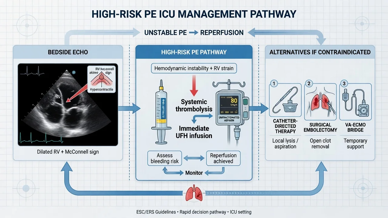
CTEPH is the long-term sequel of acute PE in 0.4-4% of survivors: organised, non-resolving clot in the pulmonary arteries → vascular obstruction + small-vessel arteriopathy → chronic pulmonary hypertension → RV failure over months to years. It is the only potentially curable form of pulmonary hypertension (via pulmonary endarterectomy), which is why post-PE screening is essential. [1]
Pathophysiology and why it follows PE
In most patients, acute emboli resolve via endogenous fibrinolysis within weeks. In CTEPH, clot fails to resolve (organises into fibrous intraluminal bands) AND a secondary small-vessel pulmonary vasculopathy develops in unobstructed areas (resembling idiopathic PAH). The result: fixed, pre-capillary pulmonary hypertension with elevated PVR and progressive RV failure. Risk factors for CTEPH: large/ recurrent PE, large perfusion defects, indwelling IVC filter, splenectomy, thyroid replacement, chronic inflammatory states, non-O blood group, factor VIII elevation, antiphospholipid syndrome. [1]
Follow-up and screening — the 6-12 month V/Q scan
- Symptom surveillance: any PE survivor with new or persistent/worsening dyspnoea (especially on exertion) at any time after PE must be evaluated for CTEPH — do not dismiss as "deconditioning".
- V/Q scan at 6-12 months: ventilation-perfusion scanning is the preferred first-line imaging for CTEPH (more sensitive than echo or CTPA for chronic thromboembolic disease). A V/Q scan is considered normal/near-normal only if no perfusion defects are present — a normal V/Q effectively excludes CTEPH.
- Echocardiography: estimate RVSP (TR jet), look for RV hypertrophy/dilation, paradoxical septal motion (chronic signs, unlike the acute dilation of PE).
- Definitive workup if V/Q abnormal: right-heart catheterisation (mPAP >20 mmHg, PAWP ≤15, PVR >2-3 WU = pre-capillary PH) + pulmonary angiography / cone-beam CT / MRI to map disease and assess surgical accessibility. [1]
Treatment of CTEPH
Treatment options for CTEPH
| Treatment | Indication | Outcome |
|---|---|---|
| Pulmonary endarterectomy (PEA) | Operable disease (proximal, segmental/lobar/main PA clot accessible) and acceptable surgical risk | Curative — the definitive treatment. Operated on CPB with deep hypothermic circulatory arrest to allow a bloodless field; the fibrous intimal peel is dissected from the PA wall. Operative mortality 2-5% in expert centres. Requires lifelong anticoagulation |
| Percutaneous balloon pulmonary angioplastomy (BPA) | Inoperable disease (distal clot) or residual PH after PEA; symptomatic despite medical therapy | Improves haemodynamics and symptoms in selected patients; staged sessions; risk of reperfusion pulmonary oedema and haemoptysis |
| Riociguat (soluble guanylate cyclase stimulator) | Inoperable CTEPH or persistent PH after PEA (CHEST-1 and CHEST-2 trials) | Improves 6-minute walk distance and PVR; the only licensed medical therapy. NOT a substitute for surgery in operable disease |
| PAH-targeted therapy (bosentan, macitentan, ambrisentan, sildenafil) | Off-label; limited evidence | Used in selected inoperable/residual disease |
| Lifelong anticoagulation | All CTEPH patients | Standard — warfarin traditionally (vitamin-K antagonist); DOACs emerging. Prevents recurrence |
| Lung transplant | End-stage CTEPH unresponsive to all therapy | Last resort |
Key principle for the ICU exam
Every patient discharged after PE must have: (a) a documented anticoagulation plan and duration; (b) a CTEPH screen — at minimum a clinical review, and a V/Q scan if any dyspnoea persists or at 6-12 months in those with risk factors (large initial clot, recurrent PE); (c) investigation for provoking factors and thrombophilia (especially antiphospholipid syndrome in CTEPH, where warfarin is preferred over DOACs per the TRAPS trial). CTEPH is curable if caught — missing it is a failure of follow-up. [1]
Clinical pearls — advanced (continuing)
Additional red flags
Additional key trials and evidence
MOPETT trial — half-dose thrombolysis for moderate PE (PMID 22458267)
Study design
Prospective randomised (single-centre) — 121 patients with moderate PE
Intervention
Low-dose alteplase (weight-adjusted, ~50% of standard) + heparin vs heparin alone
Primary outcome
Reduced pulmonary hypertension at 28 months (16% vs 57%)
Key finding
No excess major bleeding; no intracranial haemorrhage
Clinical bottom line
Underpins reduced-dose (50 mg) alteplase for submassive PE where bleeding risk is a concern — a middle path between full-dose lysis and anticoagulation alone
EINSTEIN-PE — rivaroxaban for symptomatic PE (PMID 22394498)
Study design
Randomised open-label non-inferiority — 4,832 patients with symptomatic PE
Intervention
Rivaroxaban (15 mg BD x 3 weeks, then 20 mg daily) vs enoxaparin + warfarin/VKA
Primary outcome
Non-inferior for recurrent VTE (rivaroxaban 2.1% vs standard 1.8%)
Key finding
Major or clinically relevant non-major bleeding LOWER with rivaroxaban; no heparin/parenteral lead-in required
Clinical bottom line
Single-drug oral rivaroxaban is first-line for stable PE — simpler than warfarin, no INR monitoring, less bleeding
CHEST-1 — riociguat for inoperable CTEPH (PMID 23982428)
Study design
Randomised, double-blind, placebo-controlled — 261 patients with inoperable or residual CTEPH
Intervention
Riociguat (soluble guanylate cyclase stimulator) vs placebo
Primary outcome
Improved 6-minute walk distance (+39 m) and reduced PVR at 16 weeks
Key finding
Improved haemodynamics and functional class; well tolerated
Clinical bottom line
Riociguat is the only licensed medical therapy for inoperable/persistent CTEPH — NOT a substitute for pulmonary endarterectomy in operable disease
PREPIC — IVC filters (long-term follow-up)
Study design
Randomised — permanent IVC filter vs no filter, all anticoagulated; 8-year follow-up
Population
Proximal DVT ± PE
Key finding
Filter REDUCED recurrent PE (6% vs 15%) at 8 years BUT INCREASED recurrent DVT (36% vs 28%); no mortality difference
Clinical bottom line
Permanent filters prevent PE at the cost of more DVT and no survival benefit — hence retrievable filters and restricted indications
Diagnosis and risk stratification — summary comparison
Diagnostic tools in suspected PE — when to use each
| Tool | Role | Strength | Limitation |
|---|---|---|---|
| Wells / Revised Geneva score | Pre-test probability (low/mod/high) | Bedside, fast | Subjective components |
| PERC rule | Rule OUT PE without D-dimer (low risk) | Avoids blood tests | Only if low risk AND all 8 criteria negative |
| D-dimer | Rule OUT in low/moderate probability (age-adjusted cutoff >50) | High NPV | Non-specific (any inflammation elevates); useless in high probability |
| CTPA | Gold standard diagnostic test (moderate/high probability) | Visualises clot, RV/LV ratio, alternative dx | Contrast nephropathy; unsafe in unstable patient; radiation |
| V/Q scan | Alternative when CTPA contraindicated (renal failure, contrast allergy, pregnancy) | No contrast, no IV dye | Reduced utility with abnormal CXR (interprets as intermediate) |
| Bedside echo | Diagnostic in unstable patient; risk-stratifies (RV strain) | No transport; fast; guides therapy | Diagnoses RV strain, not the clot (except clot-in-transit) |
| Lower-limb venous Doppler | Indirect — DVT supports PE diagnosis if CTPA not possible | Useful in pregnancy/unstable patient | Negative does not exclude PE (clot may have embolised fully) |
| Troponin / NT-proBNP | Risk stratification (RV injury/strain) | Identifies intermediate-risk PE | Non-specific (elevated in sepsis, ACS, renal failure) |
| Arterial blood gas | Confirms hypoxia/hypocapnia; severity | Always available | Non-specific; ~20% of PE have normal SaO2 and PaO2 |
Anticoagulation — practical ICU protocol
Anticoagulant choice in acute PE — ICU context
| Agent | Dose | Onset/reversibility | Use in PE |
|---|---|---|---|
| Unfractionated heparin (UFH) | 80 IU/kg bolus then 18 IU/kg/h, target APTT 1.5-2.5x | Rapid; protamine-reversible | Preferred when thrombolysis given or may be needed, renal failure, high bleeding risk, obesity (monitor anti-Xa) |
| LMWH (enoxaparin) | 1 mg/kg SC BD (or 1.5 mg/kg OD) | Long; partial protamine reversal | Preferred over UFH for stable PE (lower bleeding/recurrent VTE); AVOID in severe renal failure (CrCl <30) |
| Fondaparinux | 5/7.5/10 mg OD by weight | Long; no reversal | Alternative to LMWH; also a HIT-safe option |
| Apixaban | 10 mg BD x 7 days → 5 mg BD | Oral; no monitoring | First-line for STABLE PE; not in massive PE, pregnancy, severe renal/hepatic impairment |
| Rivaroxaban | 15 mg BD x 3 weeks → 20 mg OD | Oral; no monitoring | First-line for STABLE PE (EINSTEIN-PE); same caveats |
| Warfarin | 5 mg OD (overlap with parenteral ≥5 days, until INR 2-3 x 2 days) | Slow onset; vitamin K + PCC reversal | APS (triple-positive), pregnancy (postpartum), mechanical valves; needs LMWH/UFH lead-in |
| Argatroban / bivalirudin | IV infusion (argatroban preferred in renal failure; bivalirudin in hepatic failure) | Short; direct thrombin inhibitors | Heparin-induced thrombocytopenia (HIT) |
Reversal of over-anticoagulation in the bleeding PE patient
- UFH: protamine 1 mg per 100 IU heparin given in the last 2-4 hours.
- LMWH: partial reversal with protamine (1 mg per 1 mg enoxaparin in last 8 h; slower second dose).
- Warfarin (INR-based): vitamin K (IV 5-10 mg) ± prothrombin complex concentrate (PCC) for life-threatening bleeding.
- DOACs (apixaban/rivaroxaban): andexanet alfa (factor Xa decoy) for life-threatening bleeding; activated charcoal if recent ingestion <2-4 h; haemodialysis is ineffective (highly protein-bound) but apixaban/rivaroxaban partially dialysable.
- Alteplase (post-thrombolysis bleeding): STOP infusion, cryoprecipitate (fibrinogen) ± tranexamic acid ± FFP; supportive. Intracranial haemorrhage post-lysis is a neurosurgical emergency. [1]
Exam practice
SAQ — High-risk PE with obstructive shock
10 minutes · 10 marks
A 67-year-old man day 6 after hip arthroplasty develops sudden dyspnoea and syncope. RR 34, SpO2 86% on 15 L NRB, HR 128, BP 76/44, lactate 4.8 mmol/L. ECG: sinus tachycardia, S1Q3T3, anterior T-wave inversion. Bedside echo: dilated RV, septal flattening, McConnell sign. He is too unstable for CTPA.
Viva / oral exam — high-yield questions
Q: A 65-year-old post-op patient collapses with sudden dyspnoea, SBP 70, SpO2 82% on room air. ECG shows sinus tach 130, S1Q3T3. What is your immediate management? This is massive (high-risk) PE with haemodynamic collapse. Do NOT send to CT. (1) Call for help, high-flow oxygen, large-bore IV access, arterial line. (2) Bedside echo — confirm RV strain (McConnell sign, RV/LV >0.9, TR jet, IVC plethora). (3) Give alteplase 100 mg IV over 2 h immediately on clinical + echo grounds (no absolute contraindication outweighs the >50% mortality of untreated massive PE). If arresting, alteplase 50 mg bolus during CPR. (4) Start UFH (80 IU/kg bolus + 18 IU/kg/h) — rapidly reversible if bleeding. (5) Noradrenaline for MAP >65; small (250 mL) fluid boluses only. (6) If refractory, VA-ECMO as bridge + activate PERT (CDT/embolectomy). Avoid dobutamine/milrinone in hypotension. Avoid large fluids. [1]
Q: A 50-year-old with proven PE is normotensive, troponin mildly raised, echo shows RV/LV 1.1 with McConnell sign. Discuss thrombolysis. This is intermediate-risk (submassive) PE with RV dysfunction AND biomarker elevation — the high-intermediate subgroup. PEITHO showed routine full-dose lysis reduces haemodynamic decompensation (2.6% vs 5.6%) at the cost of more stroke (2.4% vs 0.2%) and major bleeding (6.3% vs 1.5%). Therefore the standard approach is anticoagulation + close ICU/HDU monitoring, with rescue thrombolysis if deterioration. For the patient with high-risk features (severe RV dysfunction, rising biomarkers, clinical decline) where systemic lysis bleeding risk is unacceptable, catheter-directed thrombolysis (EKOS) delivers ~10-24 mg alteplase directly into the clot with far lower bleeding (ULTIMA, SEATTLE II). Reduced-dose systemic alteplase (50 mg, "MOPETT") is an alternative. [1]
Q: When would you choose surgical embolectomy over thrombolysis? When systemic thrombolysis is absolutely contraindicated (recent intracranial surgery/haemorrhagic stroke, active life-threatening bleeding, intracranial neoplasm), when lysis has failed, in clot-in-transit with a PFO (to remove clot and close the defect), in PE-induced arrest on VA-ECMO where lysis is too risky, and when no interventional radiology is available but cardiac surgery is. Modern embolectomy in expert centres has ~6-12% mortality — far better than historical series — provided it is done EARLY before irreversible RV failure. [1]
Q: How do you investigate a PE survivor for CTEPH? CTEPH develops in 0.4-4% of PE survivors. Screen all PE survivors with symptom surveillance; perform a V/Q scan (the most sensitive imaging for chronic thromboembolic disease) at 6-12 months or sooner if new/worsening dyspnoea. An abnormal V/Q → right-heart catheterisation (pre-capillary PH: mPAP >20, PAWP ≤15, PVR >2-3 WU) + pulmonary angiography/cone-beam CT to map the disease. Operable (proximal) disease → pulmonary endarterectomy (curative). Inoperable or residual disease → riociguat (CHEST-1) and/or balloon pulmonary angioplastomy. All patients need lifelong anticoagulation; investigate for antiphospholipid syndrome (warfarin preferred over DOACs in triple-positive APS per TRAPS). [1]
References
- [1]Konstantinides SV, et al. Molecular diagnostics: the changing culture of medical microbiology Bioeng Bugs, 2012.PMID 22179143
- [2]Meyer G, et al. Initiation of poliovirus negative-strand RNA synthesis requires precursor forms of p2 proteins J Virol, 2003.PMID 12502823
- [3]Kearon C, et al. [The mobile geriatric team of Bretonneau Hospital and nursing home professionals] Soins Gerontol, 2015.PMID 26574127
- [4]Kucher N, et al. Workforce Issues in Pediatric Surgery Am Surg, 2017.PMID 28637571
- [5]Aujesky D, et al. αTAT1 catalyses microtubule acetylation at clathrin-coated pits Nature, 2013.PMID 24097348
- [6]Pasrija C, et al. Swine Vagina Under Planar Biaxial Loads: An Investigation of Large Deformations and Tears J Biomech Eng, 2019.PMID 30615053
- [7]Piazza G, et al. Ischaemic stroke and ST-segment elevation myocardial infarction: fast-track single-stop approach Eur Heart J, 2015.PMID 26038589
- [8]Sharifi M, et al. Quality of life in men receiving radiotherapy and neo-adjuvant androgen deprivation for prostate cancer: results from a prospective longitudinal study J Adv Nurs, 2013.PMID 22458267
- [9]Kuo WT, et al. Educational resources for patients considering a left ventricular assist device: a cross-sectional review of internet, print, and multimedia materials Circ Cardiovasc Qual Outcomes, 2014.PMID 25316772
- [10]Ghofrani HA, et al. Molecular plasmonics for nanoscale spectroscopy Chem Soc Rev, 2014.PMID 23982428
- [11]Tapson VF, et al. Surgical Management of the Recent Orbital War Injury J Craniofac Surg, 2018.PMID 29742577