ICU · Cardiovascular
Acute severe valvular emergencies: critical aortic stenosis, acute MR and AR
Also known as Critical aortic stenosis · Acute mitral regurgitation · Acute aortic regurgitation · Valvular emergency · Cardiogenic shock valvular · Balloon aortic valvuloplasty
Acute valvular emergencies present as cardiogenic shock, pulmonary oedema, or cardiac arrest. CRITICAL AORTIC STENOSIS (severe AS — area <1.0 cm², mean gradient 40): fixed outflow obstruction — preload-dependent, intolerance of AF/hypovolaemia, vasodilators dangerous. Definitive: aortic valve replacement (surgical or TAVI). Bridge: balloon aortic valvuloplasty (BAV) or VA-ECMO. ACUTE MITRAL REGURGITATION (papillary muscle rupture post-MI, endocarditis, chordal rupture): sudden volume overload on LA + pulmonary veins - flash pulmonary oedema + cardiogenic shock. Murmur may be SOFT (rapid equalisation of pressures). Echo diagnostic. Treatment: afterload reduction (nitroprusside, IABP), vasopressors/inotropes cautiously, definitive: urgent MV repair/replacement. ACUTE AORTIC REGURGITATION (endocarditis, aortic dissection, trauma): diastolic run-off - wide pulse pressure, bounding pulses, but in ACUTE, the murmur may be SHORT/SOFT (rapid pressure equalisation) and pulse pressure may be NORMAL (ventricle not yet adapted). Echo diagnostic. Treatment: vasopressors (noradrenaline — increase diastolic pressure), avoid beta-blockers alone (need tachycardia to shorten diastole), definitive: urgent AVR.
On this page & tools
Your progress
Saved locally on this device.
Target exams
Red flags
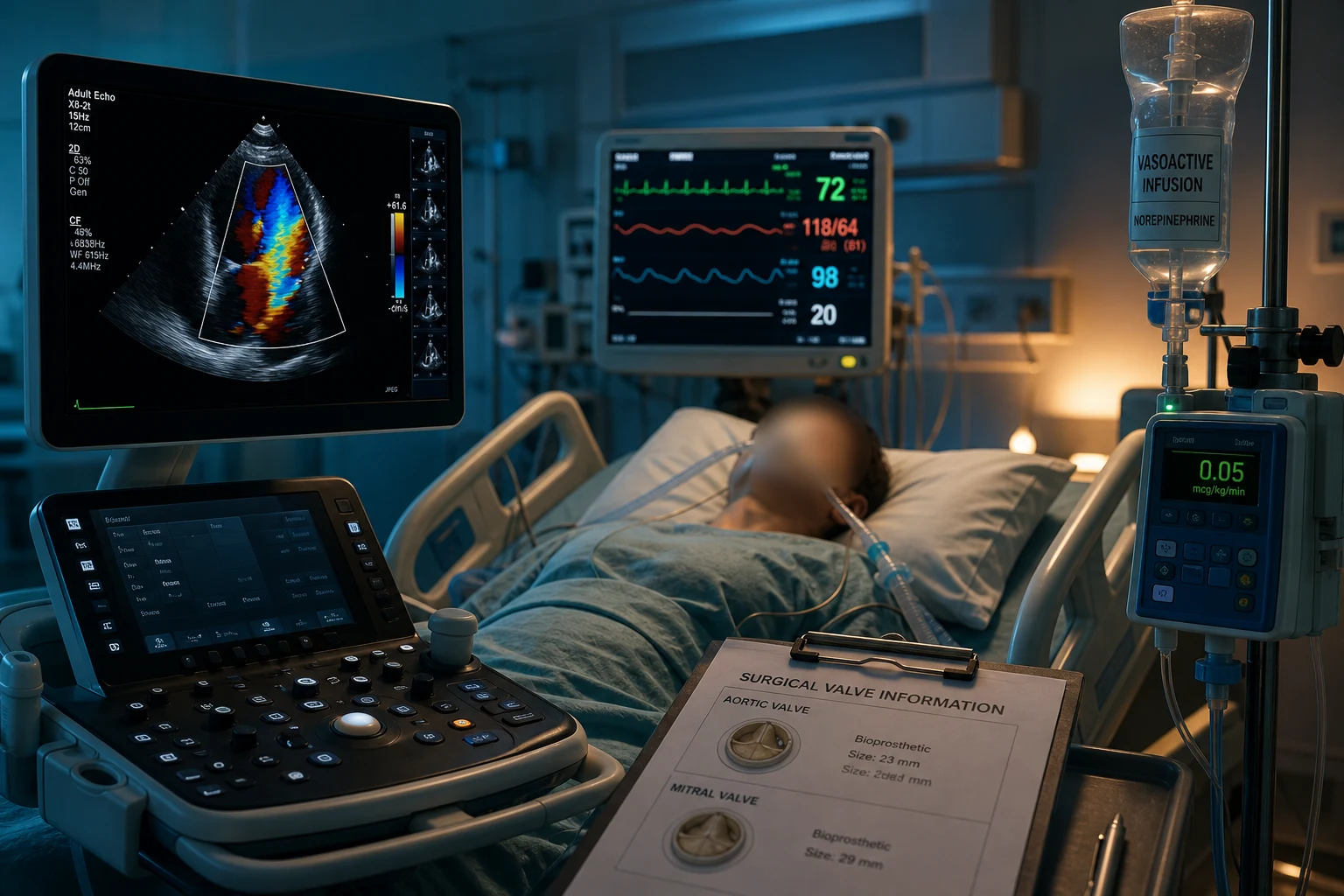
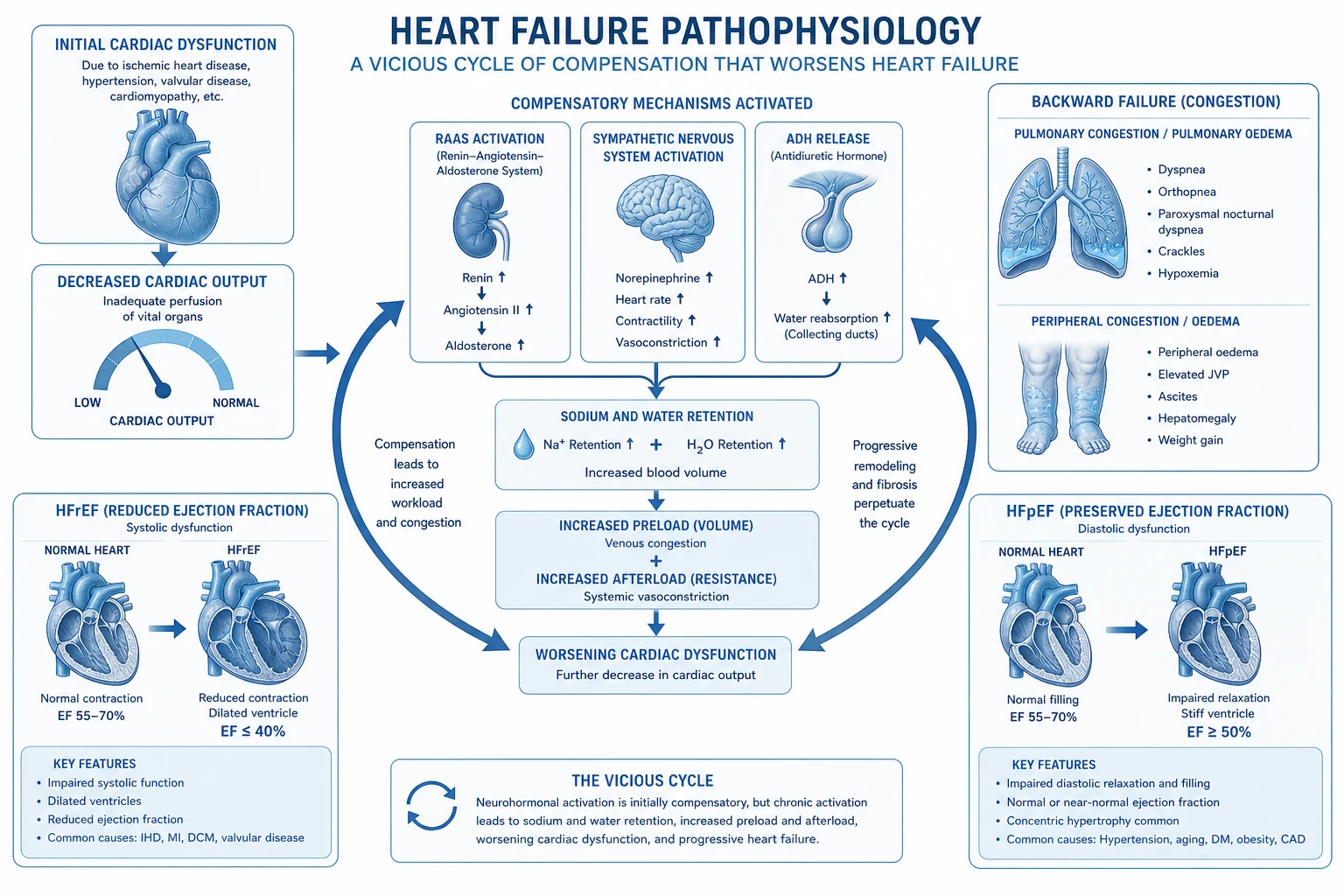
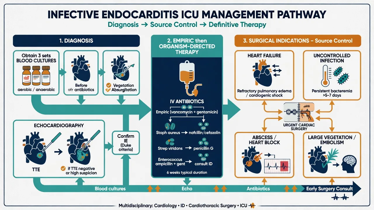
Three valvular emergencies compared
| Feature | Critical Aortic Stenosis | Acute Mitral Regurgitation | Acute Aortic Regurgitation |
|---|---|---|---|
| Pathophysiology | Fixed LV outflow obstruction | Sudden volume back into LA/pulmonary veins | Diastolic run-off from aorta into LV |
| Presentation | Syncope, angina, heart failure, cardiogenic shock | Flash pulmonary oedema, shock | Pulmonary oedema, shock, often from cause (dissection, endocarditis) |
| Murmur | Ejection systolic (crescendo-decrescendo), ejection click, soft S2 | Pansystolic, but may be SOFT in acute | Early diastolic decrescendo, but may be SHORT in acute |
| Pulse | Slow-rising (pulsus parvus et tardus), narrow pulse pressure | Normal (or AF) | Bounding (water-hammer) in chronic; may be normal in acute |
| Haemodynamic support | Maintain preload, AV synchrony, avoid vasodilators | Afterload reduction (nitroprusside, IABP) | Vasopressors (raise diastolic), avoid pure beta-blocker |
| Definitive treatment | AVR (surgical/TAVI), BAV bridge | Urgent MV repair/replacement | Urgent AVR |
Management of critical aortic stenosis in cardiogenic shock
- RECOGNISE + ECHOCARDIOGRAM — Symptoms: syncope, angina, heart failure. Murmur: ejection systolic (crescendo-decrescendo), radiation to carotids, soft/absent S2, ejection click. ECG: LVH, LBBB. Echo diagnostic: area <1.0 cm², mean gradient >40 mmHg, peak velocity >4 m/s, dimensionless index <0.25. CRITICAL AS = cardiogenic shock/hypotension in known AS
- HAEMODYNAMIC PRINCIPLES (critical) — (a) MAINTAIN PRELOAD (volume — don't diurese aggressively; AS is preload-dependent — fixed obstruction needs high preload to maintain stroke volume). (b) MAINTAIN SINUS RHYTHM (AF devastating — loses atrial kick which is 30-40% of filling in stiff LV — cardiovert if haemodynamically unstable). (c) MAINTAIN AFTERLOAD (vasopressors — noradrenaline — DON'T vasodilate — vasodilation reduces diastolic pressure -> subendocardial ischaemia + can't maintain coronary perfusion). (d) AVOID excessive tachycardia (less filling time) AND bradycardia (less cardiac output). Rate 60-80 ideal
- PHARMACOLOGICAL SUPPORT — (a) NORADRENALINE (vasopressor — maintain afterload + coronary perfusion). (b) CAREFUL inotrope (dobutamine — if LV dysfunction, but can worsen outflow gradient in HOCM-like physiology; milrinone alternative). (c) DIURESE cautiously (only if clearly overloaded — frusemide small doses). (d) AVOID: nitrates (vasodilation -> catastrophic), ACEi/ARB (vasodilation), high-dose beta-blockers (bradycardia -> low output), rate control for AF if unstable (cardiovert)
- CARDIOVERSION FOR AF — AF in critical AS is DEVASTATING (loss of atrial kick). If haemodynamically unstable: SYNCHRONISED DC CARDIOVERSION (not pharmacological rate control — too slow). Maintain AV synchrony. If AF persists after cardioversion: consider amiodarone (cautiously — avoid hypotension)
- BRIDGE TO DEFINITIVE THERAPY — (a) BALLOON AORTIC VALVULOPLASTY (BAV): percutaneous balloon dilation of calcified valve — temporary improvement (gradient reduced for weeks-months) — bridge to AVR/TAVI or palliative. (b) VA-ECMO: if cardiogenic shock refractory — bridge to AVR/TAVI. (c) TAVI (transcatheter aortic valve implantation): for patients too high-risk for surgery — increasingly first-line in elderly/comorbid. (d) SURGICAL AVR (SAVR): for younger, lower-risk, or specific anatomy
- DEFINITIVE THERAPY — Aortic valve replacement is the ONLY definitive treatment. (a) SAVR: open surgery (younger, lower-risk, bicuspid valve, concomitant CABG). (b) TAVI: transcatheter (elderly, high-risk, comorbid — now approved for low-risk too — PARTNER 3, Evolut Low Risk trials). WITHOUT valve replacement: mortality near 100% in critical AS with shock. Don't delay — multidisciplinary heart team urgent review
Clinical pearls
Red flags
Prognosis
Valvular emergency evidence and outcomes
Critical AS: without AVR, mortality near 100% in shock. AVR (SAVR or TAVI) — survival improves dramatically. PARTNER 3, Evolut Low Risk: TAVI non-inferior to SAVR in low-risk. BAV: bridge — temporary (restenosis 6-12 months). Useful for palliation or bridge. Acute MR: without surgery, mortality near 100%. Urgent MV repair/replacement — survival 70-90% (depends on cause — ischaemic worse). Acute AR: without AVR, mortality high (endocarditis, dissection). Urgent AVR — survival depends on cause + timing. Endocarditis surgery: early (within 48h) for heart failure/abscess — improves outcomes. Aortic dissection with AR: mortality 1-2%/h untreated -> surgical emergency. Anaesthesia in critical AS: extremely high-risk — cardiac arrest may be refractory (CPR ineffective across stenotic valve).
Acute severe mitral stenosis (MS) — new AF trigger
[1]Acute decompensated MS vs chronic compensated MS
| Feature | Chronic compensated MS | Acute decompensated MS (ICU) |
|---|---|---|
| Valve area | <1.5 cm² (moderate), <1.0 cm² (severe) | Same — but decompensated |
| Trigger | Slowly progressive exertional dyspnoea | New AF, pregnancy, infection, anaemia |
| Symptoms | Exertional dyspnoea, fatigue, palpitations | Flash pulmonary oedema, shock |
| Atrial kick | Lost gradually (chronic AF, adapted) | Lost SUDDENLY (new AF) -> catastrophic LA pressure spike |
| Murmur | Loud S1, opening snap, mid-diastolic rumble, presystolic accentuation | Same but tachycardic — rumble hard to hear, opening snap subtle |
| Immediate management | Diurese, rate control, anticoagulate | Cardiovert unstable AF; rate control; BMV/surgery |
| Definitive | BMV or elective MVR | Urgent BMV (if suitable) or MVR |
Management of acute severe MS with new AF and pulmonary oedema
- RECOGNISE + ECHOCARDIOGRAM — MS murmur (mid-diastolic rumble, opening snap, loud S1, presystolic accentuation — but in AF the presystolic accentuation is lost). ECG: AF (common), RVH, P mitrale if sinus rhythm. Echo diagnostic: area <1.0 cm² (planimetry, PHT/pressure half-time, PISA, continuity), mean gradient >10 mmHg, RVSP/pulmonary artery systolic >50 mmHg, Wilkins score for valve morphology (pliability, calcification, subvalvular involvement, leaflet thickness). TOE if TTE poor, to exclude LA/LAA thrombus before cardioversion or BMV
- HAEMODYNAMIC PRINCIPLES (critical) — (a) RATE CONTROL paramount: diastolic filling time is everything in MS — tachycardia (especially AF) shortens diastole -> less flow across stenotic MV -> LA pressure rises -> pulmonary oedema. Target HR 60-80. (b) MAINTAIN PRELOAD — don't diurese too aggressively (preload is needed to DRIVE flow across the stenotic valve) but diurese enough to clear pulmonary oedema. (c) MAINTAIN AFTERLOAD (vasopressors if hypotensive — avoid vasodilators which reduce coronary perfusion and the pressure head driving flow across MV). (d) AVOID tachycardia above all else — every extra beat shortens filling
- RATE CONTROL — (a) BETA-BLOCKER (metoprolol IV — first-line; prolongs diastole; titrate to HR 60-80). (b) NON-DIHYDROPYRIDINE CCB (diltiazem or verapamil IV — alternative if beta-blocker contraindicated, e.g. asthma). (c) DIGOXIN (if AF — slower onset but useful adjunct, especially with heart failure). (d) AMIODARONE (if AF — combined rate + rhythm control; preferred in critically ill, pregnancy, heart failure). (e) AVOID pure AV nodal blockers in pre-excitation (rare in MS)
- CARDIOVERSION FOR UNSTABLE AF — If haemodynamically unstable (hypotension, worsening pulmonary oedema, shock): SYNCHRONISED DC CARDIOVERSION. If AF onset >48h or unknown — TOE FIRST to exclude LA/LAA thrombus, anticoagulate 3 weeks before OR TOE-guided immediate cardioversion with peri-procedural heparin. RESTORE SINUS RHYTHM — atrial kick contributes 25-30% of LV filling in MS (catastrophic if lost). Maintain rhythm with amiodarone, sotalol, or flecainide (caution — structural disease)
- DIURESIS + SUPPORT — (a) FRUSEMIDE IV (reduce LA/pulmonary venous pressure — cautiously — preload dependent). (b) OXYGEN; NIV (CPAP/BiPAP) if pulmonary oedema — careful of vasodilation-induced hypotension. (c) AVOID NITRATES (venodilation -> preload drop -> collapse across stenotic MV). (d) AVOID ACEi/ARB (vasodilation, no benefit in MS). (e) VASOPRESSOR (noradrenaline) if hypotensive after rate control. (f) TREAT TRIGGER (infection — antibiotics; anaemia — transfuse cautiously; thyrotoxicosis — beta-blocker, antithyroid). (g) ANTICOAGULATE (AF — high embolic risk in MS)
- DEFINITIVE THERAPY — (a) BALLOON MITRAL VALVOTOMY (BMV/PMBV/PMC): percutaneous transseptal Inoue-balloon commissurotomy — first-line if PLIABLE NON-CALCIFIED valve, no LA thrombus, no significant MR (Wilkins score <8). Immediate gradient reduction, symptom relief. (b) SURGICAL MV REPLACEMENT (mechanical or bioprosthetic): if calcified valve, severe subvalvular disease, significant MR, LA thrombus, failed BMV. (c) OPEN SURGICAL COMMISSUROTOMY (rare now — replaced by BMV). (d) TIMING: urgent BMV if shock refractory to medical therapy
Acute severe aortic regurgitation — deep dive (dissection and endocarditis)
Management of acute severe AR (dissection, endocarditis, trauma)
- RECOGNISE THE CAUSE — Acute severe AR causes: (a) AORTIC DISSECTION (Type A — annular disruption, dissection flap prolapsing through valve, leaflet detachment, dilatation preventing coaptation) — surgical emergency. (b) INFECTIVE ENDOCARDITIS (leaflet destruction, perforation, large vegetation preventing coaptation, paravalvular extension) — medical + surgical. (c) TRAUMA (blunt chest, deceleration — rare). (d) IATROGENIC (post-AVR, post-BAV, TAVI complication, post-aortic cannulation). (e) SPONTANEOUS leaflet rupture (degenerative, myxomatous). Echo URGENT — TTE then TOE for aortic root, vegetations, mechanism
- PATHOPHYSIOLOGY OF ACUTE AR — Sudden diastolic run-off from aorta into LV -> LV (small, non-compliant, NOT adapted — unlike chronic AR where LV dilates and accommodates) cannot accommodate regurgitant volume -> LV end-diastolic pressure SPIKES rapidly -> EQUALISES with aortic diastolic pressure early -> diastolic gradient disappears -> regurgitation ceases early -> forward output falls catastrophically -> cardiogenic shock + pulmonary oedema. Tachycardia is COMPENSATORY (shorter diastole = less time for regurgitation). LVEF may appear preserved on echo early — but forward output is dire
- PHARMACOLOGICAL SUPPORT — (a) VASOPRESSOR (NORADRENALINE — raise diastolic pressure -> improve coronary perfusion + reduce gradient — first-line). (b) INOTROPE cautious (dobutamine or milrinone if LV dysfunction — but may worsen tachycardia/ischaemia). (c) DIURESIS cautious (frusemide if pulmonary oedema — preload dependent). (d) AVOID pure beta-blocker monotherapy in isolated AR (slows HR -> longer diastole -> MORE regurgitation -> worse). (e) AVOID primary vasodilators (nitroprusside) as monotherapy in non-dissection AR — diastolic already low, further vasodilation worsens coronary perfusion. (f) Vasodilator only useful if DILATED LV with high pressure + adequate BP — uncommon in acute AR
- IABP IS ABSOLUTELY CONTRAINDICATED IN AORTIC REGURGITATION (high-yield exam point) — (a) IABP mechanism: inflates in diastole (raises diastolic pressure / coronary perfusion), deflates in systole (reduces afterload / systolic unloading). (b) In AR, diastolic AUGMENTATION by IABP WORSENS the regurgitant fraction (raises aortic diastolic pressure -> INCREASES the gradient driving blood back from aorta into LV during diastole). (c) IABP is ABSOLUTELY CONTRAINDICATED in moderate-severe AR AND in aortic dissection. (d) ALTERNATIVE mechanical support: VA-ECMO (preferred — supports circulation + oxygenation without worsening regurgitant fraction), Impella (axial flow across AV — but risky if AR severe as it may pull blood back into LV), CentriMag, TandemHeart. (e) Never place IABP in AR — classic exam question
- AORTIC DISSECTION WITH AR — special pathway — (a) DIFFERENT from isolated AR: beta-blocker IS given first (reduce dp/dt shear stress on the dissected aorta — esmolol IV first-line, target HR <60, then SBP 100-120). (b) THEN add vasodilator (labetalol, nicardipine, sodium nitroprusside) — NEVER vasodilator before beta-blocker (unopposed alpha stimulation worsens shear force -> propagation/rupture). (c) URGENT surgery (ascending aorta replacement ± AVR ± root repair ± hemiarch). (d) MORTALITY 1-2% per hour untreated — surgical emergency. (e) Type B dissection with AR (rare — usually from retrograde extension) — usually medical therapy, TEVAR if complicated
- ENDOCARDITIS WITH AR — special pathway — (a) BLOOD CULTURES (3 sets from different sites) before antibiotics if possible — don't delay antibiotics if septic/shock. (b) EMPIRICAL IV antibiotics (cover S. aureus, streptococci, enterococci — adapt to native vs prosthetic, community vs healthcare-associated). (c) EARLY SURGERY (within 48h) if: heart failure from acute AR (THE major indication — operate regardless of antibiotic duration), abscess, paravalvular extension, fungal or multidrug-resistant organism, embolisation with large vegetation, prosthetic dehiscence. (d) Surgery: AVR (mechanical or bioprosthetic), repair of abscess with pericardial patch, homograft preferred for destructive aortic root endocarditis (resistant to reinfection). (e) TIMING after stroke: intracranial haemorrhage = absolute contraindication (wait >4 weeks); silent/small ischaemic stroke — early surgery supported by modern data
- DEFINITIVE TREATMENT — URGENT AORTIC VALVE REPLACEMENT (surgical AVR — mechanical or bioprosthetic) ± aortic root/ascending repair (dissection) ± radical debridement of infected tissue (endocarditis). TAVI has limited role in acute AR (native AR — valve may not anchor well — off-label/experimental). Mortality without surgery high — especially endocarditis with AR + heart failure (approaches 100%)
Acute severe mitral regurgitation — deep dive (papillary muscle rupture)
Management of acute severe MR from papillary muscle rupture post-MI
- RECOGNISE + ECHOCARDIOGRAM — Patient typically 2-7 days post-MI (often INFERIOR MI — the POSTEROMEDIAL papillary muscle has a SINGLE blood supply from the PDA and is far more vulnerable to ischaemia/rupture than the anterolateral, which has DUAL supply from LAD and LCx). Sudden flash pulmonary oedema + cardiogenic shock ± SOFT or ABSENT pansystolic murmur (LA pressure equalises rapidly with LV, abolishing the gradient that produces the murmur). Echo URGENT (TTE ± TOE): flail leaflet (posteromedial more common), eccentric regurgitant jet (often anteriorly directed if posterior leaflet flail), severe MR (ERO >0.4 cm², regurgitant volume >60 mL, regurgitant fraction >50%, vena contracta >0.7 cm). SHOCK + NEW MR post-MI = papillary muscle rupture until proven otherwise (also consider VSD, free wall rupture)
- HAEMODYNAMIC SUPPORT — afterload reduction is central — (a) NITROPRUSSIDE IV (potent arteriolar vasodilator — reduces SVR -> more forward flow, less regurgitation into LA — first-line IF blood pressure permits). (b) NITROGLYCERIN IV (venodilator + mild arteriolar — reduces preload + afterload, helps pulmonary oedema — less potent than nitroprusside). (c) If HYPOTENSIVE: cannot pharmacologically vasodilate -> use IABP (mechanical afterload reduction + diastolic augmentation — EXCELLENT bridge in acute MR). (d) INOTROPE (dobutamine or milrinone) if LV dysfunction. (e) VASOPRESSOR (noradrenaline) cautiously — may increase SVR and worsen regurgitant fraction, but necessary for organ perfusion
- IABP — first-line mechanical support in acute MR — (a) MECHANISM: inflates in diastole (raises diastolic pressure / coronary perfusion), deflates in systole (reduces afterload / systolic unloading). (b) EFFECT IN MR: systolic unloading -> reduces LV systolic pressure -> reduces the gradient between LV and LA -> LESS regurgitation -> MORE forward output. (c) INDICATION: acute MR with shock — BRIDGE to surgery. (d) CONTRAINDICATIONS: aortic regurgitation (WORSENS regurgitant fraction), aortic dissection, severe peripheral vascular disease, sepsis (relative), uncontrolled bleeding. (e) IABP is a BRIDGE, not destination — definitive surgery required
- VA-ECMO — for refractory shock — If IABP insufficient (profound cardiogenic shock, biventricular failure, severe hypoxaemia from pulmonary oedema) -> VA-ECMO. (a) Drains venous blood, returns oxygenated arterial blood — supports both ventricles + oxygenation. (b) BRIDGE to definitive surgery (MV repair/replacement). (c) COMPLICATIONS: limb ischaemia (femoral cannulation), bleeding (anticoagulation), haemolysis, LV distension (may need LV vent — Impella or pulmonary artery vent), stroke. (d) Consider central cannulation if peripheral access poor or patient very small
- PERCUTANEOUS OPTIONS (MitraClip / TEER) — selected cases only — (a) MitraClip (TEER — transcatheter edge-to-edge repair): clips together opposing leaflets to reduce MR — modelled after the surgical Alfieri stitch. (b) ROLE IN ACUTE MR: LIMITED — approved for severe symptomatic degenerative or functional MR despite guideline-directed medical therapy (selected chronic patients). (c) Off-label RESCUE use in critically ill with prohibitive surgical risk. (d) EVEREST II, COAPT (functional MR, NEJM 2018 — mortality/hospitalisation benefit), MITRA-FR (neutral) trials — predominantly CHRONIC MR. (e) Acute papillary muscle rupture: SURGICAL repair/replacement is standard of care; MitraClip is a rescue option if the patient is too sick for surgery
- SURGICAL DEFINITIVE TREATMENT — (a) MV REPAIR preferred (leaflet resection, chordal transfer, artificial chords, annuloplasty ring — lower operative mortality than replacement, preserves LV function and the submitral apparatus). (b) MV REPLACEMENT (mechanical or bioprosthetic — if repair not feasible, e.g. extensive destruction) — preserve chordal attachments where possible to maintain LV geometry. (c) CONCOMITANT CABG if ischaemic (revascularise the culprit — but the rupture itself requires surgical correction). (d) TIMING: urgent (within 24-48h) — delay increases mortality. (e) Surgical mortality in papillary muscle rupture post-MI: 10-25% (era- and centre-dependent — high but far better than no surgery, ~100%). (f) Activate the cardiogenic shock / heart team early
- POST-MI MECHANICAL COMPLICATIONS — recognise the constellation — Acute MR (papillary muscle rupture) is one of the THREE classic post-MI mechanical complications (with VENTRICULAR SEPTAL DEFECT and FREE WALL RUPTURE), all presenting days after MI with sudden collapse/shock and a new murmur. Echo distinguishes: MR (eccentric jet, flail leaflet, pulmonary oedema dominant) vs VSD (high-velocity left-to-right shunt at ventricular level, harsh holosystolic murmur at lower sternal border, oxygen step-up on right heart catheter) vs tamponade (free wall rupture — echo-free pericardial effusion with tamponade physiology, electrical alternans, pulsus paradoxus). ALL THREE need urgent surgery
Prosthetic valve thrombosis
[1]Management of prosthetic valve thrombosis by clinical scenario
| Scenario | First-line | Second-line | Notes |
|---|---|---|---|
| Left-sided obstructive + unstable | URGENT SURGERY | Fibrinolysis if surgery unavailable | Fibrinolysis stroke risk 10-15% left-sided |
| Left-sided obstructive + stable | IV UFH; reassess 24-72h | Surgery if no improvement | Bridging strategy, monitor gradients |
| Right-sided obstructive | FIBRINOLYSIS (first-line) | Surgery if failed/contraindicated | Lower systemic embolic risk |
| Non-obstructive thrombus (small) | Anticoagulation (UFH + warfarin) | Surgery if large/mobile | Repeat echo to confirm resolution |
| Non-obstructive, large/mobile | SURGERY | Fibrinolysis (controversial) | High systemic embolic risk |
| Pannus (fibrous, chronic) | SURGERY (always) | — | Fibrinolysis INEFFECTIVE — organised tissue |
| Pregnancy + obstructive | Surgery (mitral: lysis cautiously) | UFH/LMWH bridge | Multidisciplinary obstetric-cardiac |
Management of suspected prosthetic valve thrombosis
- SUSPECT IT — Any patient with a prosthetic valve + new dyspnoea, MUFFLED or ABSENT closing clicks, embolic event (stroke, peripheral), or cardiogenic shock/pulmonary oedema -> suspect thrombosis. Risk factors: SUBTHERAPEUTIC INR (mechanical valve — most common), pregnancy (hypercoagulable + warfarin problematic), poor adherence, MITRAL position (higher thrombosis risk than aortic), old-generation valve (caged-ball, tilting disc), low-flow state. Auscultate: muffled clicks, new murmur (stenotic — murmur of MS/AS; regurgitant — murmur of MR/AR)
- DIAGNOSIS — multimodal imaging — (a) TTE: elevated transvalvular gradients (compare to the patient's BASELINE post-implant gradient — mean gradient >2x normal suggests obstruction), reduced leaflet mobility (often missed due to acoustic shadowing from the prosthetic), new regurgitation, elevated pulmonary artery pressure. (b) TOE/TEE: BEST for visualising thrombus, pannus, leaflet motion — essential before deciding surgery vs lysis. (c) FLUOROSCOPY: cinefluoroscopy quickly shows mechanical leaflet excursion — a leaflet that does not move = obstructed; cheap and fast. (d) 4D CT: distinguishes thrombus (low Hounsfield units) from pannus (tissue-density, higher HU), shows pannus location and extent. (e) Distinguish THROMBUS (soft, recent, low-attenuation, responds to lysis) from PANNUS (hard, chronic, organised fibrous tissue, surgical only)
- STRATIFY — obstructive vs non-obstructive, stable vs unstable — (a) OBSTRUCTIVE: thrombus prevents leaflet opening/closing -> severe stenosis or regurgitation -> haemodynamic compromise. (b) NON-OBSTRUCTIVE: small thrombus, leaflets still move but embolic risk. (c) CLINICAL STATE: stable (no shock, no severe symptoms, controlled) vs unstable (cardiogenic shock, pulmonary oedema, cardiac arrest). (d) SIDE: LEFT (aortic/mitral — high systemic embolic risk, lysis dangerous) vs RIGHT (tricuspid/pulmonary — lower systemic embolic risk, lysis safer). These four axes determine the management pathway
- LEFT-SIDED OBSTRUCTIVE + UNSTABLE — surgery preferred — (a) URGENT SURGERY: valve re-replacement (thrombectomy ± new valve) — PREFERRED for unstable left-sided thrombosis because fibrinolysis carries 10-15% systemic embolic (stroke) risk + intracranial haemorrhage risk in left-sided disease. (b) Surgical mortality 5-15% (high but acceptable vs lysis embolic risk in left-sided). (c) BRIDGE to surgery: VA-ECMO or IABP (IABP only if no AR) if profound shock while awaiting theatre. (d) Multidisciplinary heart team decision
- FIBRINOLYSIS — selected cases — (a) INDICATIONS: surgery UNAVAILABLE (no cardiothoracic surgeon on site), patient too high-risk for surgery, RIGHT-SIDED thrombosis (tricuspid/pulmonary — lower systemic embolic risk — lysis is FIRST-LINE). (b) REGIMEN: alteplase (rt-PA) — slow infusion 10 mg IV bolus then 90 mg over 2 h (or weight-adjusted 25 mg bolus then infusion), OR streptokinase (historic, anaphylaxis risk). (c) MONITOR: transvalvular gradient by echo during/after infusion — improvement = success. (d) COMPLICATIONS: systemic embolism/stroke (10-15% left-sided, much lower right-sided), major bleeding (5-10%), intracranial haemorrhage (1-2%), recurrent thrombosis, anaphylaxis (streptokinase). (e) CONFOUNDERS: lysis is INEFFECTIVE for PANNUS (organised fibrous tissue) — imaging must distinguish
- NON-OBSTRUCTIVE THROMBUS / STABLE OBSTRUCTIVE — anticoagulation — (a) IV UNFRACTIONATED HEPARIN (UFH — fast-acting, titratable, fully reversible with protamine) + bridge to warfarin (target INR per valve position/type). (b) Reassess at 24-72 h with echo — if gradient falls + thrombus shrinks = success; continue anticoagulation, repeat echo at intervals. (c) If fails (no improvement) or large mobile clot: surgery. (d) Enoxaparin NOT preferred in critical illness (variable pharmacokinetics, renal accumulation, anti-Xa monitoring). (e) DOACs generally NOT first-line in mechanical valves — RE-ALIGN (Eikelboom 2013, NEJM) showed dabigatran WORSE than warfarin (more thromboembolism + bleeding). (f) PROACT Xa (2024) — apixaban met non-inferiority for the On-X aortic mechanical valve only (NOT mitral, NOT other valves) — emerging but warfarin remains standard
- PREVENTION — the real cure — (a) Strict INR control (mechanical — target INR: 2.5 for mitral/caged-ball/tricuspid, 2.0 for aortic bileaflet; per specific valve recommendations). (b) PATIENT EDUCATION on adherence + importance of INR monitoring. (c) Home INR self-monitoring (improves time-in-range). (d) PREGNANCY: high thrombosis risk — switch from warfarin to LMWH (with anti-Xa monitoring 4h post-dose, target 0.8-1.2 U/mL) from weeks 6-12 to 36 (warfarin teratogenic weeks 6-12; warfarin acceptable weeks 13-36 if dose <5 mg/day; LMWH peripartum; UFH if renal failure). (e) Bridge anticoagulation around invasive procedures (never stop entirely). (f) Address pannus risk (older-generation valves — consider elective re-operation). (g) Treat intercurrent infection/low-output states promptly
Endocarditis with valvular destruction
Surgical decision-making in infective endocarditis with valvular destruction
- RECOGNISE DESTRUCTIVE ENDOCARDITIS — Aggressive organisms destroy valves rapidly: S. aureus (WORST — necrotising, destructive, metastatic), S. lugdunensis (virulent despite coagulase-negative), beta-haemolytic streptococci (S. pyogenes, S. agalactiae), enterococci (especially VRE), HACEK, fungi (Candida, Aspergillus — large friable vegetations). Echo features of destruction: large (>10 mm) mobile vegetation, leaflet perforation, flail leaflet, ABSCESS (aortic root — echo-bright ring or septated cavity, prosthetic dehiscence with rocking motion), paravalvular involvement, fistula (aorto-RV/RA, aorto-LA), aneurysm of Valsalva rupture. Multiple valve involvement. Persistent bacteraemia >5-7 days despite appropriate antibiotics
- INDICATIONS FOR URGENT SURGERY (within 48 h) — (a) HEART FAILURE from acute valve failure — THE most common and strongest indication (acute AR or MR — operate regardless of antibiotic duration; medical therapy alone mortality approaches 100%). (b) UNCONTROLLED INFECTION: abscess, paravalvular extension, persistent bacteraemia >5-7 days on appropriate antibiotics, fungal or multidrug-resistant organisms, S. aureus PVE. (c) EMBOLISATION: large mobile vegetation (>10 mm) with prior embolic event, or >15 mm even without prior embolism (high embolic risk — operate to prevent stroke). (d) CONDUCTION BLOCK: new AV block = aortic root abscess extending into the interventricular septum — urgent surgery. (e) PROSTHETIC valve endocarditis with dehiscence or obstruction. (f) Relapse after adequate therapy
- SURGICAL PROCEDURE — (a) RADICAL DEBRIDEMENT of all infected and necrotic tissue (vegetations, abscess cavity, infected annulus — the principle is to leave only healthy tissue). (b) VALVE REPAIR preferred when feasible (especially MV — repair has better outcomes than replacement in endocarditis). (c) VALVE REPLACEMENT (mechanical, bioprosthetic, or homograft): BIOPROSTHETIC often preferred in endocarditis (avoid lifelong anticoagulation in patients at risk of further infection/bleeding; no clear reinfection difference vs mechanical); HOMOGRAFT for destructive aortic ROOT endocarditis (resistant to reinfection, handles abscess cavities well). (d) RECONSTRUCTION of aortic root or mitral annulus with pericardial patch if abscess destroyed the annulus. (e) CLOSURE of fistula, repair of ventricular septal defect, reconstruction of fibrous skeleton. (f) Send intraoperative tissue for culture, histology, and molecular (16S/18S PCR). (g) Mechanical valve acceptable in young patients with good adherence
- TIMING AFTER STROKE — (a) ISCHAEMIC STROKE: previously surgery was delayed >4 weeks; modern data (Emery 2018, Croft 2019 systematic reviews) supports EARLY surgery (within 7 days, often within 48 h if heart failure) after SILENT or SMALL ischaemic stroke — haemorrhagic transformation risk acceptable if infarct <2/3 MCA territory and no haemorrhage on imaging. (b) INTRACRANIAL HAEMORRHAGE: ABSOLUTE contraindication to early cardiac surgery (wait >4 weeks; mortality of early surgery after ICH is very high). (c) MYCOTIC ANEURYSM: treat first (neurosurgical clipping or endovascular coiling) before cardiac surgery. (d) COMA or massive stroke (coma, large MCA): palliative or delayed surgery. (e) Multidisciplinary decision (cardiac surgery + neurology + infectious diseases + anaesthesia)
- ANTIBIOTIC DURATION AFTER SURGERY — (a) Native valve endocarditis (NVE): usually 4-6 weeks TOTAL (combined pre- and post-operative days). (b) Prosthetic valve endocarditis (PVE): 6 weeks total. (c) CULTURE-NEGATIVE post-surgery: tailor to organism if identified intra-operatively; otherwise 6 weeks empirical broad-spectrum. (d) FUNGAL: lifelong suppressive therapy often needed (e.g. fluconazole after initial amphotericin/flucytosine). (e) RESTART antibiotics immediately post-op — do not wait for intraoperative cultures. (f) Infectious diseases + microbiology guide therapy and duration. (g) Follow inflammatory markers (CRP, procalcitonin) and repeat blood cultures
- PROGNOSIS AND FOLLOW-UP — In-hospital mortality: NVE 15-20%, PVE 20-30%, S. aureus PVE up to 40%, fungal and multiresistant higher. Heart failure from endocarditis is an indication for surgery regardless of antibiotic duration — without surgery mortality approaches 100%. Embolic stroke worsens prognosis. Long-term: REINFECTION risk 5-10% (higher in IV drug users), lifelong surveillance with periodic echo, complete dental clearance, ID follow-up, address IV drug use if applicable. Six-month and 12-month echo + clinical review
Additional clinical pearls — acute MS, AR, MR deep dive, prosthetic thrombosis, endocarditis
Expanded red flags — deep dive
Prognosis and evidence — deep dive
Evidence: BMV, prosthetic thrombosis, endocarditis surgery, MitraClip
Mitral stenosis / BMV: Inoue balloon BMV established 1980s-90s; first-line for pliable rheumatic MS. Echo-guided (Wilkins score <8 ideal — pliable, non-calcified). Long-term patency comparable to open commissurotomy in suitable valves; restenosis ~10-20% at 5 years. RE-ALIGN (Eikelboom 2013, NEJM): dabigatran in mechanical valves — MORE thromboembolism and bleeding than warfarin. DON'T use dabigatran in mechanical valves. Warfarin remains standard. PROACT Xa (2024): apixaban vs warfarin in On-X aortic mechanical valve — apixaban met non-inferiority. Apixaban may be acceptable for On-X AORTIC only (not mitral, not other valves). Emerging — warfarin still standard. Emery (2018, Eur J Cardiothorac Surg): early cardiac surgery after ischaemic stroke in endocarditis — safe and improves outcomes vs conservative waiting. Changed practice from old >4-week delay dogma. Croft (2019, Heart): systematic review supports early surgery after silent/small ischaemic stroke; haemorrhagic stroke still contraindication. COAPT (2018, NEJM): MitraClip in functional MR (heart failure) — reduced mortality + hospitalisation. Selected patients benefit. MITRA-FR (2018, NEJM): MitraClip in functional MR — neutral. Selection matters (disproportionate vs proportionate MR). EVEREST II: MitraClip safety/efficacy — less effective than surgery but lower procedural risk — niche role in high-risk surgical patients. Staphylococcal endocarditis (S. aureus): in-hospital mortality 20-40%; early surgery for heart failure reduces mortality. Fowler 2005 (JAMA) — S. aureus bacteraemia/endocarditis. Papillary muscle rupture post-MI: surgical mortality 10-25% (era- and centre-dependent) — far better than no surgery (mortality approaches 100%). Type A dissection with AR (IRAD): mortality 1-2% per hour untreated — surgery is the only cure. Surgical mortality 17-26% (IRAD registry, contemporary). Acute AR (endocarditis) mortality: untreated approaches 100% — urgent AVR transforms prognosis. Heart failure is the surgical trigger. Prosthetic valve thrombosis surgery: operative mortality 5-15% — acceptable vs lysis embolic risk in left-sided unstable disease. Pregnancy + mechanical valve: thrombosis risk up to 10% (mitral higher) — anti-Xa-monitored LMWH or carefully managed warfarin strategy; multidisciplinary essential.
Exam-style SAQs — acute severe AR and critical MS
SAQ — Acute severe aortic regurgitation in cardiogenic shock (Type A dissection)
10 minutes · 10 marks
A 58-year-old man presents with sudden tearing chest pain radiating to the back, dyspnoea and presyncope. Examination: HR 110, BP 78/45, SpO2 90% on 15 L O2, bilateral crackles, a soft early diastolic murmur at the left sternal edge and equal radial pulses. CT angiography confirms a Type A aortic dissection with severe acute aortic regurgitation. He is intubated for pulmonary oedema and is awaiting theatre.
SAQ — Critical mitral stenosis with flash pulmonary oedema and new AF
10 minutes · 10 marks
A 44-year-old woman with known rheumatic mitral stenosis presents to the emergency department with acute breathlessness over two hours. Examination: HR 148 in new-onset atrial fibrillation, BP 88/58, SpO2 82% on room air, she is diaphoretic and using accessory muscles, and chest X-ray shows bilateral alveolar pulmonary oedema. Bedside echocardiography confirms severe mitral stenosis (valve area 0.8 cm²).
Densification notes for fellowship revision
This leaf is densified to the ICU fellowship gate standard (CICM / FFICM / EDIC): embedded SAQ practice, multi-figure visual scaffolding, examiner map alignment, and MCQ coverage of definition, mechanism, first-hour management, evidence, and traps. [1]
- Revision checkpoint 1: restate definition, one number examiners expect, and one absolute do-not-miss action.
- Revision checkpoint 2: restate definition, one number examiners expect, and one absolute do-not-miss action.
- Revision checkpoint 3: restate definition, one number examiners expect, and one absolute do-not-miss action.
- Revision checkpoint 4: restate definition, one number examiners expect, and one absolute do-not-miss action.
- Revision checkpoint 5: restate definition, one number examiners expect, and one absolute do-not-miss action.
- Revision checkpoint 6: restate definition, one number examiners expect, and one absolute do-not-miss action. [1]
References
- [1]Baumgartner H, et al. Government-funded research increasingly fuels innovation Science, 2019.PMID 31221848
- [2]Nishimura RA, et al. Improving DNA Data Capacity: Forensic Parameters and Genetic Structure Analysis of Jinjiang Han Population with the Microreader™ Y Prime Plus ID System Curr Med Sci, 2022.PMID 35403953
- [3]Thompson JL, et al. Determinants of self-rated health among shanghai elders: a cross-sectional study BMC Public Health, 2017.PMID 29029627
- [4]Siu SC, et al. Can sand nourishment material affect dune vegetation through nutrient addition? Sci Total Environ, 2020.PMID 32278174
- [5]Picard MH, et al. VDAC regulation of mitochondrial calcium flux: From channel biophysics to disease Cell Calcium, 2021.PMID 33529977
- [6]Estrada A, et al. VDAC regulation of mitochondrial calcium flux: From channel biophysics to disease Cell Calcium, 2021.PMID 33529977