ICU · Cardiovascular
Arrhythmia, cardiac arrest and post-arrest care
Also known as Cardiac arrest · Advanced Life Support (ALS) · Post-cardiac arrest syndrome · Targeted temperature management (TTM) · Ventricular fibrillation (VF) · Refractory VF · Extracorporeal CPR (ECPR)
Cardiac arrest in the ICU requires immediate Advanced Life Support per ERC 2021 guidelines: high-quality CPR (rate 100-120, depth 5-6cm, full recoil), early defibrillation for shockable rhythms (VF/pVT), adrenaline 1 mg IV every 3-5 minutes, amiodarone 300 mg for refractory VF. Post-arrest care: targeted temperature management (TTM2: normothermia 37.5C is as good as 33C; avoid fever), percutaneous coronary intervention for cardiac cause, lung-protective ventilation, haemodynamic optimisation (MAP =65, lactate clearance), neuroprognostication at =72 hours. ECPR for refractory in-hospital arrest in selected centres.
On this page & tools
Your progress
Saved locally on this device.
Target exams
Red flags
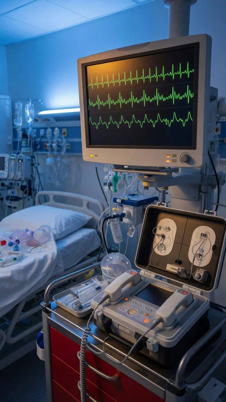
The ALS algorithm (ERC 2021)
Shockable rhythms (VF / pulseless VT)
ALS algorithm for VF/pVT — shockable rhythm
Start high-quality CPR immediately
Rate 100-120/min, depth 5-6cm, full recoil between compressions, minimise interruptions (<10s for rhythm check). Switch compressor every 2 minutes to prevent fatigue.
Defibrillate — biphasic 150-200J
First shock: 150-200J biphasic (or manufacturer recommended). Subsequent shocks: same or higher energy. Resume CPR immediately after shock — do NOT check pulse first.
Continue CPR for 2 minutes
After each shock, continue CPR for 2 minutes (one cycle) before rhythm check. Minimise interruptions to <10 seconds. Switch compressor.
Give adrenaline 1 mg IV
After the 3rd shock, give adrenaline 1 mg IV every 3-5 minutes (alternate cycles). PARAMEDIC2: adrenaline improves survival to discharge but not neurological outcomes at 3 months.
Give amiodarone 300 mg IV
After the 3rd shock, give amiodarone 300 mg IV bolus. Further dose: 150 mg after 5 shocks. Alternative: lidocaine 100 mg (if amiodarone unavailable).
Identify and treat reversible causes
4Hs: Hypoxia, Hypovolaemia, Hypo/Hyperkalaemia, Hypothermia. 4Ts: Tension pneumothorax, Tamponade, Thrombosis (PE/MI), Toxins.
Consider ECPR
For refractory in-hospital VF arrest (>10 min despite standard ALS) in a centre with ECPR capability. Must be initiated within 60 minutes of arrest onset.
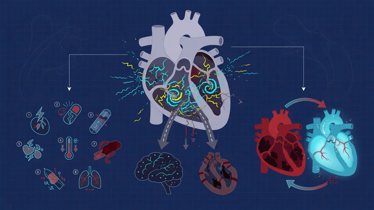
Non-shockable rhythms (asystole / PEA)
- Give adrenaline 1 mg IV as soon as IV/IO access is available (do NOT wait for 3rd shock — give immediately)
- Continue CPR for 2 minutes between rhythm checks
- Identify and treat the 4Hs and 4Ts — PEA has an organised rhythm but no pulse, meaning there IS electrical activity but NO mechanical output. Find the cause. [1]
4Hs
Reversible causes
- Hypoxia — check oxygenation, ventilate
- Hypovolaemia — give fluid bolus, check for bleeding
- Hypo/Hyperkalaemia — check VBG, treat accordingly
- Hypothermia — active rewarming
4Ts
Reversible causes
- Tension pneumothorax — needle decompression
- Tamponade (cardiac) — pericardiocentesis
- Thrombosis (PE or MI) — thrombolysis / PCI
- Toxins — identify drug, give antidote
Post-cardiac arrest care
Targeted temperature management (TTM)
The TTM2 trial (NEJM 2021) is the definitive study:[1]
TTM2 trial results (NEJM 2021)
Current recommendation (ERC/ESICM 2021): Maintain normothermia (target core temperature 36-37.5C). Prevent and treat fever aggressively (paracetamol, cooling blankets, ice packs). Therapeutic hypothermia at 33C is NOT recommended — it does not improve outcomes and increases complications.[8]
Prior evidence:
- TTM trial (NEJM 2013): 33C was NOT superior to 36C. Set the stage for TTM2.[10]
- HYPERION (NEJM 2019): TTM at 33C may benefit non-shockable rhythm arrest.[2]
Other post-arrest interventions
Post-arrest care bundle (ERC/ESICM 2021)
Airway and ventilation
Intubate if not already. Lung-protective ventilation (Vt 6-8 mL/kg, PEEP 5-8, plateau <30). Target PaO2 75-100 mmHg (avoid hyperoxia — may worsen neurological injury). Target PaCO2 35-45 (normocapnia).
Haemodynamic optimisation
Target MAP >=65 mmHg (or >=80 if chronic HTN for CPP). Use noradrenaline if hypotensive. Monitor lactate clearance. Echocardiography to assess cardiac function (post-arrest myocardial stunning is common).
Coronary angiography
If suspected cardiac cause (STEMI, new LBBB, history of IHD): emergency coronary angiography within 120 minutes of ROSC, even if coma (do NOT use coma as reason to withhold PCI).
Glycaemic control
Target glucose 7-10 mmol/L. Avoid hypoglycaemia (neurologically devastating). Avoid intensive insulin therapy (increased hypoglycaemic episodes).
Seizure prophylaxis and monitoring
Routine prophylactic anticonvulsants NOT recommended. Continuous EEG monitoring if available — seizures occur in 20-30% of comatose post-arrest patients and worsen prognosis.
Temperature management
Normothermia 36-37.5C. Aggressively treat fever (>37.8C). TTM2: 33C is NOT superior to normothermia. Avoid hyperthermia (worsens neurological injury).
Neuroprognostication
Do NOT prognosticate before 72 hours post-ROSC. Use multimodal approach: clinical examination (brainstem reflexes, motor response, myoclonus), EEG (malignant patterns), somatosensory evoked potentials (N20), biomarkers (NSE), imaging (CT/MRI).
Rehabilitation planning
Early involvement of rehabilitation team. Screen for cognitive impairment, emotional distress, and physical deconditioning in survivors.
ICU arrhythmia management
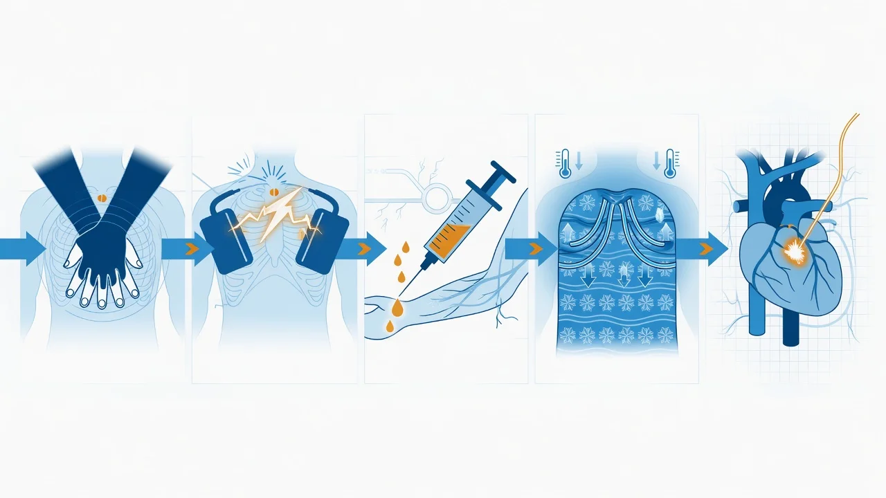
Critically ill patients are predisposed to arrhythmia via multiple convergent mechanisms: sympathetic surge and high circulating catecholamines (endogenous and exogenous vasopressors), electrolyte disturbance (hypo/hyperkalaemia, hypomagnesaemia, hypocalcaemia), acid-base derangement, hypoxia, myocardial ischaemia, sepsis and inflammatory cytokines (interleukin-6–mediated atrial electrical remodelling), drug effects (antiarrhythmics, theophylline, haloperidol), autonomic imbalance, and mechanical irritation (central venous catheters, pulmonary artery catheters, chest tubes). Atrial fibrillation is the most common new-onset ICU arrhythmia (10–30% of medical ICU, 30–50% after cardiac surgery) and independently predicts longer ICU stay and mortality. Ventricular arrhythmias in ICU are usually secondary to ischaemia, electrolytes, or drugs rather than primary electrical disease. [1]
Atrial fibrillation in ICU
Rate vs rhythm control
Two parallel strategies exist — neither has shown consistent mortality superiority over the other in critical illness. The choice is driven by haemodynamic stability, duration of AF, and underlying substrate. The landmark AFFIRM and RACE trials established rate control as an acceptable default in older patients with structural heart disease.[11][12]
Rate control
Default first-line
- Target ventricular rate <110 bpm (lenient) or <80 (strict); lenient non-inferior in stable AF
- Drug: IV metoprolol / esmolol / landiolol, OR IV diltiazem (avoid verapamil if LV systolic dysfunction)
- Digoxin — useful adjunct when beta-blocker/CCB inadequate or contraindicated; less effective in high sympathetic tone (sepsis)
- Use when haemodynamically stable and AF >48h (low chance of sustained sinus rhythm after cardioversion)
Rhythm control
Selected patients
- Pharmacological: IV amiodarone 300 mg over 1h then 900 mg/24h, OR vernakalant 3 mg/kg IV over 10 min (new-onset AF <7 days only)
- Electrical: synchronized cardioversion 120–200J biphasic — first-line if haemodynamically unstable
- Preferred when: new-onset AF (<48h), post-cardiac surgery AF, AF precipitating cardiogenic shock
- Anticoagulation: assess CHA2DS2-VASc; cardioversion requires ≥3 weeks therapeutic anticoagulation if AF >48h OR exclude LAA thrombus on TOE
Drug properties for ICU AF rate control
Beta-blockers
IV options
- Esmolol: t1/2 ~9 min, titratable, ideal for trial in unstable AF
- Landiolol: ultra-short acting (t1/2 ~4 min), beta-1 selective, minimal negative inotropy — emerging evidence in sepsis-related AF
- Metoprolol: longer acting, IV boluses 5 mg q5min up to 15 mg
- CAUTION: avoid in acute decompensated HF, severe bronchospasm, high-grade AV block, hypotension on vasopressor
Calcium-channel blockers
Non-dihydropyridine
- Diltiazem: IV bolus 0.25 mg/kg then infusion 5–15 mg/h
- Verapamil: contraindicated in LV systolic dysfunction and ANY wide-complex tachycardia (could be VT)
- Avoid combining with beta-blocker (risk of asystole/severe bradycardia)
Digoxin
Adjunct only
- IV loading 0.5–1 mg in divided doses over 24h, then 0.0625–0.25 mg/day
- Less effective in high sympathetic tone (sepsis, exercise, postoperative state) — never first-line in ICU
- Toxicity: arrhythmias (atrial tachycardia with block, bidirectional VT), nausea, visual disturbance (yellow halos, xanthopsia), confusion
- Antidote: digoxin-specific Fab fragments (Digibind). Hypokalaemia precipitates and worsens toxicity — keep K+ >4.0
- Useful in AF with HF when beta-blocker/CCB not tolerated
Amiodarone
Rate + rhythm
- IV: 300 mg over 1h, then 900 mg over 24h infusion via central line
- Effective in sepsis-associated AF where beta-blocker causes hypotension
- CAUTION: hypotension (solvent-related), bradycardia, QT prolongation, phlebitis (use central access for prolonged infusion)
- Onset over hours — NOT first-line for acute rate control when immediate effect needed
Special situations
- New-onset AF in sepsis: amiodarone is preferred where beta-blockade causes hypotension; landiolol has supportive trial evidence. Magnesium sulfate (2–4 g IV) is a useful adjunct for rate control and reduces AF recurrence.
- AF in cardiogenic shock: synchronized electrical cardioversion (120–200J biphasic) is first-line — do not attempt pharmacological conversion in a shocked patient.
- Post-cardiac surgery AF: peak incidence day 2–3 (overall 30–50%). Beta-blocker prophylaxis reduces incidence. Amiodarone if AF develops despite prophylaxis. Usually self-limiting; anticoagulate if AF persists >48h.
- Electrolyte targets: maintain K+ >4.0 mmol/L and Mg2+ >1.0 mmol/L to reduce AF threshold and recurrence — the most commonly overlooked correctable precipitant.
- Anticoagulation: balance CHA2DS2-VASc stroke risk against ICU bleeding risk (frequently elevated). Avoid routine heparin in immediate post-arrest/postoperative period. [1]
ICU management of new-onset AF
Assess haemodynamic stability
Unstable (hypotension on vasopressor, ischaemia, acute pulmonary oedema, altered consciousness): synchronize cardiovert immediately (120–200J biphasic, escalate). Stable: proceed to pharmacological management.
Identify and treat precipitant
Check electrolytes (K+, Mg2+, Ca2+, PO4), ABG (hypoxia, acidosis), septic source, myocardial ischaemia (troponin/ECG), drug cause (vasopressor excess, theophylline, withdrawal of chronic beta-blocker), thyroid function, CVC/mass effect.
Correct K+ to >4.0 mmol/L and Mg2+ >1.0 mmol/L
Low K+/Mg2+ is the most common correctable precipitant of AF in ICU and reduces efficacy of all antiarrhythmics. Replace aggressively; consider IV magnesium 2–4 g even if Mg2+ normal.
Choose rate vs rhythm strategy
AF <48h or postoperative: rhythm control (amiodarone, vernakalant, or electrical cardioversion). AF >48h or chronic: rate control (beta-blocker, CCB, digoxin, or combination).
Anticoagulation
Assess CHA2DS2-VASc vs bleeding risk. If cardioversion planned and AF >48h: therapeutic anticoagulation for ≥3 weeks OR TOE to exclude LAA thrombus. Bleeding risk in ICU often limits anticoagulation — individualise.
Prevent recurrence
Continue rate/rhythm agent; maintain electrolytes; treat sepsis; wean vasopressor; ensure adequate analgesia/sedation. Consider amiodarone prophylaxis in high-risk postoperative patients.
Ventricular tachycardia (VT)
VT in ICU is life-threatening and demands immediate stratification by haemodynamic stability and presence of pulse. Regular monomorphic wide-complex tachycardia (>120 ms QRS) in a patient with structural heart disease is VT until proven otherwise (90% in this population). [1]
Stable VT (with pulse)
Pharmacological
- Wide QRS (>120 ms), regular, AV dissociation, capture/fusion beats
- Amiodarone 150 mg IV over 10 min, repeat as needed up to 2.2 g/24h, then infusion
- Alternative: lidocaine 1–1.5 mg/kg IV, repeat to 3 mg/kg max
- Correct K+ (>4.0), Mg2+ (>1.0), ischaemia, acidosis first
- If persists: synchronized cardioversion 100J biphasic (escalate)
Unstable VT (with pulse)
Electrical
- Signs: hypotension, altered consciousness, chest pain, acute pulmonary oedema, shock
- Synchronized cardioversion 100J biphasic, escalating (150J, 200J)
- If synchronisation fails or patient deteriorates: switch to UNSYNCHRONIZED shock (defibrillation) 200J
- Prepare for sedation/anaesthesia if conscious
- Treat precipitant: ischaemia, electrolytes, drugs
Pulseless VT / VF
Cardiac arrest
- Treat as cardiac arrest — start ALS algorithm immediately
- Immediate unsynchronized defibrillation 150–200J biphasic
- Adrenaline 1 mg IV q3–5 min after 3rd shock
- Amiodarone 300 mg IV after 3rd shock (lidocaine alternative) — ROC trial showed equivalent to placebo overall but benefit in witnessed arrests
Torsades de pointes
Special case
- Polymorphic VT with prolonged QT — QRS amplitude rotates around isoelectric line
- STOP all QT-prolonging drugs (amiodarone, sotalol, macrolides, fluoroquinolones, haloperidol, methadone, ondansetron)
- IV magnesium sulfate 2 g bolus — first-line; may repeat
- Correct K+ to 4.5–5.0 mmol/L (aggressive)
- If bradycardia-induced: isoprenaline infusion or overdrive pacing to rate 90–100
Synchronized vs unsynchronized shock — a critical exam distinction that decides rhythm-cardiac arrest:
- Synchronized: delivered to coincide with R wave (avoids R-on-T phenomenon). Used for stable/unstable AF, AFL, regular monomorphic VT with pulse. Energy: AF 120–200J, AFL 50–100J, VT 100J biphasic.
- Unsynchronized (defibrillation): delivered immediately regardless of cardiac cycle. Used for VF, pulseless VT, polymorphic VT (Torsades) where the sync circuit cannot lock onto an R wave. Energy: 150–200J biphasic. If a synchronized shock is programmed but the machine cannot synchronise, deliver an unsynchronized shock rather than delay. [1]
Wide-complex tachycardia in ICU — decision tree
Confirm pulse and assess stability
If NO pulse → defibrillate as cardiac arrest (unsynchronized 150–200J). If pulse present and unstable (shock, ischaemia, altered mental state) → synchronized cardioversion.
Determine if regular or irregular wide-complex
Regular monomorphic → likely VT (90% of regular WCT in structural heart disease is VT). Irregular polymorphic → Torsades if long QT; AF with aberrancy; pre-excited AF (WPW).
NEVER give IV verapamil/diltiazem to undiagnosed WCT
Risk of cardiovascular collapse if it is actually VT — verapamil is negative inotropic and can precipitate VF. Treat all regular WCT as VT until proven otherwise.
Stable regular monomorphic WCT
Treat as VT. Amiodarone 150 mg IV over 10 min. Lidocaine 1–1.5 mg/kg alternative. If fails → synchronized cardioversion.
Investigate underlying cause
12-lead ECG, electrolytes (K+, Mg2+, Ca2+), troponin, drug levels, tox screen. Consider cardiology input for electrophysiology, ICD assessment.
Bradyarrhythmias
Bradycardia in ICU commonly arises from: sinus node dysfunction, AV nodal block (intrinsic — ischaemia, drugs, post-valve surgery; extrinsic — high vagal tone, raised ICP), drug effects (beta-blockers, CCBs, digoxin, amiodarone, ivabradine), hypothyroidism, hypothermia, hyperkalaemia, hypoxia, raised intracranial pressure (Cushing reflex). [1]
Bradycardia management algorithm
Assess for adverse signs
Shock, syncope, myocardial ischaemia, acute HF, altered consciousness. If adverse signs present → treat immediately (atropine 0.5 mg IV). If asymptomatic (e.g. sleeping athlete) → observe.
Give atropine 0.5 mg IV
Repeat every 3–5 min to maximum 3 mg. Effective for symptomatic sinus brady and Mobitz I (Wenckebach). INEFFECTIVE for Mobitz II and complete heart block (block is below AV node — go straight to pacing).
Identify and treat reversible cause
Stop offending drug (beta-blocker, CCB, digoxin, amiodarone). Treat ischaemia, hypothyroidism, hyperkalaemia, hypothermia, raised ICP. Check glycaemic control, drug levels.
Initiate pacing if atropine ineffective or high-grade block
Transcutaneous (immediate but requires sedation), transvenous (definitive temporary, bedside via right IJV to RV apex), epicardial (post-cardiac surgery via temporary wires).
Pharmacological bridge
Adrenaline infusion 2–10 mcg/min OR dopamine 5–20 mcg/kg/min as bridge to definitive pacing. Glucagon 2–10 mg/h for beta-blocker/CCB overdose. High-dose insulin euglycaemic therapy (HIET) for severe CCB toxicity.
Definitive management
Permanent pacemaker indication: symptomatic bradycardia (syncope, dizziness, HF), Mobitz II, third-degree AV block, sick sinus syndrome, AF pause >3s with symptoms. Post-MI complete heart block → usually permanent indication.
Transcutaneous
First bridge
- Pads anterior + posterior chest wall, set rate 60–80, mA above capture threshold
- Immediate availability — every ICU bed
- Painful — sedate/anaesthetise; ineffective capture in obese/oedematous
- Use only as bridge to transvenous (max 1–2 hours)
Transvenous
Definitive temporary
- Float balloon-tipped pacing wire via right IJV to RV apex under fluoroscopy or ECG guidance
- Set rate 60–80, output just above threshold (typically 1–2 mA), sensitivity 2–3 mV
- Risks: pneumothorax, infection, RV perforation/tamponade, ventricular ectopy on insertion
- Remove within 7 days to reduce infection; antibiotic prophylaxis not routine
Epicardial
Post-cardiac surgery
- Temporary wires placed on RV epicardium at sternotomy
- Used post-CABG/valve surgery — usually removed day 4–5
- Indicates block from oedema/trauma to conduction tissue — usually resolves
Permanent
Definitive
- Indications: symptomatic sinus node dysfunction, Mobitz II, third-degree AV block, AF pause >3s symptomatic
- MRI-compatible systems available
- ICD (defibrillator) for VT/VF secondary prevention or ejection fraction <35%
Pharmacological considerations in bradycardia:
- Atropine 0.5 mg IV q3–5 min, max 3 mg. Paradoxical bradycardia if dose <0.5 mg. Caution in raised ICP and in Mobitz II/complete heart block (ineffective — go to pacing).
- Glucagon for beta-blocker/CCB toxicity: 3–5 mg IV bolus over 1 min then infusion 1–5 mg/h. Side effects: nausea/vomiting (give antiemetic), hyperglycaemia.
- High-dose insulin euglycaemic therapy (HIET) for CCB/beta-blocker poisoning: insulin 1 U/kg bolus then 0.5–1 U/kg/h with concomitant dextrose, monitor and replace K+ aggressively.
- Lipid emulsion (Intralipid) 20% 1.5 mL/kg bolus for severe lipophilic drug toxicity (bupivacaine, verapamil, amitriptyline) — acts as a lipid sink.
- Isoprenaline infusion for torsades with bradycardia and for beta-blocker overdose with severe bradycardia. [1]
Drug-induced arrhythmia in ICU
QT-prolonging drugs
Torsades risk
- Amiodarone, sotalol, haloperidol, methadone, macrolides (azithromycin/clarithromycin), fluoroquinolones (ciprofloxacin/moxifloxacin), ondansetron, antipsychotics
- Risk amplified by hypokalaemia, hypomagnesaemia, bradycardia, structural heart disease, hepatic impairment
- Monitor QTc; stop or switch offending agent if QTc >500 ms. Correct electrolytes aggressively.ATHENA: dronedarone reduced AF burden but PALLAS showed harm in permanent AF
Vasopressor-related
Sympathomimetic
- Noradrenaline, adrenaline, dobutamine, dopamine, salbutamol can precipitate AF, VT, ischaemia
- Titrate to minimum effective dose; consider combination therapy to lower individual drug dose
- Dobutamine paradoxically can lower SVR and worsen hypotension; adrenaline causes lactate rise (anaerobic glucose metabolism)
Electrolyte-driven
Correct first
- Hypokalaemia: AF, VT, digoxin toxicity — target K+ >4.0
- Hypomagnesaemia: AF, Torsades, refractory VT — give empirically even if "normal"
- Hypocalcaemia: prolonged QT, Torsades — correct with calcium gluconate
- Hyperkalaemia: bradyarrhythmias, sine wave, asystole — peaked T waves, widened QRS, then cardiac arrest. Treat: calcium gluconate 10 mL 10% IV first (membrane stabiliser), then insulin/dextrose, salbutamol nebs, bicarbonate if acidotic
Antiarrhythmic toxicity
Recognise
- Digoxin: nausea, xanthopsia, confusion, atrial tachycardia with block, bidirectional VT — give Fab fragments
- Amiodarone: acute pulmonary toxicity (rare), hypotension, QT prolongation, thyroid dysfunction (hypo or hyper) with long-term use
- Beta-blocker/CCB overdose: bradycardia, hypotension, AV block — glucagon, HIET, lipid emulsion, pacing
Neuroprognostication in comatose post-arrest patients
Modern prognostication is multimodal and deferred to ≥72 hours post-ROSC (longer if sedatives are being metabolised, especially if TTM or continuous infusions used). No single predictor is sufficient to predict poor outcome reliably.[8][14]
Clinical
Bedside
- Brainstem reflexes (pupil, corneal, gag/cough) — bilaterally absent at ≥72h is a strong predictor
- Motor response to pain — absent or extensor at ≥72h poor prognostic sign
- Myoclonus (NOT myoclonic status) — historically overvalued; do NOT use alone
- Confounded by sedatives, neuromuscular blockers, hypothermia, metabolic disturbance
Electrophysiology
Objective
- EEG: malignant patterns — suppression, burst-suppression, status epilepticus, absence of reactivity
- SSEP (somatosensory evoked potentials): bilateral absence of N20 cortical response is the MOST RELIABLE single predictor (specificity >95%)
- Continuous EEG recommended if available — seizures occur in 20–30% of comatose post-arrest patients
Biomarkers
NSE
- Neuron-specific enolase (NSE) >60 ng/mL at 48–72h supports poor prognosis
- Confounded by haemolysis (NSE on red cells) — sample carefully
- Trend over time more useful than single value
Imaging
CT/MRI
- CT: diffuse cerebral oedema, loss of grey-white differentiation, sulcal effacement
- MRI: diffuse cortical diffusion restriction (cortical laminar necrosis), basal ganglia/thalamic injury
- Imaging alone rarely sufficient — used adjunctively
Routine prophylactic anticonvulsants are NOT recommended for comatose post-arrest patients (no outcome benefit in trials). Treat clinical and electrographic seizures when they occur with levetiracetam or valproate (avoid phenytoin — negative inotropy and poor outcome signal in observational data).[14]
Evidence and landmark trials
TTM2
NEJM 2021
1900 pts post-OHCA coma — hypothermia 33C for 24h vs normothermia 37.5C
Key finding
No difference in death or poor functional outcome at 6 months (48% vs 47%). More arrhythmias and bleeding with 33C.
Practice change
Normothermia is the standard — 33C hypothermia NOT recommended
TTM
NEJM 2013
950 pts post-OHCA — 33C vs 36C for 36h
Key finding
No difference in mortality or neurological outcome. Set stage for abandoning 33C.
Practice change
36C became acceptable — led to TTM2
HYPERION
NEJM 2019
584 pts non-shockable rhythm arrest coma — 33C vs 37C
Key finding
Favourable outcome at 90 days: 10.2% hypothermia vs 5.7% normothermia (p=0.04). Small absolute benefit in non-shockable arrest.
Practice change
May consider 33C for non-shockable rhythm arrest (uncommon scenario)
PARAMEDIC2
NEJM 2018
8014 pts OHCA — adrenaline vs placebo
Key finding
Increased survival to discharge (3.2% vs 1.9%, p<0.001). BUT: more survivors in severe neurological impairment. No difference in favourable outcome.
Practice change
Adrenaline still recommended but discussion about quality of survival
AFFIRM
NEJM 2002
4060 pts ≥65y with AF and stroke risk — rate control vs rhythm control (antiarrhythmics ± cardioversion)
Key finding
No difference in mortality (rate control non-inferior). Trend towards better outcome with rate control; fewer hospitalisations and adverse drug events.
Practice change
Rate control is acceptable first-line in most AF, especially older patients with structural heart disease
RACE
NEJM 2002
522 pts persistent AF after cardioversion — rate vs rhythm control (rhythm with serial cardioversion + antiarrhythmics)
Key finding
Rate control non-inferior to rhythm control for composite cardiovascular outcome at 2–3 years.
Practice change
Reinforced rate control as default; rhythm control reserved for symptomatic/selected patients
ALPS/ROC
NEJM 2016
3026 pts OHCA shock-refractory VF/pVT — amiodarone vs lidocaine vs placebo
Key finding
No significant difference in survival to discharge or favourable neurological outcome vs placebo. Subgroup with witnessed arrest showed modest survival benefit with both active drugs.
Practice change
Reaffirms amiodarone and lidocaine as equivalent options after 3rd shock
ALIVE
NEJM 2002
347 pts OHCA VF resistant to ≥3 shocks — IV amiodarone vs lidocaine
Key finding
Higher proportion surviving to hospital admission with amiodarone (22.8% vs 12.0%, p=0.009).
Practice change
Amiodarone preferred over lidocaine in refractory VF in many guidelines
ATHENA
NEJM 2009
4628 pts AF/AFL with risk factors — dronedarone vs placebo
Key finding
Reduced composite of CV hospitalisation or death (HR 0.76). Reduced stroke.
Practice change
Dronedarone emerged as safer than amiodarone for paroxysmal/persistent AF
PALLAS
NEJM 2011
3236 pts permanent AF with risk factors — dronedarone vs placebo
Key finding
Trial terminated early: increased CV death, stroke, and heart failure hospitalisation with dronedarone.
Practice change
Dronedarone CONTRAINDICATED in permanent AF and in heart failure
Prognosis
Cardiac arrest outcomes
- Prognostic factors: initial rhythm (VF > asystole), witnessed arrest, bystander CPR, time to defibrillation, time to ROSC, lactate at ROSC, age, comorbidities
- Neuroprognostication: must be multimodal and performed at >=72 hours post-ROSC. NEVER based on a single modality. Factors: absent brainstem reflexes, absent motor response to pain, bilateral absence of N20 SSEP, malignant EEG patterns, NSE >60 ng/mL, diffuse cerebral oedema on CT/MRI.[8]
Exam practice
SAQ — Post-arrest care
10 minutes · 10 marks
A 55-year-old man is admitted to ICU after out-of-hospital VF cardiac arrest. Bystander CPR was started immediately, and ROSC was achieved after 15 minutes (3 shocks, adrenaline 1 mg, amiodarone 300 mg). On arrival: GCS 4 (E1V1M2), BP 85/50, HR 55 sinus, temp 37.2C. ECG shows ST elevation in V1-V4 (anterior STEMI). Lactate 6.2.
SAQ — ICU atrial fibrillation in septic shock
10 minutes · 10 marks
A 72-year-old man, day 3 of ICU admission for severe community-acquired pneumonia with septic shock (now on noradrenaline 0.2 mcg/kg/min), develops rapid AF at ventricular rate 145 with BP 88/50. K+ 3.3, Mg2+ 0.7, lactate 2.8, temperature 37.8C. Previously fit, on no cardiac medications.
SAQ — Wide-complex tachycardia
10 minutes · 10 marks
A 64-year-old woman, day 5 ICU post-out-of-hospital cardiac arrest with hypoxic brain injury, develops a regular wide-complex tachycardia at rate 180, QRS 160 ms. BP 75/40, GCS unchanged from baseline (E1VtM4). K+ 4.1, Mg2+ 0.9. 12-lead ECG shows AV dissociation with capture beats.
Clinical pearls
Red flags
References
- [1]Kirkegaard H, Soreide E, de Haas I, et al. Hypothermia versus Normothermia after Out-of-Hospital Cardiac Arrest N Engl J Med, 2021.PMID 34133859
- [2]Lascarrou JB, Merdji H, Le Gouge A, et al. Targeted Temperature Management for Cardiac Arrest with Nonshockable Rhythm N Engl J Med, 2019.PMID 31577396
- [3]Lott C, Truhlar A, Alfonzo A, et al. European Resuscitation Council Guidelines 2021: Cardiac arrest in special circumstances Resuscitation, 2021.PMID 33773826
- [4]Cheskes S, Dainty LN, Taillon M, et al. Double (dual) sequential defibrillation for refractory ventricular fibrillation cardiac arrest: A systematic review Resuscitation, 2020.PMID 32561473
- [5]Sander M, Spindler M, Biermann H, et al. Extracorporeal Cardiopulmonary Resuscitation Dtsch Arztebl Int, 2023.PMID 37656466
- [6]Michels G, Wengenmayer T, Hagl C, et al. Post-Cardiac Arrest Care in Adult Patients After Extracorporeal Cardiopulmonary Resuscitation Crit Care Med, 2024.PMID 37921532
- [7]Koczor CA, Radosevich JW, Mak AT, et al. Emergency medicine updates: Cardiac arrest medications Am J Emerg Med, 2025.PMID 40107124
- [8]Nolan JP, Sandroni C, Bottiger BW, et al. Saliva testing as a means to monitor therapeutic lithium levels in patients with psychiatric disorders: Identification of clinical and environmental covariates, and their incorporation into a prediction model Bipolar Disord, 2021.PMID 34536974
- [9]Soar J, Perkins GD, Maconochie I, et al. Hierarchy and scaling behavior of multi-rank domain patterns in ferroelectric K(0.9)Na(0.1)NbO(3) strained films Nanotechnology, 2018.PMID 29185437
- [10]Nielsen N, Wetterslev J, Cronberg T, et al. Training models for enhancing psychiatric care in primary care Int J Psychiatry Med, 2015.PMID 26142289
- [11]Wyse DG, Waldo AL, DiMarco JP, et al. A comparison of rate control and rhythm control in patients with atrial fibrillation N Engl J Med, 2002.PMID 12466506
- [12]Van Gelder IC, Hagens VE, Bosker HA, et al. A comparison of rate control and rhythm control in patients with recurrent persistent atrial fibrillation N Engl J Med, 2002.PMID 12466507
- [13]Kudenchuk PJ, Brown SP, Daya M, et al. Amiodarone, Lidocaine, or Placebo in Out-of-Hospital Cardiac Arrest N Engl J Med, 2016.PMID 27557314
- [14]Sandroni C, Cariou A, Cavallaro F, et al. Prognostication in comatose survivors of cardiac arrest: an advisory statement from the European Resuscitation Council and the European Society of Intensive Care Medicine Resuscitation, 2014.PMID 25438253
- [15]Hohnloser SH, Crijns HJ, van Eickels M, et al. Effect of dronedarone on cardiovascular events in atrial fibrillation N Engl J Med, 2009.PMID 19213680
- [16]Connolly SJ, Camm AJ, Halperin JL, et al. Dronedarone in high-risk permanent atrial fibrillation N Engl J Med, 2011.PMID 22082198
- [17]Dorian P, Cass D, Schwartz B, et al. Amiodarone as compared with lidocaine for shock-resistant ventricular fibrillation N Engl J Med, 2002.PMID 11907287
- [18]Roy D, Pratt CM, Torp-Pedersen C, et al. Vernakalant hydrochloride for rapid conversion of atrial fibrillation: a phase 3, randomized, placebo-controlled trial Circulation, 2008.PMID 18332267