ICU · Cardiovascular
Cardiac tamponade and pericardial disease
Also known as Cardiac tamponade · Pericardial effusion · Beck triad · Pericardiocentesis · Pulsus paradoxus
Cardiac tamponade is a life-threatening emergency caused by accumulation of fluid in the pericardial space, compressing the heart and impairing filling. Beck triad: hypotension + muffled heart sounds + raised JVP. Pulsus paradoxus: systolic BP drop 10 mmHg on inspiration. ECG: electrical alternans (swinging heart), low voltage, sinus tachycardia. Echo: pericardial effusion + right atrial/right ventricular diastolic collapse. Treatment: emergent pericardiocentesis (drain fluid). Causes: malignancy (1 in adults), idiopathic/viral, uraemia, post-MI, TB, trauma, aortic dissection. Do NOT delay pericardiocentesis for extensive workup if patient is haemodynamically unstable.
On this page & tools
Your progress
Saved locally on this device.
Target exams
Red flags
Pathophysiology — the haemodynamic vicious cycle
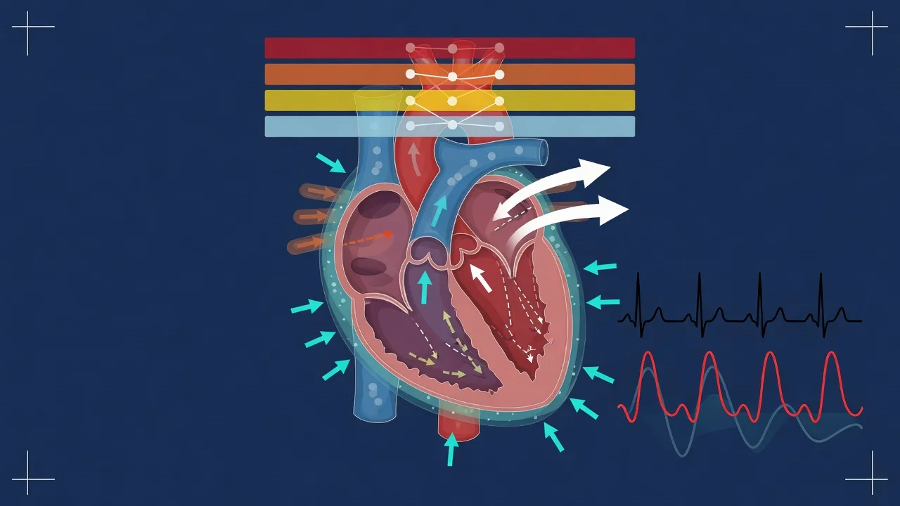
Cardiac tamponade is the clinical syndrome produced by rising intra-pericardial pressure that compresses the cardiac chambers and limits diastolic filling. It is fundamentally a problem of impaired ventricular filling rather than impaired contractility — the heart can pump perfectly well but cannot fill. Understanding the pressure-volume sequence is the key to every sign, every echo finding, and every treatment decision.[2][7]
The tamponade vicious cycle — from fluid accumulation to circulatory collapse
1. Pericardial fluid accumulates
Fluid (serous, sero-sanguinous, blood, pus) enters the relatively **non-compliant** pericardial space. The pericardium can stretch only slowly — the pericardial **stress-strain (volume-pressure) curve** is J-shaped: small volume rises are tolerated until a critical point, after which pressure climbs steeply with every additional millilitre. This is why a SMALL, RAPIDLY accumulating effusion (e.g., 200 mL of blood over minutes from an aortic dissection or cardiac perforation) can cause tamponade, while a LARGE, SLOW effusion (2-3 litres over months, e.g., malignancy) may be well tolerated — the pericardium has had time to stretch.
2. Intra-pericardial pressure rises above ventricular filling pressure
Once intra-pericardial pressure exceeds the transmural distending pressure of the thin-walled, low-pressure chambers (the right atrium and right ventricle fill first), those chambers begin to **collapse**. The critical pressure is around 15-30 mmHg (the level at which intra-pericardial pressure equals right atrial pressure). At this point cardiac chambers are competing for a fixed, non-expandable pericardial volume — the chambers become **interdependent** (filling one steals volume from another).
3. Impaired ventricular filling → reduced stroke volume
Compression of the right heart limits pulmonary venous return and therefore LV preload. Because the pericardial volume is fixed, the inspiratory increase in right-sided filling (the normal ~7 mmHg systolic BP rise) becomes **exaggerated** — the augmented RV filling on inspiration pushes the septum leftward and transiently impairs LV filling, producing **pulsus paradoxus**. Net stroke volume falls.
4. Compensatory tachycardia and vasoconstriction
Fallen cardiac output triggers sympathetic compensation: **sinus tachycardia** (to maintain cardiac output when stroke volume is fixed low) and peripheral **vasoconstriction** (to maintain blood pressure). The patient looks shocked, peripherally shut down, and tachycardic. This compensation can maintain the BP for a time — the patient is "compensated tamponade" — but the reserve is exhausted abruptly.
5. Eventual circulatory collapse
Once compensation fails, cardiac output falls precipitously, coronary perfusion drops, and a spiral of worsening ischaemia and bradycardia/asystole (or pulseless electrical activity) ensues. The decompensation is **sudden and nonlinear** — the steep part of the pericardial volume-pressure curve means a single additional millilitre can convert a stable patient into an arrest. This is why tamponade must be drained BEFORE the patient arrests: once the heart has stopped filling, CPR cannot generate output (it cannot fill a compressed heart).
Clinical features
Beck triad
Classic (not always complete)
- Hypotension (impaired LV filling → reduced cardiac output)
- Muffled heart sounds (fluid around heart)
- Raised JVP (impaired RA filling → venous congestion)
- Note: all 3 present in only ~30% of cases. Have high index of suspicion.
Other signs
Supportive
- Pulsus paradoxus: systolic BP drops >10 mmHg on inspiration (exaggerated normal physiology)
- Sinus tachycardia
- Dyspnoea, tachypnoea
- Pericardial friction rub (if pericarditis)
- Raised JVP with prominent x descent (normal y descent absent)
Beck triad — mechanism of each component
The Beck triad (Claude Beck, 1935) was originally described in the surgical setting of acute traumatic haemopericardium. Each component maps directly onto the pathophysiology. The full triad is present in only ~10-40% of cases — absence of the complete triad does not exclude tamponade, and most modern presentations are detected earlier by echocardiography.[2]
Beck triad — each sign and its mechanism
| Component | Sign | Mechanism |
|---|---|---|
| Hypotension | Systolic BP low, narrow pulse pressure | Compressed ventricles cannot fill → fixed-low stroke volume → falling cardiac output. The narrowed pulse pressure reflects the fixed stroke volume (most of the arterial pressure trace is sustained by vasoconstriction). |
| Muffled heart sounds | Distant, quiet heart sounds on auscultation | The fluid layer surrounding the heart attenuates the acoustic transmission of valve closures and myocardial contraction to the chest wall. The same physics that produces the low-voltage QRS on ECG. |
| Raised JVP | Distended neck veins, with a prominent x descent and absent y descent | Impaired atrial and ventricular filling raises right atrial and systemic venous pressure. The x descent is preserved (atrial relaxation and tricuspid descent during systole still occur), but the y descent disappears because the ventricle cannot fill in early diastole (it is compressed). This abnormal venous waveform is the jugular signature of tamponade. |
Pulsus paradoxus — the pathognomonic exaggeration of normal physiology
Pulsus paradoxus is an exaggeration of the normal inspiratory fall in systolic blood pressure. In health, inspiration increases venous return to the right heart; the expanded right heart bulges the septum slightly leftward and pulmonary capacitance increases, producing a small (up to ~10 mmHg) drop in systolic BP on inspiration. In tamponade, the fixed pericardial volume amplifies this interdependence: the augmented RV filling on inspiration steals volume from the LV and the LV stroke volume falls markedly.[2][7]
How to measure pulsus paradoxus at the bedside
Set up the cuff
Inflate the sphygmomanometer cuff above systolic pressure. The patient should be breathing normally (NOT held breath — the test depends on the respiratory cycle).
Slowly deflate, listening for the first Korotkoff sound
Deflate slowly (~2-3 mmHg per heartbeat). Note the pressure at which the Korotkoff sounds FIRST appear — at this pressure they will be audible only during expiration (the stronger beats), disappearing during inspiration.
Continue deflating until sounds are audible through BOTH phases
Note the pressure at which Korotkoff sounds become audible throughout the respiratory cycle (i.e., on inspiration AND expiration).
The difference is the pulsus paradoxus
The difference between the two pressures is the paradox. **>10 mmHg is abnormal** (sensitive for tamponade); **>20-25 mmHg is severe** and predicts haemodynamic compromise.
Kussmaul sign — and why it is usually ABSENT in tamponade
Kussmaul sign is an inspiratory rise in the JVP (rather than the normal inspiratory fall). It signals that the right heart cannot accommodate the increased venous return of inspiration. Kussmaul sign is characteristic of constrictive pericarditis and severe right ventricular failure / RV infarction, NOT of classic tamponade — because in a free-flowing circumferential effusion, inspiratory augmentation of venous return is still transmitted to the heart. A positive Kussmaul sign in suspected tamponade should raise the question of an effusive-constrictive syndrome (concurrent constriction + effusion) or a loculated/organised effusion behaving mechanically like constriction.[1]
Diagnosis
Tamponade diagnostic approach
Clinical suspicion
Hypotension + raised JVP + muffled heart sounds. Or: unexplained shock with raised JVP and clear lung fields. Pulsus paradoxus: measure BP during expiration and inspiration. If SBP drops >10 mmHg on inspiration = positive.
ECG
Low voltage QRS (<5 mm in limb leads — fluid attenuates signal). Electrical alternans: QRS amplitude varies beat-to-beat (heart swinging in fluid). Sinus tachycardia. PR depression + diffuse ST elevation (if concurrent pericarditis).
Echocardiogram (gold standard)
Shows: pericardial effusion (anechoic space around heart). Right atrial collapse (during late diastole — sensitive). Right ventricular diastolic collapse (during early diastole — specific). IVC plethora (dilated, non-collapsing — >50% collapse on sniff is normal; <50% = tamponade). Respiratory variation in mitral/tricuspid inflow (increased tricuspid, decreased mitral on inspiration).
If unstable: treat FIRST, image LATER
If patient is haemodynamically unstable (hypotensive, shocked): perform emergent pericardiocentesis without waiting for echo (can be done at bedside, ultrasound-guided if available). Do NOT delay for CT or formal echo.
Echocardiographic findings — the gold standard in detail
Echocardiography is both diagnostic and haemodynamically decisive — it confirms the effusion, demonstrates the physiological consequences of compression, and guides pericardiocentesis. The diagnosis is never made on the size of the effusion alone; it rests on demonstrating chamber collapse and the reciprocal inflow changes that indicate intra-pericardial pressure is elevated.[1][2]
Echocardiographic signs of tamponade — sensitivity, specificity and mechanism
| Echo sign | Phase / timing | Sensitivity / specificity | Mechanism / interpretation |
|---|---|---|---|
| Pericardial effusion | Throughout cycle | Variable | Anechoic (echo-free) space separating visceral and parietal pericardium. Size does NOT predict tamponade — but no effusion makes tamponade unlikely (except loculated post-surgical haematoma). |
| Right atrial collapse (inversion) | Late diastole / early systole | High sensitivity (~90%), lower specificity | The thin-walled RA is the first chamber to be compressed as intra-pericardial pressure rises. Collapse lasting >1/3 of the cardiac cycle increases specificity. |
| Right ventricular diastolic collapse (inversion) | Early diastole | High specificity (~85-100%), lower sensitivity | RV free wall invagination in early diastole indicates intra-pericardial pressure exceeds RV diastolic pressure — a clear haemodynamic tamponade. Less sensitive in RV hypertrophy / pulmonary hypertension (RV pressure is higher). |
| IVC plethora | Throughout respiratory cycle | High sensitivity, moderate specificity | Inferior vena cava dilated (>2.1 cm) with <50% collapse on sniff — reflects the elevated right-sided filling pressure. A collapsible IVC argues strongly AGAINST tamponade. |
| Respiratory variation in mitral / tricuspid inflow | Across the respiratory cycle | Specific (when marked) | On inspiration, augmented RV filling reduces LV filling: mitral E-wave velocity drops >25-30% on inspiration; tricuspid E-wave velocity increases >40-60%. Demonstrates the exaggerated ventricular interdependence that is the echo equivalent of pulsus paradoxus. Best measured by PW Doppler at the mitral/tricuspid leaflet tips. |
| Swinging heart | Throughout cycle | Specific when present | The heart oscillates within a large effusion — the substrate for electrical alternans on ECG. |
Management
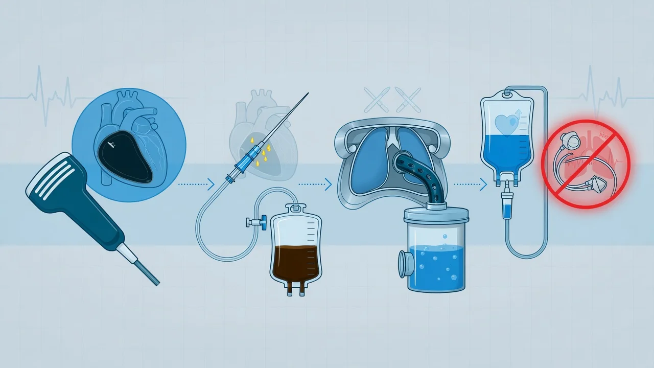
Tamponade management
Pericardiocentesis (EMERGENT)
Subxiphoid approach (ultrasound-guided if possible). Insert needle 1-2 cm below xiphoid, aim towards left shoulder at 30-45 degrees, aspirate while advancing. Send fluid for: culture, cytology, biochemistry (protein, LDH, glucose), cell count. Leave drain (pigtail catheter) for ongoing drainage.
Fluid resuscitation (CAUTIOUS)
Give IV crystalloid (250-500 mL bolus) to increase preload and maintain cardiac output. CAUTION: do NOT over-resuscitate (right heart is compressed — excess fluid worsens the situation). Goal: maintain BP until pericardiocentesis performed.
Avoid vasodilators and diuretics
Do NOT give nitrates (reduce preload → worsen tamponade). Do NOT give diuretics (reduce preload). Do NOT give positive pressure ventilation unless necessary (increases intrathoracic pressure → reduces venous return). Maintain spontaneous breathing if possible.
If pericardiocentesis unsuccessful or recurrent
Surgical pericardial window (subxiphoid pericardiotomy — creates a permanent drainage route into pleural/peritoneal space). Pericardiectomy (stripping of pericardium — for constrictive pericarditis).
AORTIC DISSECTION causing tamponade
Do NOT pericardiocentese (the fluid is blood from the dissection — removing it may cause catastrophic re-bleeding). Instead: immediate surgery (ascending aorta repair). This is a critical exception to the pericardiocentesis rule.
Pericardiocentesis technique — the echo-guided Seldinger approach
Modern pericardiocentesis is echo-guided wherever possible — the echo identifies the largest, safest access window (usually where the effusion is closest to the transducer and the path avoids liver, lung, and internal mammary vessels), confirms the needle tip in the pericardial space, and documents re-expansion of the chambers after drainage. The technique has evolved from the blind anatomical approach to a point-of-care ultrasound-guided procedure with markedly lower complication rates.[3]
Echo-guided pericardiocentesis — step by step
1. Echo-mapped site selection
Scan subxiphoid, apical, and parasternal windows. Choose the site where the effusion is **maximal and closest** to the transducer and the needle path is **extrapleural** (avoids lung) and **extraperitoneal** (avoids liver/peritoneum). The **apical approach** is now often preferred over the classic subxiphoid — shorter, more direct, and avoids the liver.
2. The classical subxiphoid approach (exam favourite)
Local anaesthetic infiltration. Insert the needle **1-2 cm inferior and to the left of the xiphoid process**, aimed **posteriorly and cephalad at ~30-45° towards the left shoulder** (the heart sits above and to the left). Advance slowly, aspirating continuously. Aspirate fluid → needle is in the pericardial space.
3. Seldinger wire and dilator
Pass a **J-tipped guidewire** through the needle into the pericardial space (confirm position by echo — wire seen in the effusion, or by agitated saline contrast). Withdraw the needle, then dilate the track. Railroad a **pigtail (8-12 Fr) catheter** over the wire into the pericardial space.
4. Drain the effusion and leave the catheter
Aspirate and allow the effusion to drain completely (the pericardial pressure should fall and chamber collapse resolve on echo). **Leave the pigtail catheter in situ** (not a one-off tap) for ongoing drainage — effusions, especially malignant and haemorrhagic, reaccumulate. Connect to a drainage bag; flush periodically to maintain patency.
5. Send the fluid
Send for **biochemistry** (protein, LDH, glucose, pH — exudate vs transudate via Light's criteria), **cell count and differential** (neutrophils → bacterial; lymphocytes → TB/malignancy), **microbiology** (Gram stain, culture, AFB, TB PCR), **cytology** (malignancy — send several samples, yield rises with repeat sampling), and consider **haematocrit** of the fluid vs blood (haemorrhagic — dissection, trauma, malignancy; if fluid haematocrit >50% of blood haematocrit → haemopericardium).
6. Confirm re-expansion and monitor
Post-procedure echo confirms resolution of chamber collapse, re-expansion of the RA/RV, and a collapsible IVC. Watch for **re-accumulation** (drain output), **pneumopericardium**, and **pleural effusion**. Continuous ECG monitoring for the first 24 h. Remove drain when output <50 mL/24 h and echo shows no re-accumulation.
Fluid resuscitation — why it is temporising, not definitive
In tamponade the heart is compressed; raising the right-sided filling pressure (transmural distending pressure) above the intra-pericardial pressure transiently restores RV filling and cardiac output. A cautious 250-500 mL crystalloid bolus is therefore a temporising measure while pericardiocentesis is organised. However, volume loading is not a substitute for drainage — over-resuscitation over-distends the compressed right heart, worsens the septal shift, and can precipitate deterioration. The classic Sagristà-Sauleda study confirmed that volume expansion raises cardiac output in tamponade, but the effect is modest and the benefit is confined to the under-filled (low-pressure) subset.[6]
Why positive pressure ventilation is dangerous in tamponade
Avoid endotracheal intubation and positive pressure ventilation (PPV) unless absolutely unavoidable. PPV raises mean intrathoracic pressure, which reduces venous return (the pressure gradient from the extrathoracic veins to the right atrium falls) and reduces the transmural filling pressure of the right heart — the very thing that is already critically low in tamponade. Intubation of a tamponade patient is frequently followed by cardiovascular collapse / cardiac arrest because the sedation abolishes the sympathetic tone and the PPV abolishes venous return. If intubation is unavoidable (profoundly agitated, respiratory failure), use the lowest mean airway pressure possible (low PEEP, low tidal volume, spontaneous-ventilation modes if feasible), pre-load with fluid, have vasopressors drawn up, and be ready to perform immediate pericardiocentesis / emergent sternotomy at the moment of induction.[2]
Causes
Tamponade causes
Aetiology — the full differential and what each cause means
The cause of the pericardial effusion determines the urgency, the fluid characteristics, the risk of recurrence, and the definitive therapy. Malignancy is the single most common cause of tamponade in adults presenting to the ICU; in younger patients and endemic regions, viral/idiopathic and tuberculosis dominate. The intensivist's job is to drain the effusion AND to identify the underlying cause, because the cause dictates the next step (surgery for dissection, chemotherapy for malignancy, dialysis for uraemia, anti-TB therapy for tuberculosis).[1][2]
Causes of cardiac tamponade — by mechanism, clue and management
| Cause | Relative frequency | Key clues / fluid characteristics | Specific management |
|---|---|---|---|
| Malignancy | #1 in adults (~30-40%) | Lung, breast, lymphoma, melanoma; recurrent, often haemorrhagic; cytology positive (multiple samples increase yield); mediastinal nodes / known primary on imaging. | Pericardiocentesis + pericardial window / sclerosis (prevents re-accumulation) + treat the cancer (chemo/radiotherapy). |
| Idiopathic / viral | Common (younger patients) | Often preceded by viral prodrome; exudative, lymphocytic fluid; usually responds to anti-inflammatory therapy. | Pericardiocentesis + NSAIDs + colchicine. |
| Uraemic | Dialysis / ESRF patients | From renal failure; often haemorrhagic (uraemic platelet dysfunction + heparin on dialysis); does NOT respond to NSAIDs. | Pericardiocentesis + intensify dialysis (the definitive treatment). |
| Tuberculosis | Common in endemic regions | Chronic, low-grade fever, night sweats, weight loss; exudative, lymphocytic, high ADA; concomitant pulmonary TB; high risk of constriction. | Pericardiocentesis + 4-drug anti-TB therapy (RIPE) + adjunctive steroids. |
| Post-MI free-wall rupture | Days 1-5 post-MI | Catastrophic — sudden haemopericardium, electromechanical dissociation, often fatal before reaching hospital; ~8-10% of MI deaths historically (rarer with early reperfusion). | Emergent surgery (rupture repair / patch) — pericardiocentesis alone is futile (bleeding is brisk). |
| Dressler syndrome | 2-10 weeks post-MI | Autoimmune post-cardiac injury; fever, pleuritic pain, friction rub, effusion (occasionally tamponade). | Pericardiocentesis if tamponade + NSAIDs + colchicine (avoid steroids). |
| Post-cardiac surgery | Days to weeks postoperatively | Bleeding / inflammation; may be loculated (clotted, regional) — easily missed on transthoracic echo; high suspicion required. | Drainage (often surgical evacuation, as clot cannot be aspirated percutaneously); reversal of anticoagulation. |
| Aortic dissection (Type A) | Acute | Haemopericardium from retrograde dissection into the pericardial space; tearing chest/back pain, BP differential, widened mediastinum. | URGENT SURGERY — do NOT pericardiocentese (removing the tamponading blood precipitates fatal re-bleeding). |
| Blunt / penetrating trauma | Acute | Haemopericardium; penetrating cardiac injury (stab wound), or blunt (steering-wheel) cardiac rupture; may present in arrest. | Emergent pericardiocentesis / resuscitative thoracotomy (penetrating) / surgery. |
| Radiation | Years after thoracic RT | Late fibrotic pericardium; effusive-constrictive physiology; often combined with coronary / myocardial damage. | Drainage; pericardiectomy (high morbidity from mediastinal fibrosis). |
| Autoimmune / connective tissue | Variable | SLE, rheumatoid arthritis, scleroderma, vasculitis; recurrent; multi-system involvement. | Pericardiocentesis + immunosuppression (steroids, steroid-sparing agents) + colchicine. |
| Purulent bacterial | Rare, emergency | S. aureus, pneumococcus; septic, high fever, purulent fluid; rapid progression to tamponade and sepsis. | IV antibiotics + immediate surgical/percutaneous drainage — life-threatening. |
| Drug-induced / iatrogenic | Procedures | Catheter perforation (PCI, pacemaker, CVC, ablation), anticoagulant/lytic-related bleeding; procainamide/hydralazine (lupus). | Stop offending agent/drug; drainage; reverse anticoagulation. |
Differential diagnosis — the shocked patient with raised JVP
The clinical picture of shock + raised JVP + clear lung fields has a focused differential. Bedside echo resolves the majority within minutes. The intensivist must distinguish tamponade from its mimics because the treatment is entirely different (drain the pericardium vs. decompress a tension pneumothorax vs. lyse a massive PE).[1][2]
Tamponade vs the other obstructive shock causes — the bedside echo decision
| Feature | Cardiac tamponade | Tension pneumothorax | Massive pulmonary embolism | Severe RV infarction | Constrictive pericarditis |
|---|---|---|---|---|---|
| Onset | Hours-days | Minutes | Hours | Acute (with MI) | Months-years |
| JVP | Raised, prominent x, absent y descent | Raised, non-pulsatile | Raised | Raised | Raised, Kussmaul sign, prominent y descent |
| Lung fields | Clear | Unilateral absent breath sounds + hyperresonance, trachea deviated AWAY | Clear | Clear (± crackles if LV failure) | Clear |
| Pulsus paradoxus | Marked | Marked | Mild-moderate | Mild | Mild or absent |
| ECG | Low voltage, electrical alternans | Sinus tachy ± small QRS in affected side | Sinus tachy, S1Q3T3, RBBB, T inversion V1-V4 | Inferior STEMI, RV involvement (V4R) | Low voltage (±) |
| Echo | Effusion + RA/RV collapse + IVC plethora | No pericardial effusion; RV underfilled; compressed heart; lung point on lung US (diagnostic) | Dilated hypokinetic RV, McConnell sign, septal bowing, D-shaped LV, tricuspid regurgitation | RV dilatation/hypokinesis in territory of infarct; no effusion | Thickened/calcified pericardium, septal bounce, pericardial knock, no chamber collapse |
| Treatment | Pericardiocentesis | Needle thoracostomy / chest drain | Thrombolysis / thrombectomy (consider in unstable) | Revascularise, fluid, inotrope, avoid nitrates/diuretics | Pericardiectomy (surgical) |
Low-pressure tamponade — the occult variant
First characterised by Sagristà-Sauleda, low-pressure tamponade is tamponade physiology occurring at lower-than-expected right atrial pressures — classically in hypovolaemic or over-diuresed patients, the elderly, and those on high-dose diuretics. The classical Beck triad may be incomplete or subtle: the JVP may be only mildly raised (because the patient is volume-depleted), and pulsus paradoxus may be borderline. Echo is the key — it shows chamber collapse and IVC plethora despite a non-impressive JVP. Volume loading (250-500 mL crystalloid) is both diagnostic (it raises RA pressure into the critical range, unmasking tamponade) and a temporising bridge to pericardiocentesis. Low-pressure tamponade is a frequently missed diagnosis because the clinician is reassured by the "not-very-high" JVP.[5][6]
The aortic dissection exception — when pericardiocentesis is dangerous
Post-cardiac surgery tamponade — the loculated trap
After cardiac surgery, pericardial effusions may be loculated (organised clot or adhesions compartmentalise the fluid), so circumferential compression and the classic Beck triad / pulsus paradoxus may be absent. Transthoracic echo may be non-diagnostic in the postoperative setting (poor windows, mediastinal air). Any unexplained fall in cardiac output, rising filling pressures, oliguria, or tachycardia in the first postoperative days demands a transoesophageal echo (TOE) to exclude a loculated clot compressing an individual chamber (classically the left or right atrium). Clotted haematomas cannot be aspirated percutaneously — they require surgical evacuation (re-sternotomy).[2]
Outcomes and prognosis
Cardiac tamponade — outcomes and key numbers
Landmark trials and evidence
ESC 2015 Guidelines for the diagnosis and management of pericardial diseases (Adler, Eur Heart J 2015; PMID 26320112)
Source
European Society of Cardiology — clinical practice guidelines (Task Force on Pericardial Diseases)
Diagnosis
Echo is the **gold standard** — RA collapse (most sensitive) + RV diastolic collapse (most specific) + IVC plethora + respiratory variation in inflow. Triage by clinical features (tachycardia, hypotension, pulsus paradoxus, raised JVP).
Management
Echo-guided pericardiocentesis (Seldinger, pigtail drain). **Avoid PPV, diuretics, vasodilators** — they reduce venous return and worsen tamponade. Cautious IV fluid as a temporising bridge.
Special situations
Aortic dissection tamponade → surgery, NOT pericardiocentesis. Loculated post-surgical tamponade → TOE + surgical evacuation. Recurrent / malignant → pericardial window or sclerosis.
Clinical bottom line
The definitive pericardial disease guideline — underpins the modern echo-guided, cause-targeted approach to tamponade.
Cardiac tamponade: an educational review (Imazio & De Ferrari, Eur Heart J Acute Cardiovasc Care 2021; PMID 32628038)
Source
Comprehensive contemporary review by the leading pericardial disease group (Turin)
Clinical features
Beck triad complete in only ~10-40%. **Pulsus paradoxus >10 mmHg** sensitive but not specific. Prominent **x descent with absent y descent** on the venous waveform. Sinus tachycardia universal.
Echo
Effusion + **RA collapse (>1/3 cycle)** + **RV diastolic collapse** (most specific) + **IVC plethora (<50% collapse)** + **respiratory variation** of mitral (>25-30%) and tricuspid (>40-60%) inflow.
Pathophysiology
J-shaped pericardial compliance curve — a small, RAPIDLY accumulating effusion is far more dangerous than a large, slow one.
Management
Echo-guided pericardiocentesis with indwelling pigtail drain; treat the underlying cause; consider surgical window for recurrence/malignancy.
Clinical bottom line
The clearest single-source summary of the modern understanding of tamponade — diagnosis, echo, drainage.
Evolution of the pericardiocentesis technique (Maggiolini, J Cardiovasc Med 2018; PMID 29553993)
Source
Italian multicentre experience review — the shift from blind to echo-guided pericardiocentesis
Key evolution
The technique moved from the **blind anatomical subxiphoid approach** (high complication rate) to **echo-guided, site-selected** pericardiocentesis (the needle enters where the effusion is closest to the probe and the path avoids liver/lung) — the apical approach is increasingly preferred.
Outcome
Echo-guided pericardiocentesis is **safer** (lower rates of pneumothorax, laceration, vessel injury) and **more effective** (higher success, lower re-accumulation with an indwelling catheter) than the historical blind technique.
Clinical bottom line
Modern pericardiocentesis is echo-guided, site-selected, and uses an indwelling pigtail drain — the blind approach is obsolete wherever ultrasound is available.
Low-pressure cardiac tamponade (Sagristà-Sauleda, Circulation 2006; PMID 16923755)
Source
Mayo Clinic-style haemodynamic series characterising the low-pressure variant
Clinical profile
Tamponade physiology at **lower-than-expected right atrial pressures** (often <15 mmHg), classically in **hypovolaemic / over-diuresed** patients. The JVP may be only mildly raised and pulsus paradoxus subtle — the diagnosis is easily missed.
Haemodynamics
Echo shows chamber collapse and IVC plethora **despite** an unimpressive JVP. **Volume loading raises RA pressure into the critical range** — it both unmasks tamponade and improves cardiac output transiently.
Clinical bottom line
Suspect low-pressure tamponade in the volume-depleted patient with unexplained low output / IVC plethora — give fluid and look for the echo signs. A normal JVP does NOT exclude tamponade.
Hemodynamic effects of volume expansion in tamponade (Sagristà-Sauleda, Circulation 2008; PMID 18332261)
Source
Haemodynamic study of the response to volume expansion in patients with tamponade
Finding
Volume expansion **raises cardiac output** in tamponade, but the effect is **modest and confined to the under-filled (low-pressure) subset**. In the fully filled classic tamponade, additional volume produces little benefit and may worsen septal shift.
Clinical bottom line
Cautious 250-500 mL crystalloid is a reasonable **temporising** bridge to pericardiocentesis, especially in the hypovolaemic/low-pressure subset — but it is NOT a substitute for drainage, and over-resuscitation is harmful.
Outcomes of idiopathic chronic large pericardial effusion (Imazio, Heart 2019; PMID 30274986)
Source
Contemporary cohort of conservatively-managed idiopathic large effusions
Finding
Most **chronic, large, idiopathic effusions** can be observed (no prophylactic drainage) UNLESS tamponade features develop. The risk of evolution to tamponade and the long-term risk of constriction guide intervention. Aspirin/colchicine may reduce size in selected cases.
Clinical bottom line
A large chronic effusion without tamponade physiology is NOT an automatic indication for pericardiocentesis — observe and treat the underlying pericarditis. Drain when tamponade features appear.
Exam practice — SAQs
SAQ — Malignant pericardial effusion with classic tamponade and echo signs
10 minutes · 10 marks
A 64-year-old woman with metastatic lung adenocarcinoma on pembrolizumab presents to the emergency department with three days of progressive dyspnoea, fatigue and presyncope. On examination: BP 84/56 (MAP 65), HR 128 (sinus), RR 30, SpO2 92% on room air, JVP raised to the angle of the jaw at 45 degrees, muffled heart sounds, clear lung fields and cool peripheries. Pulsus paradoxus is 22 mmHg. ECG shows low-voltage QRS complexes with electrical alternans. Bedside focused echo: a 2.4 cm circumferential pericardial effusion with right atrial collapse lasting 40% of the cardiac cycle, right ventricular free-wall diastolic collapse in early diastole, IVC 2.4 cm with <10% collapse on sniff, and a 35% inspiratory fall in mitral E-wave velocity on pulse-wave Doppler.
SAQ — Post-cardiac surgery loculated tamponade with absent classic signs
10 minutes · 10 marks
A 72-year-old man is day 3 after an elective three-vessel on-pump coronary artery bypass graft. He was extubated on day 1 and initially did well, but overnight has developed a falling cardiac index (1.8 L/min/m2), a rising CVP (now 18 mmHg), oliguria (0.3 mL/kg/h) and new sinus tachycardia (HR 116). MAP 60 on escalating noradrenaline (now 0.3 mcg/kg/min). There is no clear pulsus paradoxus and the JVP is difficult to assess beneath surgical emphysema. The mediastinal and pleural drains were removed on day 2 after minimal output. Transthoracic echo shows poor windows with a 1 cm loculated posterior collection adjacent to the right atrium and no convincing chamber collapse.
Clinical pearls
Red flags
Exam one-liners — the rapid recall set
[1]Comparison with adjacent topics — high-yield cross-references
- Acute pericarditis: the cause; tamponade is the complication. Treat the pericarditis (NSAIDs + colchicine) AND drain if tamponade.
- Constrictive pericarditis: the chronic cousin — thickened/calcified pericadium, Kussmaul sign, pericardial knock, prominent y descent (vs absent y in tamponade), months-years onset. Treatment is pericardiectomy.
- Aortic dissection: the critical exception — haemopericardium from Type A dissection is a surgical emergency, NOT a pericardiocentesis candidate.
- Massive pulmonary embolism: shares the shocked + raised JVP + clear lungs presentation — echo distinguishes (dilated RV, McConnell sign, no effusion).
- Cardiogenic shock: low output from pump failure (not filling failure) — the LV is failing, the JVP is raised but there is pulmonary oedema (unlike the clear lungs of tamponade). [1]
References
- [1]Adler Y, Charron P, Imazio M, et al. 2015 ESC Guidelines for the diagnosis and management of pericardial diseases: The Task Force for the Diagnosis and Management of Pericardial Diseases of the European Society of Cardiology (ESC)Endorsed by: The European Association for Cardio-Thoracic Surgery (EACTS) Eur Heart J, 2015.PMID 26320112
- [2]Imazio M, De Ferrari GM. Cardiac tamponade: an educational review Eur Heart J Acute Cardiovasc Care, 2021.PMID 32628038
- [3]Maggiolini S, De Carlini CC, Imazio M. Evolution of the pericardiocentesis technique J Cardiovasc Med (Hagerstown), 2018.PMID 29553993
- [4]Imazio M, Lazaros G, Valenti A, et al. Outcomes of idiopathic chronic large pericardial effusion Heart, 2019.PMID 30274986
- [5]Sagristà-Sauleda J, Angel J, Sambola A, et al. Low-pressure cardiac tamponade: clinical and hemodynamic profile Circulation, 2006.PMID 16923755
- [6]Sagristà-Sauleda J, Angel J, Sambola A, et al. Hemodynamic effects of volume expansion in patients with cardiac tamponade Circulation, 2008.PMID 18332261
- [7]Sagristà-Sauleda J, Mercé AS, Soler-Soler J. Diagnosis and management of pericardial effusion World J Cardiol, 2011.PMID 21666814