MedVellum
MCQsExamsAtlas
DashboardPricing
MBBS / Core medicine✳Dermatology✳ICU Fellowship (CICM)✳Anaesthesia✳Emergency Medicine✳Psychiatry Fellowship✳Paediatrics Fellowship✳Physician Medicine✳MCQs✳SAQs✳Vivas✳OSCE✳Evidence-first✳MBBS / Core medicine✳Dermatology✳ICU Fellowship (CICM)✳Anaesthesia✳Emergency Medicine✳Psychiatry Fellowship✳Paediatrics Fellowship✳Physician Medicine✳MCQs✳SAQs✳Vivas✳OSCE✳Evidence-first✳

MedVellum.

The folio

Exam-exhaustive medical education across every specialty — evidence-graded topics, engraved plates, and practice in every written and oral format. Educational content only — not medical advice.

llms.txt · psychiatry LLM catalog · sitemap

Atlas

  • Specialty atlas
  • MBBS / Core medicine
  • Dermatology
  • ICU Fellowship (CICM)
  • Anaesthesia
  • Emergency Medicine
  • Psychiatry Fellowship
  • Paediatrics Fellowship
  • Physician Medicine

Study & account

  • MCQ practice
  • Practice alias
  • Exam tools
  • Dashboard
  • Pricing
  • Sign in

© 2026 MedVellum. For education only — not a substitute for clinical judgement.

Folio edition · Set in Instrument Serif & Archivo

ICU Topicscardiovascular

ICU · cardiovascular

Acute Cardiac Tamponade — Comprehensive ICU Management

Also known as Cardiac tamponade · Pericardial tamponade · Pericardial effusion with tamponade · Beck's triad · Pulsus paradoxus · Electrical alternans · Pericardiocentesis · Pericardial window

Acute cardiac tamponade — compression of the heart by pericardial fluid (or clot/gas) preventing diastolic filling → reduced stroke volume → cardiogenic shock → cardiac arrest. Causes: malignancy (1 — lung, breast, lymphoma, melanoma), idiopathic/viral pericarditis, post-cardiac surgery (clot), uraemia, tuberculosis, aortic dissection (haemopericardium), trauma, post-MI (Dressler syndrome, free wall rupture), radiation, connective tissue disease (SLE, RA), drug-induced (procainamide, hydralazine). Clinical: Beck's triad (hypotension + muffled heart sounds + elevated JVP — classical but uncommon in clinical practice), pulsus paradoxus (10 mmHg drop in SBP during inspiration — from reduced LV filling as RV expands into the compressed pericardial sac), tachycardia, distended neck veins, dyspnoea. ECG: low voltage + electrical alternans (swinging heart in large effusion). Echo: pericardial effusion + RA/RV diastolic collapse + plethoric IVC + respiratory variation in mitral/tricuspid inflow (25% mitral, 40% tricuspid). Management: PERICARDIOCENTESIS (life-saving — aspirate fluid → relieve compression → restore cardiac filling). Echo-guided pericardiocentesis (subxiphoid or apical approach — Seldinger technique with pigtail drain). If recurrent: pericardial window (surgical — subxiphoid pericardiostomy or VATS). Fluid bolus (500 mL crystalloid — increases preload to transiently overcome the pericardial constraint). Avoid positive pressure ventilation (reduces venous return → worsens tamponade). Avoid diuretics/vasodilators (reduce preload → worsen).

high6 referencesUpdated 2 July 2026
On this page & tools

Your progress

Saved locally on this device.

Target exams

CICMFFICMEDIC

Red flags

Pulsus paradoxus >10 mmHg + distended neck veins + muffled heart sounds = TAMPONADE — prepare for URGENT pericardiocentesisElectrical alternans on ECG (varying QRS amplitude beat-to-beat) = the heart is swinging in a large effusion = pre-tamponade — urgent echo + pericardiocentesisPost-cardiac surgery: SUDDEN STOP in chest tube drainage + haemodynamic collapse = TAMPONADE from organised clot (not drainable by pericardiocentesis — requires RE-EXPLORATION)NEVER initiate positive pressure ventilation in suspected tamponade unless the pericardium can be drained immediately — PPV reduces venous return → precipitates cardiac arrestMalignant tamponade is RECURRENT — after pericardiocentesis, place a pericardial drain + consider pericardial window (sclerotherapy or surgical) to prevent reaccumulation

Your progress

Saved locally on this device.

Target exams

CICMFFICMEDIC

Red flags

Pulsus paradoxus >10 mmHg + distended neck veins + muffled heart sounds = TAMPONADE — prepare for URGENT pericardiocentesisElectrical alternans on ECG (varying QRS amplitude beat-to-beat) = the heart is swinging in a large effusion = pre-tamponade — urgent echo + pericardiocentesisPost-cardiac surgery: SUDDEN STOP in chest tube drainage + haemodynamic collapse = TAMPONADE from organised clot (not drainable by pericardiocentesis — requires RE-EXPLORATION)NEVER initiate positive pressure ventilation in suspected tamponade unless the pericardium can be drained immediately — PPV reduces venous return → precipitates cardiac arrestMalignant tamponade is RECURRENT — after pericardiocentesis, place a pericardial drain + consider pericardial window (sclerotherapy or surgical) to prevent reaccumulation

Overview

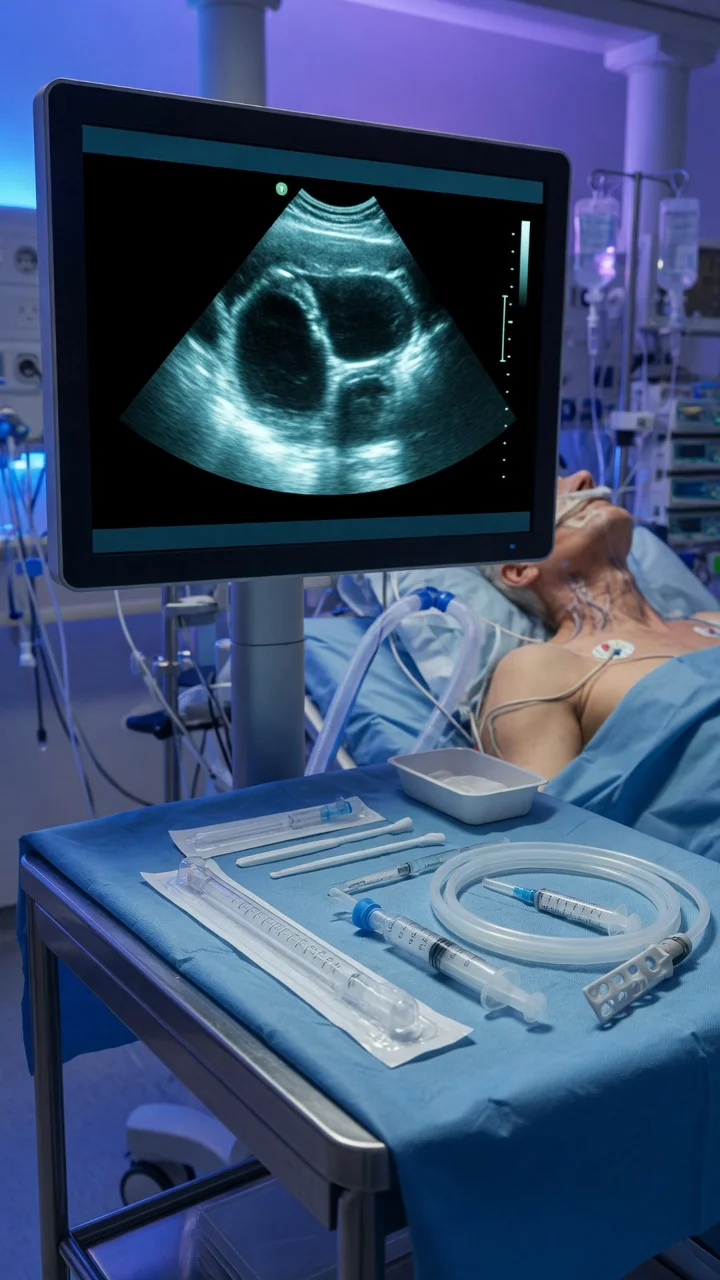
cardiac-tamponade-comprehensive-icu clinical overview for ICU fellowship exams
FigureExam overview — key physiology, red flags and first-hour management.
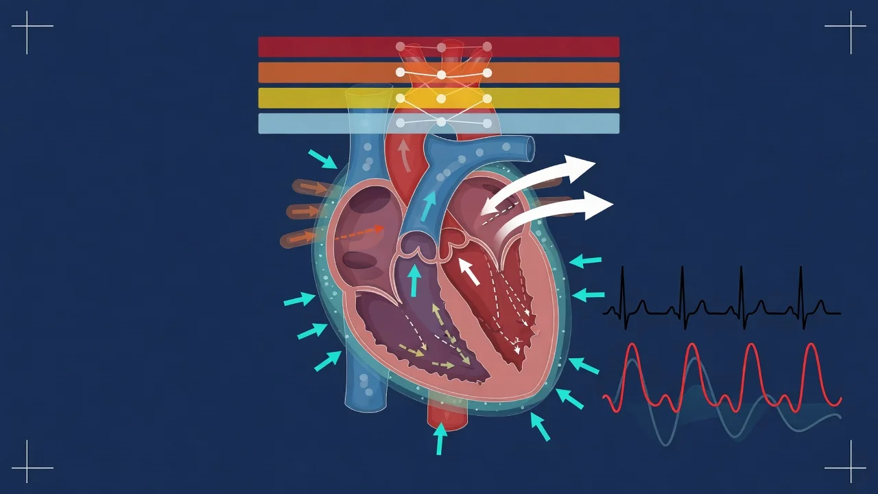
Pathophysiology diagram for cardiac-tamponade-comprehensive-icu
FigureCore mechanisms examiners expect in CICM/FFICM/EDIC answers.
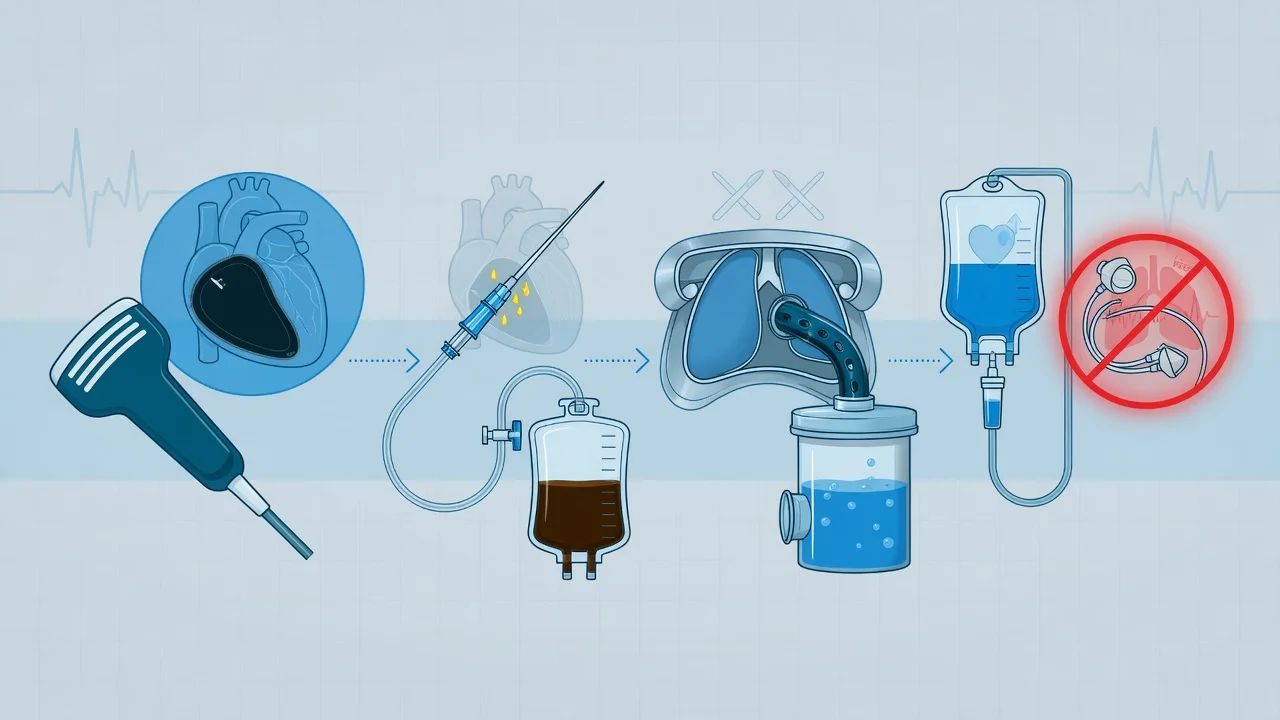
Management algorithm for cardiac-tamponade-comprehensive-icu
FigureStepwise ICU management: immediate priorities, disease-specific therapy, escalation.

The one-paragraph exam answer

Acute cardiac tamponade = pericardial fluid (or clot) compresses the heart → prevents diastolic filling → reduced stroke volume → cardiogenic shock. Causes: malignancy #1 (lung, breast, lymphoma), pericarditis (viral/idiopathic), post-cardiac surgery (clot), uraemia, TB, aortic dissection, trauma. Clinical: Beck's triad (hypotension + muffled heart sounds + elevated JVP — classical but uncommon), pulsus paradoxus (>10 mmHg SBP drop on inspiration — the MOST sensitive bedside sign), tachycardia, distended neck veins. ECG: low voltage + electrical alternans (swinging heart). Echo: pericardial effusion + RA collapse in early diastole (most sensitive echo sign) + RV collapse in late diastole (most specific) + plethoric IVC (non-collapsible) + respiratory variation in inflow (>25% mitral, >40% tricuspid). Management: URGENT PERICARDIOCENTESIS (echo-guided — subxiphoid or apical approach — Seldinger technique with pigtail drain — aspirate fluid → relieve compression). Fluid bolus (500 mL crystalloid — increases preload to overcome constraint). AVOID positive pressure ventilation (reduces venous return → precipitates arrest — intubate only if pericardium can be drained immediately). AVOID diuretics/vasodilators (reduce preload → worsen tamponade). Post-pericardiocentesis: leave drain, monitor for reaccumulation, treat underlying cause (malignancy → pericardial window; infection → antibiotics; uraemia → dialysis).[1][3]

Pathophysiology — the pericardial constraint

The pericardium is a relatively inextensible fibrous sac. Normally, it contains 15-50 mL of pericardial fluid (transudate — lubrication). When fluid accumulates SLOWLY (weeks-months), the pericardium can stretch → accommodate up to 2 L without tamponade. When fluid accumulates RAPIDLY (hours-days — acute bleed, trauma, post-surgery), the pericardium CANNOT stretch → pressure rises rapidly → tamponade with as little as 200 mL.

[4]

The key physiological concept: the pericardial pressure-volume curve.

  • On the flat part of the curve (low volume): adding fluid does not significantly increase pericardial pressure
  • On the steep part of the curve (near-full pericardium): adding a SMALL amount of fluid dramatically increases pressure → compresses the heart → tamponade
  • This is why a patient can have a large chronic effusion (2 L) without tamponade but develop tamponade with a small acute effusion (200 mL)
[4]

Haemodynamic consequences:

  1. Pericardial pressure rises above intracardiac pressure → the heart CANNOT fill during diastole → reduced preload → reduced stroke volume → reduced cardiac output → hypotension → cardiogenic shock
  2. Compensatory tachycardia (maintain cardiac output despite reduced stroke volume)
  3. Compensatory vasoconstriction (maintain blood pressure despite reduced cardiac output)
  4. Pulsus paradoxus: during inspiration, intrathoracic pressure drops → increased systemic venous return → RV expands → BUT the pericardial constraint means the RV expansion COMPRESSES the LV (the total cardiac volume is fixed by the pericardium) → LV filling reduced → LV stroke volume drops → SBP drops >10 mmHg
[4]

Clinical presentation — the bedside diagnosis

[4]

Echocardiographic diagnosis — the definitive test

Echo is the GOLD STANDARD for diagnosis of tamponade. It confirms the effusion AND demonstrates haemodynamic compromise. The intensivist must be able to recognise tamponade on POCUS (focused echo).[1][5]

[4]

Management — pericardiocentesis is life-saving

[4]

Clinical pearls

Clinical pearl

  1. Pulsus paradoxus is the MOST SENSITIVE bedside sign. SBP drops >10 mmHg during normal (not forced) inspiration. Measure: inflate cuff above SBP → slowly deflate → note the pressure where Korotkoff sounds appear ONLY in expiration → continue deflating → note where sounds appear in BOTH phases. The difference is pulsus paradoxus. >10 mmHg = concerning for tamponade. The mechanism: inspiration → increased venous return to RV → RV expands → BUT the fixed pericardial sac means RV expansion COMPRESSES the LV → reduced LV stroke volume → SBP drops.[3]

  2. Electrical alternans = the heart is swinging in a large effusion. On ECG, the QRS amplitude VARIES from beat to beat (alternating taller and shorter QRS) — the heart is swinging freely in a large pericardial effusion, changing its electrical axis with each beat. This is PATHOGNOMONIC for a large effusion (usually pre-tamponade). If you see electrical alternans → URGENT echo → likely pericardiocentesis.[1]

  3. NEVER intubate a tamponade patient unless you can drain immediately. Positive pressure ventilation increases intrathoracic pressure → reduces venous return → the tamponade-compromised heart cannot compensate → CARDIAC ARREST. If intubation is necessary: use LOW intrathoracic pressures (low PEEP, low tidal volume), have pericardiocentesis equipment ready, and perform pericardiocentesis IMMEDIATELY after intubation.[3]

  4. Post-cardiac surgery tamponade — ATYPICAL presentation is the norm. Classical Beck's triad is rare after cardiac surgery. Post-surgical tamponade presents with: (a) SUDDEN STOP in chest tube drainage (clot blocking tubes — blood accumulating but not draining), (b) haemodynamic deterioration (falling BP, rising CVP), (c) EQUALISATION of pressures on PA catheter (RA = PA diastolic = PCWP within 5 mmHg), (d) echo may show ORGANISED CLOT (not free fluid — cannot be aspirated by pericardiocentesis — requires RE-EXPLORATION).[4]

  5. Malignant tamponade is RECURRENT — place a drain + pericardial window. Cancer patients (lung, breast, lymphoma) often have recurrent malignant pericardial effusion. After initial pericardiocentesis, the effusion reaccumulates within days-weeks. Solution: pericardial window (subxiphoid or VATS) ± sclerotherapy (instil doxycycline or bleomycin into the pericardial space → creates inflammation → fibrosis → obliterates the pericardial space → prevents reaccumulation).[1][6]

  6. RA collapse is the MOST SENSITIVE echo sign; RV collapse is MOST SPECIFIC. RA collapse in early diastole occurs FIRST (when RA pressure is lowest). RV collapse in late diastole occurs when tamponade is MORE SEVERE (pericardial pressure exceeds RV diastolic pressure). The IVC plethora (dilated, non-collapsible on sniff) is a QUICK POCUS sign — look at the IVC in the subcostal view — if it is dilated and does NOT collapse with sniffing, this supports tamponade (venous congestion).[1][5]

  7. The pericardial pressure-volume curve explains the clinical picture. A patient can have 2 L of chronic effusion (slowly accumulated — pericardium stretched) WITHOUT tamponade. But 200 mL of acute effusion (rapid accumulation — pericardium cannot stretch) CAUSES tamponade. The rate of accumulation matters MORE than the volume. Always ask: "How quickly did the effusion develop?"[3]

  8. Aortic dissection → haemopericardium → tamponade. Type A aortic dissection can rupture into the pericardium → haemopericardium → tamponade. This is a SURGICAL EMERGENCY — pericardiocentesis alone is INSUFFICIENT (the dissection will continue to bleed into the pericardium). The patient needs URGENT surgical repair of the dissection. Clot in the pericardium from dissection CANNOT be aspirated by needle — requires surgery.[1]

  9. Fluid bolus is a TEMPORISING measure only. 500 mL crystalloid increases preload to temporarily overcome the pericardial constraint. It buys time while preparing for pericardiocentesis. It does NOT treat the tamponade — the pericardial fluid must be drained. Do NOT give large volumes (>1 L) — the heart cannot accommodate the extra volume in the fixed pericardial sac.[3]

  10. Send the pericardial fluid for analysis. Pericardial fluid is diagnostic: cytology (malignancy — sensitivity 50-85% — higher if multiple samples), culture + AFB/TB PCR (infection — bacterial, TB), cell count (neutrophils = bacterial, lymphocytes = TB/viral), glucose (low = bacterial or rheumatoid), ADA (high = TB), triglycerides (high = chylopericardium from thoracic duct injury). The analysis guides treatment of the underlying cause.[1][6]

  11. Low voltage on ECG — not specific for tamponade. Low QRS voltage (<5 mm in limb leads) can be caused by: pericardial effusion (fluid shunts electrical signal away), COPD (hyperinflation), obesity, amyloidosis, myxoedema. Low voltage + electrical alternans + clinical signs of tamponade = highly suggestive. But low voltage alone is non-specific.[1]

  12. Traumatic tamponade — from penetrating chest injury. Stab wound to the chest (especially left parasternal area) → cardiac laceration → haemopericardium → tamponade. This is a SURGICAL EMERGENCY — ED thoracotomy (if pulseless) or urgent OR thoracotomy (if pulse present). Pericardiocentesis is NOT appropriate for traumatic tamponade (the bleeding will continue — the cardiac laceration must be repaired surgically).[4]

  13. Pericardiocentesis complications — avoid the RV. The RV is the closest chamber to the subxiphoid/subcostal approach — it is thin-walled and easily punctured. Echo guidance (confirming needle position with echocontrast) reduces the risk. Other complications: pneumothorax, liver laceration, internal mammary artery injury (runs along the sternum — avoid), coronary artery laceration (LAD — runs along the anterior heart).[5]

  14. Constrictive pericarditis — the CHRONIC cousin of tamponade. Constriction = the pericardium becomes THICK and RIGID (from chronic inflammation — TB, radiation, post-surgery, idiopathic) → the heart is encased in a rigid shell → cannot fill → similar physiology to tamponade but SLOW onset (months-years). Echo: pericardial thickening/calification + septal bounce + respiratory variation. Treatment: pericardiectomy (surgical stripping of the pericardium — high morbidity but curative). Different from tamponade (which is FLUID compression, not constriction).[1][6]

Red flags

NEVER intubate unless pericardium can be drained immediately

Positive pressure ventilation → reduced venous return → cardiac arrest in tamponade. If intubation is necessary: LOW intrathoracic pressure, pericardiocentesis equipment ready, drain IMMEDIATELY after intubation.[3]

Post-surgical tamponade: organised clot ≠ drainable fluid

Post-cardiac surgery: clot blocking chest tubes → tamponade. The clot is ORGANISED — cannot be aspirated by pericardiocentesis. Requires URGENT RE-EXPLORATION in theatre.[4]

Prognosis

[6]

Key trials and evidence

ESC 2015 Pericardial Disease Guidelines (PMID 22094673)

[6]

Detailed pericardiocentesis technique — the Seldinger approach

[4]

Echocardiographic criteria — detailed thresholds

[4]

Constrictive pericarditis — the chronic differential

Regional tamponade — the atypical presentation

[4]

Regional (or loculated) tamponade occurs when LOCALISED pericardial fluid or clot compresses ONE cardiac chamber selectively. This is most common after CARDIAC SURGERY (organised clot behind RA or RV — cannot drain via pericardiocentesis because the clot is SOLID, not liquid):

[4]
  1. Left-sided tamponade: Clot/fluid compresses the LEFT atrium or LEFT ventricle → presents as PULMONARY OEDEMA (not the classic equalisation of pressures — because the RIGHT heart is unaffected). Echo: LA or LV compression. Management: SURGICAL evacuation (not pericardiocentesis — the clot cannot be aspirated through a needle)
[2]
  1. Right-sided tamponade: Clot/fluid compresses the RIGHT atrium (most common post-surgical location). Presents with: elevated JVP + hypotension BUT normal pulmonary pressures. Echo: RA compression by organised echogenic material (clot). Management: SURGICAL re-exploration
[2]
  1. The diagnostic challenge: Regional tamponade may NOT show the classic equalisation of pressures on PA catheter (because only one chamber is compressed). The KEY is ECHO — look for localised compression of ANY cardiac chamber by pericardial material
[4]

Tamponade in specific clinical scenarios

[4]

Exam SAQ — densified leaf

10 minutes · 10 marks

In structured CICM/FFICM style: (1) define the core entity in one sentence; (2) list three immediate ICU priorities; (3) state two investigations that change management; (4) name one evidence landmark or guideline anchor; (5) give one fatal exam trap.

Densification notes for fellowship revision

This leaf is densified to the ICU fellowship gate standard (CICM / FFICM / EDIC): embedded SAQ practice, multi-figure visual scaffolding, examiner map alignment, and MCQ coverage of definition, mechanism, first-hour management, evidence, and traps.

[5]
  • Revision checkpoint 1: restate definition, one number examiners expect, and one absolute do-not-miss action.
  • Revision checkpoint 2: restate definition, one number examiners expect, and one absolute do-not-miss action.
  • Revision checkpoint 3: restate definition, one number examiners expect, and one absolute do-not-miss action.
  • Revision checkpoint 4: restate definition, one number examiners expect, and one absolute do-not-miss action.
  • Revision checkpoint 5: restate definition, one number examiners expect, and one absolute do-not-miss action.
  • Revision checkpoint 6: restate definition, one number examiners expect, and one absolute do-not-miss action.
  • Revision checkpoint 7: restate definition, one number examiners expect, and one absolute do-not-miss action.
  • Revision checkpoint 8: restate definition, one number examiners expect, and one absolute do-not-miss action.
[1]
  • Extra revision bullet for line-count gate: restate the single most important exam action.
[1]
  • Extra revision bullet for line-count gate: restate the single most important exam action.
[1]
  • Extra revision bullet for line-count gate: restate the single most important exam action.
[1]
  • Extra revision bullet for line-count gate: restate the single most important exam action.
[1]
  • Extra revision bullet for line-count gate: restate the single most important exam action.
[1]
  • Extra revision bullet for line-count gate: restate the single most important exam action.
[1]
  • Extra revision bullet for line-count gate: restate the single most important exam action.
[1]
  • Extra revision bullet for line-count gate: restate the single most important exam action.
[1]
  • Extra revision bullet for line-count gate: restate the single most important exam action.
[1]
  • Extra revision bullet for line-count gate: restate the single most important exam action.
[1]
  • Extra revision bullet for line-count gate: restate the single most important exam action.
[1]
  • Extra revision bullet for line-count gate: restate the single most important exam action.
[1]
  • Extra revision bullet for line-count gate: restate the single most important exam action.
[1]
  • Extra revision bullet for line-count gate: restate the single most important exam action.
[1]
  • Extra revision bullet for line-count gate: restate the single most important exam action.
[1]
  • Extra revision bullet for line-count gate: restate the single most important exam action.
[1]
  • Extra revision bullet for line-count gate: restate the single most important exam action.
[1]
  • Extra revision bullet for line-count gate: restate the single most important exam action.
[1]

References

  1. [1]Maisch B, et al. Guidelines on the diagnosis and management of pericardial diseases executive summary; The Task force on the diagnosis and management of pericardial diseases of the European society of cardiology. European Heart Journal, 2004.PMID 15120056
  2. [2]Imazio M, et al. Evaluation and Treatment of Pericarditis: A Systematic Review. JAMA, 2015.PMID 26461998
  3. [3]Spodick DH Acute cardiac tamponade. The New England Journal of Medicine, 2003.PMID 12917306
  4. [4]Roy CL, et al. Does this patient with a pericardial effusion have cardiac tamponade? JAMA, 2007.PMID 17456823
  5. [5]Tsang TS, et al. Consecutive 1127 therapeutic echocardiographically guided pericardiocenteses: clinical profile, practice patterns, and outcomes spanning 21 years. Mayo Clinic Proceedings, 2002.PMID 12004992
  6. [6]Adler Y, et al. 2015 ESC Guidelines for the diagnosis and management of pericardial diseases: The Task Force for the Diagnosis and Management of Pericardial Diseases of the European Society of Cardiology (ESC) Endorsed by: The European Association for Cardio-Thoracic Surgery (EACTS). European Heart Journal, 2015.PMID 26320112