ICU · Cardiovascular
Cardiogenic shock and mechanical circulatory support
Also known as Cardiogenic shock · Mechanical circulatory support (MCS) · Intra-aortic balloon pump (IABP) · Impella · VA-ECMO · Counterpulsation · SCAI shock staging
Cardiogenic shock is a life-threatening state of end-organ hypoperfusion due to cardiac pump failure, with mortality of 40-60%. The SCAI classification (Stages A-E) guides severity assessment. Management escalates from pharmacological support (noradrenaline first-line vasopressor, dobutamine/milrinone as inotropes) through to mechanical circulatory support: IABP (0.5-1 L/min counterpulsation, no routine benefit per IABP-SHOCK II), Impella (2.5-5 L/min active LV unloading), and VA-ECMO (3-5 L/min full cardiopulmonary bypass). VA-ECMO does NOT unload the LV — the combination with Impella (ECPELLA) addresses both systemic flow and ventricular unloading.
On this page & tools
Your progress
Saved locally on this device.
Target exams
Red flags
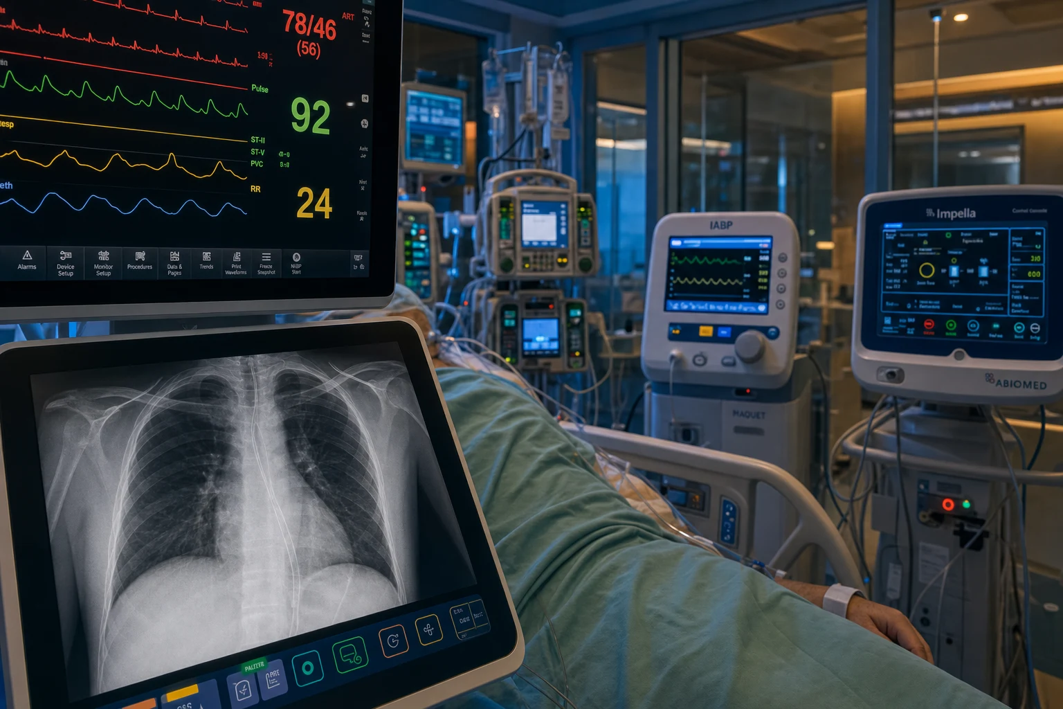
Definition and SCAI classification [1]
Definition
Cardiogenic shock is a clinical syndrome of end-organ hypoperfusion due to cardiac pump dysfunction. The diagnostic criteria require ALL of the following:[1][2]
- Hypotension: SBP <90 mmHg (or MAP <65 mmHg) for ≥30 minutes, or requiring vasopressor/inotrope support to maintain SBP ≥90
- End-organ hypoperfusion: at least one of:
- Lactate >2 mmol/L (with metabolic acidosis)
- Oliguria (<0.5 mL/kg/hr)
- Altered mental state (confusion, agitation)
- Cold extremities (mottled, prolonged capillary refill >3 seconds)
- Cardiac cause: clinical or echocardiographic evidence of cardiac dysfunction (e.g., LV failure, RV failure, arrhythmia, mechanical complication) [1]
Exclude: hypovolaemia (assess volume responsiveness), septic shock (warm peripheries, normal/high cardiac output), obstructive shock (tension pneumothorax, massive PE, tamponade). [1]
SCAI shock staging
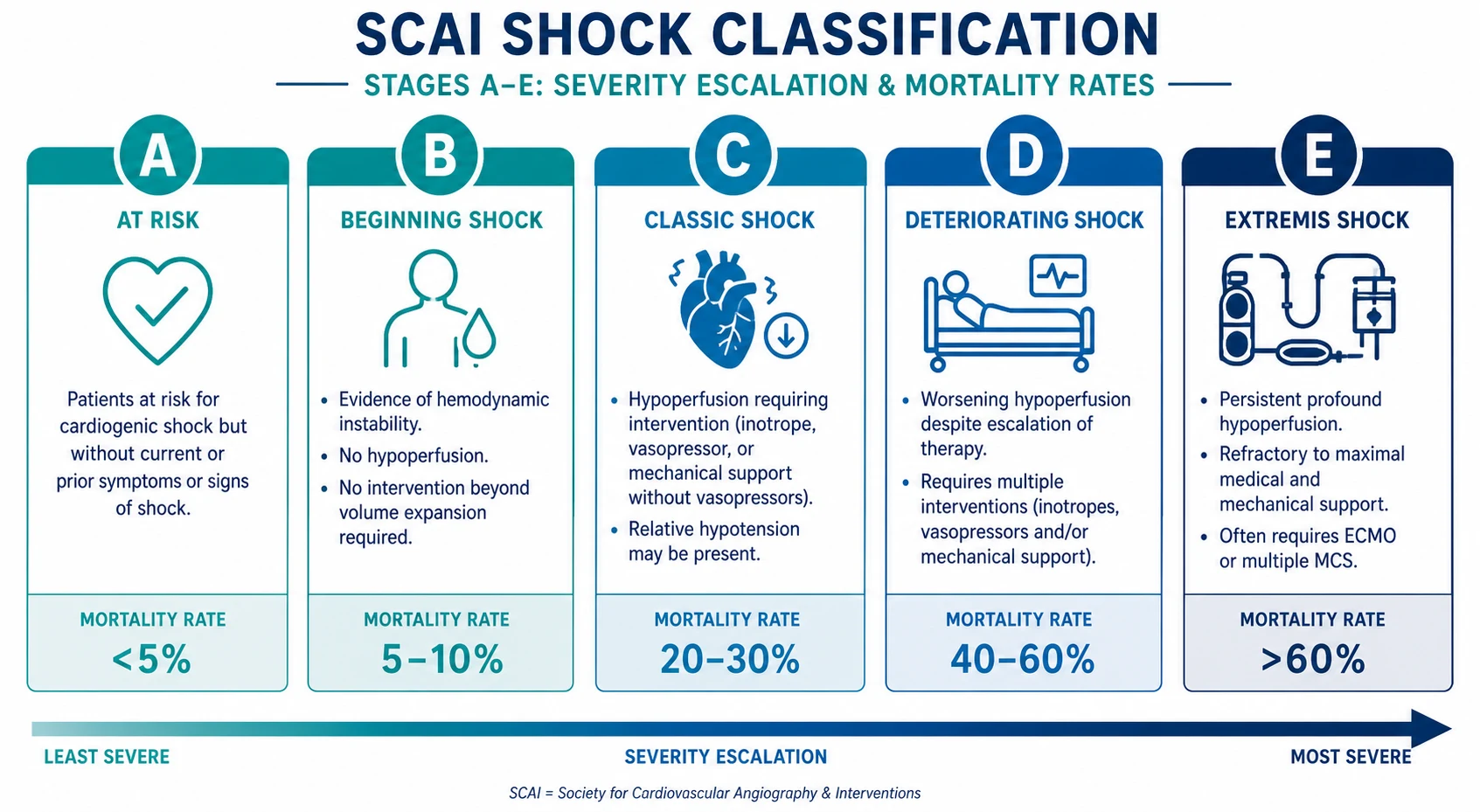
The Society for Cardiovascular Angiography and Interventions (SCAI) classification provides a standardised severity assessment:[3][10]
SCAI shock staging (click each stage)
Classic
Hypotension requiring vasoactive support (MAP <65 on agents). End-organ hypoperfusion (lactate >2, oliguria). Standard shock management protocol.
Causes
| Cause category | Examples |
|---|---|
| AMI (most common, ~80%) | LV failure (anterior STEMI), RV infarct (inferior STEMI), mechanical complication (VSD, papillary muscle rupture, free wall rupture)[14] |
| Acute decompensated heart failure | Chronic HF exacerbation, end-stage cardiomyopathy |
| Myocarditis | Giant cell, lymphocytic, fulminant |
| Valvular | Acute severe MR (papillary muscle rupture), acute severe AR, prosthetic valve thrombosis |
| Arrhythmia | Sustained VT, complete heart block, atrial fibrillation with rapid ventricular response in compromised ventricle |
| Drug toxicity | Beta-blocker overdose, calcium channel blocker overdose, anthracycline cardiotoxicity |
| Post-cardiotomy | Following cardiac surgery |
| Other | Stress cardiomyopathy (Takotsubo), pulmonary embolism (causing RV failure), peripartum cardiomyopathy |
Pathophysiology
The cardiogenic shock vicious cycle
The shock state is a self-perpetuating downward spiral:[13][10]
- Pump failure → cardiac output falls
- Blood pressure drops → compensatory sympathetic activation (tachycardia, vasoconstriction)
- Afterload increases (from vasoconstriction) → the failing ventricle cannot pump against the high SVR → stroke volume falls further
- Coronary perfusion pressure drops (diastolic BP falls) → worsening ischaemia → further pump dysfunction
- End-organ hypoperfusion → anaerobic metabolism → lactate rises → metabolic acidosis → myocardial depression → the spiral accelerates [1]
Breaking the cycle: reduce the afterload (vasodilators — risky if hypotensive), support the contractility (inotropes), support the blood pressure (vasopressors), unload the ventricle directly (MCS), and restore the coronary perfusion (revascularisation). [1]
Why noradrenaline is preferred over dopamine
The SOAP-2 trial (De Backer, NEJM 2010) randomised 1,679 patients with any shock type to noradrenaline vs dopamine. Key findings:[7]
- 28-day mortality: no significant difference (48.5% NA vs 52.5% dopamine; p=0.10)
- Arrhythmic events: significantly more with dopamine (12.4% NA vs 24.1% dopamine; p<0.001)
- Subgroup — cardiogenic shock: a trend toward higher mortality with dopamine (p=0.05)
- Conclusion: noradrenaline is preferred because dopamine causes more arrhythmias with no mortality benefit. Dopamine's theoretical "renal protective" effect (DA1 receptor → renal vasodilation) has never translated into clinical renal protection. [1]
Why VA-ECMO increases LV afterload
VA-ECMO drains venous blood and returns it retrogradely into the femoral artery (or centrally into the aorta). The retrograde flow raises aortic pressure — against which the failing LV must eject. If the LV cannot overcome this increased afterload:[11][12]
- Blood accumulates in the LV → LV distension
- Wall stress increases → subendocardial ischaemia
- Pulmonary oedema worsens (the LV cannot eject, so LA and pulmonary venous pressure rises)
- Aortic valve may not open (if ECMO flow exceeds native cardiac output) → stasis in the aortic root → thrombus formation [1]
Solution: add an Impella (ECPELLA approach) to actively drain the LV, or use a venting strategy (iatrogenic atrial septostomy, pulmonary artery vent, or surgical LV apical vent). Monitor LV size on serial echocardiography. [1]
Clinical presentation and bedside assessment
Symptoms and signs
- Hypotension: SBP <90, MAP <65 (on or off vasopressors)
- End-organ hypoperfusion:
- Skin: cold, clammy, mottled, capillary refill >3 seconds (peripheral vasoconstriction)
- Renal: oliguria (<0.5 mL/kg/hr) or anuria
- Neurological: altered mental state, agitation, confusion, reduced GCS
- Metabolic: rising lactate, worsening metabolic acidosis
- Cardiac: tachycardia (compensatory), narrow pulse pressure, pulsus alternans (alternating strong/weak pulse — severe LV failure), S3 gallop, elevated JVP (if biventricular failure)
- Respiratory: tachypnoea, pulmonary crackles (if LV failure), or clear lungs (if pure RV failure or early) [1]
Bedside assessment framework (CICM hot case approach)
When assessing a shocked patient at the bedside, use a systematic approach: [1]
- Hands: palpate — cold/clammy (vasoconstriction), warm (distributive shock)? Check capillary refill.
- Pulses: radial → femoral → carotid — assess rate, rhythm, volume. Thready pulse suggests low stroke volume.
- JVP: elevated (right heart failure, tamponade) or low/flat (hypovolaemia)?
- Precordium: displaced apex (LV dilatation), RV heave (RV pressure overload), palpable thrills (VSD/MR), auscultate for murmurs (new pansystolic murmur → papillary muscle rupture/VSD post-MI), gallop rhythm (S3).
- Lungs: bilateral crackles (pulmonary oedema) or clear? Wheeze (cardiac asthma)?
- Abdomen: hepatic congestion (tender, enlarged liver), pulsatile liver (tricuspid regurgitation), ascites.
- Legs: bilateral oedema (heart failure), asymmetric (DVT → PE), cold/pulseless (limb ischaemia from vascular device).
- Neuro: GCS, pupils, focal deficits. [1]
Formulate your impression: "This patient is in cardiogenic shock — they have hypotension, end-organ hypoperfusion evidenced by the rising lactate and oliguria, and clinical evidence of a cardiac cause (the cool peripheries, S3 gallop, and elevated JVP). My immediate priorities are to secure the airway and breathing, establish arterial and central venous access, start noradrenaline to achieve a MAP of 65, and arrange an urgent echocardiogram to determine the mechanism." [1]
Investigations and monitoring
Blood tests
| Test | Expected finding | Significance |
|---|---|---|
| Arterial blood gas | Metabolic acidosis (low pH, low HCO3), high lactate, low SvO2 | Severity of hypoperfusion; serial lactate for monitoring |
| Lactate | >2 mmol/L (often >4 in severe shock) | Key prognostic marker — clearance ≥10%/2hr = good prognosis |
| Troponin | Elevated (if AMI cause) | Confirms myocardial injury; does NOT guide management in real-time |
| BNP/NT-proBNP | Elevated | Supports cardiac cause; helps differentiate from sepsis |
| Mixed venous O2 saturation (SvO2) | <65% (low cardiac output → more O2 extraction) | Key monitoring parameter; target ≥65% |
| Renal function | Rising creatinine, AKI | End-organ damage marker |
| Liver function | Elevated ALT/AST (shock liver), coagulopathy | Hepatic hypoperfusion |
| CK | Elevated (if rhabdomyolysis or myocardial injury) | Differentiate cause |
Echocardiography (the most important investigation)
Bedside echocardiography (FoCUS/FATE protocol) is the single most important diagnostic tool in cardiogenic shock. It answers the key questions:[2]
- Is the LV systolic function impaired? (EF <30-40% suggests pump failure)
- Is there a mechanical complication? (VSD, papillary muscle rupture causing severe MR, free wall rupture causing tamponade)[14]
- Is there RV failure? (RV dilatation, McConnell's sign → PE or RV infarct)
- Is there tamponade? (Pericardial effusion with RA/RV diastolic collapse)
- Is the patient volume-responsive? (IVC assessment — collapsed IVC suggests hypovolaemia component)
Haemodynamic monitoring
| Monitoring | What it measures | Target values |
|---|---|---|
| Arterial line | Beat-to-beat BP, pulse pressure variation | MAP ≥65 mmHg (≥70 if chronic HTN) |
| CVC (central venous pressure) | RA pressure (volume status surrogate) | CVP 8-12 mmHg (not reliable alone) |
| PA catheter (Swan-Ganz) | CO/CI, PA pressure, PAOP, SVR, SvO2 | CI ≥2.2 L/min/m²; PAOP 12-18; SvO2 ≥65% |
| PiCCO/LiDCO | Continuous CO, GEDV, ITBV, EVLW | Continuous CO trending; EVLW <10 mL/kg |
| Lactate | Tissue perfusion | Clearance ≥10% over 2 hours |
| Urine output | Renal perfusion | ≥0.5 mL/kg/hr |
| ScvO2 (central venous) | O2 delivery vs consumption balance | ≥70% |
When to use a PA catheter: not routinely required (FOCUS catheter trial showed no outcome benefit), but useful in complex shock (differentiating LV vs RV failure, assessing response to MCS, guiding weaning). [1]
Management
Immediate management (first 15 minutes)
Immediate management priorities (first 15 minutes)
Airway and breathing
If respiratory failure or reduced GCS — intubate and ventilate. Lung-protective ventilation (Vt 6 mL/kg, PEEP 5-8, plateau pressure <30). Reduces work of breathing and O2 consumption of respiratory muscles.
Vascular access
Arterial line (beat-to-beat BP) + central venous catheter (vasopressor delivery, ScvO2, CVP). Essential before starting vasoactive agents.
Vasopressor — noradrenaline
First-line vasopressor. Start 0.05 mcg/kg/min, titrate to MAP >=65 mmHg. Central line only — extravasation causes necrosis. Preferred over dopamine (SOAP-2: fewer arrhythmias).
Assess volume status
Clinically (JVP, IVC on echo) + passive leg raise. Give 250 mL crystalloid only if hypovolaemia component. Avoid excessive fluid — failing LV is on flat Frank-Starling curve.
Inotrope — dobutamine
Add if hypotension persists despite vasopressor + features of low CO (cold peripheries, low SvO2, echo confirmation). Dobutamine 2.5-5 mcg/kg/min is first-choice. Milrinone if beta-blocked.
Urgent echocardiogram
Determine the mechanism (LV failure? RV failure? mechanical complication? tamponade?). The single most important investigation in cardiogenic shock.
12-lead ECG
Identify AMI, arrhythmia, or mechanical complication (new murmur + ECG changes post-MI).
Treat the cause
If AMI — urgent coronary angiography and revascularisation (SHOCK trial: early revasc improves 6-month survival). If arrhythmia — cardiovert. If mechanical complication — surgical referral.
Ongoing management
| Intervention | Details |
|---|---|
| MAP target | ≥65 mmHg (consider ≥70 if chronic hypertension) |
| Lactate monitoring | Every 2-4 hours; target clearance ≥10%/2hr |
| Urine output | Hourly; target ≥0.5 mL/kg/hr |
| SvO2/ScvO2 | Every 4-6 hours; target ≥65% (SvO2) or ≥70% (ScvO2) |
| Echocardiography | Serial assessments (every 6-12 hours or after each intervention) |
| Ventilation | Lung-protective if intubated; avoid high PEEP if RV failure (reduces venous return) |
| Renal replacement therapy | If AKI with refractory acidosis, hyperkalaemia, or fluid overload |
| Glycaemic control | Target glucose 7-10 mmol/L (avoid hypoglycaemia; no intensive insulin therapy — NICE-SUGAR) |
| Thromboprophylaxis | LMWH (unless contraindicated); mechanical prophylaxis if high bleeding risk |
| Temperature | Avoid fever (increases metabolic demand); target normothermia (36-37°C) |
| Stress ulcer prophylaxis | PPI if mechanically ventilated >48 hours or coagulopathy |
Pharmacological support
Noradrenaline
First-line vasopressor
- Dose: 0.05–1.0 mcg/kg/min
- Mechanism: α1 agonist → vasoconstriction
- Adverse: peripheral ischaemia, extravasation necrosis
- SOAP-2: fewer arrhythmias than dopamine
Adrenaline
Second-line vasopressor/inotrope
- Dose: 0.05–1.0 mcg/kg/min
- Mechanism: α1 + β1 + β2 agonist
- ↑ lactate (β2 glycolysis — NOT worsening perfusion)
- Risk: arrhythmias, ↑ myocardial O2 demand
Vasopressin
Catecholamine-sparing
- Dose: 0.01–0.04 units/min (fixed)
- Mechanism: V1 receptor (NO-independent)
- Does NOT increase lactate
- Risk: digital/mesenteric ischaemia
Dobutamine
First-choice inotrope
- Dose: 2.5–20 mcg/kg/min
- Mechanism: β1 agonist (inotropy + mild vasodilation)
- Risk: tachyarrhythmia, hypotension
- May cause hypotension without vasopressor cover
Milrinone
Non-catecholamine inotrope
- Dose: 0.125–0.75 mcg/kg/min (load 50 mcg/kg)
- Mechanism: PDE-3 inhibitor → ↑ cAMP
- Useful in beta-blocked patients (downstream of receptor)
- Risk: hypotension, thrombocytopenia
Dopamine
NOT recommended first-line
- Dose: 2.5–20 mcg/kg/min
- Mechanism: DA1/β1/α1 (dose-dependent)
- SOAP-2: significantly MORE arrhythmias than NA
- 'Renal-dose dopamine' concept is obsolete
Mechanical circulatory support
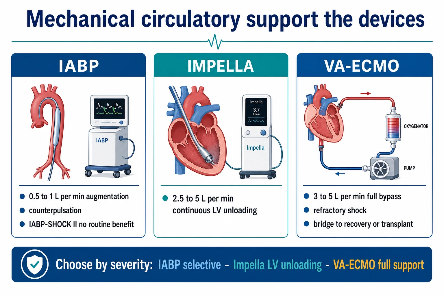
When pharmacological support fails (progressive end-organ dysfunction despite inotropes/vasopressors), mechanical circulatory support (MCS) is the next step. Three devices provide escalating levels of support:[11][12]
Intra-aortic balloon pump (IABP)
- Mechanism: balloon-tipped catheter in the descending thoracic aorta (tip at the level of L2, just distal to the left subclavian artery). Inflated in diastole (augmenting coronary perfusion pressure and systemic BP) and deflated in systole (reducing afterload and LV workload) — counterpulsation.
- Haemodynamic effect: augmentation of cardiac output by ~0.5–1.0 L/min.
- IABP-SHOCK II trial (Thiele, NEJM 2012): randomised 600 patients with ACS cardiogenic shock to IABP vs no IABP. No mortality benefit at 30 days (39.7% IABP vs 41.3% no IABP, p=0.69) or 12 months.[4]
- Current recommendation: not routinely recommended for ACS cardiogenic shock. Used selectively for: mechanical complications (VSD, MR), bridge to more advanced MCS, acute stabilisation in selected centres, or RV infarct (augments RCA perfusion in diastole).
- Contraindications: aortic regurgitation (≥moderate — diastolic balloon inflation worsens regurgitation), aortic dissection, severe peripheral vascular disease (femoral access), uncontrolled sepsis, bleeding diathesis.
- Complications: limb ischaemia (~3%), bleeding, infection, balloon leak/rupture, thrombocytopenia (shear stress), renal injury (renal artery occlusion if balloon too low).
- Timing errors: early inflation (premature — reduces SV and CO), late inflation (reduced coronary augmentation), early deflation (reduced afterload reduction), late deflation (increased afterload — balloon inflated during systole).
Impella
- Mechanism: percutaneous axial-flow micro-pump placed across the aortic valve (via femoral artery), continuously aspirating blood from the LV and expelling it into the descending aorta — active LV unloading.
- Flow rates: Impella CP 2.5 L/min; 3.5 L/min; 5.0 L/min (5.5 LD up to 5.5 L/min).[12]
- Indications: severe LV failure (cardiogenic shock), high-risk PCI support, and unloading the LV on VA-ECMO (the ECPELLA approach).[12]
- Advantages over IABP: active LV unloading (not just passive augmentation), higher flow, directly reduces LV preload and wall stress.
- Complications: haemolysis (shear stress — monitor LDH, haptoglobin, free haemoglobin), limb ischaemia, device migration/malposition, aortic valve injury, ventricular arrhythmia (catheter irritation), access site bleeding.[12]
- Weaning: reduce flow in 0.5 L/min increments; monitor lactate, urine output, BP, and echo (native LV function recovering?).
VA-ECMO (veno-arterial extracorporeal membrane oxygenation)
- Mechanism: full cardiopulmonary bypass. Drains venous blood from the femoral vein (or RA via centrall cannulation), passes it through a pump and oxygenator, and returns oxygenated blood to the femoral artery (peripheral) or aorta (central).
- Flow rates: 3–5 L/min (full circulatory support).
- Indications: refractory cardiogenic shock (Stage D/E), peri-arrest, cardiac arrest (ECPR), bridge to recovery/transplant/LVAD.[8][11]
- ECLS-SHOCK trial (Thiele, NEJM 2023): randomised patients with AMI-CS to early VA-ECMO vs no VA-ECMO. No significant reduction in 30-day composite of death/ECMO dependency (63.5% ECMO vs 61.0% control). BUT: high crossover rate (32% of control group received ECMO) limits interpretation.[8]
- Does NOT unload the LV: retrograde femoral return raises aortic pressure against which the failing LV cannot eject → LV distension → pulmonary oedema. If the LV is dilating on echo, add an Impella (ECPELLA) or use a venting strategy.[11]
- North-South syndrome (differential hypoxia): in peripheral VA-ECMO with a recovering LV, the oxygenated blood from the native lungs (returning via the LV to the aortic arch) mixes poorly with the ECMO return in the descending aorta. If native lung function is impaired (pneumonia, pulmonary oedema), the upper body (brain, heart) receives deoxygenated blood while the lower body is oxygenated by ECMO. Recognise by differential cyanosis (pink lips, blue fingers — or monitor cerebral oximetry). Solution: switch to central cannulation, axillary artery return, or veno-venous ECMO for the lungs plus VA for the heart.
- Complications:
- Limb ischaemia (~25% with peripheral cannulation): insert a distal perfusion cannula at the time of femoral arterial cannulation.
- Bleeding (~30–40%): from anticoagulation + large-bore cannulation.
- Stroke (~5%): both ischaemic and haemorrhagic.
- Haemolysis: pump shear stress (monitor LDH, plasma free haemoglobin).
- AKI: from haemolysis, microthrombi, and inflammatory response.
- Infection: line/cannula-related.
- Weaning: reduce ECMO flow in 0.5 L/min increments; assess native cardiac function on echo; if stable at 1.5 L/min for 6-12 hours with recovering echo and stable lactate/urine/BP → trial off → decannulate.
MCS device selection
IABP
0.5–1.0 L/min · Counterpulsation
- Diastolic inflation augments coronary perfusion
- Systolic deflation reduces afterload
- IABP-SHOCK II: NO routine benefit in ACS-CS
- Use selectively: VSD, RV infarct, bridge
- Contraindicated: aortic regurgitation, dissection
Impella
2.5–5.0 L/min · Active LV unloading
- Axial-flow pump across the aortic valve
- Actively drains LV → reduces preload & wall stress
- Indicated: severe LV failure, high-risk PCI
- ECPELLA: unloads LV on VA-ECMO
- Risk: haemolysis, limb ischaemia, migration
VA-ECMO
3–5 L/min · Full bypass
- Provides systemic flow AND oxygenation
- Does NOT unload LV (retrograde flow increases afterload)
- ECLS-SHOCK: no benefit in early CS
- Risk: limb ischaemia (25%), bleeding, stroke
- Bridge to recovery/decision/transplant/LVAD
Evidence and landmark trials
SHOCK
NEJM 1999
302 pts with AMI-CS — early revascularisation vs initial medical stabilisation
Key finding
6-month mortality: 50% early revasc vs 63% delayed (p=0.027)
Practice change
Early revascularisation became the standard of care in AMI-CS
IABP-SHOCK II
NEJM 2012
600 pts with ACS-CS — IABP vs no IABP
Key finding
30-day mortality: 39.7% vs 41.3% (p=0.69) — NO benefit
Practice change
Routine IABP no longer recommended in ACS cardiogenic shock
CULPRIT-SHOCK
NEJM 2017
1075 pts with AMI-CS — culprit-only PCI vs immediate multivessel PCI
Key finding
30-day death/severe renal failure: 45.9% culprit vs 55.4% multivessel (p=0.01)
Practice change
Culprit-only PCI preferred over multivessel in AMI-CS
SOAP-2
NEJM 2010
1679 pts with any shock — noradrenaline vs dopamine
Key finding
Mortality: no difference (p=0.10). Arrhythmias: 12.4% NA vs 24.1% dopamine (p<0.001)
Practice change
Noradrenaline preferred over dopamine (fewer arrhythmias)
ECLS-SHOCK
NEJM 2023
420 pts with AMI-CS — early VA-ECMO vs no VA-ECMO
Key finding
30-day composite death/ECMO dependency: 63.5% vs 61.0% (not significant)
Practice change
Early routine VA-ECMO not recommended (but 32% crossover limits interpretation)
Complications and troubleshooting
| Problem | Recognition | Management |
|---|---|---|
| Limb ischaemia (ECMO/IABP) | Cold/pulseless leg, pain, mottling | Insert distal perfusion cannula; reduce device flow if possible; vascular surgery review; fasciotomy if compartment syndrome |
| Bleeding (anticoagulation + cannulation) | Dropping Hb, visible bleeding, ∆Hb >2 g/dL/24hr | Reduce/hold anticoagulation; transfuse (target Hb ≥70 g/L); surgical control; consider heparin-free circuit if life-threatening |
| Haemolysis (Impella/ECMO pump) | ↑ LDH, ↓ haptoglobin, ↑ bilirubin, haemoglobinuria, ∆Hb | Check circuit for clot/malposition; consider device exchange; renal protection (maintain urine output) |
| LV distension (VA-ECMO) | Echo: dilating LV, worsening pulmonary oedema, aortic valve not opening | Add Impella (ECPELLA); or venting strategy; reduce ECMO flow if tolerated |
| North-South syndrome (peripheral VA-ECMO) | Differential cyanosis (SpO2 right hand < legs); low cerebral oximetry | Switch to central/axillary cannulation; consider VV-ECMO addition |
| Thromboembolism | Stroke, limb/DVT, circuit clot | Optimise anticoagulation (target ACT 1.5x baseline or anti-Xa 0.3-0.7 IU/mL); circuit inspection |
| Infection (lines/cannulae) | Fever, ↑ WBC/CRP, positive cultures | Remove/change lines; empirical antibiotics; source control |
| Renal failure | Rising creatinine, oliguria | Optimise haemodynamics; RRT (CVVHDF); avoid nephrotoxins |
Communication and ethics
Family discussion framework
- Set the scene: quiet room, sit down, all team members present.
- Explain the diagnosis: "Your loved one is critically ill. The heart is not pumping enough blood to the organs — this is called cardiogenic shock."
- Explain the plan: "We are using medications to support the blood pressure and the heart. If these are not enough, we may need mechanical support devices that temporarily take over the heart's work."
- Discuss prognosis honestly: "Even with the best treatment, approximately half of patients with this condition do not survive."
- Discuss escalation limits: "If the heart does not recover, we need to consider whether ongoing aggressive support is appropriate, or whether we should focus on comfort and dignity."
- Involve palliative care early if the trajectory is poor or if withdrawal of life-sustaining therapy (WLST) is being considered.
- Document all discussions in the medical record. [1]
Organ donation
If the patient progresses to brain death or WLST is planned:
- Make an early referral to the donation team (before withdrawal for DCD — donation after circulatory death).
- The treating team should be separate from the donation team (conflict of interest).
- Discuss donation at an appropriate time and in a supportive manner. [1]
Prognosis
- Overall mortality: 40–60% (has improved with early revascularisation from >80% in the pre-SHOCK era).[1][2]
- By SCAI stage: A ~5%, B ~10%, C ~25–35%, D ~50–70%, E ~80–95%.[3]
- Prognostic factors: age, comorbidities, lactate clearance, time to revascularisation, SCAI stage at presentation, need for MCS.[10]
- Recovery: LV function may recover over days to weeks (stunning resolves); some require durable LVAD or transplant.
Exam practice
SAQ — AMI cardiogenic shock
10 minutes · 10 marks
A 65-year-old man is admitted 8 hours after an anterior STEMI. He had primary PCI with stenting of the proximal LAD. He is now intubated and ventilated. BP 78/45 (MAP 56), HR 110 (sinus), SpO2 95% on FiO2 0.6. Lactate 4.2 mmol/L. Urine output 15 mL/hr. On noradrenaline 0.3 mcg/kg/min. Echo: EF 25%, dilated LV, no mechanical complication.
Sample Viva 1 — Data interpretation
Examiner: "This is the arterial waveform from a patient on an IABP. Describe what you see and explain the timing." [1]
Expected response: "This is an IABP arterial waveform showing diastolic augmentation. The balloon inflates at the dicrotic notch (onset of diastole), producing a sharp rise in diastolic pressure (the augmentation peak) that exceeds systolic pressure. This augments coronary perfusion during diastole. The balloon then deflates rapidly at the onset of the next systole, producing a reduction in end-diastolic pressure (afterload reduction) and a slightly lower systolic pressure than the unaugmented beat. The net effects are: increased coronary perfusion pressure, reduced LV afterload, and a modest increase in cardiac output (0.5–1 L/min)." [1]
Follow-up: "What are the contraindications to IABP?" [1]
Response: "Moderate-to-severe aortic regurgitation (the diastolic balloon inflation would worsen the regurgitant volume), aortic dissection (the balloon could extend the dissection flap), and severe peripheral vascular disease (femoral access would be difficult and risk limb ischaemia)." [1]
Sample Viva 2 — Equipment
Examiner: "Explain the mechanism of action of the Impella device and its key advantage over the IABP." [1]
Expected response: "The Impella is a percutaneous axial-flow micro-pump placed across the aortic valve via the femoral artery. It continuously aspirates blood from the left ventricle and expels it into the ascending aorta, providing active left ventricular unloading at flow rates of 2.5–5.0 L/min depending on the model. Its key advantage over the IABP is that it provides active LV unloading — it directly reduces LV preload, LV end-diastolic volume, and wall stress — whereas the IABP only provides passive counterpulsation with a modest 0.5–1.0 L/min effect. The Impella is particularly useful for unloading the LV in patients on VA-ECMO, where the retrograde ECMO flow can worsen LV distension — this combination is called ECPELLA." [1]
Clinical pearls
Red flags
References
- [1]van Diepen S, Katz JN, Albert NM, et al. Cardiogenic shock Lancet, 2024.PMID 39550175
- [2]Samsky MD, Morrow DA, Proudfoot AG, et al. Cardiogenic Shock After Acute Myocardial Infarction: A Review JAMA, 2021.PMID 34751704
- [3]Baran DA, Grines CL, Bailey S, et al. SCAI clinical expert consensus statement on the classification of cardiogenic shock: This document was endorsed by the American College of Cardiology (ACC), the American Heart Association (AHA), the Society of Critical Care Medicine (SCCM), and the Society of Thoracic Surgeons (STS) in April 2019 Catheter Cardiovasc Interv, 2019.PMID 31104355
- [4]Thiele H, Zeymer U, Neumann FJ, et al. Intraaortic balloon support for myocardial infarction with cardiogenic shock N Engl J Med, 2012.PMID 22920912
- [5]Thiele H, Akin I, Sandri M, et al. PCI Strategies in Patients with Acute Myocardial Infarction and Cardiogenic Shock N Engl J Med, 2017.PMID 29083953
- [6]Hochman JS, Sleeper LA, Webb JG, et al. Early revascularization in acute myocardial infarction complicated by cardiogenic shock. SHOCK Investigators. Should We Emergently Revascularize Occluded Coronaries for Cardiogenic Shock N Engl J Med, 1999.PMID 10460813
- [7]De Backer D, Biston P, Devriendt J, et al. Comparison of dopamine and norepinephrine in the treatment of shock N Engl J Med, 2010.PMID 20200382
- [8]Thiele H, Zeymer U, Neumann FJ, et al. Extracorporeal Life Support in Infarct-Related Cardiogenic Shock N Engl J Med, 2023.PMID 37634145
- [9]Toda N, Taguchi I, Toda R, et al. Inotrope and vasopressor use in cardiogenic shock: what, when and why? Curr Opin Crit Care, 2022.PMID 35792520
- [10]Sarma D, Jentzer JC, et al. Cardiogenic Shock: Pathogenesis, Classification, and Management Crit Care Clin, 2024.PMID 37973356
- [11]Enger T, Philipp A, et al. Mechanical circulatory support in cardiogenic shock J Intensive Care, 2023.PMID 38115065
- [12]Masiero G, Arturi F, et al. Mechanical Circulatory Support with Impella: Principles, Evidence, and Daily Practice J Clin Med, 2024.PMID 39200728
- [13]Kislitsina ON, Rich JD, et al. Shock - Classification and Pathophysiological Principles of Therapeutics Curr Cardiol Rev, 2019.PMID 30543176
- [14]Gong FF, Vaitenas I, et al. Mechanical Complications of Acute Myocardial Infarction: A Review JAMA Cardiol, 2021.PMID 33295949