ICU · Cardiovascular / cardiomyopathy
Cardiomyopathies & Myocarditis
Also known as Cardiomyopathy · Dilated cardiomyopathy · Hypertrophic cardiomyopathy · Restrictive cardiomyopathy · Arrhythmogenic right ventricular cardiomyopathy · ARVC · Takotsubo cardiomyopathy · Stress cardiomyopathy · Broken heart syndrome · Myocarditis · Giant cell myocarditis · Eosinophilic myocarditis · Fulminant myocarditis · Cardiac amyloidosis · SAM · Systolic anterior motion · LVOT obstruction · Mavacamten · Tafamidis · Cardiac MRI late gadolinium enhancement · Lake Louise criteria · Dallas criteria · HCM Risk-SCD
The four cardiomyopathies are dilated (the commonest — an enlarged LV with a reduced EF, treated with heart-failure therapy), hypertrophic (asymmetric septal LVH with a dynamic LVOT obstruction and a risk of sudden cardiac death — treat with beta-blockers, avoid vasodilators, an ICD for prevention), restrictive (amyloid, sarcoid — stiff ventricles with impaired filling, treated for the cause), and arrhythmogenic (a genetic right-ventricular cardiomyopathy). Takotsubo is a transient stress-mediated apical ballooning that mimics an anterior STEMI. Myocarditis is an inflammation of the myocardium (most commonly viral — coxsackie, parvovirus, SARS-CoV-2), presenting with chest pain, arrhythmia, heart failure, and a rising troponin with normal coronary arteries. The cardiac MRI (mid-wall late gadolinium enhancement) and the biopsy (the Dallas criteria) confirm. Treatment is supportive; most recover, but giant-cell and a fulminant case may need immunosuppression, mechanical support or a transplant.
On this page & tools
Your progress
Saved locally on this device.
Target exams
Red flags
Overview & definition
The cardiomyopathies are diseases of the heart muscle, classified into four morphological types — dilated (the commonest), hypertrophic, restrictive, and arrhythmogenic right ventricular — to which Takotsubo (stress) cardiomyopathy is often added as a fifth, reversible, mimic. Myocarditis is an inflammation of the myocardium (most commonly viral) that can mimic an ACS and may progress to a dilated cardiomyopathy.[1][1]
The 2023 ESC cardiomyopathy guideline reframes these around a phenotype-first, genotype-driven approach: define the phenotype (dilated, hypertrophic, restrictive, non-compaction, ARVC) from imaging, then hunt systematically for the cause (genetic, acquired), and cascade-screen first-degree relatives.[1]
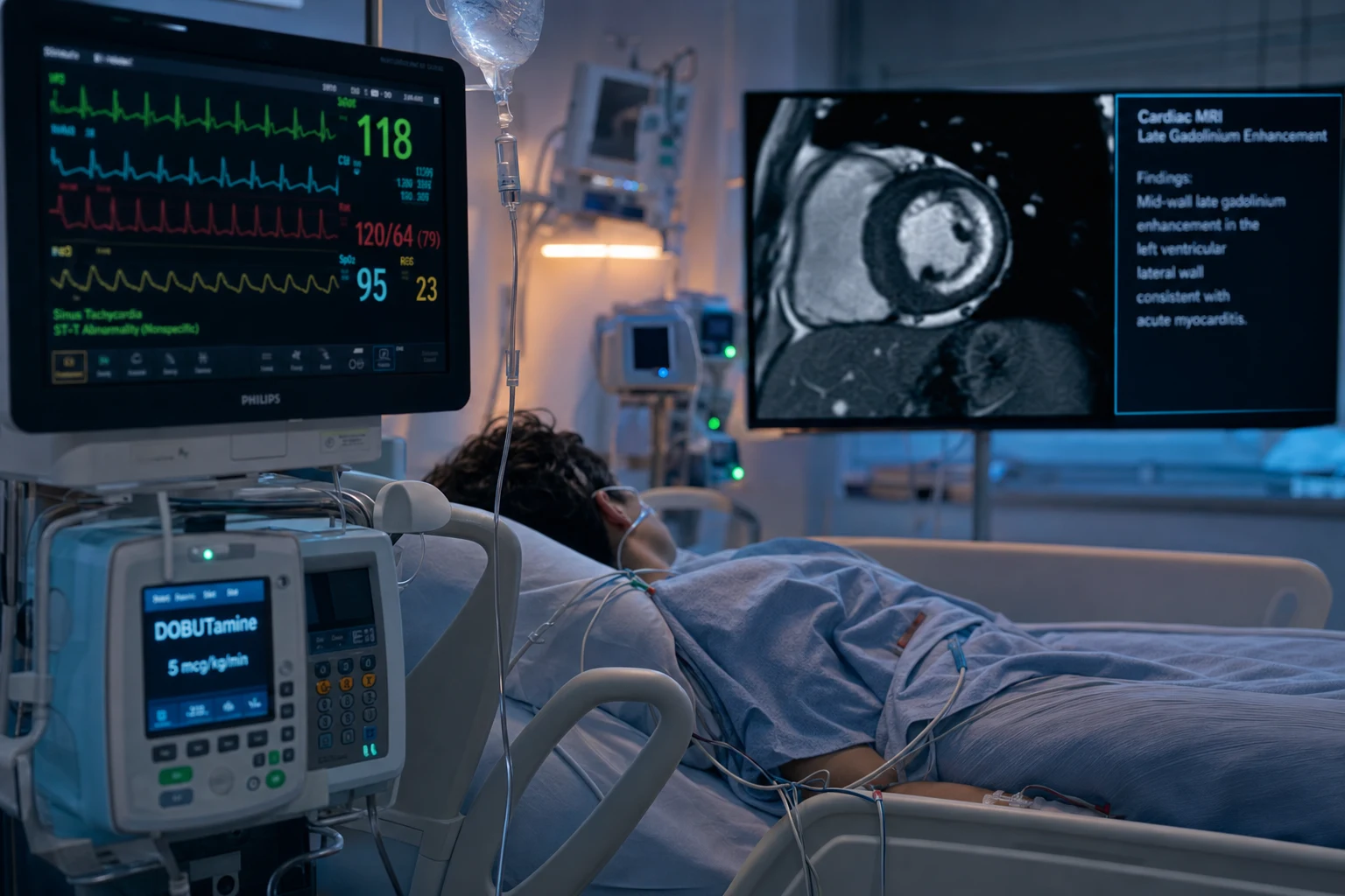
The phenotype-first approach (2023 ESC)[1]
| Phenotype | Hallmark on imaging | Commonest cause to seek |
|---|---|---|
| Dilated | LV dilation, EF <40% | Genetic (30-40%), post-viral, toxic, peripartum |
| Hypertrophic | LV wall ≥15 mm, asymmetric septum | Sarcomeric gene mutation (MYH7, MYBPC3) |
| Restrictive | Bilateral atrial enlargement, normal/slightly reduced EF, diastolic dysfunction | Amyloidosis (AL/ATTR), sarcoid, storage disease |
| Arrhythmogenic (ARVC) | RV regional akinesia/dyskinesia, RV dilation | Desmosomal genes (PKP2, DSG2, DSC2) |
| Non-compaction | Prominent trabeculae, deep intertrabecular recesses | Genetic (MYH7, TAZ) |
Dilated cardiomyopathy (DCM)
The commonest cardiomyopathy. An enlarged, thin-walled LV with a reduced ejection fraction (a systolic failure) — LVEDD usually >6 cm with an EF <40%.[1][6]
Causes (the mnemonic "ABCD GET SMASHED" is overkill — learn the high-yield six):[1][6]
- Genetic — a family history in 30-40 per cent of "idiopathic" cases; autosomal dominant truncating variants in titin (TTNtv) are the single commonest genetic cause (found in ~15-20 per cent of familial and ~10 per cent of sporadic DCM). Truncate screening of first-degree relatives is now standard.[1]
- Post-viral myocarditis — the progression to DCM (the "burnt-out" myocarditis); a viral prodrome weeks to months earlier is the clue.
- Toxic — alcohol (a direct myocardial toxin; >7-8 drinks/day over ≥5 years), anthracycline chemotherapy (doxorubicin — dose-dependent, cumulative, via iron-mediated free-radical injury; dexrazoxane is cardioprotective; the newer anti-HER2 trastuzumab causes a usually-reversible dysfunction), cocaine/methamphetamines, and clozapine (myocarditis/cardiomyopathy within the first 8 weeks).
- Peripartum — the last month of pregnancy to 5 months postpartum; the diagnosis requires the timing plus an EF <45%; higher rates in pre-eclampsia, older maternal age, and multiparity. It recovers more often than other DCMs (≈50-70 per cent recover LV function by 12 months with GDMT). A subsequent pregnancy carries a recurrence risk of ~30-50 per cent if the EF has not normalised.
- Tachycardia-induced — chronic AF/SVT with a rate >110-130 for weeks-months; the EF recovers with rate control — the only DCM that is essentially curable.
- Endocrine/nutritional — thyrotoxicosis, hypothyroidism, selenium deficiency (Keshan disease in parts of China), carnitine deficiency, thiamine (beriberi — high-output).
- Idiopathic — a diagnosis of exclusion after the above are excluded; ~30 per cent.
Pathophysiology: the cardinal defects are loss of myocytes (necrosis, apoptosis), interstitial fibrosis, and adverse remodelling — leading to wall thinning, chamber dilation, reduced contractility, and functional mitral/tricuspid regurgitation from annular dilation. Neurohormonal activation (RAAS, sympathetic) drives progressive remodelling — which is exactly what GDMT blocks. [1]
Investigations:
- Echocardiography — the first test: LV dilation, global or regional hypokinesia, reduced EF, functional MR. Strain imaging (reduced global longitudinal strain) is more sensitive than EF.
- ECG — non-specific; may show AF, LBBB (a CRT consideration), low voltage, or Q waves (mimicking old infarction).
- Cardiac MRI — the most informative single test: confirms LV/RV size and function, shows mid-wall LGE (the non-ischaemic pattern, typically in the septum — distinguishes from the subendocardial LGE of infarction), and identifies myocarditis or sarcoid patterns. Should be done in every new "idiopathic" DCM.[1]
- Coronary angiography — to exclude ischaemia as the cause (ischaemic vs non-ischaemic DCM).
- Genetic testing — multigene panel; cascade screening of first-degree relatives with clinical assessment + echo ± genetics.[1]
Management — the four pillars of GDMT (+ ARNI, loop diuretic, devices):[1][6]
- ACE inhibitor / ARB / ARNI — ARNI (sacubitril/valsartan) preferred over ACEi in HFrEF (PARADIGM-HF); use ACEi first then switch once stable.
- Beta-blocker — one of the three mortality-reducing ones: bisoprolol, carvedilol, or nebivolol (metoprolol tartrate is NOT — use succinate, but evidence base is the three).
- Mineralocorticoid receptor antagonist — spironolactone or eplerenone (for symptomatic HFrEF or post-MI EF <30).
- SGLT2 inhibitor — dapagliflozin or empagliflozin (DAPA-HF, EMPEROR-Reduced) — now first-line, mortality- and hospitalisation-reducing, irrespective of diabetes.
- Loop diuretic — furosemide/bumetanide for congestion relief (symptomatic only; no mortality benefit).
- Device therapy (for selected patients): ICD for the primary prevention of sudden cardiac death (EF ≤35%, NYHA II-III, ≥3 months of optimal GDMT, ≥1-year survival expectation); CRT (biventricular pacemaker) for QRS ≥150 ms with LBBB morphology.
- Advanced — LVAD (destination or bridge-to-transplant) or cardiac transplantation for refractory end-stage disease.
- Anticoagulation — for an LV thrombus, prior embolus, or AF; NOT routinely for low EF alone (no net benefit in sinus rhythm).
Prognosis: 5-year mortality ~20 per cent on modern GDMT (historically ~50 per cent); reversible causes (peripartum, tachycardia-induced, alcoholic with abstinence) do better. The predictors of a poor outcome are a low EF, RV dysfunction, advanced NYHA class, high NT-proBNP, and renal dysfunction.[6]
Hypertrophic cardiomyopathy (HCM)
A genetic sarcomeric disease (most commonly a beta-myosin heavy chain MYH7 or myosin-binding protein C MYBPC3 mutation) producing an asymmetric septal hypertrophy (wall ≥15 mm, or ≥13 mm in a first-degree relative) — inherited autosomal dominant, prevalence ~1 in 500.[1][2][4]
The pathophysiology of the dynamic LVOT obstruction: the thickened septum narrows the LV outflow tract, and the systolic anterior motion (SAM) of the mitral valve (the anterior leaflet is pulled into the narrowed outflow tract by the Venturi effect) worsens the obstruction and produces a mitral regurgitation. The dynamic LVOT obstruction — it varies with the preload, the afterload, and the contractility — is the hallmark. The obstruction is present at rest in ~one-third, labile/latent in one-third (provokable by Valsalva, standing, amyl nitrite, or exercise echo), and absent in one-third.[2]
The "four D's" that worsen the LVOT gradient — and the four that relieve it (an exam favourite): [1]
Worsens obstruction (AVOID)
↑ gradient
- ↓ Preload — dehydration, diuretics, Valsalva, standing, nitroglycerin
- ↓ Afterload — vasodilators (GTN), sepsis, anaesthesia-induced vasodilation
- ↑ Contractility — inotropes (dobutamine, digoxin), exercise, tachycardia
- ↑ SAM — anything that increases the contractile state of the hyperdynamic LV
Relieves obstruction (USE)
↓ gradient
- ↑ Preload — fluids, squatting, leg-raising, beta-blockade-induced slower filling
- ↑ Afterload — pure alpha-agonists (phenylephrine, metaraminol)
- ↓ Contractility — beta-blockers, disopyramide, mavacamten
- ↑ LV filling time — rate control (the slower the rate, the more filling, the less SAM)
Clinical features:
- The murmur — a crescendo-decrescendo systolic ejection murmur at the left sternal edge, radiating to the apex (not the neck), that intensifies with Valsalva/standing (less preload) and softens with squatting/handgrip (more preload/afterload). The opposite of aortic stenosis (which softens with Valsalva).
- Symptoms — exertional dyspnoea (the commonest, from diastolic dysfunction and/or MR), angina (despite clean coronaries — microvascular dysfunction, increased muscle mass), pre-syncope/syncope (the LVOT gradient, arrhythmia), and sudden cardiac death — HCM is the commonest cause of SCD in the young athlete.[4]
Risk stratification for sudden death — the HCM Risk-SCD score:[3] a validated model (O'Mahony 2014) estimating 5-year SCD risk from age, maximal wall thickness, LA diameter, LVOT gradient, family history of SCD, NSVT, and unexplained syncope. An estimated 5-year risk ≥6 per cent warrants an ICD for primary prevention; 4-6 per cent is a shared decision; <4 per cent generally not. The 2020 AHA/ACC guideline supplements this with major risk factors (massive LVH ≥30 mm, family history of ≥1 SCD <50, unexplained syncope, NSVT, abnormal BP response).[2]
- Beta-blockers (the first-line — they reduce the contractility and the heart rate, lowering the LVOT gradient). Non-dihydropyridine calcium-channel blockers (verapamil/diltiazem) are second-line (caution: avoid in heart block, severe gradient where the negative inotropy can transiently worsen filling).
- Disopyramide — a class Ia antiarrhythmic with strong negative inotropy, added for refractory symptoms/gradient.
- Mavacamten — a first-in-class cardiac myosin inhibitor that reduces actin-myosin cross-bridging, lowering the gradient; EXPLORER-HCM showed it improved symptoms and reduced the LVOT gradient in symptomatic obstructive HCM.[5]
- Avoid vasodilators and inotropes — they reduce the afterload and the preload, worsening the dynamic obstruction (the LVOT narrows further). This is the opposite of the dilated cardiomyopathy management.
- Avoid dehydration — maintain the preload (the LVOT is less obstructed with a fuller LV).
- Septal reduction therapy for the refractory symptomatic patient with a gradient ≥50 mmHg: surgical septal myectomy (the gold standard, performed at high-volume centres) or alcohol septal ablation (a percutaneous alternative inducing a controlled septal infarct; reserved for those unsuitable for surgery, higher pacemaker dependency).
- An ICD for the primary prevention of sudden cardiac death (HCM is the commonest cause of sudden cardiac death in the young athlete; risk stratify with the HCM Risk-SCD score).[3]
Restrictive cardiomyopathy
Stiff, non-compliant ventricles that cannot relax and fill in diastole (a diastolic failure) — small ventricles with massively enlarged atria, a near-normal EF, and high filling pressures.[1]
Causes:[1]
- Amyloidosis — the commonest and most important: AL (light-chain, from a plasma-cell dyscrasia; multisystem — macroglossia, periorbital purpura, nephrotic-range proteinuria) and ATTR (transthyretin; wild-type/senile in elderly men, or hereditary from a TTR mutation). The red-flag clue on echo is a thick LV with low voltages on the ECG (the "discordance" between echo and ECG) and a global longitudinal strain with apical sparing ("cherry-on-the-top" pattern) on strain imaging.[7]
- Sarcoidosis — granulomatous infiltration; may cause heart block, ventricular arrhythmia, and a sarcoid cardiomyopathy. Cardiac MRI shows focal LGE in a basal inferolateral/subepicardial distribution; PET-CT shows active inflammation.
- Eosinophilic (Loeffler endocarditis) — eosinophilic myocarditis with mural thrombus and endocardial fibrosis; in the tropics (endomyocardial fibrosis) or with hypereosinophilic syndrome, Churg-Strauss, or parasitic infestation.
- Storage diseases — haemochromatosis (iron; also a DCM phenotype), Fabry disease (alpha-galactosidase A deficiency; an X-linked mimicker of HCM with concentric LVH), glycogen storage diseases (Danon, Pompe).
- Radiation — a late consequence of mediastinal radiotherapy (years to decades); may cause concomitant constriction, valvular disease, and coronary ostial disease.
- Idiopathic.
The cardiac amyloidosis workup (the workflow examiners love):
- Index of suspicion — elderly man with "hypertension" that has become refractory, heart failure with preserved EF, carpal tunnel syndrome (often bilateral, often preceding the cardiac disease by years), biceps tendon rupture (the "Popeye sign"), spinal stenosis, and orthostatic hypotension.
- ECG + echo discordance — thick LV on echo but low voltages on ECG (especially in AL).
- Strain imaging — apical sparing pattern.
- Scintigraphy (the non-biopsy route for ATTR) — ^99mTc-DPD, PYP or HMDP uptake grade 2-3, with no monoclonal protein on serum/urine immunofixation, makes ATTR without biopsy (Perugini criteria — avoid the endomyocardial biopsy for ATTR).
- Serum/urine immunofixation + serum free light chains — for AL amyloid.
- Endomyocardial biopsy — the gold standard for AL when scintigraphy is not diagnostic (Congo red apple-green birefringence). [1]
Treatment of cardiac amyloidosis (ATTR-ACT changed the landscape):[7]
- Tafamidis — a transthyretin stabiliser that slows ATTR progression (ATTR-ACT, Maurer 2018, reduced all-cause mortality and cardiovascular hospitalisations in ATTR-CM).[7]
- AL amyloid — treat the underlying plasma-cell dyscrasia with chemotherapy/bortezomib ± autologous stem-cell transplant (haematology-led).
- Heart failure — cautious diuretics (the stiff ventricles are preload-dependent — over-diuresis causes a catastrophic fall in cardiac output); AVOID ACE inhibitors/ARBs/ARNI (often intolerant — hypotension), calcium-channel blockers (negatively inotropic, bind amyloid fibrils), and digoxin (binds amyloid fibrils → toxicity).
- Anticoagulation for atrial fibrillation (high thromboembolic risk).
Sarcoid cardiomyopathy — corticosteroids (prednisolone 0.5-1 mg/kg) ± steroid-sparing agents (methotrexate) for active inflammation; an ICD (even primary prevention) for the arrhythmic risk, which is disproportionate to the EF.[1]
The key distinction — restriction vs constriction: restrictive cardiomyopathy is distinguished from constrictive pericarditis (both present with right heart failure and equalised diastolic pressures — see the pericardial-disease topic). Restriction has a normal pericardium and a thick, infiltrated LV (amyloid on the MRI or the biopsy); constriction has a thickened pericardium and a normal LV.[1]
Restrictive cardiomyopathy
Myocardial disease
- Normal/thin pericardium on CT/MRI
- Thick, infiltrated LV (amyloid, sarcoid); large atria
- Equalised diastolic pressures (LVEDP ≈ RVEDP, but the dip-and-plateau is blunted)
- NO pericardial knock; prominent S3, Kussmaul sign may be present
- NO septal bounce; preserved tissue Doppler e' (unless amyloid)
- Treatment: medical — treat cause (tafamidis, chemotherapy); cautious diuretics
Constrictive pericarditis
Pericardial disease
- Thickened pericardium (≥3-4 mm) on CT/MRI (though thickness can be normal in ~20%)
- Normal LV; tethered, small ventricles; large atria
- Equalised diastolic pressures; pronounced dip-and-plateau ("square-root sign")
- Pericardial knock; prominent Kussmaul sign; paradoxical septal motion
- Septal BOUNCE on echo (ventricular interdependence) — the key discriminator
- Treatment: surgical pericardiectomy (curative)
Management: treat the underlying cause (amyloid — treat the underlying plasma cell dyscrasia or the ATTR; sarcoid — corticosteroids). The heart failure is diastolic — cautious diuretics, avoid over-diuresis (the stiff ventricles are preload-dependent).[1]
Arrhythmogenic right ventricular cardiomyopathy (ARVC)
A genetic desmosomal cardiomyopathy (most commonly PKP2 encoding plakophilin-2; also DSG2, DSC2, JUP) in which the right-ventricular myocardium is progressively replaced by fibrofatty tissue — leading to RV dilation, regional akinesia/dyskinesia, ventricular arrhythmia, and a risk of sudden death. Autosomal dominant, prevalence ~1 in 2000-5000.[1][1]
The 2010 revised Task Force Criteria diagnose ARVC from a combination of:[1]
- Imaging — RV regional akinesia/dyskinesia/aneurysm + RV dilation/dysfunction (echo, MRI, or angiography).
- Cardiac MRI — RV regional wall-motion abnormalities + fibrosis (late gadolinium enhancement); fatty infiltration is suggestive but hard to confirm reliably.
- Biopsy — residual myocytes <60 per cent by area with fibrous replacement ± fat (endomyocardial biopsy).
- ECG repolarisation — T-wave inversion in V1-V3 (without RBBB).
- ECG depolarisation — the epsilon wave (a small deflection at the end of the QRS in V1-V3 — a near-pathognomonic but late sign), late potentials on signal-averaged ECG, a prolonged terminal activation duration.
- Arrhythmia — NSVT or sustained VT of left-bundle-branch-block morphology (RV origin) with a superior axis.
- Family history / genetics — a confirmed disease-causing desmosomal mutation.
Management:[1]
- Sports restriction — competitive and endurance sport is prohibited (it accelerates the disease and precipitates arrhythmia).
- Beta-blocker first-line for symptomatic arrhythmia.
- ICD for the secondary prevention of VF/VT and for primary prevention in high-risk phenotypes (sustained VT, syncope, extensive RV involvement).
- Catheter ablation for recurrent VT (often recurrent at a new focus — the disease is diffuse).
- Heart failure therapy for the biventricular failure of advanced disease; transplantation for end-stage.
Takotsubo (stress) cardiomyopathy
A transient, reversible regional systolic dysfunction, typically apical ballooning, triggered by intense emotional or physical stress ("broken-heart syndrome"), in the absence of a culprit coronary occlusion.[12][13]
Pathophysiology: catecholamine surge (the apex is rich in beta-receptors and more vulnerable to calcium overload and microvascular spasm); the basal hyperkinesis squeezes against a stunned apex, producing the characteristic akinesia of the apex with hyperkinesis of the base (the octopus-pot shape, tako-tsubo).[12]
The Mayo/InterTAK diagnostic criteria (all four required):[13]
- Transient regional wall-motion abnormality (apical, mid-ventricular, basal, or focal) that usually extends beyond a single epicardial vascular territory.
- No obstructive epicardial coronary disease (or acute plaque rupture) on angiography.
- A new ECG abnormality (ST elevation, T-wave inversion, or QTc prolongation) or a modest troponin rise (disproportionately small compared with the wall-motion abnormality).
- The syndrome is not explained by myocarditis or pheochromocytoma.
Presentation: post-menopausal women (>90 per cent) hours-days after a trigger (grief, sepsis, surgery, subarachnoid haemorrhage, an asthma attack, or even a surprise party); chest pain, dyspnoea, syncope, or an arrhythmia. The InterTAK registry (Templin 2015) showed an in-hospital mortality of ~4 per cent and serious complications in ~20 per cent — it is NOT always benign.[12]
The variants — not all are apical:
- Apical ballooning (~80 per cent) — the classic.
- Mid-ventricular — a "mitral systolic motion" variant; can co-apical-ballooning with HCM-like SAM and an LVOT gradient.
- Basal (reverse Takotsubo) — basal akinesia, hyperkinetic apex.
- Focal. [1]
Complications that demand ICU vigilance:
- LV apical thrombus (stasis in the akinetic apex) — anticoagulate during the acute phase (echo to detect; anticoagulate for ~3 months until the LV recovers).
- LVOT obstruction (from the basal hyperkinesis + SAM) — avoid inotropes; treat with a beta-blocker and fluids, as in HCM.
- Acute heart failure and cardiogenic shock.
- Arrhythmia — Torsades de pointes (the QTc is markedly prolonged), VT/VF, high-grade AV block.
- Free-wall rupture (rare but lethal; typically day 3-7). [1]
Management:[13]
- Supportive — treat as acute heart failure; diuretics and nitrates for pulmonary oedema (provided there is no LVOT obstruction — echo first).
- ACE inhibitor/ARB, beta-blocker — conventional, though beta-blocker benefit is debated; most continue it for 3-6 months then stop once the EF normalises.
- Anticoagulation for an LV thrombus or severe apical akinesia.
- Avoid inotropes if there is an LVOT gradient; if shock with NO LVOT obstruction, cautious low-dose inotrope or MCS.
- Recovery within weeks to months in the majority (~95 per cent recover the EF); recurrence ~5-10 per cent over years.
Myocarditis
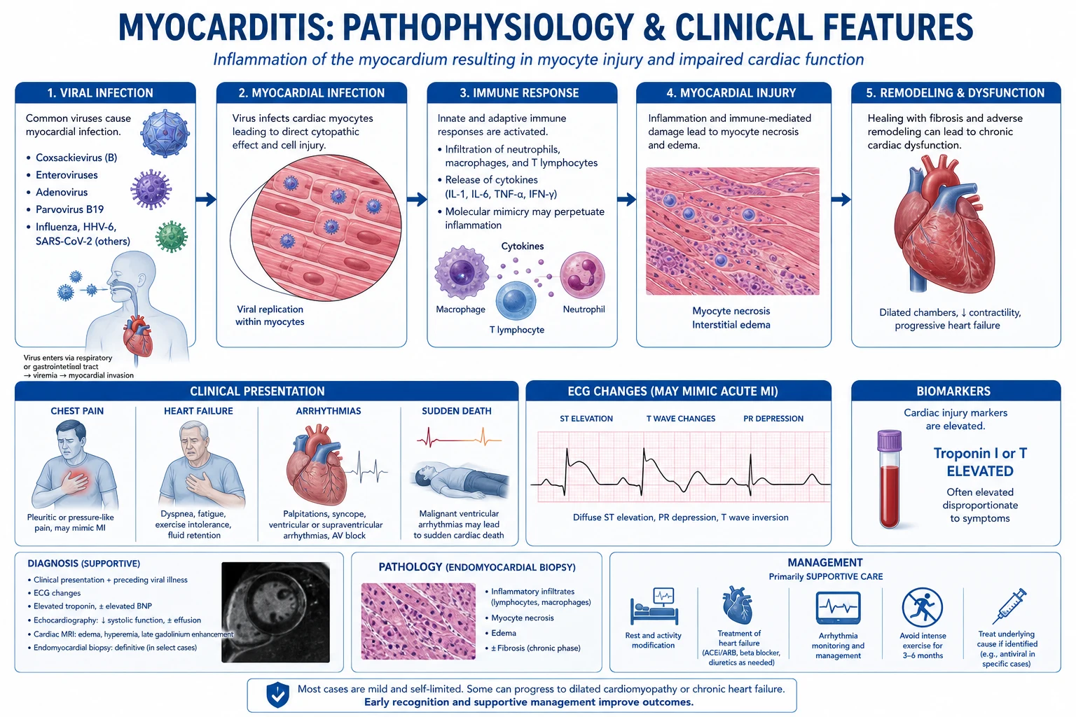
An inflammation of the myocardium, most commonly viral (coxsackie B, parvovirus B19, SARS-CoV-2, influenza, adenovirus, HHV-6, Epstein-Barr, HIV), but also autoimmune, drug (anthracyclines, clozapine, immune-checkpoint inhibitors), toxic, and hypersensitivity (DRESS, eosinophilic).[1][9]
Aetiology — the exam-friendly breakdown:[9]
Viral (commonest)
Lymphocytic on biopsy
- Coxsackie B (the classic "is it the heart or the gut" enterovirus)
- Parvovirus B19 (the commonest PCR-identified virus in adults)
- SARS-CoV-2 / COVID-19 mRNA vaccines (young men, within 1 week of dose 2 — mild, self-limiting)
- Influenza, adenovirus, HHV-6, EBV, HIV, Hep C
- Biopsy: lymphocytic infiltrate; usually supportive treatment
Giant cell (the killer)
Aggressive, needs immunosuppression
- Middle-aged adults; fulminant, rapidly progressive heart failure + VT
- Multinucleated giant cells + granulomas on biopsy
- Untreated: near-universal death within weeks-months
- Treat: cyclosporine + azathioprine + corticosteroids ± transplant
- Better prognosis than historical now that immunosuppression is used (Kandolin 2015)
Eosinophilic
Hypersensitivity / parasitic
- Drug hypersensitivity (clozapine, penicillin, sulfonamide, smallpox vaccine)
- Loeffler endocarditis (hypereosinophilic syndrome, parasitic)
- DRESS syndrome (Drug Reaction with Eosinophilia and Systemic Symptoms)
- Peripheral eosinophilia; often curable by stopping the drug + steroids
- Differentiate from the eosinophilic restrictive cardiomyopathy
Autoimmune
Steroid-responsive in selected cases
- Systemic lupus erythematosus, rheumatoid, scleroderma
- Cardiac sarcoidosis (often grouped here)
- Biopsy-proven virus-negative, antibody-positive cases
- Treat the underlying disease + corticosteroids ± steroid-sparing agent
- Chest pain (mimics an ACS), a flu-like prodrome in the preceding 1-2 weeks.
- Arrhythmia (atrial and ventricular — from the inflamed myocardium; the most common cause of death in the acute phase).
- Heart failure (from the LV dysfunction — may be fulminant).
- Sudden death in young athletes — myocarditis is the third commonest cause of SCD in the under-35s (after HCM and arrhythmogenic syndromes).
- A rising troponin with normal coronary arteries on the angiogram.
- A rub or pericardial features if there is a concomitant pericarditis (myopericarditis).
- Echocardiography — an LV dysfunction (may be regional or global; may be normal early). Look specifically for RV involvement (a poor prognostic sign) and a pericardial effusion.
- Cardiac MRI — the gold-standard non-invasive test. The Lake Louise criteria (updated 2018) require both a tissue-characterisation abnormality (T1 mapping, ECV, late gadolinium enhancement) and an inflammatory signal (T2 mapping/STIR oedema). The classic pattern is mid-wall or subepicardial late gadolinium enhancement in the lateral/inferolateral wall (the non-ischaemic pattern — distinguishes from the subendocardial LGE of an infarct).[10]
- Endomyocardial biopsy — the historical gold standard; the Dallas criteria (a lymphocytic infiltration with myocyte necrosis = active myocarditis; borderline if infiltrate without necrosis). Rarely done in clinical practice (the MRI has largely replaced it for diagnosis), but remains indicated for the fulminant case in a middle-aged adult to exclude giant-cell myocarditis — because giant-cell changes the management to immunosuppression/transplant.[9][11]
- Viral PCR on blood and (if biopsied) myocardial tissue.
- Coronary angiography — to exclude an ACS as the cause of the troponin rise.
The fulminant vs acute distinction (McCarthy, NEJM 2000):[8] fulminant myocarditis presents with sudden severe haemodynamic compromise needing inotropes/MCS within hours-days, often with a distinct viral prodrome and fever; non-fulminant acute myocarditis is more insidious. Paradoxically, fulminant myocarditis has a BETTER long-term survival (93 per cent at 11 years vs 45 per cent for acute) — because the fulminant inflammation is a single, robust, self-limited immune response, whereas the indolent form smolders into a dilated cardiomyopathy.[8]
- Supportive — heart-failure therapy (ACEi, beta-blocker, diuretic), treat the arrhythmia, anticoagulate if an LV thrombus.
- Rest — avoid exercise for 6 months (exercise during the acute phase increases the viral replication and the mortality; the classic example is athletes who collapse on returning to sport).
- NSAIDs for the pericarditis component (myopericarditis) — colchicine may be added.
- Treat the cause — antiviral for the specific virus if available; immunosuppression for the giant-cell or the autoimmune myocarditis (confirmed on the biopsy) — Kandolin 2015 showed combined immunosuppression (cyclosporine + azathioprine + steroids) improved survival in giant-cell myocarditis; a trial of corticosteroids for virus-negative, biopsy-proven lymphocytic myocarditis may be considered.[11]
- Most recover fully; some progress to a dilated cardiomyopathy; the fulminant case (a severe acute presentation with cardiogenic shock) may need mechanical support (VA-ECMO, an Impella) as a bridge to recovery, or a transplant.[1][8]
COVID-19 and vaccine myocarditis
SARS-CoV-2 can cause myocarditis directly (viral tropism via the ACE2 receptor) or indirectly (the cytokine storm, the multisystem inflammatory syndrome in children [MIS-C], and critical illness). The mRNA vaccines (BNT162b2, mRNA-1273) carry a small but real risk of myopericarditis, predominantly in young males within 1 week of the second dose, that is generally mild and self-limiting — distinct from classical viral myocarditis, with a prompt recovery of the EF and a low complication rate.[14] The overall risk-benefit strongly favours vaccination.
Mechanical circulatory support for fulminant myocarditis
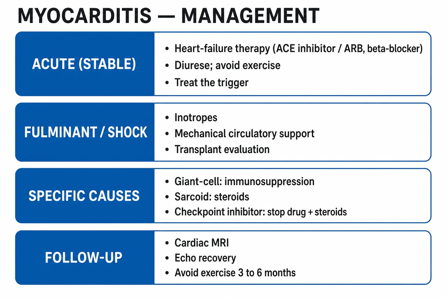
The principle: support the failing ventricle while the inflammation resolves (most fulminant cases recover within days to weeks), with a defined exit strategy — bridge to recovery, to decision, to a durable LVAD, or to transplant.[1][8]
The escalation pathway for fulminant myocarditis / refractory cardiogenic shock
Recognise the shock early
Hypotension (SBP <90), cold peripheries, oliguria, rising lactate, pulmonary oedema — in a young patient with a viral prodrome, think fulminant myocarditis. Bedside echo: a small, thick, hypocontractile LV with a preserved RV size (unlike chronic DCM).
Inotrope ± vasopressor
Noradrenaline for the MAP, dobutamine/milrinone for the cardiac output. AVOID high-dose catecholamines for prolonged periods — they increase myocardial oxygen demand and arrhythmia in the inflamed myocardium. Use the lowest effective dose.
Intra-aortic balloon pump
Reasonable first-line MCS in the haemodynamically unstable patient — diastolic augmentation, reduced afterload. Contraindicated in significant aortic regurgitation and aortic dissection.
VA-ECMO for refractory shock
Indicated for cardiac arrest (ECPR), a lactate rising despite inotropes, or a MAP that cannot be maintained. Provides full cardiopulmonary bypass (3-5 L/min). It does NOT unload the LV — if the LV dilates (aortic valve not opening on echo, worsening pulmonary oedema), add an Impella or a venting strategy.
Impella (percutaneous LVAD)
2.5-5 L/min of active forward flow with LV unloading — ideal when VA-ECMO is causing LV distension (the ECPELLA configuration). Monitor for haemolysis (LDH, haptoglobin, free haemoglobin) and limb ischaemia.
Endomyocardial biopsy
For any fulminant case (especially middle-aged), to exclude giant-cell myocarditis — which mandates immunosuppression and consideration of early transplant listing. The MRI can be deferred in extremis.
Define the goal and the exit
Bridge to recovery (the commonest outcome in fulminant lymphocytic myocarditis — wean MCS as the EF recovers over days-weeks), bridge to decision (if the trajectory is unclear), bridge to transplant (for the giant-cell or non-recovering case), or bridge to a durable LVAD.
Trials that changed practice
HCM Risk-SCD (O'Mahony)
Eur Heart J 2014
3675 pts with HCM — derivation/validation of a continuous risk model for 5-year SCD
Key finding
A validated equation (age, max wall thickness, LA size, LVOT gradient, family history of SCD, NSVT, syncope) estimates 5-year SCD risk
Practice change
≥6% 5-year risk → ICD; 4-6% shared decision; underpins the 2014 and 2023 ESC HCM recommendations
EXPLORER-HCM
Lancet 2020
251 pts with symptomatic obstructive HCM — mavacamten vs placebo, 30 weeks
Key finding
36% of mavacamten pts improved ≥1 NYHA class + ≥10% peak VO2 vs 17% placebo; LVOT gradient fell ~36 mmHg
Practice change
Mavacamten became the first cardiac myosin inhibitor approved for symptomatic obstructive HCM
ATTR-ACT
NEJM 2018
441 pts with ATTR amyloid cardiomyopathy — tafamidis vs placebo, 30 months
Key finding
All-cause mortality 42.9% tafamidis vs 57.9% placebo; fewer cardiovascular hospitalisations
Practice change
Tafamidis became standard disease-modifying therapy for ATTR-CM (both wild-type and hereditary)
McCarthy — fulminant vs acute myocarditis
NEJM 2000
147 pts with biopsy-proven myocarditis — fulminant (n=15) vs acute non-fulminant (n=132)
Key finding
11-year survival 93% fulminant vs 45% acute — fulminant paradoxically has BETTER long-term survival
Practice change
Fulminant myocarditis warrants aggressive MCS as a bridge to recovery, because the prognosis with support is excellent
InterTAK registry (Templin)
NEJM 2015
1750 pts with Takotsubo across 26 centres — international registry
Key finding
90% female; a physical or emotional trigger in ~70%; in-hospital mortality ~4%; serious complications (heart failure, shock, thrombus, rupture) ~20%
Practice change
Reframed Takotsubo as not-always-benign; drove standardised InterTAK diagnostic criteria and the InterTAK score
Kandolin — giant-cell myocarditis
Circulation 2015
Mayo Clinic cohort — giant-cell myocarditis in the era of combined immunosuppression (cyclosporine + azathioprine ± steroids)
Key finding
Combined immunosuppression improved survival and transplant-free survival over historical eras
Practice change
Confirmed combined immunosuppression as the standard of care for biopsy-proven giant-cell myocarditis, with early transplant for non-responders
2023 ESC Cardiomyopathy Guideline
Eur Heart J 2023
Multidisciplinary task force — phenotype-first, genotype-driven framework for all cardiomyopathies
Key finding
Recommends defining phenotype (dilated, HCM, restrictive, non-compaction, ARVC) then systematic aetiological hunt + cascade genetic screening of first-degree relatives
Practice change
Unified cardiomyopathy care around phenotype + genotype; replaced disease-siloed guidance
Lake Louise (2018 update)
JACC 2009 / JCMR 2018
Expert consensus on CMR criteria for myocarditis
Key finding
Updated criteria require BOTH a tissue-characterisation signal (T1/ECV/LGE) AND an inflammatory signal (T2) — improves diagnostic accuracy
Practice change
CMR became the non-invasive gold standard for myocarditis; biopsy reserved for fulminant/atypical cases

SAQ — Peripartum cardiomyopathy in acute heart failure
10 minutes · 10 marks
A 33-year-old woman (gravida 2, para 2) presents three weeks after an uneventful vaginal delivery with progressive dyspnoea, orthopnoea and bilateral leg swelling. She is tachycardic (HR 118), BP 96/62, JVP to the angle of the jaw, with bibasal crackles and a third heart sound. NT-proBNP is 6,800 ng/L. Echocardiography shows a dilated LV (LVEDD 6.2 cm) with an EF of 28% and global hypokinesia.
SAQ — Peripartum cardiomyopathy with refractory cardiogenic shock
10 minutes · 10 marks
A 29-year-old woman is admitted ten days postpartum with severe breathlessness. She is in florid pulmonary oedema, HR 132, BP 80/55, cold peripheries, lactate 3.8 mmol/L. Bedside echo shows a dilated LV with an EF of 18% and a large mobile apical LV thrombus. She is breastfeeding her infant.
Red flags
References
- [1]Arbelo E, Protonotarios A, Gimeno JR, et al. Corrigendum to Successful kidney-alone transplantation in a patient with PH1 on combination RNA-interference therapy. Kidney International, 2023;104:203-204 Kidney Int, 2023.PMID 37863627
- [2]Ommen SR, Mital S, Burke MA, et al. 2020 AHA/ACC Guideline for the Diagnosis and Treatment of Patients With Hypertrophic Cardiomyopathy: Executive Summary: A Report of the American College of Cardiology/American Heart Association Joint Committee on Clinical Practice Guidelines Circulation, 2020.PMID 33215938
- [3]O'Mahony C, Jichi F, Pavlou M, et al. Compound and digenic heterozygosity predicts lifetime arrhythmic outcome and sudden cardiac death in desmosomal gene-related arrhythmogenic right ventricular cardiomyopathy Circ Cardiovasc Genet, 2013.PMID 24070718
- [4]Maron BJ, Maron MS IRMA Code II: unique annotation of medical images for access and retrieval Stud Health Technol Inform, 2012.PMID 22874172
- [5]Olivotto I, Oreziak A, Barriales-Villa R, et al. Investigation of the phenomenology, physiology and impact of spiritually transformative experiences - kundalini awakening Explore (NY), 2021.PMID 32753262
- [6]Weintraub RG, Semsarian C, Macdonald P Antimalarial activity of novel 4-aminoquinolines active against drug resistant strains Bioorg Chem, 2017.PMID 27908538
- [7]Maurer MS, Schwartz JH, Gundapaneni B, et al. Laparoscopic Redo Pull-Through for Hirschsprung Disease Due to Innervation Disorders J Laparoendosc Adv Surg Tech A, 2019.PMID 30461345
- [8]McCarthy RE, Boehmer JP, Hruban RH, et al. Intravenous nesiritide, a natriuretic peptide, in the treatment of decompensated congestive heart failure. Nesiritide Study Group N Engl J Med, 2000.PMID 10911006
- [9]Caforio AL, Pankuweit S, Arbustini E, et al. Development and validation of a new radiographic scoring system to evaluate bone and cartilage destruction and healing of large joints with rheumatoid arthritis: ARASHI (Assessment of rheumatoid arthritis by scoring of large joint destruction and healing in radiographic imaging) study Mod Rheumatol, 2013.PMID 23624939
- [10]Friedrich MG, Sechtem U, Schulz-Menger J, et al. Chronic estradiol replacement impairs performance on an operant delayed spatial alternation task in young, middle-aged, and old rats Horm Behav, 2009.PMID 19631212
- [11]Kandolin R, Lehtonen J, Salmenkivi K, et al. Recruitment of HIV-1 target cells at topical mucosal sites: a sensitive and early marker for determining the safety of microbicide candidates Emerg Microbes Infect, 2013.PMID 26038476
- [12]Templin C, Ghadri JR, Diekmann J, et al. Cytomegalovirus Preventive Strategies in Liver Transplant Am J Transplant, 2015.PMID 25908256
- [13]Ghadri JR, Wittstein IS, Prasad A, et al. Addressing Risks: Mental Health, Work-Related Stress, and Occupational Disease Management to Enhance Well-Being Biomed Res Int, 2018.PMID 29850529
- [14]Bozkurt B, Kamat I, Hotez PJ Mycobacterium tuberculosis H(2)S Functions as a Sink to Modulate Central Metabolism, Bioenergetics, and Drug Susceptibility Antioxidants (Basel), 2021.PMID 34439535