ICU · Cardiovascular
Acute cardiorenal syndrome in ICU
Also known as Cardiorenal syndrome · CRS · Type 1 CRS · Worsening renal function in heart failure · Venous congestion · Type 2 CRS · Type 3 CRS · Type 4 CRS · Type 5 CRS · Renocardial syndrome · Sequential nephron blockade · Diuretic resistance
Cardiorenal syndrome (CRS): disorders of heart and kidneys whereby acute/chronic dysfunction of one causes dysfunction of the other. FIVE TYPES — TYPE 1 (acute cardiorenal): acute heart failure → AKI (most ICU-relevant). TYPE 2 (chronic cardiorenal): chronic heart failure → CKD. TYPE 3 (acute renocardial): AKI → acute heart failure. TYPE 4 (chronic renocardial): CKD → chronic heart failure. TYPE 5 (secondary): systemic disease (sepsis, diabetes, amyloid) → both. Pathophysiology: VENOUS CONGESTION (raised CVP → renal interstitial pressure → reduced GFR) is as important as low cardiac output; RAAS and sympathetic overactivation drive a vicious cycle; intra-abdominal hypertension, inflammation and oxidative stress amplify injury. Management: DECONGESTION (high-dose loop diuretics ± thiazide sequential nephron blockade), vasodilators (nitrates), inotropes (if low output), SGLT2 INHIBITORS (disease-modifying — DAPA-HF, EMPEROR, DAPA-CKD), avoid nephrotoxins, ultrafiltration only for refractory cases (CARRESS-HF — worse than pharmacological therapy), renal replacement therapy if refractory. KEY INSIGHT: VENOUS CONGESTION (not just low output) drives AKI — decongestion is the primary target; a creatinine rise during effective decongestion is acceptable and prognostically favourable.
On this page & tools
Your progress
Saved locally on this device.
3 MCQs with explanations
Target exams
Red flags
The five types of cardiorenal syndrome
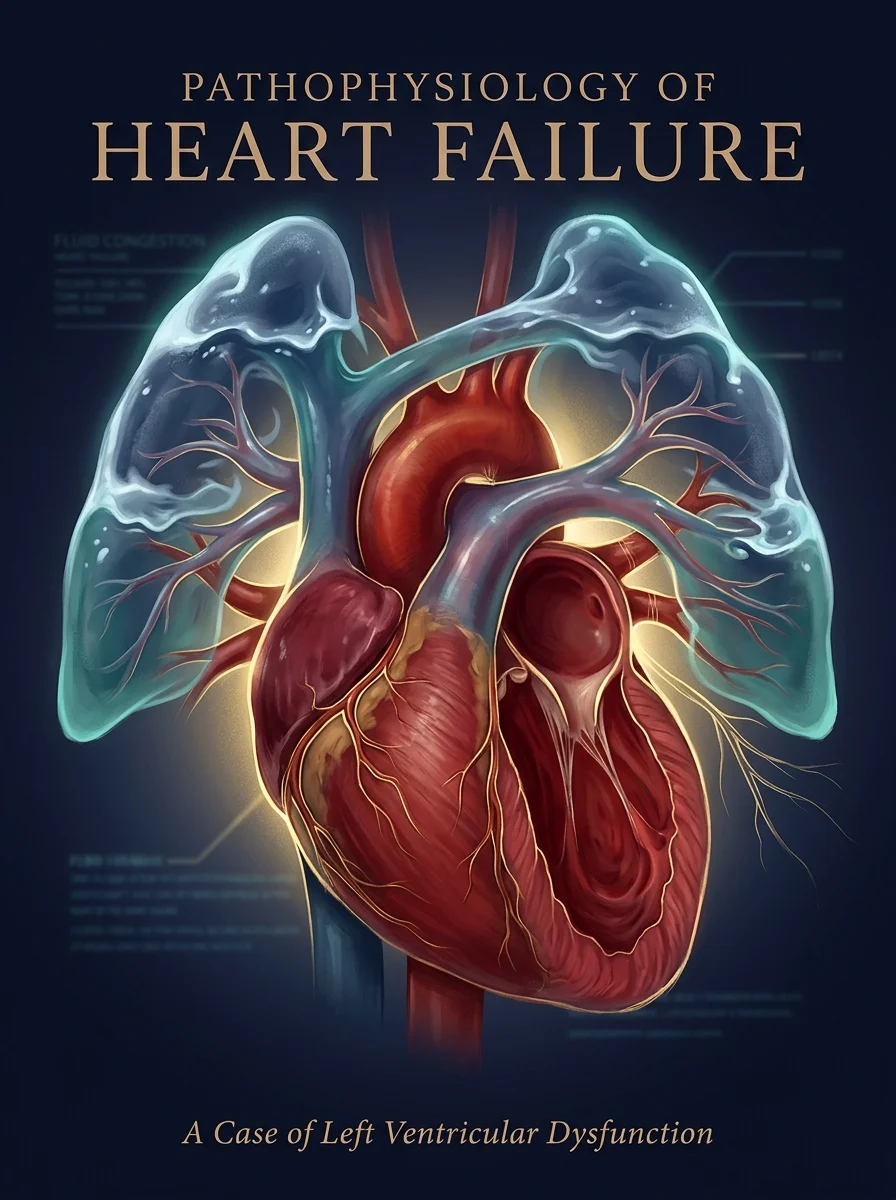
The Ronco classification distinguishes five CRS subtypes by acuity (acute vs chronic) and direction (heart → kidney, kidney → heart, or systemic). Distinguishing the type drives both the workup and the therapy.[6][1]
The five types of cardiorenal syndrome (Ronco/Rangaswami classification)
| Type | Name | Primary insult | Secondary effect | Setting | Typical ICU relevance |
|---|---|---|---|---|---|
| 1 | Acute cardiorenal | Acute heart failure / ACS / flash pulmonary oedema | AKI develops hours-days | ICU, ED, CCU | Highest — most encountered in ICU |
| 2 | Chronic cardiorenal | Chronic heart failure (HFrEF/HFpEF) | Progressive CKD | Outpatient, cardiology | Moderate — seen during exacerbations |
| 3 | Acute renocardial | AKI (contrast nephropathy, ATN, obstruction) | Acute heart failure (volume, K+, acidosis, uraemia) | ICU, nephrology | High — common in ICU AKI |
| 4 | Chronic renocardial | CKD / ESRD | Chronic heart failure (uraemic cardiomyopathy, LVH, vascular calcification) | Renal clinic, dialysis unit | Moderate — dialysis patients in ICU |
| 5 | Secondary (systemic) | Systemic disease (sepsis, diabetes, amyloid, SLE, cirrhosis) | Simultaneous heart AND kidney dysfunction | ICU, multisystem disease | High — sepsis is the prototype |
Type 1 (acute cardiorenal) is the prototype ICU problem: a patient with acute decompensated heart failure (ADHF), flash pulmonary oedema or cardiogenic shock develops a rising creatinine within hours to days. The dominant driver is venous congestion raising renal interstitial pressure, compounded by low forward flow when output is impaired.[2][5]
Type 2 (chronic cardiorenal) describes the slow, progressive CKD that accompanies chronic HFrEF or HFpEF. Renal dysfunction here is a marker of advanced disease and predicts mortality; it is not, by itself, a reason for ICU admission unless the patient decompensates into a type 1 episode.[5]
Type 3 (acute renocardial) is renal-first: an AKI (post-contrast, ATN, obstructive, rhabdomyolysis) precipitates volume overload, hyperkalaemic arrhythmia, acidosis-driven myocardial depression, or uraemic pericarditis. Management is of the AKI first (fluids if hypovolaemic, K+ control, RRT for refractory overload/hyperkalaemia/acidosis).[1]
Type 4 (chronic renocardial) is the uraemic cardiomyopathy of CKD/ESRD — left ventricular hypertrophy, diastolic dysfunction, vascular and valvular calcification, accelerated atherosclerosis. Dialysis patients presenting to ICU have high cardiovascular mortality.[13]
Type 5 (secondary) is simultaneous heart and kidney injury from a systemic process: sepsis (the most common ICU cause — cytokine storm, microcirculatory failure, mitochondrial dysfunction), diabetes, amyloidosis, SLE, vasculitis, cirrhosis (hepatorenal plus cardiomyopathy). Treat the underlying disease; the heart and kidney recover together.[6]
Pathophysiology in depth
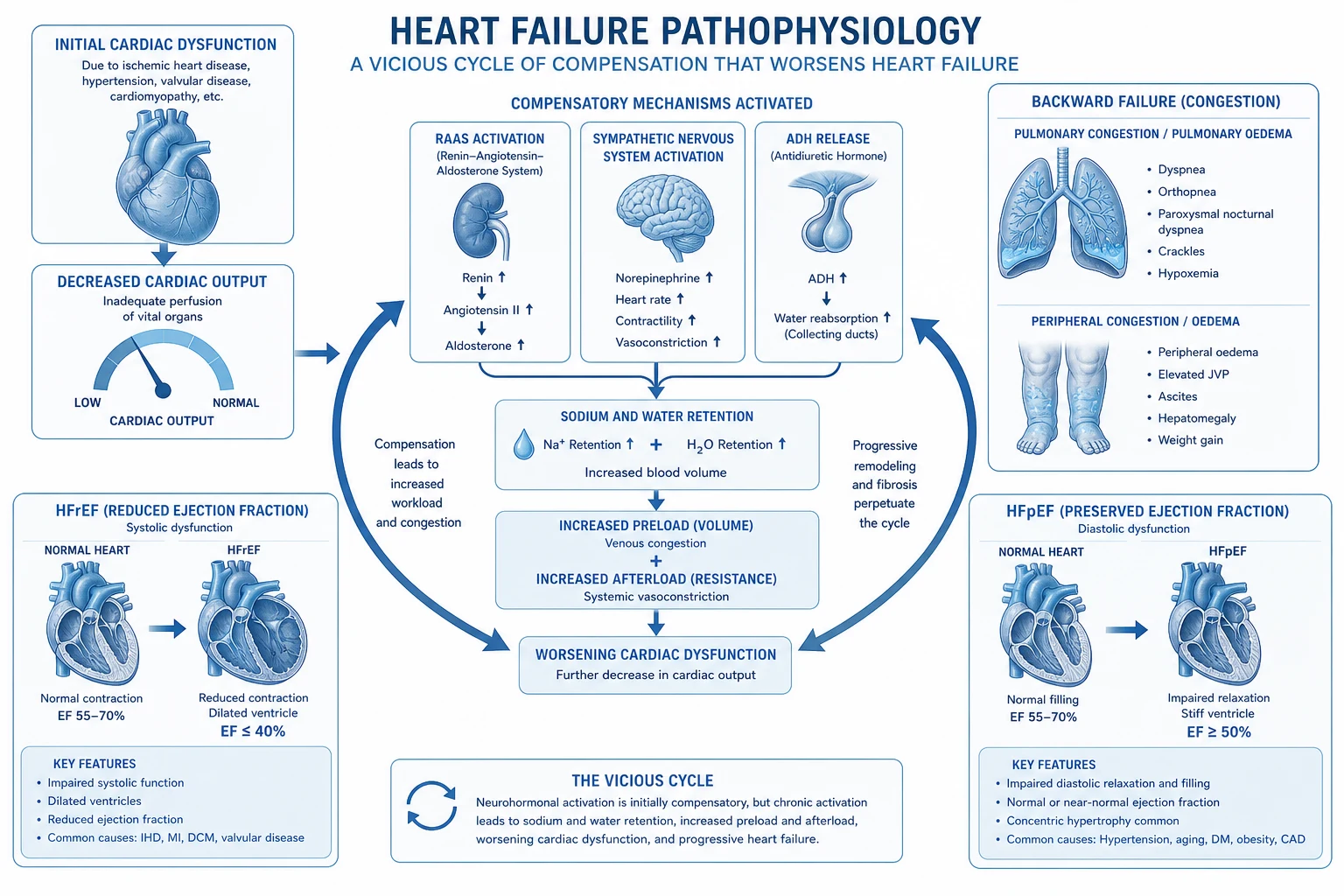
CRS is not a single mechanism — it is a convergent disorder in which four overlapping pathways drive a self-reinforcing vicious cycle. Understanding which pathway dominates in a given patient directs therapy.[1][13]
1. Venous congestion — the dominant driver in decompensated HF
Elevated central venous pressure (CVP) is transmitted retrograde to the renal veins, raising renal interstitial and Bowman's capsule hydrostatic pressure. This compresses the peritubular capillaries and tubules, opposes glomerular filtration, and reduces the hydrostatic gradient that drives filtration — GFR falls even when renal arterial inflow is intact. The landmark Mullens/ESCAPE analyses showed CVP is the strongest haemodynamic predictor of worsening renal function in ADHF, stronger than cardiac output, ejection fraction, or systolic blood pressure. A CVP above ~12–14 mmHg is associated with a steep rise in AKI incidence; above ~18 mmHg AKI is nearly universal.[2][5]
This single insight underpins modern therapy: decongestion is the primary target, even when creatinine rises during diuresis — a congested patient with a slightly higher creatinine does better than a less-congested patient with a stable creatinine. [1]
2. Low forward flow — reduced renal perfusion
When cardiac output falls (cardiogenic shock, end-stage HFrEF, right ventricular failure), mean arterial pressure and renal perfusion fall. The kidney is vulnerable because it receives ~20% of cardiac output and because the afferent arteriole is supplied by relatively low-pressure flow. Reduced perfusion activates intrarenal autoregulation and, when MAP falls below ~65 mmHg, autoregulation fails and GFR drops sharply. Inotropes and vasopressors restore forward flow but at the cost of increased myocardial oxygen demand and arrhythmia risk — they are bridges, not destinations.[5]
3. Neurohormonal activation — the vicious cycle
Reduced renal perfusion and sympathetic sensing trigger the renin-angiotensin-aldosterone system (RAAS) and the sympathetic nervous system (SNS):
- Angiotensin II → efferent arteriolar vasoconstriction (maintains GFR acutely — the basis for the ACEi/ARB creatinine rise), systemic vasoconstriction, aldosterone release, sodium retention, inflammation and fibrosis.
- Aldosterone → distal sodium reabsorption, potassium/magnesium loss, myocardial fibrosis.
- Sympathetic activation → vasoconstriction, tachycardia, renin release, arrhythmias. [1]
The cycle is self-reinforcing: each effector worsens both heart failure and renal perfusion, driving further activation. Breaking the cycle (vasodilators, ACEi/ARB/ARNI once stable, beta-blocker once stable, MRA, SGLT2 inhibitor) is the foundation of chronic disease modification.[5][13]
4. Inflammation, oxidative stress and endothelial dysfunction
Acute heart failure and AKI both release cytokines (TNF-α, IL-6), reactive oxygen species, and damage-associated molecular patterns that cross-talk between the two organs. Endothelial dysfunction reduces nitric oxide bioavailability in the renal vasculature, blunting the vasodilation that protects the kidney. This pathway is dominant in type 5 CRS (sepsis) but contributes to all types.[1]
5. Intra-abdominal hypertension (IAH) and the cardio-abdominal-renal syndrome
Severe congestion causes hepatic and bowel wall oedema, raising intra-abdominal pressure (IAP). IAH (IAP >12 mmHg) compresses the renal veins, the renal parenchyma, and the abdominal aorta/renal arteries, raising CVP and reducing renal perfusion simultaneously. A bladder pressure >20 mmHg with new organ dysfunction defines abdominal compartment syndrome — a reversible contributor to CRS that responds to decongestion and paracentesis/surgical decompression.[2]
Venous congestion vs low forward flow — two mechanisms, different therapy
| Feature | Venous congestion dominant | Low forward flow dominant |
|---|---|---|
| Typical patient | ADHF, volume overload, raised JVP, oedema, hepatomegaly | Cardiogenic shock, cool peripheries, oliguria, low CO |
| CVP | High (>12–14 mmHg) | Variable — may be low or normal |
| Cardiac output | Preserved or high (warm, wet) | Low (cold, dry or cold, wet) |
| Blood pressure | Often high | Often low |
| Primary therapy | Decongestion — high-dose loop ± thiazide, nitrates | Restore output — inotrope, vasopressor, MCS |
| Creatinine on diuresis | May rise — continue if decongesting | Worsens with low flow — fix output |
| Prognostic marker | CVP is the strongest predictor of AKI | MAP, lactate, mixed venous saturation |
Clinical assessment
CRS is a clinical diagnosis supported by bedside investigations. The single most important question is: is the patient congested (wet) or dry, and is perfusion adequate (warm) or impaired (cold)? The four haemodynamic profiles (wet/dry × warm/cold) drive therapy.[2]
Bedside assessment of congestion:
- Jugular venous pressure — the single most useful bedside sign. Raised JVP = venous congestion.
- Lung auscultation — crackles (but crackles may be absent in chronic HF).
- Peripheral oedema, sacral oedema, ascites.
- Hepatomegaly, hepatojugular reflux.
- Lung ultrasound — B-lines (comet-tail artefacts) quantify extravascular lung water; multiple bilateral B-lines = interstitial pulmonary oedema.
- Point-of-care echo — IVC size and collapsibility (a plethoric, non-collapsing IVC = high CVP); right heart assessment for pulmonary hypertension, RV failure, TR; LV systolic and diastolic function.
- Daily weights — the most sensitive bedside measure of net fluid balance.
- Hemodynamic monitoring — CVP (central line), arterial line for MAP; advanced monitors (PiCCO, pulmonary artery catheter) when peripheral perfusion or cardiac output is in question. [1]
Laboratory and imaging:
- Urea, creatinine, eGFR, electrolytes (K+, Na+, Mg2+, phosphate), venous blood gas (lactate, acidosis).
- Troponin (myocardial injury), NT-proBNP/BNP (heart failure severity — but BNP rises with renal dysfunction alone).
- Urine sodium (low <30 mmol/L on diuretics suggests under-diuresis; FENa if pre-renal vs ATN unclear).
- Urinalysis — bland in CRS; casts/proteinuria suggest intrinsic renal disease (type 3 or concurrent).
- Echocardiography — EF, diastolic function, RV size and function, valvular lesions, pericardial effusion, IVC.
- Renal ultrasound — to exclude obstruction (especially type 3), assess kidney size/corticomedullary differentiation for chronicity. [1]
Management of acute cardiorenal syndrome
Management of acute cardiorenal syndrome (type 1)
- Assess volume status and perfusion — clinical (JVP, oedema, crackles, hepatomegaly, peripheral temperature, capillary refill, urine output), ultrasound (IVC, B-lines), haemodynamics (CVP, MAP, cardiac output if monitored). Classify as WARM/WET, WARM/DRY, COLD/WET, COLD/DRY.
- Decongest (primary target for wet patients) — HIGH-DOSE loop diuretics (frusemide IV — 2-2.5x oral dose). If inadequate: ADD thiazide (metolazone/hydrochlorothiazide — sequential nephron blockade). TARGET: 2-3 L negative balance/day (or adequate urine output). Monitor renal function — may rise, continue if congested
- Optimise cardiac output (for cold patients) — if low output (cardiogenic shock): inotropes (dobutamine, milrinone). Vasopressors (noradrenaline) if MAP <65. Vasodilators (GTN infusion) if hypertensive and warm. Mechanical circulatory support (IABP, Impella, VA-ECMO) if refractory shock. Avoid hypotension (worsens renal perfusion)
- Avoid nephrotoxins — STOP NSAIDs, ACEi/ARB/ARNI (temporarily if hypotensive), contrast (use pre-procedural hydration if unavoidable), aminoglycosides, iodinated contrast
- Treat electrolyte derangements — replace K+ (IV/oral), Mg2+; manage hyponatraemia (fluid restriction, tolvaptan for severe); correct acidosis
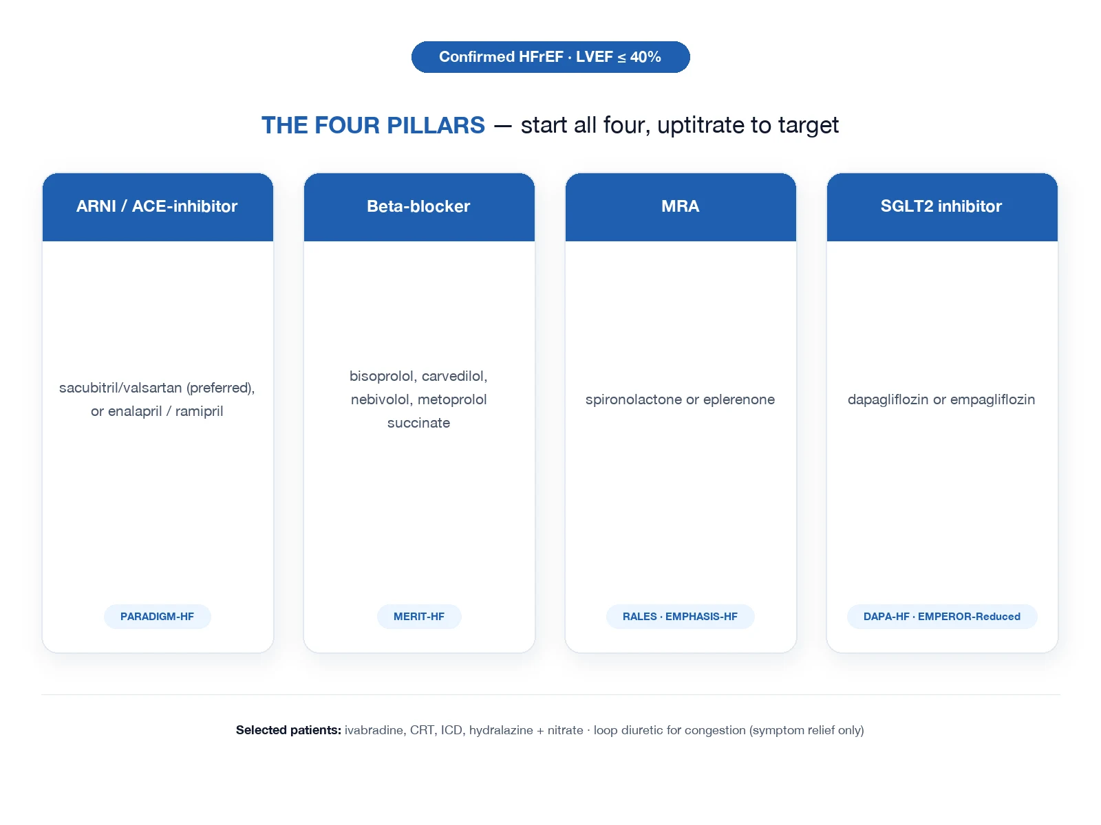
- Start disease-modifying therapy once stable — SGLT2 inhibitor (dapagliflozin 10 mg OD) as soon as euvolaemic and stable; beta-blocker, MRA, ARNI once stable and not in cardiogenic shock
- Monitor — daily weight, fluid balance, renal function, electrolytes (hypokalaemia, hyponatraemia from diuretics), BP, signs of decongestion (JVP falling, B-lines clearing, weight falling, IVC collapsing)
- Renal replacement therapy — if refractory overload (despite high-dose diuretics + thiazide), refractory hyperkalaemia, severe metabolic acidosis, or uraemic complications. Slow continuous ultrafiltration, CVVHDF, or intermittent HD depending on haemodynamic stability
Stepped approach to diuretic resistance in CRS
- Confirm resistance — <100–150 mL/h urine output after an adequate IV loop bolus, or failure to achieve a 1–2 L/day negative balance despite escalating doses.
- Exclude correctable causes — high sodium intake (diet, IV fluids, saline flushes), NSAIDs (block diuretic effect), non-adherence, hypotension (low perfusion), hypoalbuminaemia (reduced diuretic delivery to nephron).
- Up-titrate the loop diuretic — frusemide up to 200–400 mg/day IV equivalent (bumetanide 1:40, torsemide 1:2 vs frusemide). DOSE trial: high-dose (2.5x home) gave more decongestion than low-dose (1x) with no significant renal harm.[7]
- Switch bolus to continuous infusion — smoother natriuresis, may overcome brake phenomena, equivalent efficacy to bolus by DOSE.
- Add a thiazide (sequential nephron blockade) — metolazone 2.5–5 mg PO, hydrochlorothiazide 25 mg, or chlorthalidone. Blocks distal Na reabsorption — powerful synergy. Monitor closely: hypokalaemia, hyponatraemia, profound volume depletion, ototoxicity.
- Add acetazolamide (250–500 mg) — proximal tubule carbonic anhydrase inhibition, complements the loop + thiazide triple-blockade.
- Add or up-titrate MRA (spironolactone/eplerenone) if not hyperkalaemic — blocks aldosterone escape.
- Albumin if serum albumin <25 g/L — increases diuretic delivery to the nephron (evidence modest but commonly used).
- Vasopressin V2 antagonist (tolvaptan) — for hypervolaemic or euvolaemic hyponatraemia; modest aquaresis.
- Ultrafiltration / RRT — only after stepped pharmacological therapy has failed (CARRESS-HF showed pharmacological therapy superior).[4]
Diuretic and decongestion strategies in CRS
| Strategy | Mechanism | Indication | Cautions / evidence |
|---|---|---|---|
| High-dose IV loop (frusemide, bumetanide, torsemide) | Inhibit Na-K-2Cl in thick ascending limb — powerful natriuresis | First-line for all congested CRS | Hypokalaemia, hyponatraemia, ototoxicity, alkalosis. DOSE: high-dose (2.5x) superior for decongestion without renal harm.[7] |
| Continuous infusion | Steady delivery, smoother natriuresis | When bolus ineffective or frequent boluses impractical | DOSE: equivalent to bolus for outcomes; infusion preferred by many units for high doses |
| Sequential nephron blockade (loop + thiazide) | Block compensatory distal Na reabsorption — synergy | Diuretic resistance on loop alone | VERY potent — monitor K+, Na+, volume daily; ototoxicity |
| Acetazolamide | Proximal tubule CA inhibition | Triple-blockade for refractory congestion; corrects alkalosis | Metabolic acidosis, hypokalaemia |
| MRA (spironolactone/eplerenone) | Aldosterone antagonism in collecting duct | Symptomatic HF, diuretic augmentation, K+ preservation | Hyperkalaemia (esp. with ACEi/ARNI), gynaecomastia (spironolactone) |
| Tolvaptan | Vasopressin V2 antagonist — aquaresis | Hypervolaemic/euvolaemic hyponatraemia | Modest volume effect; costly; hepatotoxicity (in ADPKD doses) |
| Low-dose dopamine (1–3 mcg/kg/min) | Renal vasodilation (DA1) | Historically for renal protection | ROSE-AHF: NO benefit in ADHF — do NOT use for CRS.[3] |
| Nesiritide (BNP) | Vasodilation, natriuresis | Historically for ADHF | ROSE-AHF: NO benefit at low dose — do NOT use for CRS.[3] |
| Ultrafiltration | Mechanical fluid removal via venous access | Refractory overload only (failed stepped diuretics) | CARRESS-HF: MORE adverse events and AKI than stepped pharmacological therapy.[4] |
| RRT (CVVHDF / HD) | Solute and volume clearance | Refractory overload, hyperkalaemia, acidosis, uraemia | Haemodynamic instability; vascular access; dose for AKI |
SGLT2 inhibitors — disease-modifying therapy for cardiorenal syndrome
Sodium-glucose cotransporter-2 (SGLT2) inhibitors are the most important therapeutic advance in cardiorenal medicine in the last decade. They reduce heart failure hospitalisation, cardiovascular death, and renal progression independent of diabetes status.[8][13]
Mechanisms relevant to CRS:
- Osmotic diuresis and natriuresis — modest loop-independent decongestion, reduced preload.
- Tubuloglomerular feedback restoration — increased distal Na delivery reactivates afferent arteriolar tone, reducing single-nephron GFR and intraglomerular pressure — renoprotective despite a small creatinine rise at initiation.
- Improved cardiac energetics (ketone body utilisation), reduced inflammation and fibrosis, weight loss, lower uric acid, reduced SNS tone. [1]
Initiation in the ICU: do NOT start during acute decompensation with hypovolaemia, ketoacidosis, or perioperative states. Once the patient is euvolaemic, haemodynamically stable, and not in cardiogenic shock, start dapagliflozin 10 mg OD or empagliflozin 10 mg OD as part of the four-pillar GDMT. Hold during acute illness, surgery, or starvation (euglycaemic DKA risk).[12]
Landmark SGLT2 inhibitor trials in cardiorenal disease
| Trial (year) | Drug | Population | n | Primary outcome | Effect | Renal signal |
|---|---|---|---|---|---|---|
| DAPA-HF (2019) | Dapagliflozin | HFrEF (EF ≤40%) | 4744 | CV death or HF hospitalisation | ↓ 26% | Slower eGFR decline; ↓ renal composite.[8] |
| EMPEROR-Reduced (2020) | Empagliflozin | HFrEF | 3730 | CV death or HF hospitalisation | ↓ 25% | ↓ 50% renal progression; smaller creatinine rise.[9] |
| EMPEROR-Preserved (2021) | Empagliflozin | HFpEF (EF >40%) | 5988 | CV death or HF hospitalisation | ↓ 21% (FIRST positive HFpEF trial) | Slower eGFR decline.[10] |
| DAPA-CKD (2020) | Dapagliflozin | CKD (with/without diabetes) | 4304 | Sustained eGFR decline, ESRD, renal/CV death | ↓ 39% | Renoprotective across CKD causes.[11] |
| EMPA-KIDNEY (2023) | Empagliflozin | CKD broad | 6609 | Sustained eGFR decline, ESRD, renal/CV death | ↓ 28% | Benefit across eGFR, including low; benefit in non-diabetic CKD |
Practical CRS use: dapagliflozin and empagliflozin both reduce HF hospitalisation and renal progression; they can be started in CKD down to eGFR ~20 mL/min/1.73 m² (DAPA-CKD) and even lower per EMPA-KIDNEY. Hold if euglycaemic DKA risk (insulin-dependent diabetes, starvation, surgery) or symptomatic volume depletion. [1]
Exam practice
SAQ — Type 1 cardiorenal syndrome in acute decompensated heart failure
10 minutes · 10 marks
A 72-year-old man with known HFrEF (EF 28%) on oral frusemide 80 mg BD, bisoprolol 5 mg OD, ramipril 5 mg OD and spironolactone 25 mg OD presents after 5 days of progressive dyspnoea, orthopnoea and a 6 kg weight gain. Examination: JVP to the angle of the jaw, bibasal crackles, sacral and pedal oedema to mid-thigh, BP 168/92, HR 102 in atrial fibrillation, RR 28, SpO2 90% on room air. Creatinine 165 μmol/L (baseline 110), eGFR 38 mL/min/1.73 m², K+ 4.8, Na+ 134, NT-proBNP 8 500 ng/L. Bedside ultrasound: bilateral B-lines, dilated IVC without collapse.
SAQ — Type 2 cardiorenal syndrome with acute-on-chronic kidney injury
10 minutes · 10 marks
A 68-year-old woman with ischaemic HFrEF (EF 25%) and CKD stage 3b (baseline creatinine 180 μmol/L, eGFR 30 mL/min/1.73 m²) on bisoprolol 5 mg OD, ramipril 5 mg OD, spironolactone 12.5 mg OD and empagliflozin 10 mg OD presents with 2 weeks of exertional dyspnoea but no orthopnoea or paroxysmal nocturnal dyspnoea. Examination: JVP raised 4 cm above the sternal angle, trace ankle oedema, clear lung fields, BP 110/65, HR 78 sinus. Creatinine now 240 μmol/L, K+ 5.6 mmol/L, Na+ 136, venous lactate 1.4 mmol/L. Spot urine Na+ 25 mmol/L.
Clinical pearls
Red flags
Prognosis and key trials
CARRESS-HF trial (Bart 2012) — ultrafiltration vs pharmacological therapy
RCT: 188 patients with ADHF + worsening renal function + persistent congestion. Stepped pharmacological therapy (high-dose loop + thiazide) vs ultrafiltration.
- Primary outcome (weight loss at 96h): similar (5.7 kg pharmacological vs 5.1 kg ultrafiltration — no difference)
- Creatinine change: WORSE with ultrafiltration (+0.23 mg/dL vs -0.04 mg/dL, p=0.003)
- Serious adverse events: HIGHER with ultrafiltration (72% vs 57%, p=0.03)
- CONCLUSION: Pharmacological therapy (stepped diuretics) SUPERIOR to ultrafiltration for CRS. Ultrafiltration reserved for refractory cases only.[4]
DOSE trial (Felker 2011): high-dose (2.5x) vs low-dose (1x) loop diuretic; bolus vs continuous infusion. High-dose: more fluid loss, faster symptom relief, similar renal function. HIGH-DOSE preferred; bolus and infusion equivalent.[7] ROSE-AHF (Chen 2013): low-dose dopamine and low-dose nesiritide did NOT improve renal function or decongestion in ADHF with renal dysfunction. Don't use either for CRS.[3]
SGLT2 inhibitor trials — the disease-modifying breakthrough
DAPA-HF (2019) — dapagliflozin vs placebo in HFrEF (with and without diabetes): ↓ primary outcome (CV death or HF hospitalisation) 26%, ↓ all-cause mortality 17%, slower eGFR decline.[8]
EMPEROR-Reduced (2020) — empagliflozin in HFrEF: ↓ primary outcome 25%, ↓ renal progression 50%, smaller creatinine rise than placebo.[9]
EMPEROR-Preserved (2021) — empagliflozin in HFpEF: ↓ HF hospitalisation 29%, FIRST positive HFpEF trial; slower eGFR decline.[10]
DAPA-CKD (2020) — dapagliflozin in CKD (with and without diabetes): ↓ composite renal / CV death 39%, benefit down to eGFR 25 mL/min/1.73 m², benefit independent of diabetes.[11]
EMPA-KIDNEY (2023) — empagliflozin in broad CKD (including low eGFR ~20 and non-diabetic): ↓ composite renal / CV death 28%. [1]
Take-home for CRS: SGLT2 inhibitors are the first drug class to simultaneously reduce heart failure events AND renal progression in HFrEF, HFpEF, and CKD — independent of diabetes. Start as part of GDMT once the patient is stable, not during the acute crisis.[12]
Mechanism and supportive trials
UNLOAD (2008) — ultrafiltration removed more fluid than diuretics at 48h; subsequently superseded by CARRESS-HF for safety. [1]
ESCAPE (2005) — pulmonary artery catheter-guided therapy did NOT improve outcomes in ADHF, but the dataset (Mullens analyses) established that CVP is the strongest haemodynamic predictor of AKI in ADHF.[2]
EVEREST (2007) — tolvaptan (V2 antagonist) for hypervolaemic/euvolaemic hyponatraemia in HF: aquaresis and Na correction, but no mortality benefit. [1]
SECRET of CRS — the four convergent pathophysiology pathways (venous congestion, low forward flow, RAAS/SNS activation, inflammation) explain why decongestion + disease-modifying neurohormonal therapy + SGLT2 + source control together improve outcomes, while isolated 'renal-protective' drugs (dopamine, nesiritide) fail.[1][13]
AnswerCard summary
References
- [1]Rangaswami J, et al. Cardiorenal Syndrome: Classification, Pathophysiology, Diagnosis, and Treatment Strategies: A Scientific Statement From the American Heart Association Circulation, 2019.PMID 30852913
- [2]Mullens W, et al. Evaluation of kidney function throughout the heart failure trajectory - a position statement from the Heart Failure Association of the European Society of Cardiology Eur J Heart Fail, 2020.PMID 31908120
- [3]Chen HH, et al. Low-dose dopamine or low-dose nesiritide in acute heart failure with renal dysfunction: the ROSE acute heart failure randomized trial JAMA, 2013.PMID 24247300
- [4]Bart BA, et al. Ultrafiltration in decompensated heart failure with cardiorenal syndrome N Engl J Med, 2012.PMID 23131078
- [5]Damman K, et al. The kidney in heart failure: an update Eur Heart J, 2015.PMID 25838436
- [6]Ronco C, et al. Cardio-renal syndromes: a systematic approach for consensus definition and classification Heart Fail Rev, 2012.PMID 21197571
- [7]Felker GM, et al. Diuretic strategies in patients with acute decompensated heart failure N Engl J Med, 2011.PMID 21366472
- [8]McMurray JJV, et al. Dapagliflozin in Patients with Heart Failure and Reduced Ejection Fraction N Engl J Med, 2019.PMID 31535829
- [9]Packer M, et al. Cardiovascular and Renal Outcomes with Empagliflozin in Heart Failure N Engl J Med, 2020.PMID 32865377
- [10]Anker SD, et al. Empagliflozin in Heart Failure with a Preserved Ejection Fraction N Engl J Med, 2021.PMID 34449189
- [11]Heerspink HJL, et al. Dapagliflozin in Patients with Chronic Kidney Disease N Engl J Med, 2020.PMID 32970396
- [12]McCallum W, et al. Updates in Cardiorenal Syndrome Med Clin North Am, 2023.PMID 37258013
- [13]Damman K, et al. Cardiorenal interactions in heart failure: insights from recent therapeutic advances Cardiovasc Res, 2024.PMID 37364186