ICU · Monitoring / echocardiography
Critical-Care Echocardiography — FOCUS & FATE
Also known as Focused cardiac ultrasound · FOCUS · Focused assessment with transthoracic echocardiography · FATE · Bedside echo · Point-of-care echocardiography · IVC assessment · Parasternal long axis · PLAX · Apical 4-chamber
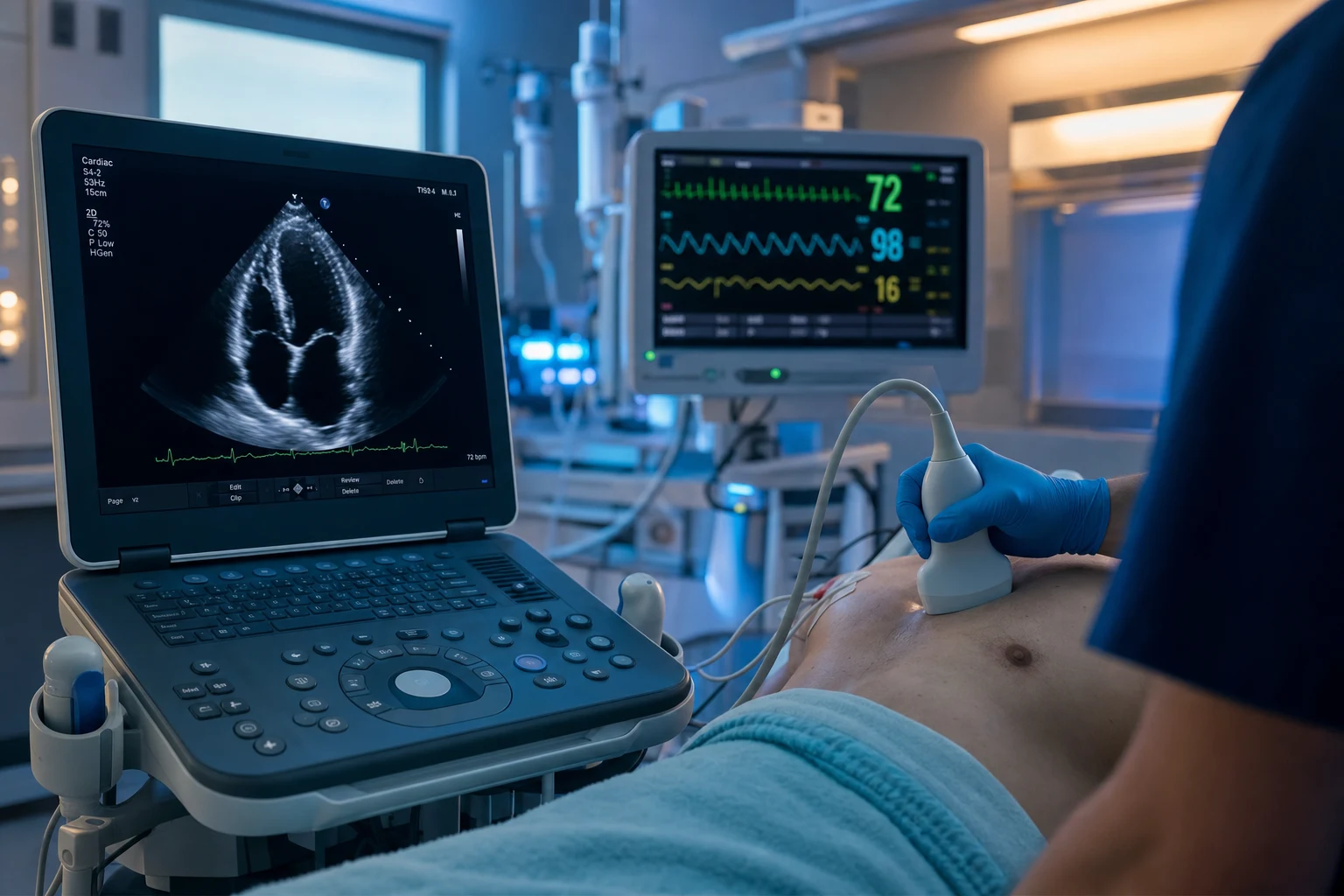
Focused cardiac ultrasound (FOCUS or FATE) is the bedside, clinician-performed transthoracic echocardiogram for the critically ill patient, designed to answer specific resuscitation questions in real time — not a comprehensive echo. The five standard views (parasternal long axis, parasternal short axis, apical 4-chamber, subcostal, and the IVC) answer: is the LV hyperdynamic (hypovolaemia) or hypokinetic (cardiogenic)? Is the RV dilated (PE, cor pulmonale)? Is there a pericardial effusion (tamponade)? Is the IVC collapsing (volume-responsive) or plethoric (high right-sided pressures)? Gross valve abnormalities? FOCUS complements — it does not replace — the formal comprehensive echo.
On this page & tools
Your progress
Saved locally on this device.
Target exams
Overview & definition
Focused cardiac ultrasound (FOCUS) or focused assessment with transthoracic echocardiography (FATE) is the bedside, clinician-performed echocardiogram for the critically ill patient. It is not a comprehensive echo (which a sonographer or cardiologist does to quantify EF, valve gradients, and diastolic function). It is a focused, goal-directed assessment to answer specific resuscitation questions at the bedside, in real time, to guide the immediate management of the unstable patient.[1][1]
The five standard views
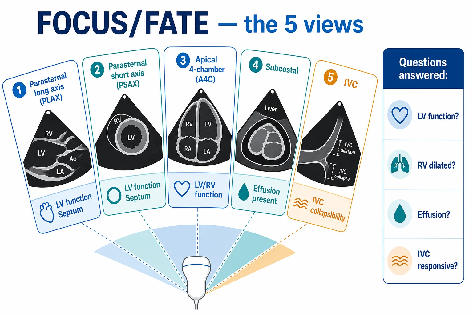
- Parasternal long axis (PLAX) — the LV (function, size), the LVOT, the aortic and mitral valves, and a slice of the RV.[1]
- Parasternal short axis (PSAX) — the LV at the papillary muscle level (regional wall motion abnormalities — coronary territory), the RV (the D-shaped septum of RV overload).[1]
- Apical 4-chamber (A4C) — all four chambers; LV and RV size and function; the tricuspid and mitral valves.[1]
- Subcostal (subxiphoid) — all four chambers (if the parasternal and apical windows are poor); the pericardial effusion (best seen here); and the IVC.[1]
- The IVC (in the subcostal or the right lateral approach) — the diameter and the collapsibility, as a marker of the right atrial pressure and the fluid responsiveness.[1]
The five questions FOCUS answers
- Is the LV hyperdynamic or hypokinetic? — a hyperdynamic, small, kissing LV suggests hypovolaemia (or vasodilatory shock); a hypokinetic, dilated LV suggests cardiogenic shock. Regional wall motion abnormalities suggest an ACS.[1]
- Is the RV dilated? — RV dilatation with septal bowing (the D-shaped septum in the PSAX) suggests acute cor pulmonale — a PE, ARDS, or RV infarct.[1]
- Is there a pericardial effusion? — an echo-free space around the heart; if there is RA or RV collapse, it is a tamponade (see the obstructive-shock topic). The subcostal view is the best for detecting and assessing a pericardial effusion.[1]
- Is the IVC collapsing or plethoric? — a small IVC (under 1.5 cm) with over 50 per cent collapse on inspiration suggests a low right atrial pressure and a fluid-responsive patient; a plethoric IVC (over 2.5 cm) with under 20 per cent collapse suggests a high right atrial pressure (heart failure, cor pulmonale, tamponade) and that fluid may harm. In the ventilated patient, the IVC distensibility (the increase on inspiration, from the positive pressure) is used.[1][1]
- Are there gross valve abnormalities? — a flail leaflet, a large vegetation, or a severely regurgitant valve (visible as a disrupted closure or a colour jet on colour Doppler).[1]
Limitations
FOCUS is not a comprehensive echo:[1][1]
- It does not quantify the EF, measure valve gradients, or assess the diastolic function to the standard of a formal echo.
- It is operator-dependent (training and practice are needed for reliable image acquisition and interpretation).
- It does not replace the formal echo — a formal study is obtained for the quantification, the valve assessment, and the serial follow-up. FOCUS complements it for the acute resuscitation question.[1]
Red flags
Basic CCE vs advanced CCE — the two levels of competence
Critical-care echocardiography (CCE) is performed in two distinct levels of competence, defined by the international consensus statements (the ACCP/SRLF and the international focused cardiac ultrasound recommendations).[2][3] Knowing which level is being practised determines what questions can be answered and what the operator may NOT claim to have excluded.
Basic CCE (FOCUS/FATE) vs Advanced CCE — the two levels of critical-care echocardiography
| Domain | Basic CCE (FOCUS / FATE) | Advanced CCE (full haemodynamic echo) |
|---|---|---|
| Operator | Any intensivist, emergency physician, anaesthetist after a short structured course (~50 supervised studies) | A small subset of intensivists with extended training (~200+ supervised studies), often holding an advanced echocardiography diploma |
| Views | The five standard views (PLAX, PSAX, A4C, subcostal, IVC) | All five + apical 5-chamber, apical 2-chamber, apical 3-chamber, right-sided views, off-axis views |
| Mode | 2D imaging + colour Doppler only | 2D + M-mode + pulsed-wave (PW) Doppler + continuous-wave (CW) Doppler + tissue Doppler (TDI) |
| Questions answered | Is the LV hyperdynamic or hypokinetic (eyeball EF)? Is the RV dilated? Is there an effusion? Is the IVC collapsing? Are there gross valve lesions? | All of basic CCE PLUS: quantified EF (Simpson's biplane), cardiac output (LVOT VTI), diastolic function (E, e', E/A, E/e'), valve gradients and regurgitant fractions, estimated pulmonary artery pressure (TR Vmax), RV function (TAPSE, S'), pericardial constriction |
| Quantification | Qualitative / semi-quantitative ("eyeball") only — "the LV is severely impaired" | Fully quantitative — "EF 28 per cent, CO 3.1 L/min, E/e' 18" |
| Time at bedside | <5 minutes | 20-45 minutes |
| What you may NOT claim | "I have excluded endocarditis / a small effusion / mild valve disease" | Comprehensive valve and structural assessment to cardiology standard (still obtain a formal echo for prosthetic valves, complex congenital, surgical decision-making) |
| Relationship to formal echo | Complements — does NOT replace — the formal comprehensive study | Closer to the formal study but in the critically ill, serial bedside assessment is its strength |
The central principle: basic CCE is qualitative and binary ("failing or not? dilated or not? effusion or not?"), advanced CCE is quantitative.[2] A clinician practising at the basic level must know the boundary — the question "what is the exact EF?" or "is this moderate or severe mitral regurgitation?" is OUTSIDE basic CCE and requires advanced CCE or a formal study. Crossing that boundary without the training is a recognised source of error in critical-care echo.[1]
The five standard TTE views — probe position, what to assess, key pathology
Each view is acquired from a specific transducer position and is chosen to answer a specific set of questions. The probe marker (the notch/index on the transducer) determines the orientation of the image on the screen; the cardiac phased-array probe is used (low frequency, small footprint, designed to fit between ribs).[1]

1. Parasternal long axis (PLAX)
- Probe position: 3rd or 4th left intercostal space, parasternal line, probe marker toward the patient's right shoulder (≈10 o'clock). The transducer dot/index points to the right side of the screen, which is why the aortic root/LVOT/aortic valve appear on the right of the image and the mitral valve/LV/apex on the left.[1]
- Image orientation: the RVOT sits at the top of the sector (anterior, closest to probe), the LV cavity is the large chamber in the centre, the LVOT and aortic root are on the right, the mitral valve and left atrium on the far right, the descending thoracic aorta in cross-section behind the LA.
- What to assess: (a) LV size and systolic function (anterior and anteroseptal walls move — inferolateral wall is perpendicular to the beam and not well seen); (b) LV chamber size in diastole (normal LV internal diameter ≤2.1 cm/m² in women, ≤3.0 cm/m² in men); (c) the aortic root and LVOT; (d) aortic and mitral valve morphology (thickening, calcification, prolapse); (e) the RV (a thin sliver at the top — RV dilatation suspected if RV >2/3 of the LV in this view); (f) pericardial and left pleural effusions (the descending aorta is the landmark separating pericardial fluid, anterior to it, from left pleural fluid, posterior to it).[1]
- Key pathology to recognise: dilated hypokinetic LV (cardiogenic shock); hyperdynamic small kissing LV (hypovolaemia/vasodilatory shock); aortic stenosis (calcified, immobile leaflets) or regurgitation (colour jet into the LVOT); mitral regurgitation/stenosis (colour jet, thickened leaflets); RV dilatation; pericardial effusion.
2. Parasternal short axis (PSAX)
- Probe position: same intercostal space as PLAX, rotate the probe 90 degrees clockwise so the marker points toward the patient's left shoulder (≈2-3 o'clock). Tilt the probe through three levels: the base (aortic valve "Mercedes-Benz" sign + RV inflow + tricuspid valve + pulmonic valve), the mid-papillary level (the LV with papillary muscles — the standard haemodynamic view), and the apical level.[1]
- Image orientation at mid-papillary level: the LV is the circular cavity in the centre with the two papillary muscles (anterolateral and posteromedial) — like a "face with two ears." The RV crescent sits at the top. The anteroseptal wall is anterior (top), the inferolateral wall is posterior (bottom).
- What to assess: (a) regional wall motion abnormalities — each segment corresponds to a coronary territory (anteroseptal = LAD; anterior = LAD; inferolateral = LCx/RCA; inferior = RCA; anterolateral = LCx); a hypokinetic/akinetic segment = ischaemia or infarction in that territory; (b) septal geometry — the interventricular septum should be round and concave toward the RV; a flattened D-shaped septum = RV pressure or volume overload (acute cor pulmonale — PE, ARDS, RV infarct); (c) RV size (the RV should be a small crescent — if >60 per cent of the LV it is dilated); (d) LV cavity size and systolic function (the change in area from diastole to systole — fractional area change).[1][1]
- Key pathology to recognise: regional wall motion abnormality (ACS); D-shaped septum with RV dilatation (massive PE / ARDS / RV infarct); hyperdynamic small cavity with obliteration (hypovolaemia); global hypokinesis (cardiogenic shock).
3. Apical 4-chamber (A4C)
- Probe position: the point of maximal impulse (5th intercostal space, mid-clavicular line). Place the patient in the left lateral decubitus position with the left arm behind the head to open the intercostal spaces; probe marker toward the patient's left flank (≈3 o'clock). Aim the beam toward the base (right shoulder). The apex is at the top of the image (closest to probe), the atria at the bottom.[1]
- Image orientation: all four chambers are displayed — LV on the left, RV on the right of the screen (a common beginner error is to swap them; the LV has the more pointed apex and thicker walls, the mitral valve is on the LV side, the tricuspid valve — attached more apically — is on the RV side). The atria are at the bottom (far field).
- What to assess: (a) chamber sizes — the RV should be smaller than the LV and the apex should be formed by the LV alone; if the RV forms the apex, it is markedly dilated (McConnell's sign context); RV:LV ratio can be estimated; (b) biventricular systolic function — eyeball EF (the change in cavity area); (c) RV free-wall thickening (RV hypertrophy if >5 mm, suggesting chronic pulmonary hypertension); (d) the tricuspid and mitral valves (morphology + colour Doppler jets for regurgitation); (e) pericardial effusion.[1]
- Key pathology to recognise: RV dilatation with free-wall hypokinesis but preserved apical contraction (McConnell's sign — acute PE); severe mitral/tricuspid regurgitation (large colour jets); pericardial effusion with chamber collapse; global LV hypokinesis; Ebstein anomaly (apical displacement of the tricuspid valve — the most common congenital finding picked up incidentally).
4. Subcostal (subxiphoid)
- Probe position: just below the xiphoid process, probe flat against the abdomen, marker toward the patient's right (3 o'clock), beam aimed toward the left shoulder. Press down and angle under the rib cage. Use the liver as an acoustic window.[1]
- Image orientation: the same four-chamber view as the A4C but obtained from below, with the liver at the top of the image (near field), the right heart closest to the liver, the pericardium behind.
- What to assess: (a) pericardial effusion — this is the single best window for detecting an effusion and its haemodynamic effect (RA/RV collapse); (b) all four chambers (if parasternal/apical windows fail); (c) the IVC (rotate the probe toward the right in the sagittal plane — see below); (d) cardiac activity during cardiac arrest (the subcostal is the recommended view during CPR because it does not interrupt compressions as much as parasternal/apical).[1]
- Key pathology to recognise: pericardial effusion with tamponade physiology (RA collapse in early diastole, RV collapse in late diastole); absence of cardiac activity (true asystole vs fine VF — confirms the futility/pulseless electrical activity assessment); gross chamber dilatation.
5. The IVC (subcostal / right lateral)
- Probe position: from the subcostal window, rotate 90 degrees into the sagittal plane (marker toward the head), identify the IVC entering the right atrium (the IVC is the vessel that enters the RA — distinguish from the aorta, which is thick-walled, pulsatile, and crosses the midline). Measure 2 cm from the RA-IVC junction (or at the left hepatic vein). Use M-mode if available.[1]
- Image orientation: the liver and IVC in the longitudinal plane, the RA at the top of the sector, the IVC descending inferiorly.
- What to assess: (a) the IVC diameter in the subcostal long axis, 1-2 cm from the RA junction; (b) the collapsibility (caval index) in the spontaneously breathing patient = (max diameter − min diameter) / max diameter; (c) in the ventilated patient, the distensibility = (max − min) / min (because positive pressure increases the diameter on inspiration).[1][1]
- Key pathology / interpretation: small IVC (<1.5 cm) with >50 per cent collapse → low RA pressure (~0-5 mmHg), fluid-responsive; IVC 1.5-2.5 cm with variable collapse → intermediate RA pressure (5-10 mmHg); plethoric IVC (>2.5 cm) with <20 per cent collapse → high RA pressure (>10-15 mmHg), fluid may harm.[1]
IVC interpretation — the bedside guide to right atrial pressure and fluid responsiveness
| IVC diameter | Collapsibility (spontaneous breather) | Estimated RA pressure | Interpretation |
|---|---|---|---|
| <1.5 cm | >50 per cent collapse | 0-5 mmHg (low) | Likely fluid-responsive — give a bolus and reassess. Common in hypovolaemic/vasodilatory shock |
| 1.5-2.5 cm | 20-50 per cent collapse | 5-10 mmHg (intermediate) | Equivocal — use a dynamic test (passive leg raise, fluid challenge, end-expiratory occlusion) rather than the static IVC |
| >2.5 cm | <20 per cent collapse | 10-20 mmHg (high) | NOT fluid-responsive — high right-sided pressures; fluid may worsen. Heart failure, cor pulmonale, tamponade, severe TR |
| >2.5 cm | No collapse | >20 mmHg (very high) | Severe right-heart failure, severe TR, tamponade. Caution with fluid |
The five views — probe position, primary purpose, and the single most important pathology each detects
| View | Probe position (marker direction) | Primary purpose | Key pathology to exclude/detect |
|---|---|---|---|
| PLAX | 3rd-4th left ICS, marker to right shoulder | LV size/function, aortic & mitral valves, RV sliver, pericardial vs pleural effusion | Cardiogenic LV failure; AS/AR; MR/MS; RV dilatation; pericardial effusion |
| PSAX (mid-papillary) | Same ICS, rotate 90° clockwise, marker to left shoulder | Regional wall motion (coronary territory), septal geometry | Regional wall motion abnormality (ACS); D-shaped septum (acute cor pulmonale — PE) |
| Apical 4-chamber | PMI, left lateral decubitus, marker to left flank | All four chambers; RV vs LV comparison; biventricular function | RV dilatation + McConnell's sign (PE); severe MR/TR; global LV hypokinesis |
| Subcostal | Below xiphoid, marker to patient's right | Effusion + tamponade; chambers if other windows fail; cardiac arrest assessment | Pericardial effusion with tamponade physiology; absent cardiac activity in arrest |
| IVC | Subcostal sagittal, marker to head | RA pressure estimate; fluid responsiveness | Small collapsing IVC (responsive) vs plethoric fixed IVC (not responsive / high RA pressure) |
Bonus view — Apical 5-chamber (A5C)
The apical 5-chamber is obtained from the A4C by tilting the probe slightly superiorly/anteriorly to bring the LVOT and aortic valve into the centre of the image (the "5th chamber" is the LVOT). It is the view used in advanced CCE for the LVOT VTI measurement (cardiac output) and to assess aortic valve flow. In basic CCE it is a confirmatory view for aortic valve pathology and the LVOT, but the LVOT VTI measurement is an advanced skill.[3][1]
The focused assessment protocol — 10 questions in 5 minutes
The structured FOCUS/FATE examination is a systematic sweep through the five views answering a fixed set of ten questions, designed to be completed in under five minutes at the bedside of the unstable patient. The discipline of a fixed question set prevents the operator from fixating on one chamber and missing a critical finding (e.g., missing a pericardial effusion while focused on LV function).[1][3]
The 10-question focused cardiac ultrasound protocol (≤5 minutes)
- Is there a pericardial effusion? (Subcostal, then PLAX/PSAX/A4C.) An echo-free space around the heart. If there is RA or RV collapse, it is tamponade — this is a life-threatening finding and takes priority over everything else.[1]
- Is the LV small and hyperdynamic (kissing walls)? → hypovolaemia or vasodilatory (distributive) shock. The LV cavity nearly obliterates in systole at the mid-papillary PSAX level.[1]
- Is the LV dilated and hypokinetic (eyeball EF <40 per cent)? → cardiogenic shock. Estimate the EF by eye: hyperdynamic (>65 per cent), normal (50-65 per cent), mild (40-49 per cent), moderate (30-39 per cent), severe (<30 per cent).[1]
- Are there regional wall motion abnormalities? (PSAX mid-papillary + PLAX.) A hypokinetic/akinetic segment in a coronary territory (anterior/anteroseptal = LAD; inferior = RCA; inferolateral = LCx) = ACS or prior infarct.[1]
- Is the RV dilated? (A4C, PSAX.) RV >2/3 of the LV size, or a D-shaped septum on PSAX. → acute cor pulmonale — massive PE, ARDS, RV infarct, severe pulmonary hypertension.[1][1]
- Is the RV hypokinetic? (A4C.) Free-wall hypokinesis with apical sparing (McConnell's sign) suggests acute PE; global RV hypokinesis with hypertrophy (>5 mm free wall) suggests chronic pulmonary hypertension or RV infarct.[1]
- Is there right ventricular hypertrophy? (A4C, RV free wall >5 mm.) → chronic pulmonary hypertension, pulmonary valve stenosis, or congenital disease. Distinguishes acute (thin-walled, dilated) from chronic (thick-walled) RV failure.[1]
- Is the IVC small and collapsing, or plethoric and fixed? (Subcostal sagittal.) Small (<1.5 cm) + >50 per cent collapse → low RA pressure, likely fluid-responsive. Plethoric (>2.5 cm) + <20 per cent collapse → high RA pressure, fluid may harm.[1]
- Are there gross valve abnormalities? (Colour Doppler on all views.) A flail leaflet, a large colour jet of regurgitation (MR, TR, AR), a thickened calcified immobile valve (aortic stenosis), or a visible mass (vegetation, thrombus). Severity grading is NOT part of basic CCE — if there is a gross lesion, request a formal study.[1]
- Are there left ventricular or right atrial thrombi, or masses? (A4C, A2C, PLAX.) An apical LV thrombus (post-MI, dilated cardiomyopathy), an RA thrombus (catheter-related, AF), or an intracardiac mass. Note: a SMALL thrombus or vegetation is easily missed on basic CCE — a negative scan does not exclude it.[1]
Categorising shock with FOCUS — the five patterns
| Echo finding | Shock category | Additional clues | Next step |
|---|---|---|---|
| Small hyperdynamic LV, small IVC, kissing walls | Hypovolaemic or vasodilatory (early septic) | Collapsed IVC; dry lung (A-lines); if vasodilatory, warm peripheries | Fluid challenge; treat the cause |
| Dilated hypokinetic LV, EF <40 per cent, pulmonary oedema | Cardiogenic | B-lines on lung US; plethoric IVC; regional wall motion abnormality = ACS | Inotrope (dobutamine); diuresis; consider MCS; coronary angiography if ACS |
| Dilated RV, D-shaped septum, McConnell's sign, dilated IVC | Obstructive (PE) | McConnell's sign; RV:LV >0.9; TR jet (advanced); dilated IVC | Thrombolysis/thrombectomy if massive; anticoagulation |
| Effusion + RA/RV collapse, dilated IVC | Obstructive (tamponade) | Pulsus paradoxus; electrical alternans; RA collapse (sensitive) + RV collapse (specific) | URGENT pericardiocentesis |
| Normal LV and RV, normal IVC, no effusion | Vasodilatory (distributive) or non-cardiac | Septic, anaphylactic, neurogenic; warm shock | Treat the cause; vasopressors; do not be falsely reassured — a "normal" echo does not exclude shock |
Basic haemodynamics — LVOT VTI for cardiac output, E-wave for diastolic function
Beyond the qualitative FOCUS questions, two simple spectral Doppler measurements bring the assessment to the threshold of advanced CCE and add enormous diagnostic value. Both require pulsed-wave (PW) Doppler, which is taught at the advanced level but is increasingly part of competence-based basic CCE.[1]
Cardiac output from the LVOT VTI (the stroke volume)
The left ventricular outflow tract velocity-time integral (LVOT VTI) is the distance the blood travels through the LVOT in one systole (units: cm). It is the cornerstone of non-invasive cardiac output monitoring in the ICU.[1]
Measuring cardiac output from the LVOT VTI — step by step
- Obtain the apical 5-chamber (A5C) — tilt the probe from the A4C to bring the LVOT and aortic valve to the centre of the image.[1]
- Measure the LVOT diameter (d) — in the PLAX, zoom on the aortic valve and measure the LVOT diameter in mid-systole, 5 mm below the aortic valve leaflets (normal 1.8-2.2 cm).[1]
- Place the PW Doppler sample volume in the LVOT, just proximal to the aortic valve, in the A5C view. Obtain the spectral trace — a sharp negative deflection (away from baseline) in systole.[1]
- Trace the VTI — the area under the velocity-time curve. A normal LVOT VTI is ~18-22 cm; a low VTI (<15 cm) suggests a low stroke volume (hypovolaemia, LV failure).[1]
- Calculate the stroke volume: SV (mL) = CSA × VTI, where CSA (cross-sectional area) = π × (d/2)² = 0.785 × d². For a typical LVOT diameter of 2.0 cm: CSA = 0.785 × 4 = 3.14 cm². SV = 3.14 × 20 cm = 62.8 mL.[1]
- Calculate the cardiac output: CO (L/min) = SV × HR / 1000. For SV 63 mL and HR 80: CO = 63 × 80 / 1000 = 5.0 L/min.[1]
- Interpret: a low CO (<4 L/min) with a high SVR suggests cardiogenic/hypovolaemic shock; a high CO (>8 L/min) with a low SVR suggests distributive (septic) shock. The LVOT VTI responds to a fluid challenge — an increase >15 per cent after a passive leg raise or 500 mL bolus confirms fluid responsiveness (this is one of the most reliable dynamic tests).[1][1]
LVOT VTI interpretation in shock
| LVOT VTI (cm) | Estimated SV (LVOT d=2 cm, CSA 3.14 cm²) | Interpretation |
|---|---|---|
| <12 | <38 mL | Severely reduced stroke volume — LV failure, severe hypovolaemia, RV failure |
| 12-15 | 38-47 mL | Reduced stroke volume — consider fluid challenge / inotrope |
| 18-22 (normal) | 57-69 mL | Normal stroke volume |
| >25 | >78 mL | High output state — sepsis, anaemia, AV fistula, thyrotoxicosis |
Diastolic function — the mitral E-wave and the E/A ratio
Diastolic dysfunction (impaired relaxation and/or increased stiffness of the LV) is common in the ICU (sepsis, ischaemia, ageing, hypertension, diabetes) and is a major, often unrecognised, contributor to pulmonary oedema and "flash pulmonary oedema" with a preserved EF (HFpEF). The simplest assessment uses PW Doppler of the mitral inflow.[1]
Assessing diastolic function with the mitral inflow (PW Doppler)
- Obtain the apical 4-chamber with colour Doppler on the mitral valve to align the inflow.
- Place the PW Doppler sample volume at the mitral leaflet tips. Obtain the spectral trace — two waves: the E-wave (early passive filling, toward the baseline) and the A-wave (atrial contraction).
- Measure the E-wave peak velocity (cm/s) and the A-wave peak velocity. Calculate the E/A ratio and the E-wave deceleration time (DT).[1]
- Interpret the pattern (see table). A normal young heart: E > A (E/A 1-2, DT 160-200 ms). Grade 1 (impaired relaxation): E < A (E/A <0.8), DT >200 ms. Grade 2 (pseudonormal): E/A 0.8-2, DT 160-200 ms (looks normal but is abnormal — needs tissue Doppler to distinguish). Grade 3 (restrictive): E >> A (E/A >2), DT <160 ms — a stiff, non-compliant LV; the worst prognosis.[1]
- Add tissue Doppler (e') for accuracy — place the PW TDI sample volume at the septal and lateral mitral annulus; measure the early diastolic velocity (e'). Calculate E/e': <8 = normal LV filling pressure; >14 = elevated LV filling pressure (a surrogate for the LV end-diastolic pressure, and the most useful single diastolic parameter in the ICU).[1]
Diastolic dysfunction grading — the mitral inflow pattern
| Grade | Pattern | E/A ratio | Deceleration time | E/e' | Clinical correlate |
|---|---|---|---|---|---|
| Normal (young) | Normal relaxation | 1.0-2.0 | 160-200 ms | <8 | Normal LV compliance |
| Grade 1 | Impaired relaxation | <0.8 | >200 ms | <8 | Common in elderly, hypertension, ischaemia. Usually low filling pressures |
| Grade 2 | Pseudonormal | 0.8-2.0 | 160-200 ms | 9-14 | Increased filling pressure masked by normal-looking E/A — needs e' to unmask |
| Grade 3 | Restrictive | >2.0 | <160 ms | >14 | Stiff non-compliant LV; worst prognosis; pulmonary oedema with preserved EF |
When to escalate to transoesophageal echocardiography (TOE)
Transthoracic echo (TTE) is limited by image quality (obesity, emphysema, surgical dressings, chest tubes, positive pressure ventilation, pneumothorax, bandages) and by the structures it cannot see well (the left atrial appendage, the prosthetic valve shadows, the descending aorta). Transoesophageal echo (TOE), with the probe in the oesophagus immediately behind the left atrium, overcomes these limitations and is the gold standard for specific questions.[1][1]
When to escalate from TTE to TOE — the indications
| Indication | Why TOE | Key finding on TOE |
|---|---|---|
| Poor TTE windows (obesity, emphysema, surgical dressings, ventilated, pneumothorax) | TTE cannot image the heart adequately; TOE sits behind the LA and images from inside | Any cardiac structure not seen on TTE |
| Suspected infective endocarditis (especially with a prosthetic valve or staphylococcal bacteraemia) | TTE misses small vegetations (sensitivity ~50-60 per cent for native valves, <40 per cent for prosthetic); TOE sensitivity >90 per cent | Vegetations (especially on the mitral valve, aortic valve, prosthetic valves), abscess, leaflet perforation, prosthetic dehiscence |
| Prosthetic valve dysfunction | Prosthetic material creates acoustic shadowing on TTE that hides the valve; TOE images from behind the LA | Prosthetic valve obstruction (thrombus, pannus), regurgitation (paravalvular vs transvalvular), dehiscence, vegetations |
| Suspected aortic dissection | TTE sees only the proximal aortic root; TOE images the entire thoracic aorta (sensitivity ~98 per cent) | Intimal flap, true and false lumen, entry tear, pericardial haemorrhage, side-branch involvement, aortic regurgitation |
| Suspected aortic source of embolus (stroke, especially cryptogenic in a young patient) | TOE visualises the aortic arch, descending aorta, and the left atrial appendage (the source of thrombus in AF) | LAA thrombus, aortic atheroma (complex/mobile), PFO with right-to-left shunt (bubble study), atrial septal aneurysm |
| Intraoperative / haemodynamic instability in theatre | TOE is the standard intraoperative monitor for cardiac and major non-cardiac surgery | LV/RV function, volume status, new regional wall motion (ischaemia), valve function after repair/replacement |
| Cardiac arrest of unclear cause (especially PEA) | TOE during CPR can identify tamponade, massive PE, severe hypovolaemia, aortic dissection as the cause | Tamponade, massive PE (clot in the main PA/RV), severe hypovolaemia, aortic dissection |
| Source of right-to-left shunt (hypoxaemia, decompression illness, cryptogenic stroke) | TOE with a bubble study (agitated saline) detects a PFO or ASD with high sensitivity | PFO, ASD, pulmonary AV malformation |
Integration with lung ultrasound and the BLUE protocol
The strength of critical-care ultrasound is the combination of cardiac and lung ultrasound — the cardiac echo explains the haemodynamics, the lung ultrasound reveals the consequences (pulmonary oedema, pneumothorax, pleural effusion, consolidation). The BLUE protocol (Bedside Lung Ultrasound in Emergency) integrates lung and venous ultrasound to diagnose the cause of acute respiratory failure, and it pairs naturally with FOCUS.[4][1]
Integrating FOCUS with lung ultrasound — the four quadrants
| Combination | Diagnosis | Why |
|---|---|---|
| Bilaterally dry lung (A-lines) + small hyperdynamic LV + small IVC | Hypovolaemic / distributive shock with no pulmonary oedema | The LV is underfilled; the lungs are dry — fluid is the treatment |
| Bilaterally wet lung (B-lines) + hypokinetic LV | Cardiogenic pulmonary oedema | LV failure → raised LA pressure → pulmonary venous congestion → B-lines (interstitial oedema) |
| Bilaterally wet lung (B-lines) + NORMAL LV (or hyperdynamic) | Non-cardiogenic pulmonary oedema (ARDS) or diastolic dysfunction | If the LV is normal/hyperdynamic but the lungs are wet, think ARDS (permeability oedema) or occult diastolic dysfunction (check E/e') |
| Unilateral absence of lung sliding + shock | Tension pneumothorax | Echo shows RV strain from the mediastinal shift; lung US shows the absent sliding + lung point — immediate decompression |
Clinical pearls
Trial cards and key evidence
ACCP/SRLF competence statement (Mayo et al, Chest 2009, PMID 19380064)
Source
American College of Chest Physicians / Societe de Reanimation de Langue Francaise — international consensus on critical-care ultrasonography
Levels
Defines TWO levels: BASIC (goal-directed, qualitative, ~50 supervised studies) and ADVANCED (full haemodynamic, quantitative, ~200 supervised studies, advanced Doppler). Clear curricula for each
Key message
Critical-care echo is a CORE competency for intensivists at the basic level; the advanced level is for a smaller group. The two levels answer different questions and have different scopes of practice
Implication
Know your level — a basic CCE operator may NOT claim to have excluded small vegetations, mild valve disease, or localised effusions. Crossing the boundary is the commonest source of error
International FOCUS recommendations (Via et al, JASE 2014, PMID 24972939)
Source
International consensus on focused cardiac ultrasound (FOCUS) — point-of-care echo recommendations
Scope
Defines FOCUS as a limited, qualitative, bedside study answering specific questions: LV systolic function, RV size and function, pericardial effusion, IVC/volume status, gross valve pathology
Standard views
Five views: PLAX, PSAX, apical 4-chamber, subcostal, IVC. Apical 5-chamber is an advanced/confirmatory view for LVOT VTI
Key message
FOCUS complements — does not replace — the comprehensive echo. It is a resuscitation tool, not a quantification tool
Acute cor pulmonale and the echo diagnosis of PE (Vieillard-Baron)
Source
Vieillard-Baron et al — defining acute cor pulmonale in the ICU by echo
Echo definition
RV dilatation (RV:LV area ratio >0.6 in the A4C) + septal dyskinesia (D-shaped septum on PSAX). Found in ~25-30 per cent of mechanically ventilated ARDS patients and in nearly all massive PE
McConnell's sign
RV free-wall hypokinesis with preserved apical contraction — ~77 per cent sensitive for acute PE but not specific (also seen in RV infarct)
Clinical use
In the shocked patient with suspected massive PE, echo findings of acute cor pulmonale support the diagnosis and support thrombolysis when CT is not immediately available
LVOT VTI as a fluid-responsiveness test
Source
Multiple ICU studies (e.g. Feissel, Monnet) — change in LVOT VTI after passive leg raise or fluid challenge
Method
Measure LVOT VTI before and after a 45-second passive leg raise (or 500 mL crystalloid bolus). Calculate percentage change
Threshold
A >12-15 per cent increase in LVOT VTI predicts fluid responsiveness with sensitivity ~80-90 per cent and specificity ~85-90 per cent — superior to the static IVC
Advantage
Fully non-invasive, repeatable, works in arrhythmia (with averaging), and gives the actual cardiac output — not just a yes/no on fluid
Differentiating the shock states — the master echo algorithm
The FOCUS algorithm for the undifferentiated shocked patient
| Step | View | Question | If yes → | If no → |
|---|---|---|---|---|
| 1 | Subcostal | Is there a pericardial effusion with chamber collapse? | Tamponade — pericardiocentesis | Go to step 2 |
| 2 | A4C / PSAX | Is the RV dilated with a D-shaped septum? | Obstructive (PE) — consider thrombolysis; check IVC | Go to step 3 |
| 3 | PSAX / PLAX | Is the LV dilated and hypokinetic (EF <40 per cent)? | Cardiogenic — inotrope, diurese, treat the cause | Go to step 4 |
| 4 | PSAX | Is the LV small and hyperdynamic (kissing walls)? | Hypovolaemic — fluid challenge; check IVC + LVOT VTI | Go to step 5 |
| 5 | All | Normal LV and RV, no effusion, normal/soft IVC | Distributive (septic/anaphylactic/neurogenic) — treat the cause; do not be falsely reassured; consider occult diastolic dysfunction |
FOCUS in specific ICU scenarios
FOCUS findings in specific ICU scenarios
| Scenario | Expected FOCUS findings | What it changes |
|---|---|---|
| Cardiac arrest (PEA) | Subcostal during the rhythm check: effusion (tamponade)? empty LV (hypovolaemia)? dilated RV (PE)? organised activity (true PEA)? | Identifies the reversible cause; confirms true asystole vs fine VF; guides whether to continue |
| Unexplained hypotension post-intubation | Small hyperdynamic LV + small IVC (loss of venous return from induction + PEEP); or hypokinetic LV (cardiogenic from the stress) | Distinguishes vasodilatory/induction hypotension (give fluid / vaso) from cardiogenic (inotrope) |
| Septic shock on escalating vasopressors | Hyperdynamic LV initially → may develop septic cardiomyopathy (LV hypokinesis + diastolic dysfunction); plethoric IVC if overloaded | Detects septic cardiomyopathy; guides fluid vs inotrope; LVOT VTI to confirm fluid responsiveness |
| Massive PE | Dilated RV, D-shaped septum, McConnell's sign, plethoric IVC, tricuspid regurgitation jet (advanced) | Supports thrombolysis/thrombectomy without waiting for CT in the peri-arrest patient |
| Acute respiratory failure (on NIV / about to intubate) | B-lines + hypokinetic LV (cardiogenic) vs A-lines + normal LV (COPD/asthma) vs unilateral absent sliding (pneumothorax) | Pairs with lung US (BLUE protocol) to determine cardiogenic vs non-cardiogenic vs obstructive |
| Post-cardiac surgery hypotension | Effusion/clot (tamponade — especially localised, needs TOE); regional wall motion (graft occlusion); hypovolaemia (small LV) | Distinguishes surgical (re-explore) from medical (inotrope/fluid) causes — TOE if any doubt |
Limitations and pitfalls (extended)
Common FOCUS pitfalls and how to avoid them
| Pitfall | What goes wrong | How to avoid |
|---|---|---|
| Calling a pleural effusion a pericardial effusion | Pericardial fluid lies anterior to the descending aorta; pleural fluid lies posterior to it | Always identify the descending aorta first in the PLAX |
| Over-calling EF from one view | The PLAX shows only anterior/anteroseptal walls; the inferolateral wall is perpendicular and not seen | Use the PSAX (all walls) and the A4C; eyeball EF from multiple views, not one |
| Misidentifying the IVC as the aorta | The aorta is thick-walled, pulsatile, and crosses the midline; the IVC is thin-walled, enters the RA, and shows respiratory variation | Trace the vessel to the RA — the IVC enters the right atrium; the aorta does not |
| Anchoring on McConnell's sign for PE | McConnell's sign is not specific — also in RV infarct, ARDS | Combine with RV dilatation, D-shaped septum, IVC, and the clinical picture |
| Using the spontaneous-breather IVC thresholds on a ventilated patient | Positive pressure distends the IVC on inspiration — collapsibility thresholds are wrong | Use the distensibility index (Dmax − Dmin)/Dmin; threshold >18 per cent |
| Missing a localised/posterior effusion | A non-circumferential effusion may be invisible in one view | Image from multiple windows; the subcostal sees the posterior pericardium best |
| Confident false negative for endocarditis | Small vegetations are missed on TTE (sensitivity ~50-60 per cent) | A negative TTE does NOT exclude endocarditis — escalate to TOE if suspicion is high |
| Calling the LV "normal" and missing diastolic dysfunction | Basic FOCUS cannot see diastolic function; HFpEF has a normal EF | If pulmonary oedema + normal EF, measure E/e' (advanced) or request a formal study |
Summary
Critical-care echocardiography is performed at two levels: basic CCE (FOCUS/FATE — five views, 2D + colour Doppler, qualitative, ~50 supervised studies) and advanced CCE (full haemodynamic echo with spectral Doppler, quantitative, ~200+ supervised studies). The five standard views — parasternal long axis, parasternal short axis, apical 4-chamber, subcostal, and the IVC — answer the ten focused questions in under five minutes: LV function (eyeball EF), RV size and function, pericardial effusion, IVC/volume status, and gross valve assessment. Basic haemodynamics extend the assessment with the LVOT VTI (cardiac output and fluid responsiveness) and the mitral E-wave/E/e' (diastolic function). The findings categorise the shock (hypovolaemic, cardiogenic, obstructive-PE, obstructive-tamponade, distributive) and guide the immediate treatment. Escalate to TOE for poor TTE windows, suspected endocarditis (especially prosthetic), prosthetic valve dysfunction, aortic dissection, and the cardiac-arrest cause. FOCUS complements — never replaces — the formal comprehensive echo.[1][1][2][3][1]
Exam practice
SAQ — FoCUS in the undifferentiated shocked patient
10 minutes · 10 marks
A 72-year-old man is admitted to ICU with confusion and hypotension of unclear cause. BP 76/48 (MAP 57), HR 128 sinus, SpO₂ 93 per cent on 15 L oxygen via a non-rebreather, lactate 5.2 mmol/L, temperature 38.9 degrees C, urine output 10 mL/hr. He has warm peripheries but a markedly elevated jugular venous pressure. A phased-array probe and a portable ultrasound machine are at the bedside. The consultant asks you to perform a focused cardiac ultrasound to categorise the shock.
SAQ — Echo assessment of acute right ventricular failure
10 minutes · 10 marks
A 64-year-old woman is admitted to ICU with sudden-onset dyspnoea, pleuritic chest pain and syncope. BP 82/52 (MAP 62), HR 116 sinus, SpO₂ 88 per cent on 15 L oxygen via a non-rebreather. She has marked jugular venous distension but clear lung fields. D-dimer 4800 micrograms/L. The consultant asks you to perform a focused echocardiogram to assess the right ventricle.
References
- [1]Nagre AS. Focus-assessed transthoracic echocardiography: Implications in perioperative and intensive care Ann Card Anaesth, 2019.PMID 31274494
- [2]Mayo PH, Beaulieu Y, Doelken P, et al. Constriction band syndrome Hand Clin, 2009.PMID 19380064
- [3]Via G, Hussain A, Wells M, et al. Quality of life after adenotonsillectomy for children with sleep-disordered breathing: a linear mixed model analysis Int J Pediatr Otorhinolaryngol, 2014.PMID 24972939
- [4]Volpicelli G, Elbarbary M, Blaivas M, et al. Albumin adsorption on CoCrMo alloy surfaces Sci Rep, 2015.PMID 26673525