ICU · Cardiovascular
Deep vein thrombosis and pulmonary embolism management in ICU
Also known as DVT · PE management · Venous thromboembolism treatment · Massive PE · Submassive PE · Catheter-directed thrombolysis
ICU management of venous thromboembolism (VTE) — DVT and PE. Severity stratification of PE: (1) MASSIVE (high-risk): sustained hypotension (SBP <90) or shock → thrombolysis or embolectomy. (2) SUBMASSIVE (intermediate-risk): RV dysfunction + biomarker elevation, normotensive → monitor closely, consider rescue thrombolysis. (3) LOW-RISK (minor): normal RV, normal biomarkers → anticoagulation, possible early discharge. Anticoagulation: DOACs (rivaroxaban, apixaban) first-line for stable; LMWH/heparin for unstable or renal failure; warfarin for long-term if antiphospholipid syndrome. Thrombolysis: alteplase 100 mg over 2h (massive PE) or 50 mg (reduced-dose, submassive — controversial). Catheter-directed thrombolysis: lower bleeding, emerging. Surgical embolectomy: if thrombolysis contraindicated.
On this page & tools
Your progress
Saved locally on this device.
Target exams
Red flags
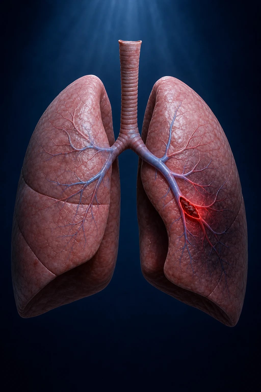
Massive vs submassive vs low-risk PE
| Feature | Massive (high-risk) | Submassive (intermediate-risk) | Low-risk |
|---|---|---|---|
| Haemodynamics | SBP <90 or shock | Normotensive | Normotensive |
| RV dysfunction (echo) | Present | Present | Absent |
| Cardiac biomarkers | Elevated | Elevated | Normal |
| Mortality | 30-60% | 5-15% | <3% |
| Initial treatment | Thrombolysis OR embolectomy | Anticoagulation + MONITOR | Anticoagulation |
| Rescue thrombolysis | Already given upfront | If deteriorates | Not needed |
| ICU admission | Yes | Yes (close monitoring) | No (ward or early discharge) |
| Risk stratification | Clinical (shock) | sPESI + echo + biomarkers | sPESI = 0 |
Acute management of massive PE
- Assess & resuscitate — ABC. Oxygen, ventilatory support if needed. Avoid excessive fluid (>500 mL bolus worsens RV). Vasopressors (noradrenaline) for shock
- Confirm diagnosis — CT pulmonary angiogram (if not delaying treatment). If unstable and high clinical suspicion → may treat empirically (echo: RV strain + DVT)
- Anticoagulation — therapeutic LMWH or IV unfractionated heparin (UFH preferred if may need procedures/thrombolysis)
- Thrombolysis — alteplase 100 mg over 2h (or 50 mg bolus if deteriorating). Indicated for massive PE. Reversal: aprotinin, cryoprecipitate, TXA if bleeding
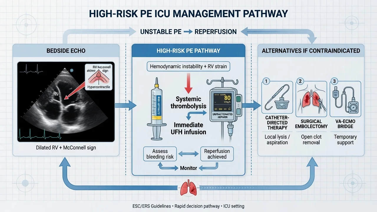
- If thrombolysis contraindicated/failed — catheter-directed thrombolysis, percutaneous embolectomy, or surgical embolectomy
- Consider IVC filter — if anticoagulation contraindicated and recurrent PE
- Escalate to ECMO — if refractory shock/collapse. VA-ECMO supports RV and perfusion while clot lyses
SAQ — Massive PE with systemic thrombolysis
10 minutes · 10 marks
A 58-year-old man, three weeks post right total hip replacement, presents with sudden-onset pleuritic chest pain and dyspnoea. On arrival he is diaphoretic and confused: GCS 14, SBP 76 mmHg, HR 132/min, RR 32/min, SpO2 88% on 15 L via non-rebreather. JVP is elevated; lung fields are clear. ECG shows sinus tachycardia with an S1Q3T3 pattern and T-wave inversion across V1 to V4. Bedside echocardiography demonstrates a dilated right ventricle (RV/LV ratio 1.2), the McConnell sign, septal flattening in systole, TAPSE 12 mm, and a plethoric IVC. CTPA confirms a saddle pulmonary embolus.
SAQ — Submassive PE risk stratification and the thrombolysis decision
10 minutes · 10 marks
A 67-year-old woman with metastatic breast cancer on weekly paclitaxel presents with two days of progressive dyspnoea and right-sided pleuritic chest pain. Vital signs: BP 112/68 mmHg, HR 108/min, RR 24/min, SpO2 94% on room air. She is not hypotensive. ECG shows sinus tachycardia only. CTPA demonstrates bilateral segmental pulmonary emboli with an RV/LV ratio of 1.1. Troponin T is 0.06 ng/mL (normal below 0.04); NT-proBNP is 1200 pg/mL (elevated). Bedside echocardiography shows mild RV dilatation, septal flattening in systole, and TAPSE 14 mm.
Clinical pearls
Red flags
Prognosis
PEITHO trial (Meyer 2014, NEJM)
RCT: 1006 patients with normotensive PE + RV dysfunction + troponin positive. Tenecteplase (weight-based single bolus) vs placebo, all received anticoagulation.
- Primary outcome (death/hemodynamic decompensation at 7 days): tenecteplase 2.6% vs placebo 5.6% (RR 0.44, NNT 33)
- Major bleeding: tenecteplase 11.5% vs placebo 2.4% (NNH 11)
- Haemorrhagic stroke: tenecteplase 2.4% vs placebo 0.2% (NNH 45)
- CONCLUSION: Routine thrombolysis in submassive PE reduces hemodynamic decompensation but at significant bleeding cost. Current practice: anticoagulation + monitor; reserve thrombolysis for clinical deterioration.
Diagnosis
Clinical probability assessment
Wells score: PE vs DVT
| Wells score for PE | Wells score for DVT | |
|---|---|---|
| Clinical signs/symptoms of DVT | 3.0 | 3.0 |
| PE more likely than alternative diagnosis | 3.0 | — |
| Heart rate >100 | 1.5 | — |
| Immobilisation ≥3 days or surgery in past 4 weeks | 1.5 | 1.0 |
| Previous DVT/PE | 1.5 | 1.0 |
| Haemoptysis | 1.0 | — |
| Malignancy (on treatment, palliative, or past 6 mo) | 1.0 | 1.0 |
| Tenderness along deep veins / ≥3 cm calf swelling | — | 1.0 each |
| Pitting oedema (symptomatic leg) | — | 1.0 |
| Collateral superficial veins | — | 1.0 |
| Alternative diagnosis at least as likely | — | −2.0 |
| Interpretation (3-tier) | 'PE likely' ≥4; 'PE unlikely' <4 (dichotomised) | High ≥3; Moderate 1-2; Low <1 |
| Interpretation (2-tier) | PE likely ≥4 / PE unlikely <4 | DVT likely ≥2 / unlikely <2 |
The dichotomised (2-tier) Wells score is preferred in modern algorithms because it simplifies the diagnostic pathway — 'PE unlikely' permits a D-dimer gate, while 'PE likely' mandates imaging regardless of D-dimer. The Wells score performs modestly (inter-observer variability, LR+ ≈5 for 'PE likely'), which is why it is never used alone — it gates D-dimer and imaging.[8] }[9] }
Diagnostic algorithm for suspected PE (Christopher / YEARS pathway)
- Apply Wells score (2-tier) — stratify into 'PE unlikely' (<4) or 'PE likely' (≥4). This is the entry point for every stable suspected PE.[9] }
- If 'PE unlikely' → check D-dimer — high-sensitivity ELISA/turbidimetric assay only. D-dimer NEGATIVE (<500 µg/L, or age-adjusted if >50 yr) → PE excluded, no imaging, discharge or seek alternative diagnosis.[16] } D-dimer POSITIVE → proceed to CTPA.
- If 'PE likely' → D-dimer is unhelpful (specificity too low) → go straight to CTPA.
- CTPA — first-line imaging in the haemodynamically stable patient. Shows filling defect in pulmonary artery, RV/LV ratio, and concomitant DVT.
- If CTPA contraindicated (renal failure, contrast allergy, pregnancy) → lower-limb compression venous ultrasound (if positive → treat as PE); if negative, V/Q scan or V/Q SPECT.
- If unstable/unable to transfer to CT → bedside echo (RV strain), bedside venous ultrasound. If high pre-test probability + RV strain → treat empirically as PE (do not delay reperfusion).[2] }
- YEARS algorithm (alternative) — assess 3 Wells items (clinical signs DVT, PE most likely, haemoptysis). If none present AND D-dimer <1000 → PE excluded; if D-dimer <500 → excluded regardless. Fewer CTPAs vs standard pathway.[21] }
D-dimer
D-dimer strategies in suspected PE
| Strategy | Rule | Use / evidence |
|---|---|---|
| Standard cutoff | <500 µg/L (FEU) excludes PE | Specificity poor (~50%), falls with age. Use only with 'PE unlikely' pre-test probability |
| PERC rule | If 'PE unlikely' + ALL 8 PERC criteria negative + age <50 → no D-dimer, no imaging | Kline 2008: safely reduces testing in low-risk young patients. Miss rate <1.5%. Must be <50 yr, no hormones, no haemoptysis, no surgery/trauma, no prior VTE, no unilateral leg swelling, HR <100, no oxygen requirement.[11] } |
| Age-adjusted D-dimer | Cutoff = age × 10 µg/L (if >50 yr) | ADJUST-PE: in >50 yr 'PE unlikely' patients, tripled the proportion in whom PE could be excluded (from 6.4% to 11.9%) with <1% 3-month VTE risk.[16] } |
| YEARS algorithm | 3-item Wells + tiered D-dimer (<1000 if 0 items, <500 if any) | YEARS study: safely reduced CTPA use ~17% vs conventional algorithm; low failure rate.[21] } |
| Pregnancy-adjusted | YEARS / adapted in pregnancy; CTPA if D-dimer positive | Avoid radiation; bilateral leg Doppler first. D-dimer physiologically rises in pregnancy so use as 'rule-out' gate only with low/moderate probability |
Imaging
CTPA vs V/Q scan in suspected PE
| Feature | CT pulmonary angiography (CTPA) | Ventilation/perfusion (V/Q) scan |
|---|---|---|
| Sensitivity/specificity | Sens ~83%, Spec ~96% (PIOPED II); misses subsegmental clots | Sens high (with SPECT); V/Q SPECT spec ~97% |
| Pros | Fast, widely available, gives alternative dx (aortic dissection, pneumonia, effusion), RV/LV ratio, concomitant DVT | No contrast nephropathy, lower radiation dose, preferred in pregnancy/renal failure/contrast allergy |
| Cons | Contrast nephropathy, radiation, overdiagnosis of subsegmental PE (incidentalomas) | Non-diagnostic 'intermediate' result in ~25-50% (especially with COPD/prior cardiopulmonary disease), no alternative diagnosis |
| PIOPED data | PPV high for segmental/proximal; low for subsegmental | High-probability scan: PPV ~88% (sufficient to treat); normal scan: PE excluded; 'intermediate' → further imaging.[7] } |
| Best use | Default first-line in stable patient | Pregnancy, renal failure, contrast allergy, follow-up of known PE |
| Reporting | Filling defect in PA; RV/LV ratio >0.9 suggests RV strain | 'High probability', 'intermediate', 'low', 'normal' (modified Prospective Investigative Study interpretation) |
Risk stratification (severity)
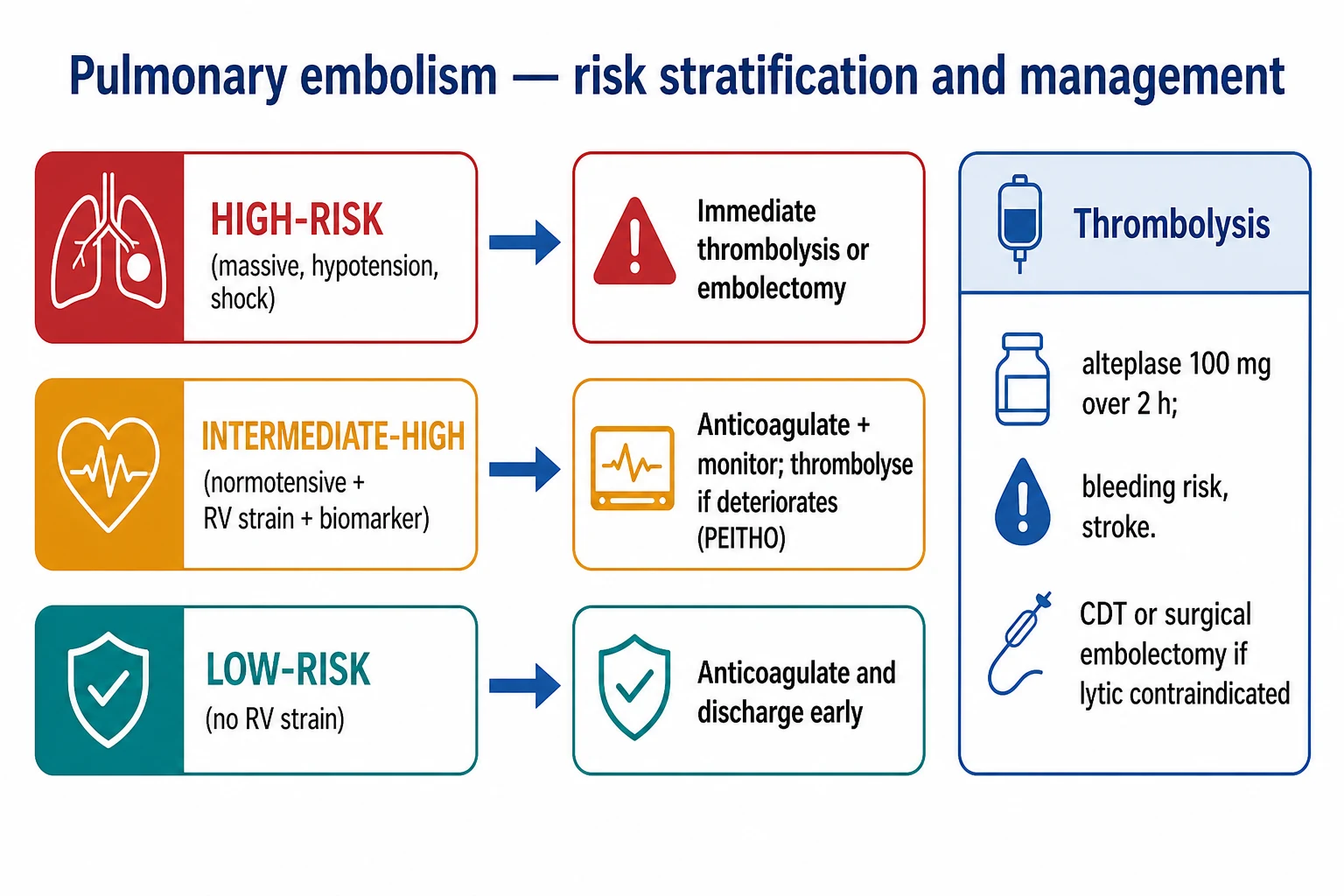
PE severity drives treatment intensity. The 2020 ESC algorithm stratifies by early mortality risk (in-hospital / 30-day):[2] }
- High-risk (massive) — clinical: shock or sustained hypotension (SBP <90 for >15 min, or needing catecholamines, or drop ≥40 mmHg). ~5% of PEs. Mortality >15%. Reperfusion mandatory (thrombolysis, surgical or percutaneous embolectomy, or catheter-directed treatment).
- Intermediate-risk (submassive) — normotensive but RV dysfunction (imaging) and/or myocardial injury (biomarkers). Subdivided:
- Intermediate-high: RV dysfunction AND biomarker elevation (both). ~20%. Monitor closely, consider rescue reperfusion.
- Intermediate-low: RV dysfunction OR biomarker elevation (one only). ~30%. Anticoagulate, monitor.
- Low-risk — sPESI = 0, no RV dysfunction, normal biomarkers. ~40-50%. Anticoagulate, possible early discharge / outpatient. [1]
sPESI / PESI
The full PESI score predicts 30-day mortality; the simplified PESI (sPESI) uses 6 equally weighted items (see SeverityGauge above).[10] } sPESI = 0 identifies low-risk patients suitable for outpatient/early-discharge management — provided they are also clinically stable, with good social support, no significant comorbidity, and (per ESC) no significant RV dysfunction or biomarker elevation. The Bova score and FAST score further risk-stratify intermediate-risk PE for haemodynamic deterioration.
Right ventricular strain
RV strain: imaging and biomarker markers
| Marker | Finding | Significance |
|---|---|---|
| Echocardiography — RV dilatation | RV/LV basal diameter ratio >0.6 (apical 4-chamber), or RV end-diastolic area > LV | Acute pressure overload; present in ~40% of normotensive PE |
| McConnell's sign | RV free-wall hypokinesis/akinesis with apical sparing (hyperdynamic apex) | Specific (~94%) but modestly sensitive (~77%) for PE; reflects differential wall stress |
| TAPSE (tricuspid annular plane systolic excursion) | <16 mm indicates RV systolic dysfunction | Per Rudski 2010 echo guidelines; independent predictor of PE mortality.[12] } |
| Septal flattening / D-shaped LV | Paradoxical septal motion in systole (pressure overload) and/or diastole (volume overload) | Indicates RV pressure >~40 mmHg |
| Tricuspid regurgitant jet velocity | >2.6 m/s (≈ >2.7-2.8) suggests pulmonary HTN | Reflects elevated pulmonary artery pressure |
| RV/LV ratio on CTPA | >0.9 (4-chamber reformats) | CT equivalent of echo RV dilatation; prognostic |
| Troponin (I/T) | Elevated = myocardial injury | RV microinfarction; risk of death/haemodynamic collapse |
| BNP / NT-proBNP | Elevated = RV wall stress/strain | Surrogate of RV dysfunction; combined with troponin identifies highest-risk patients |
| Lactate | Elevated = tissue hypoperfusion | Marker of haemodynamic compromise; prognostic in intermediate-risk PE |
The presence of both RV dysfunction (echo) and biomarker elevation (troponin/BNP) defines intermediate-high-risk PE — the group that warrants ICU admission for close haemodynamic monitoring and for whom a rescue thrombolysis strategy is reserved (treat with anticoagulation; give systemic lysis only if clinical deterioration).[1] } Either marker alone = intermediate-low risk; neither = low risk.
Thrombolysis
Alteplase for massive PE
Standard regimen: alteplase 100 mg IV over 2 hours (approved FDA regimen for massive PE). In cardiac arrest or peri-arrest, a 50 mg IV bolus is widely used. Stop UFH during infusion in many protocols (controversial — some centres continue without bolus). Resume heparin (no bolus) when aPTT <1.5× control (typically at end of infusion). Reversal of life-threatening bleeding: cryoprecipitate (fibrinogen), TXA / aminocaproic acid, and if available aprotinin; activate massive transfusion protocol. [1]
Contraindications to systemic thrombolysis
Absolute vs relative contraindications to alteplase
| Absolute | Relative (weigh benefit in massive PE) |
|---|---|
| Haemorrhagic stroke or stroke of unknown origin at any time | TIA in preceding 6 months |
| Ischaemic stroke in preceding 6 months | Central nervous system neoplasm |
| CNS neoplasm | Recent major surgery (<3 weeks) |
| Major trauma/surgery/head injury in preceding 3 weeks | Recent internal bleeding (<4 weeks) — non-CNS |
| Active bleeding / bleeding diathesis | Non-compressible vascular puncture |
| Structural cerebral vascular lesion | Pregnancy / early postpartum |
| Recent brain/spinal surgery | Age >75 yr |
| Active intracranial / spinal lesion | Uncontrolled HTN (SBP >180) |
| Recent (≤2 wk) intracranial/spinal surgery | Anticoagulant use (INR elevated) |
| Thrombocytopenia (<100 × 10⁹/L) | Peptic ulcer, severe liver disease, infective endocarditis |
In massive PE, the only true absolute contraindication is active life-threatening bleeding / recent haemorrhagic stroke — the high mortality of untreated massive PE justifies lysis even with relative contraindications. Where lysis is contraindicated, surgical embolectomy or percutaneous catheter-directed thrombolysis/embolectomy are the alternatives.[2] }
Catheter-directed thrombolysis (CDT)
Low-dose alteplase (typically 1 mg/hour per catheter, total 20-24 mg over 12-24 h) infused directly into the pulmonary artery via ultrasound-assisted catheter (EKOS) or standard multi-side-hole catheter. Advantages: fraction of systemic alteplase dose → markedly lower major bleeding; rapid reduction in RV/LV ratio and pulmonary artery pressure. [1]
- ULTIMA trial (Kucher 2014, Circulation): ultrasound-assisted CDT vs systemic heparin in intermediate-risk PE (RV/LV ≥1.0). CDT reduced RV/LV ratio at 24 h significantly more than heparin (0.99 ± 0.16 vs 1.08 ± 0.23); no major bleeding. Small but pivotal trial establishing efficacy in submassive PE.[17] }
- SEATTLE II (Piazza 2015, JACC): 150 patients with massive or submassive PE. US-facilitated CDT reduced RV/LV ratio by 0.42 and PA pressure by 7 mmHg; major bleeding 10% but zero intracranial haemorrhage.[18] }
- Indication: submassive (intermediate-risk) PE with RV dysfunction where systemic lysis is high-risk/contraindicated, and in centres with interventional radiology. AHA guidelines support CDT for selected patients with intermediate-high-risk PE failing anticoagulation.
Catheter-directed therapy for DVT — ATTRACT
Phlegmasia / iliofemoral DVT — to lyse or not
- Confirm iliofemoral (proximal) DVT with ultrasound ± venography; assess limb viability (pulse, capillary refill, sensation).
- Phlegmasia cerulea dolens (blue, painful, swollen limb ± pulseless) → emergency: catheter-directed thrombolysis ± pharmacomechanical thrombolysis (rheolytic/PMT), or surgical thrombectomy if lysis contraindicated.
- Stable iliofemoral DVT (no limb threat) — ATTRACT trial (Vedantham 2017): CDT + PMT vs anticoagulation alone reduced post-thrombotic syndrome (PTS) severity (especially severe PTS) but did NOT reduce overall PTS rate, at the cost of more bleeding. Practice: selective use in severe proximal DVT with high PTS risk; not routine.[22] }
- May-Thurner syndrome (left common iliac vein compressed by right common iliac artery) → consider venoplasty + stenting after thrombolysis.
- Post-procedure: resume anticoagulation; surveillance ultrasound; assess for underlying thrombophilia/malignancy.
Anticoagulation
DOAC vs warfarin vs LMWH for VTE
| Agent | Acute dose | Maintenance | Renal / special | Trial evidence |
|---|---|---|---|---|
| Rivaroxaban (Xa) | 15 mg BD × 21 days | 20 mg OD | Avoid CrCl <15; caution <30 | EINSTEIN-DVT/PE — non-inferior to warfarin, less major bleeding. Single-drug (no parenteral lead-in).[14] } |
| Apixaban (Xa) | 10 mg BD × 7 days | 5 mg BD | Avoid CrCl <15; dose-adjust if ≥80 yr or ≤60 kg or Cr ≥133 | AMPLIFY — non-inferior, less bleeding. Single-drug. |
| Dabigatran (IIa) | Parenteral lead-in 5-10 days (LMWH/UFH) then | 150 mg BD | Avoid CrCl <30 | RE-COVER — non-inferior to warfarin, less major bleeding; requires heparin lead-in.[13] } |
| Edoxaban (Xa) | Parenteral lead-in 5-10 days then | 60 mg OD (30 mg if CrCl 15-50 or ≤60 kg) | Contraindicated CrCl <15; ineffective in CrCl >95 | Hokusai-VTE — non-inferior to warfarin, less clinically relevant bleeding.[15] } |
| Warfarin (Vit K epoxide reductase) | Overlap parenteral ≥5 days AND until INR 2-3 × 24 h | INR 2-3 | Preferred for antiphospholipid syndrome, mechanical valve, severe renal failure | Long-standing standard; DOACs preferred for most others |
| LMWH (enoxaparin) | 1 mg/kg SC BD (or 1.5 mg/kg OD) | Same | Preferred for cancer-associated VTE and pregnancy; avoid CrCl <30 (dose-adjust) | CATCH, SELECT-D, Caravaggio — DOACs non-inferior in cancer too |
| IV UFH | 80 U/kg bolus, 18 U/kg/h infusion (weight-based, aPTT 1.5-2.5×) | — | Preferred in massive PE / pre-thrombolysis (short half-life, reversible), severe renal failure, obesity | — |
| Fondaparinux | 5-10 mg SC OD (weight-based) | — | Avoid CrCl <30; HIT-safe alternative | — |
Selection principles: [1]
- Stable, otherwise well → rivaroxaban or apixaban (single-drug, oral from day 1, no monitoring).
- Massive PE / pre-thrombolysis / unstable / likely procedures → IV UFH (titratable, short half-life, fully reversible with protamine).
- Cancer-associated VTE → apixaban, rivaroxaban, or LMWH per current guidance (Caravaggio: apixaban non-inferior to dalteparin; SELECT-D: rivaroxaban reduced recurrence vs LMWH with more non-major bleeding).
- Antiphospholipid syndrome (triple-positive) → warfarin (DOACs inferior in APS — TRAPS trial).
- Severe renal failure (CrCl <15-30) → IV UFH or warfarin.
- Pregnancy / breastfeeding → LMWH throughout; switch to warfarin postpartum (breastfeeding OK).
- Extremes of weight → IV UFH (reliable) or LMWH with anti-Xa monitoring. [1]
Duration of anticoagulation
Duration of anticoagulation by VTE provocation
| Scenario | Duration |
|---|---|
| Provoked by major transient risk (surgery, trauma, immobility) | 3 months |
| Provoked by non-surgical transient risk (oestrogen, pregnancy, transient immobility) | 3 months |
| First unprovoked proximal DVT/PE | ≥3 months, then reassess bleed vs recurrence risk; extend if low bleeding risk |
| Second unprovoked VTE | Lifelong |
| Cancer-associated VTE | Extended while cancer active |
| Antiphospholipid syndrome | Lifelong warfarin |
| Distal (calf) DVT, symptomatic | 3 months |
Recurrent VTE despite therapeutic anticoagulation → switch LMWH (if on warfarin/DOAC), check adherence/compliance, screen for malignancy and thrombophilia, consider IVC filter. [1]
Inferior vena cava filters
Indications (per SIR and CHEST/ESC guidance)
IVC filter decision pathway
- Absolute indication: acute VTE (proximal DVT or PE) with a CONTRAINDICATION to anticoagulation (active bleeding, imminent surgery) — place retrievable filter.[19] }
- Absolute indication: recurrent PE despite adequate therapeutic anticoagulation → place filter AND continue/escalate anticoagulation.[1] }
- Relative indications (case-by-case): massive PE with residual DVT and high re-embolisation risk; large free-floating proximal DVT; thrombolysis/embolectomy planned; severe cardiopulmonary compromise where a further embolus would be fatal; iliofemoral DVT in pregnancy near delivery.
- NOT recommended: routine prophylaxis in trauma/surgery patients who CAN receive anticoagulation (PREPIC2 showed no benefit when added to anticoagulation).[20] }
- Use retrievable filters — remove within 3-6 months once anticoagulation can be resumed. Track every filter in a registry to avoid permanent retention.
- Complications: filter migration/embolisation, IVC thrombosis (causes filter dysfunction + leg swelling), IVC penetration (through wall → adjacent structures), retrieval failure, access-site thrombosis.
PREPIC trials
- PREPIC (1998, NEJM): permanent IVC filter + anticoagulation vs anticoagulation alone → filter reduced recurrent PE (1.1% vs 4.8% at 12 days) but increased DVT recurrence at 2 years (21% vs 12%); no mortality difference. At 8 years PE reduction persisted, DVT excess persisted, no survival benefit.[19] }
- PREPIC2 (2015, JAMA): retrievable filter + anticoagulation vs anticoagulation alone in patients with PE + DVT + risk factors → NO reduction in recurrent PE at 3 or 6 months. Filters confer no benefit when patients can receive anticoagulation.[20] }
Subsegmental PE management
Subsegmental PE (isolated) — management
- Confirm isolated subsegmental PE on CTPA (clot in a subsegmental branch, no more proximal clot). Increasingly detected with modern multidetector CT — many are clinically insignificant.
- Assess whether symptomatic: presentation with PE symptoms (pleuritic pain, dyspnoea) vs incidental finding on CT performed for another reason (e.g., cancer staging).
- Risk-stratify recurrence / proximal DVT: perform bilateral lower-limb compression venous ultrasound — if proximal DVT present, treat as proximal VTE (full-dose, 3+ months).
- Decision per CHEST 2016 guidance: with risk factors for recurrence (cancer, previous VTE, hospitalisation, proximal DVT) OR symptomatic → anticoagulate (DOAC, full duration); with no risk factors AND asymptomatic → clinical surveillance (serial imaging/clinical review), anticoagulation may be withheld. Discuss risk-benefit with patient.
- Reassess for provoking cause (occult malignancy screen if unprovoked).
- Do NOT routinely image follow-up unless recurrent symptoms — residual thrombus is common and does not guide duration.
The rise in isolated subsegmental PE diagnosis (driven by sensitive multidetector CT) has not been matched by a fall in PE mortality, fuelling concern about overdiagnosis of clinically unimportant clots. The principle: an isolated subsegmental PE in an asymptomatic patient with no provoking risk factor may reasonably be observed rather than anticoagulated — individualise.[3] }
Clinical pearls (extended)
Red flags (extended)
Prognosis and key trials
RE-COVER (Schulman 2009, NEJM) — dabigatran for acute VTE
RCT: 2564 patients with acute VTE after 5-10 days parenteral anticoagulation, randomised to dabigatran 150 mg BD vs warfarin (INR 2-3), 6 months.
- Primary outcome (recurrent VTE + related death): dabigatran 2.4% vs warfarin 2.1% — non-inferior (HR 1.10).
- Major bleeding: similar (1.6% vs 1.9%).
- CONCLUSION: dabigatran (after parenteral lead-in) is as effective and as safe as warfarin for acute VTE. Established DOACs as warfarin alternatives.[13] }
EINSTEIN-PE (Buller 2012, NEJM) — rivaroxaban for PE
RCT: 4832 patients with acute symptomatic PE randomised to rivaroxaban (15 mg BD ×3 wk, then 20 mg OD) vs warfarin (INR 2-3) with enoxaparin lead-in.
- Primary efficacy (recurrent VTE): rivaroxaban 2.1% vs warfarin 1.8% — non-inferior.
- Major or clinically relevant non-major bleeding: rivaroxaban 10.3% vs warfarin 11.4% — significantly less.
- CONCLUSION: rivaroxaban (single-drug oral from day 1, no heparin lead-in) non-inferior to warfarin with reduced bleeding. Foundation of single-drug DOAC therapy for PE.[14] }
Hokusai-VTE (Buller 2013, NEJM) — edoxaban for VTE
RCT: 8240 patients with acute VTE after 5-10 days parenteral anticoagulation, randomised to edoxaban 60 mg OD vs warfarin (INR 2-3), 3-12 months.
- Primary efficacy (recurrent VTE + related death): edoxaban 3.2% vs warfarin 3.5% — non-inferior.
- Clinically relevant bleeding: edoxaban 8.5% vs warfarin 10.3% — less bleeding.
- Subgroup caveat: edoxaban less effective in CrCl >95 mL/min → contraindicated in renal hyperfiltration.
- CONCLUSION: edoxaban (once-daily, post-lead-in) non-inferior with less bleeding. Reinforces DOAC class; defined renal exclusion.[15] }
ULTIMA (Kucher 2014, Circulation) — ultrasound-assisted CDT for intermediate-risk PE
RCT: 59 patients with acute intermediate-risk PE (RV/LV ≥1.0) randomised to ultrasound-assisted catheter-directed alteplase (~20 mg over 15 h) + heparin vs heparin alone.
- Primary outcome (RV/LV ratio reduction at 24 h): CDT 0.99 ± 0.16 vs heparin 1.08 ± 0.23 (P=0.002) — greater RV unloading.
- Major bleeding: none in either group.
- CONCLUSION: low-dose catheter-directed alteplase reverses RV dilatation more than anticoagulation alone, with low bleeding — pivotal for CDT in intermediate-risk PE. Small sample; no outcome endpoint.[17] }
SEATTLE II (Piazza 2015, JACC) — ultrasound-facilitated CDT for PE
Single-arm multicentre trial: 150 patients with acute massive (31) or submassive (119) PE receiving ultrasound-facilitated catheter-directed low-dose alteplase (~24 mg over ~15 h per lung, max).
- RV/LV ratio: reduced from 1.54 to 1.13 (mean reduction 0.42) at 48 h.
- PA systolic pressure: reduced by ~7 mmHg.
- Major bleeding: 16% (10%), intracranial haemorrhage: 0%.
- CONCLUSION: substantial RV unloading with very low intracranial bleeding. No control arm; supports CDT safety/efficacy signal.[18] }
ATTRACT (Vedantham 2017, NEJM) — pharmacomechanical CDT for DVT
RCT: 692 patients with acute iliofemoral or femoral-popliteal DVT randomised to pharmacomechanical catheter-directed thrombolysis (PMT) + anticoagulation vs anticoagulation alone.
- Primary outcome (post-thrombotic syndrome at 24 mo): PMT 47% vs control 48% — no difference in overall PTS.
- Severe PTS: PMT 4% vs control 6% — small reduction in severe PTS.
- Major bleeding: PMT 1.7% vs control 0.3% — more bleeding.
- CONCLUSION: routine CDT for proximal DVT does NOT reduce overall PTS and increases bleeding. Selective use for phlegmasia and severe iliofemoral DVT; not routine.[22] }
PREPIC (1998, NEJM) & PREPIC2 (2015, JAMA) — IVC filters
PREPIC: 400 patients with proximal DVT ± PE, randomised to permanent IVC filter + anticoagulation vs anticoagulation alone.
- Recurrent PE at 12 days: filter 1.1% vs control 4.8% (reduced); DVT at 2 yr: filter 21% vs control 12% (increased); no mortality difference at 2 or 8 years.[19] }
PREPIC2: 399 patients with PE + DVT + risk factors, randomised to retrievable filter + anticoagulation vs anticoagulation alone.
- Recurrent PE at 6 months: filter 1% vs control 3% — no significant difference.
- CONCLUSION: IVC filters prevent PE when anticoagulation is impossible, but confer NO benefit (and raise DVT risk) when added to effective anticoagulation.[20] }
ADJUST-PE (Righini 2014, JAMA) — age-adjusted D-dimer
Prospective multicentre management study: 3346 patients >50 yr with 'PE unlikely' (revised Geneva score) and age-adjusted D-dimer (age × 10 µg/L, max 1000) used to rule out PE.
- PE exclusion rate: 11.9% (age-adjusted) vs 6.4% with standard 500 cutoff — nearly tripled.
- 3-month thromboembolic risk in non-imaged patients: 0.3% (below the <1% safety threshold).
- CONCLUSION: age-adjusted D-dimer safely increases the proportion of older patients in whom PE can be excluded without imaging.[16] }
YEARS (van der Hulle 2017, Lancet) — simplified PE algorithm
Prospective multicentre diagnostic management study: 3465 consecutive patients with suspected PE assessed by 3-item Wells (DVT signs, PE most likely, haemoptysis) + tiered D-dimer cutoff (<1000 if no items, <500 if any item present).
- PE excluded without imaging in 48% of patients.
- Failure rate (VTE at 3 months in non-imaged): 0.15% (95% CI 0.05-0.45) — well below safety threshold.
- CONCLUSION: YEARS safely reduced CTPA use vs conventional algorithms while maintaining diagnostic safety.[21] }
Quick reference
At-a-glance: PE severity and action
| Severity | Haemodynamics | RV/echo | Biomarkers | sPESI | Action |
|---|---|---|---|---|---|
| High (massive) | Shock / SBP <90 | Variable | Variable | Any | Thrombolysis OR surgical/percutaneous embolectomy OR CDT; ICU; VA-ECMO if refractory |
| Intermediate-high | Normotensive | Dysfunction | Elevated | ≥1 | Anticoagulation + ICU monitoring; rescue thrombolysis if deterioration |
| Intermediate-low | Normotensive | Dysfunction OR biomarker | (one only) | ≥1 | Anticoagulation + ward monitoring |
| Low | Normotensive | Normal | Normal | 0 | Anticoagulation; consider early discharge/outpatient |
One-minute ICU PE management checklist
- ABCDE — oxygen, vasopressors (noradrenaline), avoid fluid >500 mL (worsens RV).
- Severity — shock/SBP <90? (massive → reperfusion) vs normotensive.
- Confirm — CTPA if stable; bedside echo + Doppler if unstable.
- Risk-stratify — sPESI, echo RV strain, troponin/BNP, lactate.
- Anticoagulate — IV UFH if massive/procedures; DOAC if stable.
- Reperfusion — alteplase 100 mg/2h (massive); CDT/rescue if intermediate-high deteriorating.
- Source/Duration — provoked (3 mo) vs unprovoked (extended) vs cancer (extended) vs APS (lifelong warfarin).
- Plan — IVC filter ONLY if anticoagulation contraindicated or failure; track retrievable filters; screen for malignancy in unprovoked.
References
- [1]Stevens SM, et al. Towards circular phosphorus: The need of inter- and transdisciplinary research to close the broken cycle Ambio, 2022.PMID 34013441
- [2]Konstantinides SV, et al. Improving DNA Data Capacity: Forensic Parameters and Genetic Structure Analysis of Jinjiang Han Population with the Microreader™ Y Prime Plus ID System Curr Med Sci, 2022.PMID 35403953
- [3]Kearon C, et al. Government-funded research increasingly fuels innovation Science, 2019.PMID 31221848
- [4]Tapson VF, et al. Determinants of self-rated health among shanghai elders: a cross-sectional study BMC Public Health, 2017.PMID 29029627
- [5]Chatterjee S, et al. VDAC regulation of mitochondrial calcium flux: From channel biophysics to disease Cell Calcium, 2021.PMID 33529977
- [6]Watson HG, et al. Can sand nourishment material affect dune vegetation through nutrient addition? Sci Total Environ, 2020.PMID 32278174
- [7]PIOPED Investigators Value of the ventilation/perfusion scan in acute pulmonary embolism. Results of the prospective investigation of pulmonary embolism diagnosis (PIOPED) JAMA, 1990.PMID 2332918
- [8]Wells PS, et al. Derivation of a simple clinical model to categorize patients probability of pulmonary embolism: increasing the models utility with the SimpliRED D-dimer Thromb Haemost, 2000.PMID 10744147
- [9]van Belle A, et al. (Christopher Study) Effectiveness of managing suspected pulmonary embolism using an algorithm combining clinical probability, D-dimer testing, and computed tomography JAMA, 2006.PMID 16403929
- [10]Aujesky D, et al. A prediction rule to identify low-risk patients with pulmonary embolism Arch Intern Med, 2006.PMID 16432084
- [11]Kline JA, et al. Prospective multicenter evaluation of the pulmonary embolism rule-out criteria J Thromb Haemost, 2008.PMID 18318689
- [12]Rudski LG, et al. Guidelines for the echocardiographic assessment of the right heart in adults: a report from the American Society of Echocardiography endorsed by the European Association of Echocardiography, a registered branch of the European Society of Cardiology, and the Canadian Society of Echocardiography J Am Soc Echocardiogr, 2010.PMID 20620859
- [13]Schulman S, et al. (RE-COVER) Dabigatran versus warfarin in the treatment of acute venous thromboembolism N Engl J Med, 2009.PMID 19966341
- [14]Buller HR, et al. (EINSTEIN-PE) Oral rivaroxaban for the treatment of symptomatic pulmonary embolism N Engl J Med, 2012.PMID 22449293
- [15]Buller HR, et al. (Hokusai-VTE) Edoxaban versus warfarin for the treatment of symptomatic venous thromboembolism N Engl J Med, 2013.PMID 23991658
- [16]Righini M, et al. (ADJUST-PE) Age-adjusted D-dimer cutoff levels to rule out pulmonary embolism: the ADJUST-PE study JAMA, 2014.PMID 24643601
- [17]Kucher N, et al. (ULTIMA) Randomized, controlled trial of ultrasound-assisted catheter-directed thrombolysis for acute intermediate-risk pulmonary embolism Circulation, 2014.PMID 24226805
- [18]Piazza G, et al. (SEATTLE II) A Prospective, Single-Arm, Multicenter Trial of Ultrasound-Facilitated, Catheter-Directed, Low-Dose Fibrinolysis for Acute Massive and Submassive Pulmonary Embolism: The SEATTLE II Study JACC Cardiovasc Interv, 2015.PMID 26315743
- [19]PREPIC Study Group (Decousus, et al.) A clinical trial of vena caval filters in the prevention of pulmonary embolism in patients with proximal deep-vein thrombosis. Prévention du Risque d'Embolie Pulmonaire par Interruption Cave Study Group N Engl J Med, 1998.PMID 9459643
- [20]Mismetti P, et al. (PREPIC2) Effect of a retrievable inferior vena cava filter plus anticoagulation vs anticoagulation alone on risk of recurrent pulmonary embolism: a randomized clinical trial JAMA, 2015.PMID 25919526
- [21]van der Hulle T, et al. (YEARS) Simplified diagnostic management of suspected pulmonary embolism (the YEARS study): a prospective, multicentre, cohort study Lancet, 2017.PMID 28549662
- [22]Vedantham S, et al. (ATTRACT) Pharmacomechanical Catheter-Directed Thrombolysis for Deep-Vein Thrombosis N Engl J Med, 2017.PMID 29211671