ICU · Cardiovascular
Advanced haemodynamic monitoring in the ICU
Also known as Haemodynamic monitoring · Pulmonary artery catheter (PAC) · Arterial line · PiCCO/LiDCO · Echocardiography in ICU · Transpulmonary thermodilution · Pulse contour analysis · Fluid responsiveness · Central venous pressure
Haemodynamic monitoring guides resuscitation in critically ill patients. From basic (clinical examination, urine output, arterial line, CVP line) to advanced (invasive cardiac output monitoring). Levels: (1) Basic: arterial line (BP, blood sampling, waveform), CVP line (right atrial pressure — a POOR predictor of fluid responsiveness). (2) Intermediate: echocardiography (non-invasive, qualitative + quantitative — the most versatile ICU tool), lactate, ScvO2. (3) Advanced: pulmonary artery catheter (PAC — gold standard but invasive, declining use after FACTT/PAC-Man/ESCAPE), transpulmonary thermodilution (PiCCO/TPTD — GEDV, ITBV, EVLW, PVPI), lithium dilution (LiDCO), pulse contour analysis (arterial line-derived CO). Fluid responsiveness must be assessed with DYNAMIC tests (passive leg raise, fluid challenge, SVV/PPV) not static markers (CVP). Choose monitoring based on the clinical question, not routinely.
On this page & tools
Your progress
Saved locally on this device.
Target exams
Red flags

The framework: matching the monitor to the question
Haemodynamic monitoring is not a hierarchy of "better" devices — it is a set of tools that each answer a different question. The art of ICU monitoring is to define the question first, then choose the least invasive monitor that answers it.[1][8]
| The clinical question | The best monitor |
|---|---|
| Is the patient hypotensive / what is the blood pressure? | Arterial line (continuous BP + sampling) |
| What is the heart rate and rhythm? | ECG |
| Is the heart failing, empty, or obstructed? | Echocardiography (FoCUS) |
| Will the patient respond to fluid? | Dynamic test — passive leg raise + real-time CO (echo LVOT VTI, PiCCO, FloTrac) |
| What is the preload (volume status)? | GEDV (PiCCO), or IVC + echo — not CVP, not PAWP alone |
| What is the cardiac output? | Thermodilution (PAC, PiCCO), lithium dilution (LiDCO), pulse contour, echo (LVOT VTI) |
| What is the left-sided filling pressure? | PAWP (PAC), or E/e' (echo) |
| Is there pulmonary oedema / lung water? | EVLW (PiCCO) — more direct than CXR or PAWP |
| Is the oxygen balance adequate? | SvO2 (PAC, mixed) or ScvO2 (central line); lactate |
| What is the vascular tone (SVR)? | Calculated from MAP, CVP, CO (any CO device) |
Principle: escalate from non-invasive to invasive only when the cheaper, safer monitor cannot answer the question, and when the answer will change management ("do not monitor what you cannot interpret"). [1]
Monitoring levels
Basic monitoring
Every ICU patient
- Clinical examination: BP, HR, capillary refill, urine output, skin temperature, mental status
- Arterial line: continuous BP, blood sampling, waveform analysis. Complications: thrombosis, infection, ischaemia
- CVP line: right atrial pressure. POOR predictor of fluid responsiveness. Useful for: access, trend monitoring (not absolute value), ScvO2 sampling, waveform diagnosis of right-heart pathology
- Pulse oximetry: SpO2. ECG: rhythm, rate, ischaemia
Intermediate monitoring
Selected patients
- Echocardiography: non-invasive, rapid. Assesses: LV/RV function, valve function, preload (IVC, LVOT VTI), pericardial effusion, cardiac output (LVOT VTI). Can be repeated. Focused (FoCUS) vs advanced (CCE, TOE).
- Lactate: marker of tissue perfusion (clearance = resuscitation adequacy)
- ScvO2 (central venous oxygen saturation): marker of oxygen extraction (target >70%)
Advanced monitoring
Complex cases
- Pulmonary artery catheter (PAC/Swan-Ganz): measures PA pressure, wedge pressure (LA pressure), CO (thermodilution), mixed venous oxygen (SvO2). Gold standard for CO measurement. But: invasive, complications (arrhythmia, perforation, infection), no proven mortality benefit (PAC-Man, FACTT, ESCAPE trials). Use selectively: pulmonary HTN, complex shock, RV failure, post-cardiac surgery, MCS titration.
- Transpulmonary thermodilution (PiCCO/TPTD): measures CO (thermodilution), GEDV (preload), ITBV, EVLW (extravascular lung water), PVPI, and a calibrated continuous pulse-contour CO + SVV. Less invasive than PAC (central venous + femoral arterial line).
- Lithium dilution (LiDCO): calibrates a pulse-contour algorithm with a lithium bolus; only needs a peripheral arterial line (radial OK).
- Pulse contour analysis: derives continuous CO from arterial waveform (requires calibration — thermodilution or lithium). LiDCO (lithium), FloTrac/Vigileo (uncalibrated). Continuous CO monitoring.
Basic vs advanced haemodynamic assessment — when to escalate
Most ICU patients need only basic monitoring. The decision to escalate is triggered by a clinical question that basic monitoring cannot answer, not by the availability of a device.[1][8]
When to escalate from non-invasive to invasive monitoring
Step 1 — Basic assessment (every patient)
History, examination (mottling, capillary refill >3 s, cold/warm peripheries), urine output, BP (NIBP or arterial line), HR, lactate, ScvO2, and a focused echo (FoCUS). This is sufficient for the majority: uncomplicated sepsis responding to fluids, postoperative patients, monitored arrhythmias.
Step 2 — Escalate to intermediate (echo + dynamic testing)
Escalate when: shock is not responding to initial resuscitation; there is diagnostic uncertainty about the shock type; you need to assess fluid responsiveness; or vasoactive drugs are being titrated. Add a focused/comprehensive echo (is the heart failing, empty, or obstructed? what is the CO?), and a dynamic fluid-responsiveness test (passive leg raise with echo LVOT VTI or PiCCO/FloTrac).
Step 3 — Escalate to advanced (invasive CO monitoring)
Reserve for: complex or mixed shock not resolved by echo; cardiogenic shock (especially RV infarct, mechanical complications, MCS titration); pulmonary hypertension / suspected RV failure; differentiation of pulmonary oedema from ARDS (EVLW or PAWP); the high-risk perioperative patient. Choose the device by the question: pressure/CO/oxygen balance → PAC; volume/lung water/responsiveness → PiCCO; simple continuous CO → LiDCO/FloTrac.
Stop / de-escalate
Invasive lines accumulate complications (infection, thrombosis, ischaemia). Remove the arterial line, CVC, PiCCO, or PAC as soon as the patient is stable and the data are no longer changing management. The most common monitoring error is leaving an advanced device in place "just in case".
Indications to escalate
Trigger
- Shock not responding to initial fluid/vasopressor therapy within the first hour
- Persistent or rising lactate despite resuscitation
- Need for >1 vasoactive infusion, or rapidly escalating doses
- Diagnostic uncertainty: which type of shock? (distributive, cardiogenic, obstructive, hypovolaemic)
- Suspected pulmonary hypertension, RV failure, or RV infarct
- Differentiating hydrostatic pulmonary oedema from ARDS / permeability oedema
- High-risk perioperative cardiac or major surgery patient (cardiac output-guided therapy)
What NOT to escalate for
Don't
- Routine placement of a PAC "for monitoring" — no mortality benefit (FACTT, PAC-Man, ESCAPE)
- Using CVP to guide fluid therapy — no predictive value (Marik 2013)
- Placing an arterial line where NIBP is adequate and stable
- Advanced monitoring you cannot interpret, or that will not change management
- Leaving devices in once the question is answered — remove to reduce line-related complications
The arterial line — insertion and waveform analysis
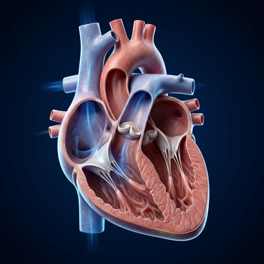
The arterial line is the workhorse of ICU monitoring: continuous beat-to-beat blood pressure, arterial blood gas sampling, and — through its waveform — derived information on stroke volume, the circulation, and fluid responsiveness. Correct interpretation requires understanding the physical system that generates the trace.[1][2]
Insertion technique and site selection
Site
Notes
- **Radial** (first choice): superficial, compressible, low complication rate. But reflects peripheral (not central) pressure — systolic is amplified ~5–15 mmHg above aortic. Allen test has poor predictive value and is no longer routinely required.
- **Femoral**: most accurate central pressure (closest to aorta), reliable in shock/vasoconstriction/hypothermia when radial under-reads; higher risk of retroperitoneal bleed/infection, and awkward for a mobile patient.
- **Brachial**: good in shock but end-artery (no collateral) — higher ischaemia risk; avoid if possible.
- **Dorsalis pedis / axillary**: alternatives; axillary useful for long-term central access.
Technique: aseptic (full barrier for femoral), ultrasound-guided where available. Modified Seldinger (catheter-over-guidewire) or catheter-over-needle for radial. Use short, stiff tubing; flush with heparinised or saline pressurised system at 3 mL/h plus a fast-flush valve. Zero the transducer to atmospheric pressure and level to the phlebostatic axis (4th intercostal space, mid-axillary line — the right atrium) for every measurement; re-level whenever the patient moves. [1]
The arterial waveform — what each part means
The normal arterial trace has a rapid systolic upstroke (the anacrotic limb, ventricular ejection), a systolic peak, the dicrotic notch (incisura — closure of the aortic valve), and the diastolic runoff down to end-diastolic pressure. Three pieces of clinical information live in the morphology: [1]
- Pulse pressure (systolic − diastolic): wide pulse pressure → high stroke volume / low SVR (aortic regurgitation, sepsis, PDA, anaemia); narrow → low stroke volume / high SVR (cardiogenic shock, severe vasoconstriction, aortic stenosis, tachycardia).
- Pulse pressure variation (PPV) / systolic pressure variation (SPV) with respiration — a marker of fluid responsiveness in ventilated patients (see below).
- Upstroke slope (dP/dt) — a rough, load-dependent index of contractility; a slow/slurred upstroke suggests poor LV function. [1]
Dynamic response — natural frequency and damping (high-yield exam topic)
The pressure trace is transmitted through a fluid-filled catheter + tubing + transducer — a resonant system that must be correctly damped to reproduce the true pressure. Two failure modes:[1]
| Problem | Mechanism | Effect on the trace | Effect on numbers |
|---|---|---|---|
| Underdamping ("resonance/ringing") | Natural frequency too low / system too stiff | Overshoot and ringing after the systolic peak; spuriously tall systolic spikes | Overestimates systolic, underestimates diastolic; MAP is preserved |
| Overdamping | Air bubbles, kinked line, soft tubing, clot | Slurred upstroke, loss of the dicrotic notch, low-amplitude trace | Underestimates systolic, overestimates diastolic; MAP is preserved |
| Optimally damped | Correct natural frequency + damping coefficient ~0.6–0.7 | Clean trace, 1–2 small oscillations after fast flush | Accurate systolic, diastolic and MAP |
The fast-flush ("square-wave") test — the bedside check: pull the fast-flush valve briefly and release.
- Optimal: 1–2 sharp oscillations before returning to baseline.
- Underdamped: many oscillations (3+) that ring before settling (high-amplitude, slowly decaying).
- Overdamped: no oscillations, slow slurred return to baseline. [1]
The cardinal rule: in both under- and over-damping the mean arterial pressure (MAP) remains accurate. So if the trace looks wrong, trust the MAP, distrust the systolic/diastolic, and fix the system (flush out bubbles, check for kinks; if underdamped, add a damping device or shorter tubing). [1]
Other pitfalls: transducer drift (re-zero hourly and after position change); wrong level (every 5 cm error = ~3.7 mmHg); radial–femoral gradient in shock (periphery vasoconstricts first — femoral reads higher); catheter whip; and cuff–line discrepancy (lines are generally more accurate; NIBP may be preferred in severe peripheral vasoconstriction if the line is over/under-damped). [1]
[1]Central venous pressure (CVP) — what it is, what it tells you, and its limits
CVP is the pressure of blood in the great thoracic veins at the entrance to the right atrium — a surrogate of right atrial pressure and, indirectly, right ventricular preload. It is measured via the distal port of a central venous catheter (tip in the SVC–RA junction), zeroed at the phlebostatic axis, and read at end-expiration (to minimise intrathoracic pressure artefact) and end-diastole (just before the R wave on the ECG). Normal range is conventionally 2–8 mmHg.[1][7]
Determinants of CVP
CVP is set by four things, which is why a single number is so hard to interpret:
- Venous return / circulating volume (the variable people assume it measures).
- Right heart compliance and function (RV infarct, pulmonary hypertension, tricuspid disease all raise CVP independent of volume).
- Intrathoracic / intrapericardial pressure — PEEP and tension pneumothorax raise the measured CVP without changing RV filling. Rule of thumb: at high PEEP the measured CVP overestimates the transmural filling pressure — subtract roughly half the applied PEEP.
- Venous tone (sympathetic activity, vasopressors). [1]
The CVP waveform
| Wave / descent | Event | Clinical clue |
|---|---|---|
| a wave | Atrial contraction (after the P wave) | Cannon a waves → AV dissociation (complete heart block, VT, junctional rhythm); absent a wave → atrial fibrillation |
| c wave | Tricuspid valve closure / bulging into RA | — |
| x descent | Atrial relaxation | Obliterated in tamponade |
| v wave | Atrial filling against closed tricuspid valve | Large v wave → tricuspid regurgitation ("regurgitant" CV wave) |
| y descent | Atrial emptying / RV filling | Steep y in constrictive pericarditis; absent/blunted in tamponade (equalisation of pressures) |
The critical limitation: CVP does NOT predict fluid responsiveness
This is the most important haemodynamic concept in critical care. Marik & Cavallazzi's 2013 meta-analysis of 43 studies showed that the area under the ROC curve for CVP predicting fluid responsiveness was ~0.56 — no better than flipping a coin, and the relationship did not improve with a change in CVP after a fluid bolus (ΔCVP was equally useless).[7] The reason is physiological: the Frank–Starling curve is non-linear, and a given CVP can sit anywhere on it depending on cardiac and vascular compliance. The Surviving Sepsis Campaign 2021 therefore recommends against using CVP alone to guide fluid therapy, and for dynamic tests.[9]
[1]What CVP IS still useful for
Despite its failure as a volume marker, the CVP line remains valuable for: vascular access (vasopressors, irritant drugs, parenteral nutrition); trend monitoring within an individual patient (a rising CVP trend); right-heart diagnosis from the waveform (cannon a waves, large v waves, equalisation in tamponade); ScvO2 sampling; and as the venous injectate port for PiCCO/transpulmonary thermodilution. It is the pressure the right heart sees — useful, just not for fluid decisions.[1]
Fluid responsiveness assessment
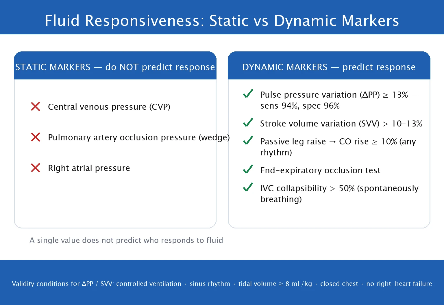
Fluid responsiveness is defined as a ≥10–15% increase in stroke volume or cardiac output after a volume challenge. The point is to predict it before giving fluid (to avoid fluid overload in non-responders). The evidence base is unanimous: use dynamic tests, not static markers (CVP).[1][7][9]
How to assess fluid responsiveness (DYNAMIC tests)
Passive leg raise (PLR)
Best bedside test. Start with head of bed 45 degrees. Lower to supine + raise legs to 45 degrees (transfers ~300 mL venous blood from legs to thorax). Measure CO/SV before and after (90 sec). If CO increases >10% = fluid responsive. Reversible — return to starting position. Advantages: self-volume challenge, reversible, does not require fluid, works in spontaneous breathing and arrhythmia. Disadvantage: requires real-time CO measurement (echo LVOT VTI, PiCCO, FloTrac).
Fluid challenge (mini-fluid bolus)
Give 250 mL crystalloid rapidly (over 1-2 min) — ideally through a large-bore cannula, not the pump. Measure CO/SV before and after. If CO increases >10-15% = fluid responsive. Gold standard but irreversible (the fluid is given — may cause overload in non-responders). Use small boluses (250 mL) and stop early if no response. The "mini-fluid challenge" (50–100 mL over 1 min) is a more conservative variant.
Stroke volume variation (SVV) / Pulse pressure variation (PPV)
In ventilated patients with regular rhythm: SVV >12-13% or PPV >12-13% predicts fluid responsiveness. Mechanism: positive pressure ventilation reduces venous return during inspiration -> SV drops in fluid-responsive patients (preload-dependent). Requirements: fully ventilated (no spontaneous breaths), regular rhythm (NOT AF), tidal volume >8 mL/kg, no right heart failure, closed chest, compliant chest wall.
IVC variability
Ultrasound: measure IVC diameter during inspiration and expiration. Collapse >50% on inspiration (sniff) = suggests fluid responsiveness. In ventilated patients: IVC distensibility >18% on inspiration. Limitations: subjective, operator-dependent, less accurate than PLR, affected by right-heart pressure. Good for screening but confirm with PLR if possible.
End-expiratory occlusion test (EEO)
In the ventilated patient, hold ventilation at end-expiration for 15 s. A rise in arterial pulse pressure (or CO) >5% predicts responsiveness. Works even in arrhythmia (unlike SVV/PPV). Requires the patient to tolerate a 15-s pause.
SVV/PPV are VALID
All must be true
- Fully controlled mechanical ventilation (no spontaneous breaths)
- Regular cardiac rhythm (sinus — NOT atrial fibrillation or frequent ectopics)
- Tidal volume >8 mL/kg (low tidal volume ventilation abolishes the effect)
- Closed chest, compliant chest wall (no open chest, no large air leak)
- No significant right heart failure (RV failure generates its own variation)
- No severe vasopressor-driven vasoconstriction distorting the pulse contour
SVV/PPV are INVALID
Use PLR instead
- Atrial fibrillation / frequent ectopics (beat-to-beat variation drowns the signal)
- Spontaneous breathing / any active inspiration (intrathoracic pressure varies independently)
- Low tidal volume ventilation (VT <8 mL/kg, e.g. ARDS lung-protective settings)
- Right heart failure / cor pulmonale / severe pulmonary hypertension
- Open chest (post-cardiac surgery) or large bronchopleural fistula
The pulmonary artery catheter (PAC / Swan-Ganz) — indications, technique, complications
The PAC is a balloon-tipped, flow-directed catheter inserted via a central vein and floated through the right heart into the pulmonary artery. It is the reference ("gold") standard for measuring cardiac output (thermodilution), pulmonary artery pressures, and the pulmonary artery wedge pressure (PAWP, an estimate of left atrial pressure), and for sampling mixed venous oxygen (SvO2). It remains a powerful diagnostic tool — but its routine use has collapsed after a series of negative trials.[2][10]
What the PAC measures
- Right atrial pressure (proximal port) = the CVP.
- Right ventricular pressure during transit (systolic 15–30, end-diastolic ~0–8 mmHg).
- Pulmonary artery pressure (distal port): systolic 15–30, diastolic 4–12, mean 9–18 mmHg.
- Pulmonary artery wedge pressure (PAWP/PCWP): with the balloon inflated (~1.5 mL air), the catheter wedges in a branch PA, creating a static column to the left atrium. PAWP reflects LA pressure and, in the absence of mitral disease and with normal pulmonary vascular resistance, LV end-diastolic pressure (LVEDP) — a pressure-based preload surrogate. Normal 6–12 mmHg.
- Cardiac output by thermodilution: a cold bolus (10 mL saline) injected in the RA is detected by the thermistor in the PA; the Stewart-Hamilton equation converts the temperature–time curve to CO. Average 3 measurements (they vary with respiration).
- Mixed venous oxygen saturation (SvO2) — sampled from the PA (true mixed venous). A continuous oximetric PAC gives real-time SvO2. [1]
Insertion and the pressure-tracing "roadmap"
The catheter is floated while watching the pressure trace (and ECG) change as it passes each chamber — this is a classic viva: [1]
| Position | Trace appearance |
|---|---|
| RA | Low, single-pressure trace (2–6 mmHg) |
| RV | Sudden rise in systolic pressure (15–30), low diastolic |
| PA | Systolic same as RV, but diastolic rises (4–12); pulse pressure narrows |
| Wedge (balloon up) | Loss of the pulse pressure, falls to a venous trace (6–12), with a/w/v waves mirroring LA |
The diastolic "step-up" from RV to PA is the key sign you have crossed the pulmonary valve; the wedge is confirmed by the waveform change and by the fall to a venous trace. [1]
Indications (current, justified use)
After the negative trials, the PAC is reserved for the complex haemodynamic problem where the question cannot be answered by echo or less-invasive devices:[8][10]
- Pulmonary hypertension and RV failure — diagnosis/characterisation (pre-capillary vs post-capillary from the wedge; calculate PVR), and acute decompensation (pulmonary hypertensive crisis). Includes connective tissue disease–associated PAH (e.g. systemic sclerosis/CREST), where severe pre-capillary PAH may decompensate in the ICU; the PAC allows diagnosis, PVR calculation, and titration of pulmonary vasodilators/inotropes. (Definitive workup is by right heart catheterisation in the catheter laboratory.)
- Cardiogenic shock — especially RV infarct, mechanical complications of MI (VSD, papillary muscle rupture), and titration of mechanical circulatory support (IABP, Impella, VA-ECMO) where knowing PA pressure, wedge, CO and SvO2 guides flow and unloading.
- Complex / mixed shock of unclear cause, or shock unresponsive to standard therapy.
- Differentiating pulmonary oedema from ARDS — a wedge >18 mmHg supports hydrostatic (cardiogenic) oedema.
- Perioperative high-risk cardiac surgery and selected major non-cardiac surgery, and cardiac tamponade vs constrictive pericarditis (equalisation of diastolic pressures).
- Heart transplantation assessment. [1]
Complications
| Category | Specific | Note |
|---|---|---|
| Vascular/access | Pneumothorax, haemothorax, arterial puncture (subclavian/IJ) | As for any CVC |
| Insertion (right heart transit) | Arrhythmia — PVCs, VT (RV passage); transient right bundle branch block | Pre-existing LBBB → risk of complete heart block: have transcutaneous/transvenous pacing ready, or avoid the PAC. |
| Catheter-related | Pulmonary artery rupture (rare, fatal — catastrophic haemoptysis); pulmonary infarction; knotting; valvular damage; catheter migration; balloon rupture | Rupture risk factors: anticoagulation, age, female sex, pulmonary hypertension, hypothermia, distal/wedge position. Never wedge with the catheter tip distal; never over-inflate the balloon; withdraw immediately on any resistance or blood return. |
| Infectious | Line-related bloodstream infection | Increases with dwell time — remove as soon as no longer needed |
| Measurement | Over-wedging, incorrect zero/level, misreading a large v wave as wedge | Read wedge at end-expiration; a giant v wave (TR) is not the wedge |
Pulmonary artery rupture is the feared, often-lethal complication — present as sudden haemoptysis. Risk is highest in anticoagulated, older, female, hypertensive patients. Prevent by never leaving the balloon inflated, inflating slowly and stopping at the wedge, avoiding distal placement, and not repositioning the catheter with the balloon up. [1]
Why PAC use has declined — the evidence
The PAC fell from routine use after a run of studies showing no benefit and possible harm:[3][5][6][4]
- Connors (JAMA 1996) — observational study of 5,735 ICU patients suggesting PAC use in the first 24 h was associated with higher 30-day mortality (OR 1.24), longer stay and higher cost. Triggered the RCTs that followed.
- PAC-Man (Lancet 2005) — UK RCT, ~1,000 ICU patients; no difference in hospital mortality whether a PAC was used or not; PAC data often changed management but this did not improve outcome.[5]
- ESCAPE (JAMA 2005) — RCT in severe heart failure; PAC-guided therapy gave no improvement in days alive out of hospital, and more adverse events (21.9% vs 11.5%).[4]
- FACTT (NEJM 2006) — NHLBI ARDS Network 2×2 factorial in acute lung injury; PAC-guided vs CVC-guided therapy gave no mortality benefit (27.4% vs 26.3%, p=0.69) and more complications.[6]
- Cochrane meta-analyses confirm no overall mortality benefit.
Bottom line for the exam: the PAC is not dead — it is redeployed. It remains the gold standard when you genuinely need PA pressure, wedge pressure, true mixed SvO2, or to characterise complex pulmonary hypertension, RV failure, or cardiogenic shock on MCS. It is not a monitoring device for the general ICU patient.[10]
Transpulmonary thermodilution (PiCCO/TPTD)
PiCCO measures cardiac output and — uniquely — volume-based preload (GEDV), extravascular lung water (EVLW), and a calibrated continuous pulse-contour CO with SVV. It needs a central venous catheter (for the cold injectate) and a femoral arterial line carrying a thermistor. A cold bolus (15–20 mL ice-cold saline) injected into the CVC traverses the right heart, lungs, left heart and aorta, and is detected in the femoral artery; the temperature–time curve yields the measurements.[1][11]
What PiCCO measures
| Parameter | What it is | Normal / use |
|---|---|---|
| CO / CI | Cardiac output by transpulmonary thermodilution (more stable than single-pass PAC thermodilution) | CI 2.5–4.0 L/min/m² |
| GEDV / GEDI | Global end-diastolic volume — the summed end-diastolic volume of all 4 chambers; a volume-based preload | GEDI 680–800 mL/m²; <680 suggests underfilled, >800 overfilled |
| ITBV / ITBI | Intrathoracic blood volume = GEDV + pulmonary blood volume (ITBV ≈ 1.25 × GEDV) | ITBI 850–1000 mL/m²; preload marker |
| EVLW / EVLWI | Extravascular lung water — water in the lungs outside the vasculature; a direct measure of pulmonary oedema | EVLWI 3–7 mL/kg PBW; >10 = oedema; >14–15 high mortality. Indexed to predicted body weight. |
| PVPI | Pulmonary vascular permeability index = EVLW / pulmonary blood volume | ~1.0–2.0 = hydrostatic (cardiogenic) oedema; >3.0 = permeability (ARDS) oedema |
| GEF | Global ejection fraction = SV/GEDV — a contractility surrogate | 25–35% |
| SVV / PPV | Pulse-contour derived, for fluid responsiveness | Same rules as arterial-line SVV (ventilated, regular rhythm, VT >8) |
Why GEDV/EVLW matter
GEDV is a more reliable preload indicator than CVP or PAWP because it measures a volume rather than a distending pressure, and is less confounded by compliance and intrathoracic pressure.[1] EVLW is the standout parameter: it is a direct measure of lung water that predicts mortality in ARDS and septic shock (each 1 mL/kg PBW rise increases mortality; Sakka 2002), guides the fluid strategy (aim for a low or falling EVLW — the conservative-fluid philosophy), and is more sensitive than the CXR or the wedge pressure for detecting pulmonary oedema.[11] The PVPI helps separate hydrostatic from permeability oedema — useful when the CXR is white-out and you need to decide between diuresis and lung-protective ventilation.
Limitations and practical points
- Requires a femoral arterial line (radial thermodilution is less accurate for GEDV/EVLW).
- Affected by intracardiac shunts (over/underestimates), severe aortic regurgitation, and extracorporeal circuits.
- The continuous pulse-contour CO must be recalibrated by thermodilution every 6–12 h, and after any major haemodynamic change (change in vascular tone, new vasopressor, fluid bolus) — the calibration "ages".
- EVLWI must be interpreted with the predicted body weight (actual weight overestimates in oedema/obesity). [1]
Lithium dilution (LiDCO)
LiDCO calibrates a pulse-contour algorithm using a lithium dilution curve rather than thermodilution. A small bolus of lithium chloride (0.002–0.003 mmol/kg) is injected via a central or peripheral venous line; the lithium concentration–time curve is detected by a lithium-selective electrode placed in a peripheral arterial line (radial is fine), and the area under the curve gives the cardiac output. That CO calibrates the PulseCO pulse-contour algorithm, which then gives continuous CO, SVV and PPV from the arterial trace.[1]
| Advantages | Limitations |
|---|---|
| No femoral line — only a radial arterial line is needed | Contraindicated in pregnancy and in patients already on lithium therapy |
| Simple, quick calibration; less invasive than PiCCO | Neuromuscular blocking agents can interfere with the lithium electrode |
| Provides continuous CO + SVV after calibration | Affected by significant shunts and aortic regurgitation |
| Recalibrate ~every 8 h and after major vascular-tone change |
LiDCO therefore suits the patient who needs calibrated continuous CO and SVV but for whom a femoral line is undesirable. Like all pulse-contour systems, it loses accuracy when vascular tone swings (sepsis, vasoactive drugs), so recalibrate after major changes. [1]
Pulse contour analysis (calibrated vs uncalibrated)
Pulse contour analysis derives a continuous stroke volume and cardiac output from the arterial pressure waveform, using the principle that the area under the systolic part of the arterial curve is proportional to stroke volume. Two families:[1]
- Calibrated (LiDCO = lithium; PiCCO = thermodilution): a dilution measurement periodically "tunes" the algorithm to the patient's vascular compliance. More accurate; requires recalibration.
- Uncalibrated (FloTrac/Vigileo, ProAQT, LiDCOrapid "auto-calibrated"): estimate vascular compliance from the waveform itself plus patient demographics (age, sex, weight). No injectate, no extra line — just the arterial catheter. Easier, but less accurate, and they drift when vascular tone changes (vasopressors, sepsis, anaesthesia). Use for trends, not absolute values, and re-check against an independent method. [1]
Echocardiography as a haemodynamic monitor
Echocardiography is the most versatile monitoring tool in the ICU: non-invasive, rapid, repeatable, and able to answer the central haemodynamic questions at the bedside — is the heart failing, is it empty, is there obstruction (tamponade/PE), and what is the cardiac output? It bridges the basic and advanced tiers.[1][8]
Basic (focused) vs advanced (quantitative) echocardiography
Basic / focused (FoCUS, FATE, FEEL)
Every intensivist
- Qualitative — categorises LV systolic function (hyperkinetic "kissing walls" = underfilled; moderate/severe = failing)
- RV size and function (RV > LV, septal shift = RV strain / PE / pulmonary HTN)
- Pericardial effusion and tamponade physiology (chamber collapse, plethoric IVC, respiratory variation)
- Gross valvular pathology (e.g. flail leaflet, destructive vegetation)
- IVC size and collapsibility (volume screening)
- Answers the four core questions: failing? empty? obstructed? what is the CO (roughly)?
Advanced / quantitative (CCE, BSE/ASE accredited)
Advanced certification
- LVOT VTI → stroke volume and cardiac output (SV = LVOT CSA × VTI)
- E/e' ratio and E-wave deceleration time → LV filling pressure
- MAPSE (mitral annular plane systolic excursion) → LV longitudinal function
- RV fractional area change (FAC), TAPSE, RVSP (from TR jet) → RV function and pressure
- RVEDA/LVEDA ratio, septal "D-sign" (flattening) → RV volume/pressure overload
- Repeatable measurements to track response (PLR, fluid bolus, inotrope titration)
Measuring cardiac output — the LVOT VTI method
SV = LVOT area × LVOT VTI, where LVOT area = π × (d/2)² from the LVOT diameter d measured in the long axis (5-chamber view), and LVOT VTI is the velocity–time integral from pulsed-wave Doppler in the 5-chamber/apical long-axis view. CO = SV × HR. Repeating the VTI before and after an intervention (PLR, fluid bolus, inotrope) tracks the response — a ≥10–15% rise in VTI after PLR = fluid responsive. This is the most practical echo-based fluid-responsiveness test and needs only basic Doppler skills.[1]
When to escalate to transoesophageal echo (TOE)
- Poor transthoracic windows (obesity, surgical emphysema, dressings, pacing wires).
- Posterior structures: mitral prosthesis, left atrial appendage thrombus, aortic dissection, endocarditis vegetations.
- Unexplained haemodynamic instability with an uninformative TTE.
- Intraoperative cardiac surgery; cardiac arrest (PEA — identify reversible cause). [1]
Limitations of echocardiography as a monitor
It is intermittent (snapshots, not continuous), operator-dependent, qualitative unless advanced-certified, and limited by image quality. It complements — but does not replace — continuous invasive monitoring in the unstable patient. The strength is the first look in any unexplained shock: failing, empty, or obstructed. [1]
Derived and advanced haemodynamic parameters
All CO devices ultimately feed the same derived variables. Knowing the equations is high-yield.[1][8]
| Parameter | Equation | Normal / meaning |
|---|---|---|
| Cardiac output (CO) | HR × SV | 4–8 L/min |
| Cardiac index (CI) | CO / BSA | 2.5–4.0 L/min/m² |
| Stroke volume (SV) | CO / HR | 60–100 mL |
| Systemic vascular resistance (SVR) | (MAP − CVP) / CO × 80 | 800–1200 dyn·s·cm⁻⁵; high = vasoconstriction (cardiogenic/hypovolaemic), low = vasodilation (septic/anaphylactic/neurogenic) |
| Pulmonary vascular resistance (PVR) | (mPAP − PAWP) / CO × 80 | <250 dyn·s·cm⁻⁵; raised in pulmonary hypertension |
| Oxygen delivery (DO2) | CO × CaO2; CaO2 = (1.34 × Hb × SaO2) + (0.003 × PaO2) | ~1000 mL/min |
| Oxygen consumption (VO2) | CO × (CaO2 − CvO2) | ~250 mL/min |
| Fick CO | VO2 / (CaO2 − CvO2) | Alternative CO method; invasive direct Fick rarely done at bedside |
| Mixed venous O2 (SvO2) | sampled from PA (PAC) | 65–75%; low = inadequate DO2 / high extraction |
| Central venous O2 (ScvO2) | sampled from SVC (CVC) | >70%; ~2–5% above SvO2 normally, but in shock can exceed SvO2 (splanchnic desaturation) |
| O2 extraction ratio (O2ER) | (SaO2 − SvO2) / SaO2 | ~25%; >50–60% = critical supply–demand imbalance |
SvO2 vs ScvO2: ScvO2 (from a central line in the SVC) is a reasonable surrogate for SvO2 (true mixed venous, from the PA via a PAC) but they diverge in shock — the splanchnic bed desaturates disproportionately, so ScvO2 may be higher than SvO2 in shock. The PAC gives the true mixed value; a central line gives an approximation adequate for most resuscitation. [1]
Landmark trials in haemodynamic monitoring
Connors (SUPPORT)
JAMA 1996
Observational propensity-matched cohort, 5,735 ICU patients — PAC in first 24 h vs no PAC
Key finding
30-day mortality higher with PAC (OR 1.24, 95% CI 1.03–1.49); longer stay, higher cost
Practice change
Triggered three decades of RCTs questioning routine PAC use
PAC-Man
Lancet 2005
UK multicentre RCT, ~1,000 general ICU patients — PAC vs no PAC
Key finding
No difference in hospital mortality; PAC data changed management but did not improve outcome
Practice change
No benefit of routine PAC in general ICU patients
ESCAPE
JAMA 2005
RCT, 433 patients with severe decompensated heart failure — PAC-guided vs clinical assessment
Key finding
No improvement in days alive/out of hospital; MORE adverse events (21.9% vs 11.5%)
Practice change
PAC not recommended to guide therapy in decompensated heart failure
FACTT (ARDSNet)
NEJM 2006
RCT, 1,000 patients with acute lung injury — PAC vs central venous catheter to guide therapy (2×2 with conservative vs liberal fluids)
Key finding
60-day mortality no different (27.4% PAC vs 26.3% CVC, p=0.69); more catheter complications with PAC
Practice change
CVC-guided therapy preferred over PAC in ALI/ARDS
Marik & Cavallazzi
Crit Care Med 2013
Meta-analysis, 43 studies — CVP to predict fluid responsiveness
Key finding
AUC ~0.56 (no better than flipping a coin); ΔCVP equally unhelpful
Practice change
CVP should NOT be used to predict fluid responsiveness or guide fluid therapy
OPTIMISE
JAMA 2014
RCT, 734 high-risk gastrointestinal surgery patients — cardiac-output-guided (dopexamine) vs usual care
Key finding
No significant reduction in complications or mortality (modest signal); supported goal-directed perioperative fluid/inotrope use
Practice change
Cardiac-output-guided therapy reasonable in high-risk surgery (effect modest)
Exam practice
SAQ — CVP-guided resuscitation and the limits of static markers
10 minutes · 10 marks
A 68-year-old woman with community-acquired pneumonia and septic shock has a CVC in the right internal jugular vein. She has received 30 mL/kg crystalloid. CVP is 12 mmHg. MAP 62 on noradrenaline 0.25 mcg/kg/min, HR 112 (sinus), lactate 3.1 mmol/L, urine output 25 mL/hr. She remains mottled with cold peripheries. The registrar proposes giving another 500 mL bolus because 'the CVP is only 12'.
Sample Viva 1 — The arterial line trace
Examiner: "You are called because the arterial line pressure looks very high. The trace is tall and spiky with ringing. How do you assess it?" [1]
Expected response: "I would perform a fast-flush (square-wave) test: briefly open the flush valve and release. If there are many oscillations that ring before settling, the system is underdamped — the systolic is spuriously high and the diastolic spuriously low, but the MAP is preserved. So I would trust the MAP, disregard the systolic, and correct the system by adding a damping device or shortening the tubing. If the test showed a slurred return with no oscillations, it would be over-damped (air bubble/kink/clot) — I would flush out bubbles, check for kinks, and aspirate for clot." [1]
Follow-up: "Where should you read the systolic pressure — radial or femoral?" [1]
Response: "In a stable patient the radial is acceptable, but it reflects peripheral pressure — systolic is amplified ~5–15 mmHg above central aortic pressure, while MAP and diastolic are close. In shock, severe vasoconstriction, or hypothermia the periphery vasoconstricts first and the radial under-reads; a femoral line gives a more reliable central pressure. I would use the femoral in this unstable patient." [1]
Sample Viva 2 — Choosing the monitor
Examiner: "A patient with severe systemic-sclerosis-associated pulmonary hypertension is admitted in pulmonary hypertensive crisis, hypotensive and hypoxic. What haemodynamic monitoring will you use and why?" [1]
Expected response: "This is one of the legitimate indications for a pulmonary artery catheter. CTD-associated PAH is pre-capillary pulmonary hypertension with a vulnerable, pressure-overloaded right ventricle; in crisis the RV fails, CO falls, and systemic hypotension compromises coronary perfusion of the RV. I need to know the mPAP, PA diastolic and wedge (to confirm pre-capillary PAH and exclude a post-capillary contribution), the PVR, the CO and the mixed SvO2 — and to titrate pulmonary vasodilators (inhaled nitric oxide, prostacyclin) and inotropes/vasopressors against these numbers. The PAC also lets me monitor RV failure and the response to therapy. An echocardiogram (RV size, septal shift, TAPSE, RVSP from the TR jet) complements but does not replace the PAC for pressure measurement. I would also place an arterial line (continuous BP) and ensure pacing is available, as right-heart catheterisation can provoke arrhythmia." [1]
Follow-up: "What are the specific risks of a PAC in this patient?" [1]
Response: "Pulmonary artery rupture is the feared complication and this patient has several risk factors: pulmonary hypertension itself, likely anticoagulation, and a stiff, hypertensive PA tree. I would avoid distal wedging, inflate the balloon slowly and stop as soon as the wedge trace appears, never leave the balloon inflated, and not advance against resistance. Arrhythmia and transient right bundle branch block are also risks — if she had a pre-existing left bundle branch block I would ensure pacing is available before insertion, because the catheter can cause complete heart block." [1]
Clinical pearls
Red flags
References
- [1]Cecconi M, De Backer D, Antonelli M, et al. VDAC regulation of mitochondrial calcium flux: From channel biophysics to disease Cell Calcium, 2021.PMID 33529977
- [2]Harvey S, et al. Notum palmitoleoyl-protein carboxylesterase regulates Fas cell surface death receptor-mediated apoptosis via the Wnt signaling pathway in colon adenocarcinoma Bioengineered, 2021.PMID 34402722
- [3]Connors AF Jr, Speroff T, Dawson NV, et al. The physician's role in helping smoke-sensitive patients to use the Americans with Disabilities Act to secure smoke-free workplaces and public spaces JAMA, 1996.PMID 8782641
- [4]Binanay C, Califf RM, Hasselblad V, et al. Evaluation study of congestive heart failure and pulmonary artery catheterization effectiveness: the ESCAPE trial JAMA, 2005.PMID 16204662
- [5]Harvey S, Harrison DA, Singer M, et al. Assessment of the clinical effectiveness of pulmonary artery catheters in management of patients in intensive care (PAC-Man): a randomised controlled trial Lancet, 2005.PMID 16084255
- [6]Wheeler AP, Bernard GR, Thompson BT, et al. Blockade of AT1 receptors by losartan did not affect renin gene expression in kidney medulla Gen Physiol Biophys, 2006.PMID 16714774
- [7]Marik PE, Cavallazzi R. Does the central venous pressure predict fluid responsiveness? An updated meta-analysis and a plea for some common sense Crit Care Med, 2013.PMID 23774337
- [8]Cecconi M, De Backer D, Antonelli M, et al. Cold fluids during cardiac arrest: faster cooling but not better outcome! Intensive Care Med, 2014.PMID 25392035
- [9]Evans L, Rhodes A, Alhazzani W, et al. Prognosis of papillary thyroid carcinoma in relation to preoperative subclinical hypothyroidism Ann R Coll Surg Engl, 2021.PMID 33682437
- [10]De Backer D, Vincent JL. The pulmonary artery catheter: is it still alive? Curr Opin Crit Care, 2018.PMID 29608456
- [11]Sakka SG, Klein M, Reinhart K, Meier-Hellmann A. The burden of infectious disease among inmates of and releasees from US correctional facilities, 1997 Am J Public Health, 2002.PMID 12406810
- [12]Pearse RM, Harrison DA, MacDonald N, et al. Genotype-guided dosing of vitamin K antagonists N Engl J Med, 2014.PMID 24785218