ICU · Cardiovascular / infection
Infective Endocarditis
Also known as Infective endocarditis · IE · Bacterial endocarditis · Duke criteria · Vegetation · Osler nodes · Janeway lesions · HACEK · Culture-negative endocarditis
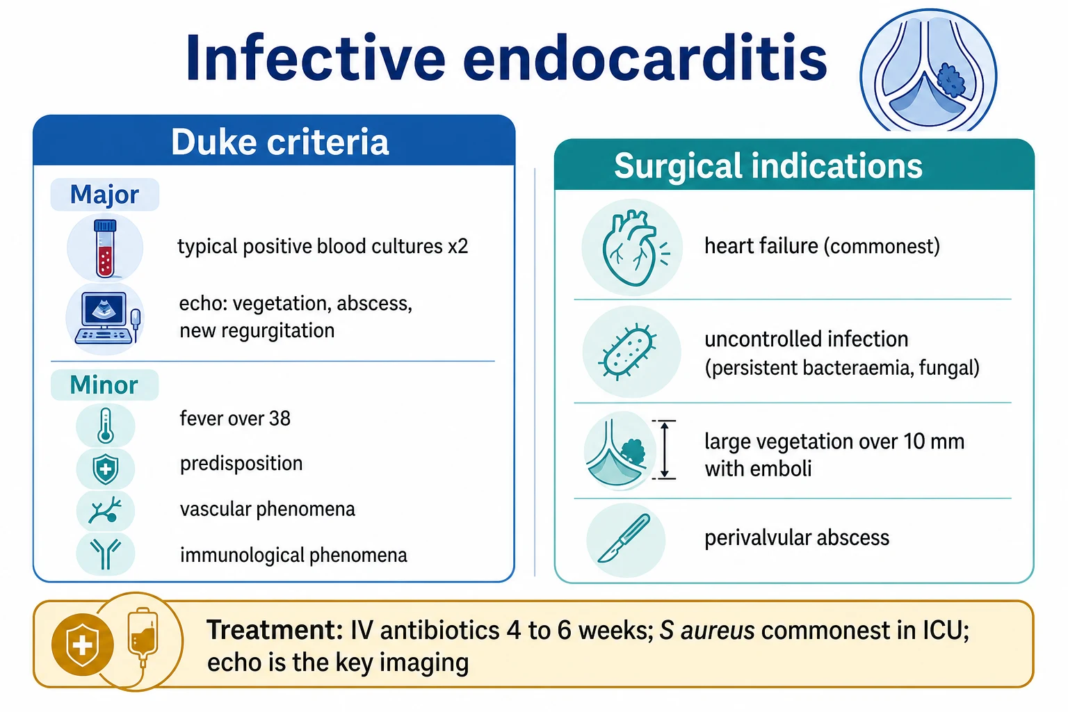
Infective endocarditis is an infection of the cardiac valves (or the endocardium), diagnosed by the modified Duke criteria: typical positive blood cultures and an echocardiographic vegetation are the major criteria. The commonest ICU organism is Staphylococcus aureus (aggressive, acute); viridans streptococci cause subacute endocarditis (dental); enterococci, the HACEK group, and culture-negative organisms (Coxiella, Bartonella, fungi) are also important. The clinical features: fever, a new or changed murmur, embolic phenomena (stroke, splenic, mesenteric, limb), immunological phenomena (Osler nodes, Roth spots, glomerulonephritis), and vascular phenomena (Janeway lesions). The treatment is 4-6 weeks of IV antibiotics (targeted to the culture). Surgery is indicated for heart failure (the commonest indication), uncontrolled infection, large vegetations with embolism, and perivalvular extension.
On this page & tools
Your progress
Saved locally on this device.
18 MCQs with explanations
Target exams
Overview & definition
Infective endocarditis (IE) is an infection of the cardiac valves (or the endocardium) by microorganisms that adhere to the valve surface and form a vegetation (a mass of platelets, fibrin, and organisms). In the ICU, it presents acutely (often with Staphylococcus aureus — aggressive, causing septic shock, acute severe valve failure, and septic emboli). The modified Duke criteria provide the diagnostic framework.[1]
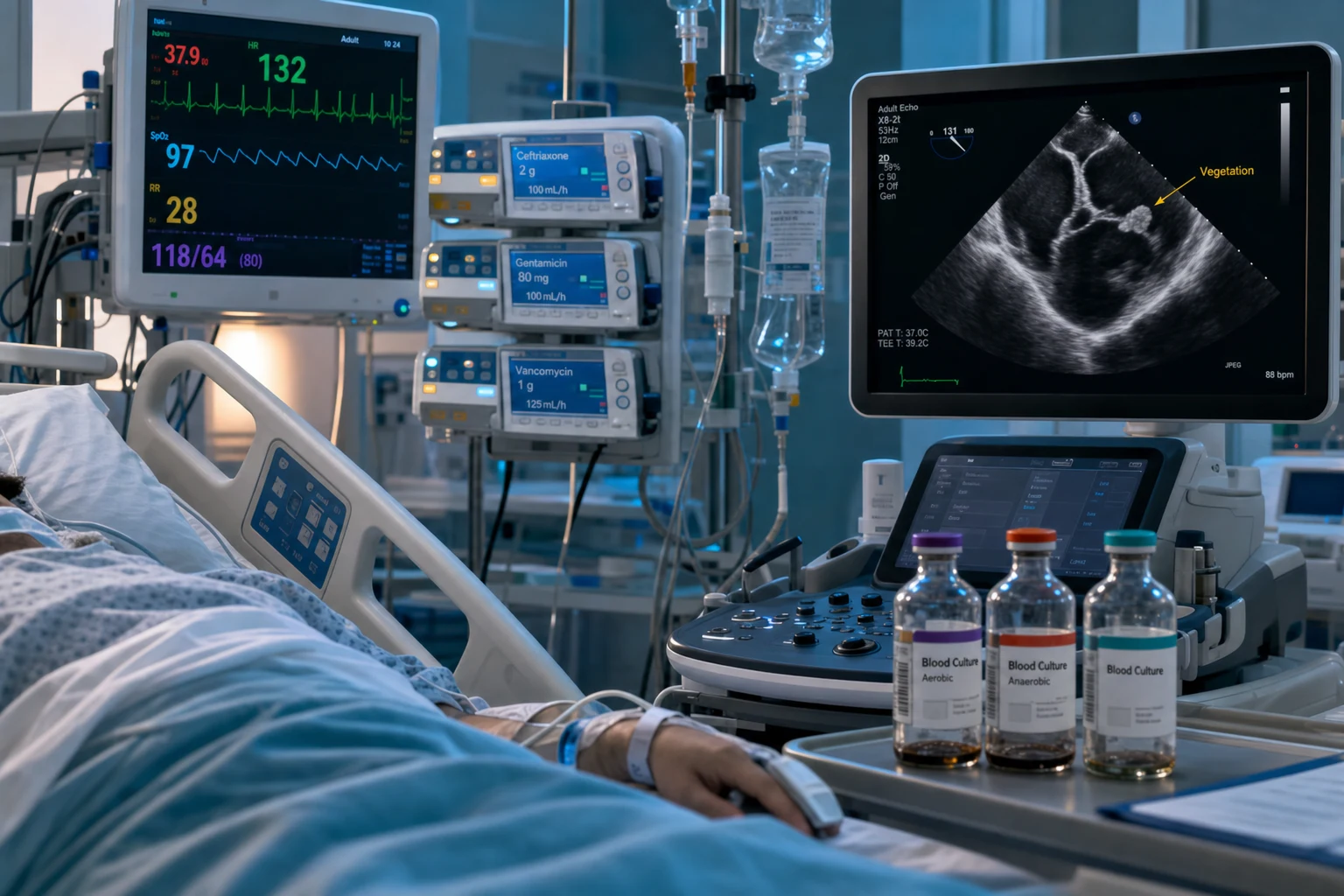
The modified Duke criteria
Definite IE: 2 major, OR 1 major + 3 minor, OR 5 minor.[1]
Major criteria:
- Typical positive blood cultures from 2 separate cultures (viridans streptococci, S. aureus, HACEK, or enterococci).
- Echocardiographic evidence — a vegetation, an abscess, or a new partial dehiscence of a prosthetic valve. [1]
Minor criteria:
- Predisposition (a predisposing heart condition — a prosthetic valve, a previous IE, a congenital heart disease, or IV drug use).
- Fever over 38 degrees Celsius.
- Vascular phenomena — arterial emboli, septic pulmonary infarcts, mycotic aneurysms, intracranial haemorrhage, Janeway lesions, conjunctival haemorrhages.
- Immunological phenomena — glomerulonephritis, Osler nodes, Roth spots, a positive rheumatoid factor.
- Microbiological evidence not meeting the major criterion (a single positive culture or a serological evidence).[1]
The organisms
- Staphylococcus aureus — the commonest in the ICU and in IV drug users; aggressive, acute; often causes acute severe valve failure and septic emboli. High mortality.[1]
- Viridans streptococci (Streptococcus sanguis, S. mutans) — subacute, from the dental plaque; the classic organism of the native-valve subacute endocarditis.[1]
- Enterococci (E. faecalis, E. faecium) — often from the gastrointestinal or the genitourinary tract; resistant organisms are increasing.[1]
- HACEK (Haemophilus, Actinobacillus, Cardiobacterium, Eikenella, Kingella) — fastidious Gram-negatives; culture-negative if the lab does not hold the cultures long enough.
- Culture-negative — Coxiella burnetii (Q fever), Bartonella, Brucella, fungi (Candida, Aspergillus); consider if the standard cultures are negative.[1]
Clinical features
- Fever (over 90 per cent of cases) and constitutional symptoms (malaise, weight loss).
- A new or changed murmur (the vegetation alters the valve function — a regurgitant murmur).
- Embolic phenomena — a stroke, a splenic infarct or abscess, a mesenteric ischaemia, a renal infarct, a limb ischaemia (from embolisation of vegetation fragments).
- Immunological phenomena — Osler nodes (tender nodules on the finger pulps), Roth spots (retinal haemorrhages with a pale centre), a glomerulonephritis.
- Vascular phenomena — Janeway lesions (painless macules on the palms or soles from septic emboli), a conjunctival haemorrhage, a mycotic aneurysm.
- Splenomegaly.[1]
Echocardiography
- Transthoracic echo (TTE) — the first test; a sensitivity of about 60 per cent for the native valve (less for the prosthetic). A vegetation appears as an oscillating mass on the valve.
- Transoesophageal echo (TOE) — if the TTE is negative or for the prosthetic valve; a sensitivity over 90 per cent; also detects the complications (an abscess, a fistula, a perforation, a prosthetic dehiscence).[1]
Treatment
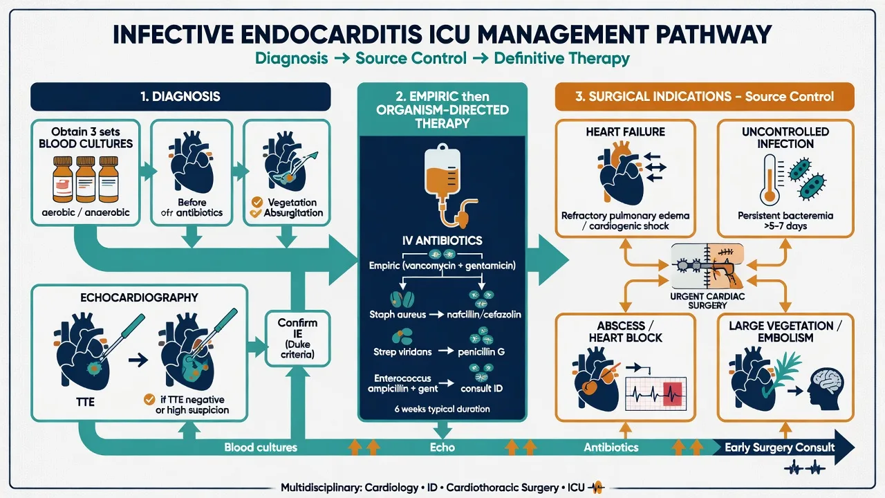
Empirical antibiotics (while awaiting the blood cultures):[1]
- For the native valve: ampicillin + gentamicin + flucloxacillin (covering the streptococci, the enterococci, and the staphylococci), OR vancomycin + gentamicin (if penicillin-allergic or if MRSA is suspected).
- For the prosthetic valve: vancomycin + gentamicin + rifampicin (covering the staphylococci including MRSA, the streptococci, and the enterococci).
Targeted therapy (once the organism and the sensitivities are known):[1]
- IV therapy for 4-6 weeks (longer for some organisms — fungi, resistant enterococci).
- Monitor the inflammatory markers (CRP), the renal function (nephrotoxicity from the gentamicin), and the repeat echo (the vegetation size, the valve function).
Surgical indications
Surgery (valve repair or replacement) is indicated for:[1]
- Heart failure from the acute severe valve dysfunction (the commonest indication — acute severe AR or MR with pulmonary oedema or cardiogenic shock).
- Uncontrolled infection — a persistent bacteraemia or fever after 5-7 days of appropriate antibiotics; fungal or resistant organisms that cannot be eradicated medically.
- Prevention of embolism — a large vegetation (over 10 mm) with one or more embolic events, or a very large vegetation (over 15 mm) even without an embolus.
- Perivalvular extension — an abscess, a fistula, a heart block (from the aortic-root abscess extending into the septum).
- Prosthetic valve endocarditis — early (within 1 year of the surgery) or complicated (dehiscence, obstruction).[1]
The ICU-specific problems
- Septic shock from S. aureus IE — resuscitate per the sepsis protocol; source control is the valve surgery.
- Acute severe valve failure (AR or MR with pulmonary oedema or cardiogenic shock) — urgent surgery; temporise with vasodilators, inotropes, or an IABP.
- Septic emboli — a stroke (may need neurosurgical input for a haemorrhagic transformation or a mycotic aneurysm); a mesenteric ischaemia; a splenic abscess (drain or splenectomy); a limb ischaemia (embolectomy).[1]
Red flags
Modified Duke criteria — interpretation and the 2023 update
The modified Duke criteria are the diagnostic standard, but the diagnostic categories (definite, possible, rejected) drive every clinical decision and are the most frequently examined nuance. The 2023 ESC update (incorporating the Duke-ISCVID 2023 criteria) added imaging findings (PET-CT uptake around a prosthetic valve, abnormal CT of a paravalvular lesion) as major criteria and re-weighted S. aureus bacteraemia.[1]
Modified Duke criteria — diagnostic categories
| Category | Rule | Practical meaning |
|---|---|---|
| Definite IE | 2 major, OR 1 major + 3 minor, OR 5 minor | Treat as IE — full antibiotic course, surgical assessment |
| Possible IE | 1 major + 1 minor, OR 3 minor | A common scenario — treat empirically while gathering more data (repeat cultures, TOE, PET-CT); do not dismiss |
| Rejected | A firm alternative diagnosis, OR resolution with under 4 days of antibiotics, OR no pathologic evidence at surgery/autopsy | Stop antibiotics, hunt for the true source |
The 2023 Duke-ISCVID additions (now a major criterion): a single positive blood culture for Coxiella burnetii (or phase I IgG titre over 1:800); imaging evidence of IE (abnormal FDG uptake on PET-CT around a prosthetic valve, paravalvular lesions on cardiac CT, a new vegetation on repeat echo); and a single positive culture for a typical IE organism is now weighted more heavily. Staphylococcus aureus bacteraemia — even nosocomial — is now a major criterion when no alternative focus is identified.[1]
The organisms — epidemiology and clinical associations
The organism predicts the syndrome, the prognosis, and the empiric therapy. The ICE-PCS cohort (over 2,700 patients, the 21st-century reference) and the EURO-ENDO registry (over 3,100 patients) define the modern epidemiology.[9][13]
IE organisms — frequency, source, and clinical signature
| Organism | Frequency | Source / portal | Clinical signature | Mortality |
|---|---|---|---|---|
| Staphylococcus aureus | 30-35 per cent (commonest overall) | Skin, IV cannulae, IV drug use, haemodialysis | Acute, aggressive, septic shock, affects NORMAL valves, highest embolic rate | 25-40 per cent |
| Viridans streptococci (S. sanguis, S. mitis, S. mutans, S. oralis) | 20-25 per cent | Oral flora — poor dentition, dental procedures | Subacute (weeks-months), native valve, penicillin-sensitive, good prognosis | 5-10 per cent |
| Streptococcus gallolyticus (formerly S. bovis) | 5-10 per cent | GI tract | Colonic neoplasia association — colonoscopy mandatory (polyps 50-60 per cent, colorectal cancer 20-30 per cent) | 10-15 per cent |
| Enterococci (E. faecalis, E. faecium) | 10 per cent | GI/GU tract — urinary catheters, bowel surgery, older men | Nosocomial, may be VRE, often subacute | 15-25 per cent |
| Coagulase-negative staphylococci (S. epidermidis, S. lugdunensis) | 5-10 per cent | Skin flora — perioperative contamination | Almost exclusively PROSTHETIC valve IE (early); S. lugdunensis behaves like S. aureus (aggressive) | 15-25 per cent |
| HACEK (Haemophilus, Aggregatibacter, Cardiobacterium, Eikenella, Kingella) | 2-5 per cent | Oropharyngeal flora | Fastidious, slow-growing (prolonged incubation), large vegetations, embolic | 5-10 per cent |
| Culture-negative | 5-10 per cent | Prior antibiotics, Coxiella, Bartonella, Brucella, Tropheryma, fungi | Diagnose by serology/PCR; needs special culture techniques | Variable |
| Fungi (Candida, Aspergillus) | under 2 per cent | IV drug use, prolonged antibiotics, immunocompromised, lines | Large vegetations, very high embolic rate, surgical valve replacement mandatory | 30-50 per cent |
Streptococcal IE — the subacute archetype
Viridans streptococci are the classic organism of native-valve, subacute endocarditis. The patient has weeks of low-grade fever, weight loss, and malaise; the valves are usually pre-damaged (rheumatic, bicuspid, degenerative). The organism is penicillin-sensitive (MIC under 0.12 mg/L), and 4 weeks of IV penicillin G or ceftriaxone gives a cure rate above 95 per cent.[1] Streptococcus gallolyticus is special — its presence mandates colonoscopy because of the strong association with colonic neoplasia (the bacteraemia arises from a mucosal breach over a tumour).[13]
Staphylococcal IE — the ICU archetype
Staphylococcus aureus is the organism the intensivist fears. Unlike viridans streptococci, it can colonise and destroy a previously normal valve; it produces a fulminant, acute illness with high fever, septic shock, and rapid valvular destruction; and it has the highest embolic rate of any organism (over 50 per cent of patients embolise). The mortality of 25-40 per cent has not improved in two decades despite better antibiotics and surgery.[6] Coagulase-negative staphylococci cause prosthetic valve IE (the organism of early PVE from perioperative contamination) — with the critical exception of S. lugdunensis, which is virulent like S. aureus and causes aggressive native-valve IE requiring the same aggressive management.[8]
Enterococcal IE
Enterococci (mainly E. faecalis) cause IE in older men after genitourinary instrumentation or in patients with bowel pathology. They are intrinsically resistant to cephalosporins (the cell wall target is altered), so therapy is ampicillin (or vancomycin) PLUS an aminoglycoside (gentamicin or streptomycin) for synergy, or ampicillin plus ceftriaxone (the double-beta-lactam regimen) for 4-6 weeks. Vancomycin-resistant enterococci (VRE) require linezolid or daptomycin.[8]
HACEK and culture-negative IE
The HACEK group are fastidious Gram-negatives of the oropharynx that need prolonged incubation (modern automated blood-culture systems usually grow them within 5 days, but they were historically a cause of "culture-negative" IE). They produce large vegetations and embolise frequently; ceftriaxone for 4 weeks is curative.[8] Culture-negative IE has a defined differential: prior antibiotics (the commonest cause — repeat cultures after a washout period), Coxiella burnetii (Q fever — phase I IgG), Bartonella ( homelessness, alcoholism, body lice), Brucella (unpasteurised dairy), Tropheryma whipplei (Whipple disease), and fungi. The workup is serology and PCR, and a history of antibiotic exposure, animal contact, or travel directs the search.[1]
Fungal endocarditis
Fungal IE (Candida most often, then Aspergillus) is rare but devastating: the vegetations are large and friable (over 50 per cent embolise), blood cultures may be negative, and medical cure is rarely achieved. Surgical valve replacement is mandatory, combined with prolonged antifungal therapy (liposomal amphotericin B plus flucytosine, then long-term fluconazole suppression; echinocandins for Candida). Risk factors are IV drug use, prolonged antibiotics, total parenteral nutrition, immunosuppression, and indwelling central lines.[8]
Echocardiography — the TTE then TOE strategy
Echocardiography is the single most important diagnostic test in suspected IE. The strategy is TTE first, then TOE — but in the ICU, with a critically ill intubated patient, TOE is low-risk and high-yield, so the threshold to proceed to TOE is low.[1]
TTE vs TOE in suspected infective endocarditis
| Feature | Transthoracic echo (TTE) | Transoesophageal echo (TOE) |
|---|---|---|
| Sensitivity — native valve | 50-60 per cent | 90-100 per cent |
| Sensitivity — prosthetic valve | 20-40 per cent | 85-95 per cent |
| Minimum vegetation detected | over 6 mm | over 2-3 mm |
| Abscess / fistula / perforation | Poor | Excellent — the test of choice |
| Prosthetic valve shadowing | Severe (limits views) | Overcomes shadowing |
| Patient burden | Non-invasive, bedside | Semi-invasive (probe), needs sedation/coagulation check |
| Role | First-line screening; detects large vegetations and new regurgitation; establishes a baseline | Definitive — always done if IE is genuinely suspected |
When TOE is mandatory (do it, regardless of the TTE result): any prosthetic valve; a suspected complication (abscess, fistula, new heart block, persistent bacteraemia); S. aureus bacteraemia; a negative TTE but a strong clinical suspicion; before surgery to define the anatomy; and after surgery to confirm eradication.[1][8] Even if the TTE shows a vegetation, a TOE is still performed to assess for abscess, perforation, fistula, and the extent of destruction — these change the surgical plan and are missed on TTE.
Beyond echo — multimodality imaging
For prosthetic valve IE, where echo is limited by shadowing, 18F-FDG PET-CT (abnormal uptake around the valve) and cardiac CT (paravalvular complications — abscess, pseudoaneurysm, dehiscence) are now major Duke criteria. PET-CT is most useful more than 3 months after surgery (early post-operative inflammation causes false positives).[1] Cerebral imaging (MRI) is performed in all patients with left-sided IE — silent cerebral emboli are found in over 50 per cent, and they alter the surgical timing.
Antimicrobial therapy — native vs prosthetic valve
IE requires prolonged, high-dose, bactericidal IV therapy. The principles: (1) culture before antibiotics (3 sets, different sites, 30 minutes apart); (2) use beta-lactams at high dose (concentration-independent time-dependent killing — the time above the MIC drives efficacy); (3) add gentamicin for synergy in staphylococcal and enterococcal IE (but limit to 2 weeks to limit nephrotoxicity); (4) add rifampicin for prosthetic-valve staphylococcal IE (biofilm penetration); (5) treat for 4-6 weeks IV.[1][8]
Empirical therapy — native vs prosthetic valve (before culture results)
| Scenario | Empirical regimen | Rationale |
|---|---|---|
| Native valve, community-acquired | Ampicillin-sulbactam (or ampicillin) + gentamicin + flucloxacillin; OR ceftriaxone + gentamicin if penicillin-allergic | Covers strep, enterococcus, HACEK, MSSA |
| Native valve, healthcare-associated or suspected MRSA | Vancomycin + gentamicin (+/- flucloxacillin if MSSA possible) | Covers MRSA, staph, strep, enterococcus |
| Native valve, IV drug user | Vancomycin + gentamicin (add gram-negative cover if septic) | MRSA common; right-sided IE |
| Prosthetic valve (within 12 months of surgery) | Vancomycin + gentamicin + rifampicin | MRSA, coagulase-negative staph; rifampicin penetrates biofilm |
| Prosthetic valve (after 12 months) | Vancomycin + gentamicin (add rifampicin if staph confirmed) | Same organisms as native valve; biofilm less dominant |
| Severe sepsis / septic shock of unknown source | Vancomycin + piperacillin-tazobactam + gentamicin | Broadest empiric cover while awaiting cultures |
Targeted therapy — by organism and valve type
| Organism / valve | Standard regimen | Duration | Notes |
|---|---|---|---|
| Viridans strep, penicillin-sensitive (native) | Penicillin G 12-18 MU/day IV OR ceftriaxone 2 g/day IV | 4 weeks | Ceftriaxone once-daily suits outpatient/HDU |
| Viridans strep, penicillin-resistant (native) | Penicillin/ceftriaxone + gentamicin (first 2 weeks) | 4 weeks | Higher MIC — add synergy |
| MSSA (native) | Flucloxacillin 12 g/day IV (+/- gentamicin first 3-5 days) | 4-6 weeks | Gentamicin synergy optional; monitor renal |
| MRSA (native) | Vancomycin 30-40 mg/kg/day IV (trough 15-20 mg/L); consider daptomycin 8-10 mg/kg/day | 6 weeks | Vancomycin MIC under 2 mg/L; daptomycin if MIC elevated or vancomycin failure |
| MSSA (prosthetic) | Flucloxacillin + rifampicin + gentamicin (first 2 weeks) | >=6 weeks | Rifampicin starts after 3-5 days (bacterial load reduction) |
| MRSA (prosthetic) | Vancomycin + rifampicin + gentamicin (first 2 weeks) | >=6 weeks | Biofilm penetration essential |
| Enterococcus (ampicillin-sensitive) | Ampicillin + gentamicin (or streptomycin) | 4-6 weeks | Check high-level aminoglycoside resistance |
| Enterococcus (ampicillin-resistant / VRE) | Vancomycin + gentamicin; or linezolid/daptomycin for VRE | 6 weeks | Infectious diseases input essential |
| HACEK | Ceftriaxone 2 g/day IV (or ampicillin-sulbactam) | 4 weeks | Beta-lactamase producers — avoid ampicillin alone |
| Fungi | Liposomal amphotericin B + flucytosine, then fluconazole suppression; surgery mandatory | >=6 weeks + lifelong suppression | Echinocandin alternative for Candida |
Antibiotic pharmacology — synergy, biofilm, and toxicity
- Gentamicin synergy — added to beta-lactams/vancomycin for enterococcal and staphylococcal IE; once-daily dosing (3 mg/kg) reduces nephrotoxicity; limit to 2 weeks (the synergy benefit is early, the toxicity accrues with time); monitor trough (under 1 mg/L) and creatinine. Stop if renal function declines.[8]
- Rifampicin and the biofilm — prosthetic material (mechanical valves, rings) develops a bacterial biofilm that beta-lactams cannot penetrate. Rifampicin is highly lipophilic and uniquely penetrates biofilm, killing the embedded staphylococci. Start it AFTER 3-5 days of vancomycin/flucloxacillin (to reduce the bacterial load and delay the emergence of rifampicin resistance, which is rapid in a high-inoculum setting).[4][12]
- Vancomycin — for MRSA and penicillin-allergic staphylococcal IE; target a trough of 15-20 mg/L (or AUC-guided dosing); slower infusion, nephrotoxicity (worsened by concurrent gentamicin/piperacillin-tazobactam). If the vancomycin MIC is over 1.5 mg/L, switch to daptomycin.[8]
- Daptomycin — bactericidal lipopeptide; an alternative for MRSA IE (especially right-sided) and VRE; not for pneumonia (inactivated by surfactant); higher doses (8-12 mg/kg/day) for IE; creatine kinase monitoring (myopathy).
- Ceftriaxone once-daily — the workhorse for streptococcal and HACEK IE; enables hospital-in-the-home and partial-oral regimens.[3]
The POET trial — partial oral switch for selected patients
The POET trial (Iversen 2019) challenged the dogma that IE must be treated entirely IV. In 400 patients with left-sided, culture-positive, streptococcal/staphylococcal/enterococcal IE who had responded to IV therapy and had no prosthesis, abscess, or heart failure, a switch to oral antibiotics (after at least 10 days IV) was non-inferior to continued IV therapy for the composite of all-cause mortality, unplanned surgery, embolic events, and relapse.[3] Caveats: the regimen was carefully selected (oral bioavailability confirmed by serum bactericidal titre), it excluded the sickest patients, and the oral drugs were chosen by an infectious-diseases specialist. The take-home: carefully selected, clinically improving, non-complicated IE can be completed orally — but this is the exception, not the default, and requires close follow-up.
Surgery — indications, timing, and the cerebral-embolism problem
Around 40-50 per cent of patients with IE need surgery. The decision is multidisciplinary (the "Endocarditis Team" — cardiology, infectious diseases, cardiac surgery) and is driven by four indications, in order of urgency: heart failure, uncontrolled infection, embolic prevention, and prosthetic/perivalvular extension.[1][2]
Surgical indications and timing in IE
| Indication | Urgency | Class/evidence (ESC) | Rationale |
|---|---|---|---|
| Heart failure from acute AR/MR | EMERGENCY (under 24 h) | I, B | The strongest predictor of mortality; medical mortality 50-70 per cent, surgical 10-15 per cent |
| Uncontrolled infection (persistent bacteraemia/fever over 5-7 days on appropriate antibiotics; fungal, MDR organisms) | URGENT (days) | I, B | Antibiotics cannot sterilise a destroyed valve or a biofilm |
| Periannular extension (abscess, fistula, heart block) | URGENT | I, B | Progression to fistula/perforation/cardiac rupture; pacing + debridement |
| Large mobile vegetation over 10 mm with embolism | URGENT (early surgery) | IIa, B | Each day with vegetation = embolic risk; early surgery prevents stroke |
| Large vegetation over 15 mm (no embolism) | URGENT (consider) | IIb, B | High embolic risk; weigh against operative risk |
| Prosthetic valve IE (early, under 12 months) | Almost always surgical | I | Biofilm cannot be eradicated medically |
| S. aureus prosthetic valve IE | Almost always surgical | I | Highest-mortality organism + biofilm |
Cerebral embolism and the timing of surgery
A cerebral embolic event is the most feared complication and the most common reason for delaying surgery. The problem: cardiopulmonary bypass requires full heparinisation, which risks haemorrhagic transformation of a recent cerebral infarct. The ESC guidance:[1]
- Ischaemic stroke, no coma, no haemorrhage — surgery can proceed, but a short delay (ideally 2 weeks) is preferred if heart failure allows. Intracranial haemorrhage must be excluded by CT/MRI first.
- Haemorrhagic stroke or coma — delay surgery for at least 3-4 weeks if at all possible.
- Heart failure is life-threatening — operate DESPITE the stroke. Heart failure, not the stroke, is the threat to life, and the mortality of unoperated heart failure exceeds the risk of bypass-related haemorrhagic transformation.
- Silent cerebral emboli on MRI — do not, by themselves, contraindicate or delay surgery. [1]
The IE management protocol — from suspicion to cure
- CULTURE BEFORE ANTIBIOTICS — 3 sets of blood cultures from different venepuncture sites, 30 minutes apart, BEFORE antibiotics. If the patient is in septic shock, draw cultures then START antibiotics within 1 hour — do not delay antibiotics for cultures in shock. Prior antibiotics reduce culture yield by 30-50 per cent.[1]
- EMPIRICAL ANTIBIOTICS — native valve (community): ampicillin + gentamicin + flucloxacillin; prosthetic valve or MRSA risk: vancomycin + gentamicin + rifampicin. Adjust for local resistance and allergy.[8]
- ECHOCARDIOGRAPHY — TTE THEN TOE — TTE first (bedside, screening), then TOE (definitive). TOE is mandatory for prosthetic valves, suspected complications, persistent bacteraemia, and S. aureus. Add PET-CT and cardiac CT for suspected prosthetic valve IE.[1]
- DEFINE THE ORGANISM — once cultures return, de-escalate to targeted therapy (4-6 weeks IV). Check MICs (penicillin for strep, vancomycin for staph), high-level aminoglycoside resistance (enterococcus), and beta-lactamase (HACEK).[8]
- SEARCH FOR THE SOURCE AND COMPLICATIONS — dental review (panorex), colonoscopy (S. gallolyticus, enterococcus), screening for emboli (cerebral MRI, abdominal CT for splenic/renal infarct), transthoracic follow-up echo.[13]
- SURGICAL DECISION (Endocarditis Team) — the four indications: heart failure (emergency), uncontrolled infection (urgent), embolic prevention (urgent), perivalvular/prosthetic extension (urgent). Weigh the cerebral-embolism timing. Refer early — the team meets daily on active cases.[1]
- MONITORING — daily: temperature, blood cultures until 3 consecutive negatives, renal function, FBC, vancomycin/gentamicin levels. Weekly: ECG (new block = abscess), CRP, repeat echo (vegetation size, valve function). New murmur, new fever, or new embolus = relapse or extension — repeat cultures and TOE.[1]
- COMPLETE THE COURSE AND PLAN PREVENTION — 4-6 weeks IV (consider POET-style oral switch only for selected, uncomplicated cases). Dental restoration. Prophylaxis for high-risk patients before dental procedures (amoxicillin 2 g PO 30-60 min pre-op). Patient education on the warning signs.[10]
Complications — embolic stroke, mycotic aneurysm, heart block
The complications of IE are the causes of death: heart failure (covered above), septic embolisation (stroke, splenic, mesenteric, limb, renal, coronary), mycotic aneurysm (especially intracranial), perivalvular extension (abscess, fistula, heart block), renal involvement (glomerulonephritis, acute kidney injury), and the septic shock of uncontrolled infection.[5][8]
Embolic stroke
Systemic embolisation occurs in 20-40 per cent of patients with left-sided IE; the brain is the commonest site (over half of emboli). The risk is highest in the first week, before antibiotics work, and is driven by vegetation size (over 10 mm), mobility, mitral location, S. aureus, and increasing CRP.[11] Management: STOP any anticoagulation; CT brain (ischaemic vs haemorrhagic); exclude a mycotic aneurysm (CT/MR angiography) before surgery; if a large mobile vegetation remains, consider early surgery to prevent further emboli.[1]
Mycotic (intracranial infectious) aneurysm
A mycotic aneurysm is a destructive arteritis caused by septic embolisation of the vasa vasorum or the arterial lumen — most often distal middle cerebral artery branches in left-sided IE. It may rupture (subarachnoid or intracerebral haemorrhage — a catastrophe) or be found incidentally on screening angiography. Suspect it in any IE patient with a sudden severe headache, a new neurologic deficit, or a haemorrhagic stroke.[14]
Mycotic intracranial aneurysm — the management dilemma
| Feature | Unruptured, distal, small | Ruptured, proximal, or enlarging | Multiple or complex |
|---|---|---|---|
| Diagnosis | CT angiography (MR angiography if CTA negative) | CT/MR angiography + digital subtraction angiography | DSA (gold standard) |
| Management | Antibiotic therapy alone; serial imaging (may regress with antibiotics) | Urgent neurosurgical or endovascular intervention (clipping/coiling) | Individualised; treat the most dangerous first |
| Surgery timing for IE | Defer cardiac surgery until aneurysm stable or treated | Treat aneurysm FIRST, then cardiac surgery | Multidisciplinary; often aneurysm first |
| Prognosis | Good with antibiotics | Ruptured aneurysm carries high mortality (over 50 per cent) | Variable |
Heart block and perivalvular extension
The aortic valve annulus is anatomically adjacent to the AV node and the bundle of His. An aortic-root or anterior mitral-annular abscess can extend into the septum, destroy the conduction tissue, and produce a new AV block (first degree progressing to complete heart block). A new conduction abnormality in aortic-valve IE is, until proven otherwise, a perivalvular abscess — confirm with TOE (the test of choice) and proceed to surgical debridement. A temporary pacing wire is placed; permanent pacing, if needed, is done at surgery (epicardial) because transvenous leads infect the new valve.[8]
Other embolic complications
- Splenic infarct or abscess — left upper quadrant pain; CT diagnosis; small infarcts need no treatment, an abscess may need drainage or splenectomy.
- Mesenteric ischaemia — abdominal pain out of proportion to examination; CT angiography; surgical resection of infarcted bowel.
- Renal infarct — flank pain, haematuria; usually managed medically; septic emboli to the kidney can also cause renal abscess.
- Limb ischaemia — a cold, painful limb; urgent embolectomy and assessment for compartment syndrome.
- Coronary embolus — embolisation to a coronary artery can cause myocardial infarction (often the right coronary, given its origin); manage per the MI protocol but the source is the vegetation. [1]
Acute kidney injury in IE
AKI in IE is multifactorial: septic acute tubular necrosis (the commonest), post-infectious glomerulonephritis (immune complex, low complement), embolic renal infarction, antibiotic nephrotoxicity (gentamicin, vancomycin), and the haemodynamics of acute valvular regurgitation. The glomerulonephritis may continue to worsen even as the infection is treated; in severe or rapidly progressive disease, consider plasma exchange and immunosuppression after the infection is controlled.[8]
Pitfalls and myths
Additional red flags
Key trials and evidence
POET — partial oral vs IV antibiotics for endocarditis (PMID 30152252)
Source
Iversen K, et al. New England Journal of Medicine, 2019
Design
Randomised, non-inferiority, 400 patients with left-sided endocarditis (strep, staph, enterococcus) who had responded to IV therapy
Intervention
Switch to oral antibiotics after at least 10 days IV (with bioavailability confirmed) vs continued IV therapy for the full course
Outcome
Composite of all-cause mortality, unplanned cardiac surgery, embolic events, relapse — non-inferior for oral switch
Caveats
Excluded prosthetic valves, abscesses, heart failure, and the sickest patients; oral regimens were ID-selected and serum-bactericidal-titre confirmed
Clinical bottom line
Carefully selected, clinically improving, non-complicated left-sided IE can complete therapy orally — the exception, not the default
Kang 2012 — early surgery for IE with large vegetations and embolic risk (PMID 23034033)
Source
Kang DH, et al. New England Journal of Medicine, 2012
Design
Randomised trial, 76 patients with left-sided IE, large vegetations over 10 mm, and severe valvular dysfunction; early surgery vs conventional treatment
Outcome
Early surgery reduced the composite of embolic events and death (3 per cent vs 23 per cent), driven mainly by fewer embolic events
Limitation
Small, single-centre Korean cohort; highly selected; patients with heart failure excluded (so the heart-failure indication was not tested)
Clinical bottom line
In selected patients with large mobile vegetations, early surgery prevents embolic stroke — the basis for the embolic-prevention surgical indication
ICE-PCS — the 21st-century epidemiology of IE (PMID 19273776)
Source
Murdoch DR, et al. Archives of Internal Medicine, 2009
Design
Prospective cohort of 2,781 patients with definite IE across 25 countries (the International Collaboration on Endocarditis)
Key findings
Staph aureus the commonest organism (31 per cent); native valve in 72 per cent, prosthetic in 21 per cent; health-care-associated IE in 26 per cent; surgery in 48 per cent; in-hospital mortality 18 per cent
Clinical bottom line
Defines the modern face of IE — S. aureus dominant, half need surgery, mortality remains high — the reference for IE epidemiology in the oral exam
EURO-ENDO — the European IE registry (PMID 31504413)
Source
Habib G, et al. European Heart Journal, 2019
Design
ESC-EORP prospective registry of 3,156 patients with IE across 156 European centres
Key findings
Staph aureus 40 per cent of native-valve IE; streptococci 30 per cent; prosthetic-valve IE 27 per cent; stroke 17 per cent; surgery in 47 per cent; in-hospital mortality 17 per cent
Clinical bottom line
Confirms ICE-PCS in a European population and shows that prognosis has not improved despite guideline-directed care — surgery remains under-used in eligible patients
Di Salvo 2001 — vegetation size predicts embolism (PMID 11263610)
Source
Di Salvo G, et al. Journal of the American College of Cardiology, 2001
Design
Prospective study of 178 patients with IE, echocardiographic vegetation size correlated with embolic events
Key findings
Vegetations over 10 mm had a significantly higher embolic rate; mobility and increasing size on serial echo added risk
Clinical bottom line
The basis for the 10 mm / 15 mm thresholds in the surgical indications — vegetation size and mobility are the modifiable embolic-risk factors
Fowler 2005 — S. aureus bacteraemia and IE (PMID 15972563)
Source
Fowler VG Jr, et al. JAMA, 2005 (the international SAB cohort)
Design
Prospective cohort of 724 patients with S. aureus bacteraemia, defining IE incidence and predictors
Key findings
IE in 25-30 per cent of SAB; nosocomial SAB increasingly common; persistent bacteraemia, community acquisition, and a prosthetic valve predicted IE
Clinical bottom line
S. aureus bacteraemia is endocarditis until a TOE excludes it — the rationale for universal echocardiography in SAB
Exam practice
SAQ — Native valve Staphylococcus aureus endocarditis with embolic stroke and haemorrhagic transformation
10 minutes · 10 marks
A 52-year-old man with poorly-controlled type 2 diabetes presents with five days of fever, rigors and progressive dyspnoea. Examination reveals a new pansystolic murmur at the apex, bilateral crackles to the mid-zones and splinter haemorrhages. Three sets of blood cultures grow methicillin-sensitive Staphylococcus aureus (MSSA). Transthoracic echo shows a 16 mm mobile mitral valve vegetation with severe mitral regurgitation. Twelve hours after admission he develops acute right hemiparesis and dysphasia; CT brain confirms a left MCA territory ischaemic infarct with a small area of haemorrhagic transformation. BP 150/85, HR 102 sinus, SpO2 95 per cent on room air, GCS E4V3M6.
The high-yield summary
References
- [1]Delgado V, Ajmone Marsan N, de Waha S, et al. 2023 ESC Guidelines for the management of endocarditis Eur Heart J, 2023.PMID 37622656
- [2]Habib G, Lancellotti P, Antunes MJ, et al. 2015 ESC Guidelines for the management of infective endocarditis: The Task Force for the Management of Infective Endocarditis of the European Society of Cardiology (ESC). Endorsed by: European Association for Cardio-Thoracic Surgery (EACTS), the European Association of Nuclear Medicine (EANM) Eur Heart J, 2015.PMID 26320109
- [3]Iversen K, Ihlemann N, Gill SU, et al. Partial Oral versus Intravenous Antibiotic Treatment of Endocarditis N Engl J Med, 2019.PMID 30152252
- [4]Kang DH, Kim YJ, Kim SH, et al. Early surgery for infective endocarditis N Engl J Med, 2012.PMID 23034033
- [5]Hasbun R, Vikram HR, Barakat LA, Buenconsejo J, Quagliarello VJ. Complicated left-sided native valve endocarditis in adults: risk classification for mortality JAMA, 2003.PMID 12697795
- [6]Fowler VG Jr, Miro JM, Hoen B, et al. Staphylococcus aureus endocarditis: a consequence of medical progress JAMA, 2005.PMID 15972563
- [7]Li JS, Sexton DJ, Mick N, et al. Proposed modifications to the Duke criteria for the diagnosis of infective endocarditis Clin Infect Dis, 2000.PMID 10770721
- [8]Baddour LM, Wilson WR, Bayer AS, et al. Infective endocarditis: diagnosis, antimicrobial therapy, and management of complications: a statement for healthcare professionals from the Committee on Rheumatic Fever, Endocarditis, and Kawasaki Disease, Council on Cardiovascular Disease in the Young, and the Councils on Clinical Cardiology, Stroke, and Cardiovascular Surgery and Anesthesia, American Heart Association: endorsed by the Infectious Diseases Society of America Circulation, 2005.PMID 15956145
- [9]Murdoch DR, Corey GR, Hoen B, et al. Clinical presentation, etiology, and outcome of infective endocarditis in the 21st century: the International Collaboration on Endocarditis-Prospective Cohort Study Arch Intern Med, 2009.PMID 19273776
- [10]Wilson W, Taubert KA, Gewitz M, et al. Prevention of infective endocarditis: guidelines from the American Heart Association: a guideline from the American Heart Association Rheumatic Fever, Endocarditis, and Kawasaki Disease Committee, Council on Cardiovascular Disease in the Young, and the Council on Clinical Cardiology, Council on Cardiovascular Surgery and Anesthesia, and the Quality of Care and Outcomes Research Interdisciplinary Working Group Circulation, 2007.PMID 17446442
- [11]Di Salvo G, Habib G, Pergola V, et al. Echocardiography predicts embolic events in infective endocarditis J Am Coll Cardiol, 2001.PMID 11263610
- [12]Chirouze C, Alla F, Fowler VG Jr, et al. Impact of early valve surgery on outcome of Staphylococcus aureus prosthetic valve infective endocarditis: analysis in the International Collaboration of Endocarditis-Prospective Cohort Study Clin Infect Dis, 2015.PMID 25389255
- [13]Habib G, Erba PA, Iung B, et al. Clinical presentation, aetiology and outcome of infective endocarditis. Results of the ESC-EORP EURO-ENDO (European infective endocarditis) registry: a prospective cohort study Eur Heart J, 2019.PMID 31504413
- [14]Cahill TJ, Prendergast BD. Infective endocarditis Lancet, 2016.PMID 26341945