ICU · Cardiovascular
Acute myocarditis and pericarditis
Also known as Acute myocarditis · Fulminant myocarditis · Giant cell myocarditis · Eosinophilic myocarditis
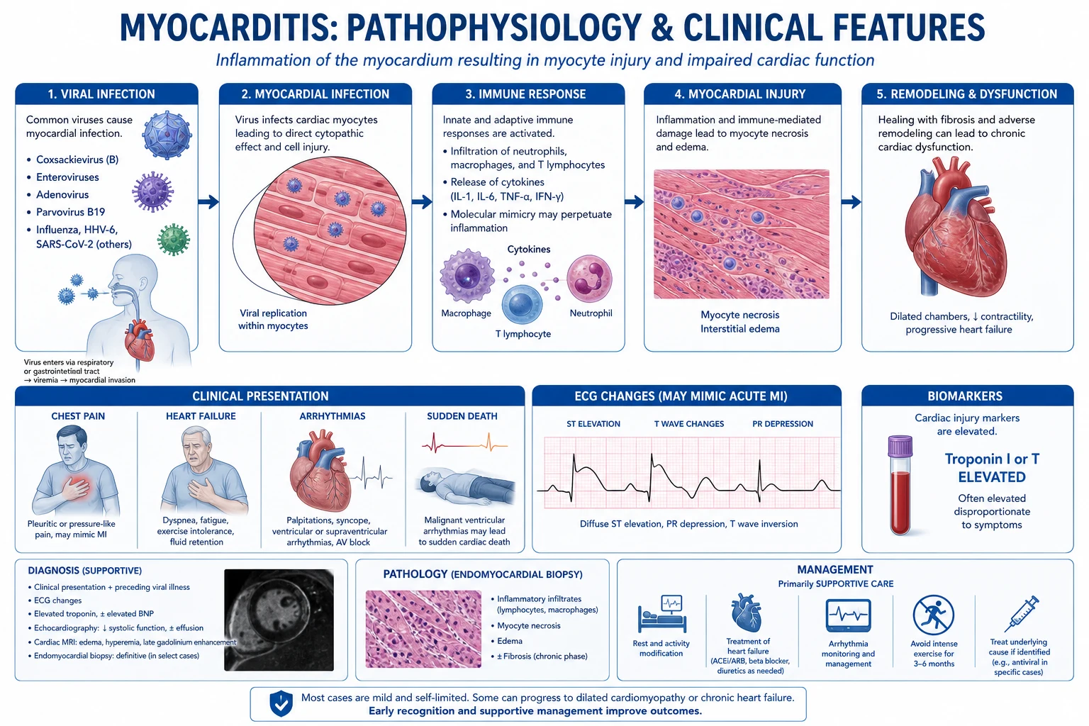
Acute myocarditis is inflammation of the myocardium — commonest causes: viral (coxsackie, parvovirus B19, COVID-19), autoimmune, drug-induced (immune checkpoint inhibitors), idiopathic. Presents with: chest pain (may mimic MI), heart failure, arrhythmia, or cardiogenic shock (fulminant myocarditis). Diagnosis: troponin elevated, ECG changes (non-specific ST/T changes, may mimic MI), echo (regional wall motion abnormalities, reduced EF), cardiac MRI (gold non-invasive — oedema, late gadolinium enhancement in subepicardial/mid-wall pattern), endomyocardial biopsy (gold standard but rarely performed). Treatment: supportive (heart failure therapy, mechanical support if fulminant), avoid NSAIDs in acute phase (may worsen inflammation). Giant cell myocarditis: urgent immunosuppression + transplant evaluation.
On this page & tools
Your progress
Saved locally on this device.
Target exams
Red flags
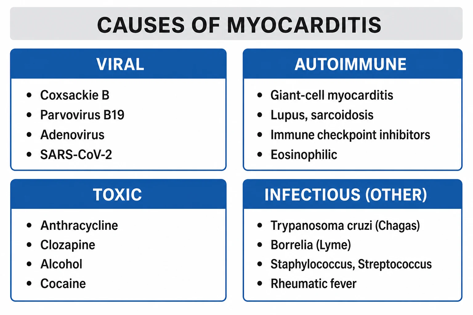
Causes
Viral (most common)
#1 cause
- Coxsackievirus B (classic)
- Parvovirus B19 (most common on biopsy)
- COVID-19 (SARS-CoV-2 — increasingly recognised)
- Influenza, EBV, CMV, HHV-6, HIV
- Enteroviruses, adenoviruses
Non-viral
Diverse
- Autoimmune: SLE, rheumatoid, sarcoidosis, Kawasaki
- Drugs: immune checkpoint inhibitors (pembrolizumab, nivolumab — CHECKPOINT inhibitor myocarditis), clozapine, chemotherapy (anthracyclines — dose-dependent)
- Toxins: cocaine, alcohol
- Hypersensitivity: penicillins, sulfonamides, diuretics (eosinophilic myocarditis)
- Giant cell myocarditis: idiopathic, rapidly progressive, often fatal
- Bacterial: Lyme disease (Borrelia burgdorferi), diphtheria
Diagnosis
Myocarditis diagnostic approach
Clinical suspicion
Young patient with MI-like chest pain but normal/abnormal coronaries. Or: new heart failure/arrhythmia in young otherwise healthy person. Or: cardiogenic shock in young patient without known cardiac disease. Viral prodrome (fever, myalgia, fatigue) preceding cardiac symptoms by days-weeks.
ECG + troponin
ECG: non-specific ST/T changes, sinus tachycardia, may mimic acute MI (ST elevation, especially in localized myocarditis — but coronary angiography shows normal coronaries). Arrhythmias: AF, heart block, ventricular arrhythmias. Troponin: elevated (sensitive but non-specific — may be normal in chronic myocarditis).
Echocardiogram
Reduced EF (global or regional), wall motion abnormalities (may mimic MI but distribution does not follow coronary territory), pericardial effusion (myopericarditis). May be normal in mild cases. Fulminant myocarditis: severely reduced EF, sometimes with preserved wall thickness.
Cardiac MRI (gold non-invasive)
Lake Louise criteria: (1) T2-weighted imaging: myocardial oedema (high signal). (2) Late gadolinium enhancement (LGE): subepicardial or mid-wall distribution (distinguishes from MI which is subendocardial). (3) T1/T2 mapping: quantitative tissue characterisation. MRI differentiates myocarditis from MI and other cardiomyopathies.
Endomyocardial biopsy (gold standard)
Histological diagnosis (Dallas criteria: inflammatory infiltrate with myocyte necrosis). Rarely performed routinely — reserved for: fulminant myocarditis (rule out giant cell), suspected specific treatable cause (sarcoidosis, eosinophilic), refractory heart failure. Complications: perforation (rare), sampling error (patchy inflammation).
Coronary angiography
Essential to EXCLUDE MI (acute coronary syndrome is the main differential). If coronaries normal + troponin elevated + regional wall motion abnormal = myocarditis. Do NOT assume young patients cannot have MI — cocaine, familial hyperlipidaemia, Kawasaki disease can cause coronary occlusion in young patients.
Management
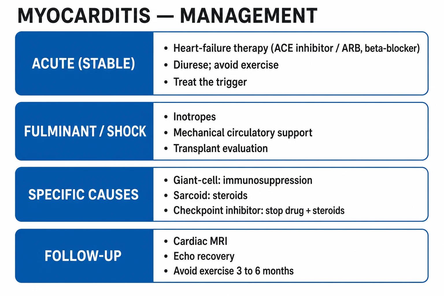
Myocarditis management
Supportive care — the foundation
Rest (avoid exercise until recovery — sudden death risk during acute phase). Monitor: continuous ECG (arrhythmia risk), daily troponin trend, serial echo (EF recovery). Heart failure therapy: ACE inhibitor/ARB, beta-blocker (once acute phase resolving, NOT during fulminant shock), diuretics, aldosterone antagonist. Treat arrhythmias.
Fulminant myocarditis — mechanical support
Cardiogenic shock in fulminant myocarditis: VA-ECMO or Impella as bridge to recovery (myocarditis often recovers within days-weeks if patient survives the acute phase). Inotropes (dobutamine, milrinone) may worsen arrhythmia — use cautiously. Good prognosis if survived to recovery (better than non-fulminant in some studies).
Immunosuppression — SELECTIVE use only
NOT routinely indicated for viral myocarditis (may worsen viral replication). Indicated for: (1) Giant cell myocarditis — urgent cyclosporine + steroids (life-saving). (2) Autoimmune-mediated (sarcoidosis, SLE) — steroids. (3) Immune checkpoint inhibitor myocarditis — stop drug, high-dose IV steroids (methylprednisolone 1g/day x 3 days). (4) Eosinophilic myocarditis — steroids. NOT for: viral lymphocytic myocarditis (no proven benefit).
Avoid NSAIDs
Do NOT give NSAIDs (ibuprofen, diclofenac) during acute myocarditis — may worsen inflammation and increase mortality. Colchicine may be beneficial for myopericarditis (anti-inflammatory, specifically for pericardial component). Aspirin only if true pericarditis (without myocardial involvement).
Activity restriction
No competitive sport or strenuous exercise for 3-6 months after myocarditis (even if recovered). Exercise can trigger fatal arrhythmia during recovery. Reassess with exercise stress test + echo + Holter before return to sport.
Giant cell myocarditis — special case
Rapidly progressive, often fatal within weeks. Suspect in: older patient, rapidly deteriorating heart failure, refractory ventricular arrhythmias, heart block. Diagnosis: biopsy (giant cells, extensive necrosis). Treatment: urgent immunosuppression (cyclosporine + azathioprine + steroids) ± heart transplant. Median survival without treatment: 3-5 months.
SAQ — Fulminant myocarditis with cardiogenic shock
10 minutes · 10 marks
A 28-year-old previously well man presents four days after a flu-like illness with dyspnoea, chest pain and presyncope. He is cool and clammy, HR 128 in sinus tachycardia, BP 78/50, JVP elevated, bilateral crackles, lactate 4.2 mmol/L. Bedside echo shows a small, thick, poorly contracting LV (EF ~20%) with a normal-sized RV and a small pericardial effusion. Coronary angiography shows normal epicardial vessels. High-sensitivity troponin is 8,200 ng/L.
SAQ — Giant-cell myocarditis masquerading as fulminant myocarditis
10 minutes · 10 marks
A 44-year-old woman with a history of autoimmune thyroiditis is admitted with rapidly progressive dyspnoea over five days. She is in cardiogenic shock (BP 72/45, lactate 5.1) with frequent runs of monomorphic VT and new complete heart block requiring transvenous pacing. Echo shows a severely impaired biventricular EF. Coronary angiography is normal. Troponin is markedly elevated.
Clinical pearls
Red flags
The aetiological framework — a structured hunt
Myocarditis is a syndrome, not a single disease. The cause drives the treatment: viral lymphocytic myocarditis is treated supportively, giant-cell and eosinophilic myocarditis are treated with immunosuppression, immune checkpoint inhibitor (ICI) myocarditis is treated with drug cessation and steroids, and autoimmune myocarditis is treated for the underlying disease. A shotgun "steroids for everyone" approach worsens viral replication and is poor medicine.[1][2]
Aetiological workup in suspected myocarditis
History — the cause is often in the story
A flu-like illness 1-4 weeks earlier (viral). A new immune checkpoint inhibitor in the last 1-6 months or a recent influenza vaccine on an ICI (ICI myocarditis, which can be potentiated by vaccination). A new drug in the last 1-8 weeks with a rash and eosinophilia (hypersensitivity/eosinophilic). An autoimmune history (SLE, sarcoid, IBD). Cocaine, alcohol, or a recent chemo cycle (anthracycline cumulative dose). A tick bite or erythema migrans (Lyme). Peripartum timing.
Viral and atypical pathogen screen
PCR/serology for enterovirus/coxsackie, parvovirus B19, adenovirus, EBV, CMV, HHV-6, influenza, and SARS-CoV-2; HIV; in the right context hepatitis C, Rickettsia, and Trypanosoma cruzi (Chagas — the leading cause of myocarditis in Latin America). Respiratory PCR for an active syndrome. A rising antibody titre (paired sera) is more specific than a single swab.
Autoimmune and toxin screen
ANA, dsDNA, extractable antigens, ANCA, complement, angiotensin-converting enzyme (sarcoid), serum and urine protein electrophoresis, and a urine drug screen (cocaine). Eosinophil count and IgE — a rising eosinophil count with a new drug is hypersensitivity myocarditis until proven otherwise.
Troponin, ECG, and echocardiography
High-sensitivity troponin (sensitive, tracks the illness), 12-lead ECG (non-specific ST/T, PR depression, conduction disease, or an infarct pattern that does not fit a territory), and transthoracic echo (reduced EF, regional or global wall motion abnormality that does not respect a coronary territory, pericardial effusion, and — in fulminant disease — a small thick non-compliant LV).
Cardiac MRI — the non-invasive arbiter
Apply the updated Lake Louise criteria (T1/ECV/LGE for tissue injury PLUS T2 for oedema/inflammation). Two positive categories confirm myocarditis with high specificity. The LGE pattern also flags the aetiology: subepicardial/mid-wall inferolateral = viral lymphocytic; patchy mid-wall septal/non-transmural with thinning = sarcoid; diffuse subendocardial with low voltages = amyloid; none of the above + a focal infarct pattern = think MINOCA or coronary.
Coronary angiography — exclude the mimic
Mandatory whenever the presentation could be an acute coronary syndrome (ST elevation, regional wall motion, haemodynamic compromise). Normal coronaries plus a raised troponin and a wall-motion abnormality that ignores a vascular territory is the working definition of clinically suspected myocarditis — but cocaine, Kawasaki, and spontaneous coronary artery dissection can occlude a coronary in a young patient.
Endomyocardial biopsy — selective, not routine
Reserved for fulminant disease (to exclude giant-cell and eosinophilic myocarditis, which change management entirely), refractory heart failure, suspected specific treatable disease (sarcoid, eosinophilic, drug hypersensitivity), and ventricular arrhythmia or high-grade heart block out of proportion to the EF. The Dallas criteria are applied to the first biopsy; PCR on biopsy tissue increases the viral yield.
The McCarthy classification — fulminant vs acute (non-fulminant)
The single most important clinical distinction is fulminant vs acute (non-fulminant) myocarditis. Fulminant myocarditis presents with abrupt, severe cardiogenic shock — but, paradoxically, has a better long-term prognosis than the indolent acute form, because the aggressive inflammatory insult that produces shock also provokes a vigorous immune clearance of virus and the myocardium often recovers completely if the patient is bridged through the acute phase. The McCarthy cohort showed 11-year survival of 93 per cent in fulminant vs 45 per cent in acute myocarditis.[3][11]
Fulminant myocarditis
Better long-term survival
- Distinct viral prodrome followed by abrupt (within hours-days) cardiogenic shock
- Severe haemodynamic compromise: hypotension, oliguria, rising lactate, pulmonary oedema
- Echo: small, thick, non-compliant LV with severe global hypokinesis; preserved RV size
- Often needs MCS (IABP, VA-ECMO, Impella) as a bridge to recovery
- Endomyocardial biopsy recommended (exclude giant-cell / eosinophilic)
- 11-year survival ~93% (McCarthy) — bridge the patient, the myocardium often recovers
Acute (non-fulminant)
Indolent but worse long-term
- More gradual onset of heart failure symptoms over days-weeks
- Preserved haemodynamics at presentation — does not need MCS up front
- Echo: dilated LV with reduced EF (looks like a new DCM)
- Higher rate of progression to dilated cardiomyopathy and late death/transplant
- 11-year survival ~45% in the McCarthy cohort — the indolent course is the danger
- Treatment: GDMT for heart failure; reassess EF at 3-6 months before declaring recovery
The Dallas criteria — histological diagnosis
The Dallas criteria (Aretz 1987) standardised the histopathological reporting of endomyocardial biopsy in suspected myocarditis. They remain the morphological reference standard, although inter-observer variability and sampling error (myocarditis is patchy) limit sensitivity to roughly 35-50 per cent — a negative biopsy does not exclude the diagnosis.[12][1]
First biopsy
Three possible reads
- Active myocarditis — inflammatory infiltrate with associated myocyte necrosis (the definitive call)
- Borderline myocarditis — inflammatory infiltrate WITHOUT myocyte necrosis (less specific)
- No myocarditis — neither infiltrate nor necrosis (does not exclude patchy disease — sampling error)
Subsequent biopsies
Tracks the disease
- Ongoing (persistent) myocarditis — infiltrate and necrosis persist
- Resolving (healing) myocarditis — infiltrate reduced, fibrosis appearing
- Resolved (healed) myocarditis — no infiltrate, residual fibrosis
Limitations
Why a negative biopsy is not the end
- Sampling error — inflammation is focal; a right-ventricular biopsy may miss a left-sided or patchy process
- Poor inter-observer agreement on the "borderline" category
- Insensitive for the underlying aetiology — add immunohistochemistry and viral PCR on the biopsy tissue
- Replaced for clinical decisions by the WHO/WHF classification, which adds immunohistochemical and molecular (PCR) layers
The Lake Louise criteria — cardiac MRI
Cardiac MRI is the non-invasive gold standard for myocarditis. The original Lake Louise criteria (Friedrich 2009) used T2 oedema imaging plus early and late gadolinium enhancement; the 2018 update (Ferreira) reframed the criteria around parametric mapping, which is more reproducible and accurate, especially in mild or chronic disease.[5][4]
Applying the updated (2018) Lake Louise criteria
Criterion 1 — tissue injury (T1-based)
Look for native T1 elevation, elevated extracellular volume (ECV), or late gadolinium enhancement (LGE). LGE is typically in a subepicardial or mid-wall inferolateral distribution (viral lymphocytic) or in the basal interventricular septum in a patchy, non-ischaemic pattern (sarcoid).
Criterion 2 — inflammation (T2-based)
Look for native T2 elevation (focal or global myocardial oedema) or a raised T2 ratio. T2 is the inflammation marker — it is the most specific signal for active myocarditis and the most useful for following recovery.
Combine — at least one from each
A positive study requires AT LEAST ONE T1-based (tissue injury) signal AND AT LEAST ONE T2-based (inflammatory) signal. Two positive categories → diagnosis confirmed with high specificity. One positive category → "possible" myocarditis; correlate clinically and reimage at 1-3 months if needed.
Report the LGE pattern
Subepicardial/mid-wall = myocarditis (differentiates from ischaemic LGE which is subendocardial and follows a coronary territory). The pattern also flags the aetiology: inferolateral subepicardial = viral lymphocytic; patchy basal septal/mid-wall = sarcoid; diffuse subendocardial with low voltages = amyloid; apical ballooning with resolving oedema = Takotsubo.
Caveats
CMR cannot reliably distinguish active from healed myocarditis without T2/T1 mapping (LGE persists for life). It is falsely negative in the first few days of fulminant disease (oedema has not yet developed) — do not exclude myocarditis on an early CMR if the clinical picture is convincing. Renal failure limits gadolinium use; rely on native T1/T2 mapping.
Late gadolinium enhancement patterns — telling the mimics apart
The distribution of LGE is the single most testable CMR observation in cardiology. The position of the enhancement in the myocardial wall (subendocardial → subepicardial) and its territory (coronary vs non-coronary) usually narrows the differential to one diagnosis.[4][5]
Subendocardial / transmural
Ischaemic (follows a coronary territory)
- Begins at the subendocardium (the watershed of the myocardium) and extends outward in proportion to infarct transmurality
- Follows a coronary vascular territory (LAD, LCx, RCA) — the key discriminator from myocarditis
- Thin, bright, "white" myocardium on LGE with adjacent wall-motion abnormality = old MI
Subepicardial / mid-wall
Myocarditis (Lake Louise)
- Spare the subendocardium — enhancement starts at the epicardial surface or sits in the mid-wall
- Classically the lateral wall (viral lymphocytic, e.g. parvovirus B19, coxsackie)
- Septal mid-wall (non-ischaemic) also seen in sarcoidosis and in late-stage dilated cardiomyopathy
Patchy mid-wall basal septum
Sarcoidosis
- Multiple focal nodular lesions, often basal septal, sometimes with thinning and aneurysm
- Look for lymphadenopathy on the same scan, conduction disease (heart block), and a raised ACE level
- Treatment: steroids — sarcoid myocarditis is one of the few that responds to immunosuppression
Diffuse subendocardial + low voltages
Cardiac amyloidosis
- Diffuse subendocardial (or transmural) LGE with difficulty nulling the blood pool
- Low QRS voltages on the ECG discordant with a thick LV on echo (the "voltage-mass discordance")
- Apical-sparing longitudinal strain; confirm with DPD/PYP/HMDP (ATTR) or serum free light chains (AL)
Mid-wall "dumbbell" septum / RV insertion points
Hypertrophic / pressure-load
- Focal enhancement at the right-ventricular insertion points in HCM, aortic stenosis, and pulmonary hypertension
- Not a myocarditis pattern — represents fibrosis from chronic pressure load
- A risk marker for sudden death in HCM when extensive
Giant-cell myocarditis — the emergency
Giant-cell myocarditis (GCM) is rare, idiopathic, and rapidly fatal without immunosuppression or transplant — untreated median survival is 3-5 months. The classical triad at presentation is rapidly progressive heart failure, refractory ventricular arrhythmias, and high-grade heart block, in a young or middle-aged adult, often with an autoimmune history. Endomyocardial biopsy is mandatory whenever GCM is on the differential (fulminant course, intractable VT, or new heart block), because the histology changes everything: combined immunosuppression (cyclosporine + azathioprine or tacrolimus + mycophenolate + steroids) is life-saving and early transplant listing is the exit strategy for non-responders.[6]
Suspected giant-cell myocarditis — the pathway
Recognise the phenotype
A fulminant or subfulminant course with intractable ventricular arrhythmia or new high-grade AV block, in a middle-aged adult, frequently with coexisting autoimmune disease (autoimmune thyroiditis, ulcerative colitis, myasthenia). GCM does not respect a coronary territory and does not respond to GDMT.
Biopsy early
Endomyocardial biopsy is the diagnostic test — histology shows multinucleated giant cells, a diffuse lymphohistiocytic infiltrate, and extensive myocyte necrosis (NOT the focal lymphocytic picture of viral myocarditis, and NOT the eosinophil-predominant picture of eosinophilic myocarditis).
Start combined immunosuppression
Cyclosporine (or tacrolimus) + azathioprine (or mycophenolate) + corticosteroids, titrated with drug levels. This combination improved survival and transplant-free survival over historical eras (the Kandolin/Mayo data). Do NOT use steroids alone — they are inadequate.
List for transplant early
For non-responders, mechanical support (VA-ECMO, Impella, durable BiVAD) as a bridge to transplant is the rescue strategy. GCM can recur in the transplanted heart but is controllable with the post-transplant immunosuppression.
Eosinophilic myocarditis — the hypersensitivity syndrome
Eosinophilic myocarditis is the great masquerader — it is frequently missed because the eosinophilia may be transient and the drug trigger is overlooked. There are three subtypes: hypersensitivity (drug-related), hypereosinophilic syndrome / parasitic, and eosinophilic granulomatosis with polyangiitis (EGPA, Churg-Strauss). The hypersensitivity form is classically triggered by clozapine, antibiotics (penicillins, sulfonamides, cephalosporins), anti-epileptics, and diuretics, and is suggested by a new drug, a rash, fever, and a rising eosinophil count 1-8 weeks after exposure. The treatment is drug cessation and corticosteroids.[10]
Hypersensitivity (drug)
Clozapine, antibiotics, AEDs
- Onset 1-8 weeks after a new drug; rash, fever, eosinophilia, occasionally a DRESS picture
- Clozapine is the classic offender — myocarditis in ~1 in 100 to 1 in 1000 exposures, usually within the first 8 weeks
- Treatment: stop the drug, high-dose corticosteroids (prednisolone 1 mg/kg or IV methylprednisolone)
- Outcomes are good if recognised early — fatal if the drug is continued
Hypereosinophilic / parasitic
Loeffler endocarditis
- Sustained eosinophilia (parasitic infestation, idiopathic hypereosinophilic syndrome, malignancy)
- Loeffler endocarditis — eosinophilic infiltration with overlying thrombus and a restrictive cardiomyopathy
- Treatment: treat the cause (anti-helminthics), corticosteroids ± cytotoxic agents, anticoagulation for apical thrombus
EGPA (Churg-Strauss)
ANCA, asthma, eosinophilia
- Adult-onset asthma, eosinophilia, sinusitis, and a systemic vasculitis (ANCA positive in ~40%)
- Cardiac involvement is a leading cause of death in EGPA — cardiomyopathy, arrhythmia, heart failure
- Treatment: high-dose corticosteroids + cyclophosphamide (or rituximab for ANCA-positive disease)
Immune checkpoint inhibitor myocarditis
ICI myocarditis (pembrolizumab, nivolumab, ipilimumab, and combination regimens) is uncommon (0.5-1 per cent) but has a case-fatality rate of 25-50 per cent — the highest of any drug-induced myocarditis. It typically presents in the first 1-6 months of therapy, can be potentiated by an influenza vaccine, and frequently overlaps with myositis (raised creatine kinase) and myasthenia (diplopia, ptosis). The treatment is permanent cessation of the ICI and high-dose corticosteroids (methylprednisolone 1 g/day for 3 days, then a slow oral prednisolone taper).[7][13]
Suspected ICI myocarditis — the pathway
Maintain a low threshold
Measure a troponin and an ECG with any new cardiac symptom (chest pain, dyspnoea, palpitation) in a patient on an ICI — and a baseline troponin before each cycle to catch silent subclinical cases. A concurrent influenza vaccine can precipitate ICI myocarditis within days.
Confirm and grade
Troponin (high-sensitivity), ECG, echo (regional or global wall motion abnormality), and cardiac MRI (oedema + LGE, often subepicardial). Check creatine kinase and acetylcholine-receptor antibodies for overlapping myositis and myasthenia. Severe cases: cardiogenic shock, complete heart block, malignant VT.
Stop the ICI permanently
Do NOT re-challenge — recurrent ICI myocarditis is more severe and frequently fatal. Hold other immunotherapy as well and consult oncology about an alternative anti-cancer strategy.
High-dose corticosteroids
Methylprednisolone 1 g IV daily for 3 days, then oral prednisolone 1 mg/kg/day with a slow taper over 4-6 weeks guided by a falling troponin. Refractory cases: add a second agent (mycophenolate, tacrolimus, infliximab, or plasmapheresis for a myasthenic crisis).
Supportive care
Treat heart failure and arrhythmia conventionally (avoid beta-blockers if there is severe myasthenia with bradycardia/heart block — pacing may be needed). MCS (VA-ECMO, Impella) for fulminant ICI myocarditis as a bridge to recovery or decision.
Surveillance
A falling troponin and a recovering EF guide the steroid taper. Long-term cardiac follow-up is essential — some patients develop a persistent cardiomyopathy despite steroids.
Drug-induced myocarditis — the offenders list
A focussed drug and toxin history is non-negotiable in any suspected myocarditis. The cause drives everything: drug cessation is the single most important intervention for the hypersensitivity and ICI forms.[1][7][10]
Immune checkpoint inhibitors
Highest mortality
- Pembrolizumab, nivolumab, ipilimumab, combination regimens (combination therapy is highest-risk)
- Onset within 1-6 months; fatality 25-50%; overlaps with myositis and myasthenia
- Stop the ICI permanently + high-dose IV steroids
Clozapine
Antipsychotic
- Risk ~1 per 1000 exposures, classically in the first 8 weeks of therapy
- Hypersensitivity (eosinophilic) mechanism — watch the eosinophil count and troponin during titration
- Stop clozapine + corticosteroids; do not re-challenge
Anthracyclines
Doxorubicin
- Dose-dependent, cumulative toxicity (risk rises sharply above 400-450 mg/m² doxorubicin)
- Mechanism: iron-mediated free-radical injury and topoisomerase-IIβ; presents as a dilated cardiomyopathy rather than acute myocarditis
- Lifelong surveillance with echo; dexrazoxane for cardio-protection in high-risk regimens
Antimalarials (chloroquine/hydroxychloroquine)
Rare
- Can cause a restrictive cardiomyopathy with conduction disease (often with biopsy storage-like change)
- Stop the drug + corticosteroids; recovery is variable
Hypersensitivity drugs
Antibiotics, diuretics, AEDs
- Penicillins, sulfonamides, cephalosporins, thiazides, furosemide, phenytoin, carbamazepine
- Eosinophilia, rash, fever 1-8 weeks after exposure; biopsy = florid eosinophilic infiltrate
- Stop the drug + corticosteroids; usually fully reversible if caught early
Toxins
Cocaine, alcohol
- Cocaine — sympathomimetic vasospasm, microvascular injury, and a true myocarditis; can also cause an acute coronary syndrome (vasospasm or thrombosis)
- Chronic alcohol — a dilated cardiomyopathy that improves with abstinence
- Stop the toxin; supportive heart-failure therapy; benzodiazepines for cocaine-related sympathetic surge
Fulminant myocarditis — mechanical circulatory support escalation
The principle in fulminant myocarditis is to support the failing ventricle while the inflammation resolves — most fulminant lymphocytic myocarditis recovers within days to weeks if the patient is bridged through the acute phase. The exit strategy is defined up front: bridge to recovery (the commonest outcome), bridge to decision, bridge to a durable LVAD, or bridge to transplant. Inotropes are a double-edged sword — they increase myocardial oxygen demand and arrhythmia in the inflamed myocardium and should be used at the lowest effective dose as a temporary measure only.[3][11]
Escalation pathway for fulminant myocarditis / refractory cardiogenic shock
Recognise the shock early
Hypotension (SBP <90), cold peripheries, oliguria, rising lactate, pulmonary oedema — in a young patient with a viral prodrome, think fulminant myocarditis. Bedside echo: a small, thick, hypocontractile LV with a preserved RV size (unlike chronic DCM).
Inotrope ± vasopressor — lowest effective dose
Noradrenaline for the MAP; dobutamine or milrinone for the cardiac output. Avoid prolonged high-dose catecholamines — they worsen arrhythmia and oxygen demand in the inflamed myocardium. Milrinone is preferred when the blood pressure tolerates it (less arrhythmogenic, pulmonary vasodilator).
Intra-aortic balloon pump
A reasonable first-line MCS — diastolic augmentation and reduced afterload improve coronary perfusion and cardiac output. Contraindicated in significant aortic regurgitation and aortic dissection.
VA-ECMO for refractory shock
Indicated for cardiac arrest (ECPR), a lactate rising despite inotropes, or a MAP that cannot be maintained. Provides full cardiopulmonary bypass (3-5 L/min). It does NOT unload the LV — if the LV dilates (aortic valve not opening on echo, worsening pulmonary oedema), add an Impella or a venting strategy (the ECPELLA configuration).
Impella (percutaneous LVAD)
2.5-5 L/min of active forward flow with LV unloading — ideal when VA-ECMO is causing LV distension. Monitor for haemolysis (LDH, haptoglobin, free haemoglobin) and limb ischaemia.
Endomyocardial biopsy
For any fulminant case (especially middle-aged), to exclude giant-cell and eosinophilic myocarditis, both of which change management entirely. The CMR can be deferred in extremis — do not let imaging delay biopsy and MCS.
Define the goal and the exit
Bridge to recovery (the commonest outcome in fulminant lymphocytic myocarditis — wean MCS as the EF recovers over days-weeks), bridge to decision (if the trajectory is unclear), bridge to transplant (for giant-cell or non-recovering cases), or bridge to a durable LVAD.
SARS-CoV-2 and mRNA vaccine myocarditis
COVID-19 infection itself is a cause of myocarditis (direct viral injury, immune-mediated, and as part of multisystem inflammatory syndrome, MIS-A/MIS-C), and the mRNA vaccines (BNT162b2 and mRNA-1273) are associated with a small but real risk of myocarditis, concentrated in young males within a week of the second dose. Vaccine myocarditis is typically mild and self-limiting — in stark contrast to ICI myocarditis — and is treated supportively, with NSAIDs and colchicine for the pericardial component in the stable patient.[8][9]
COVID-19 infection-related
Multisystem inflammatory
- Direct cardiomyocyte injury (ACE2 receptor), immune-mediated cytokine storm, and microvascular thrombosis
- May present as part of MIS-A/MIS-C (multisystem inflammatory syndrome) weeks after infection
- Can be fulminant; treat per the underlying syndrome (supportive, MCS, IVIG/steroids for MIS-C)
- Outcomes variable — long-COVID cardiomyopathy recognised
mRNA vaccine myocarditis
Young males, second dose
- Risk highest in males aged 16-24, within 1 week of the second dose (BNT162b2 or mRNA-1273)
- Estimated risk 1-10 per 100 000 doses (higher for mRNA-1273 than BNT162b2); benefit of vaccination far outweighs risk
- Typically mild: chest pain, raised troponin, subepicardial LGE; supportive care, NSAIDs, colchicine
- Witberg and Mevorach NEJM 2021 — large Israeli cohorts defined the risk-benefit
Differential diagnosis — raised troponin with normal coronaries
Once the coronaries are angiographically normal, a structured differential is run. The CMR is the single most useful second-line test.[1][4]
Acute myocarditis
Lake Louise positive
- Subepicardial/mid-wall LGE, raised T1/T2, viral prodrome, young patient
- Troponin elevated, ECG non-specific or infarct-like, EF variable
Takotsubo cardiomyopathy
Apical ballooning
- Emotional or physical stress, post-menopausal woman, apical ballooning with hyperkinetic base
- Transient ST elevation and troponin rise that is out of proportion to the (often mild) wall-motion abnormality
- Recovery over days-weeks; watch for apical thrombus, prolonged QTc with Torsades risk, and LVOT obstruction
MINOCA
Myocardial infarction with non-obstructive coronaries
- A working diagnosis — subendocardial LGE suggesting true infarction with normal epicardial coronaries
- Causes: plaque rupture/erosion, coronary spasm, spontaneous coronary artery dissection, coronary embolism, microvascular dysfunction
- Investigate with intracoronary imaging (OCT/IVUS) and a vasospasm provocation test
Pulmonary embolism
Right-heart strain
- Raised troponin from RV ischaemia/strain; ECG S1Q3T3 or right-axis; echo shows RV dilation and McConnell sign
- D-dimer, CTPA; risk-stratify — thrombolysis for high-risk (massive) PE
Sepsis / critical illness
Cytokine-mediated
- Mild troponin rise is common in any critically ill patient; sepsis-induced cardiomyopathy is reversible
- Diffuse, non-territorial wall-motion abnormality that recovers as the sepsis resolves
Renal failure
Reduced clearance
- Chronic troponin elevation from reduced renal clearance — the troponin is sensitive but non-specific in CKD
- Use a delta troponin (rise/fall pattern) to distinguish acute injury from chronic elevation
The young patient with chest pain — a diagnostic pathway
A young patient (under 35-40) presenting with chest pain and a raised troponin is a high-yield exam scenario and a high-stakes bedside decision. The goal is to separate true ischaemia (cocaine, Kawasaki, familial hyperlipidaemia, spontaneous dissection) from the non-ischaemic causes (myocarditis, Takotsubo), because the management diverges sharply.[1]
Young patient, chest pain, raised troponin
ECG + high-sensitivity troponin
ST elevation or depression, T-wave inversion, or non-specific changes. A troponin delta (rise and fall) confirms an acute injury pattern. Do NOT anchor on "young = benign" — cocaine and Kawasaki can occlude a coronary in a 25-year-old.
Urine drug screen + lipid panel + history
Cocaine (sympathomimetic vasospasm and thrombosis), amphetamines, a strong family history of premature coronary disease, prior Kawasaki disease, peripartum timing, and a recent viral illness or vaccination.
Coronary angiography if any ischaemic feature
ST elevation, regional wall-motion abnormality, haemodynamic instability, or ongoing chest pain → angiography to exclude an acute coronary syndrome. Cocaine-induced STEMI: benzodiazepines, nitrates, and aspirin; avoid beta-blockers (unopposed alpha agonism).
If coronaries are normal — CMR
Apply the updated Lake Louise criteria (T1/ECV/LGE + T2). The LGE pattern defines the diagnosis: subepicardial/mid-wall = myocarditis; apical ballooning with resolving oedema = Takotsubo; subendocardial in a territory = MINOCA (intracoronary imaging).
Echo for EF and wall motion
Regional wall-motion abnormality that ignores a coronary territory = myocarditis; apical ballooning with basal hyperkinesis = Takotsubo; global hypokinesis with LV dilation = evolving DCM from acute myocarditis.
Biopsy only if fulminant, atypical, or refractory
Reserve endomyocardial biopsy for fulminant disease, intractable VT, new high-grade heart block, or refractory heart failure — to exclude giant-cell and eosinophilic myocarditis, which mandate immunosuppression.
Pharmacology at the bedside
The drug therapy of myocarditis is a frequent source of error. The headline rule — avoid NSAIDs in the acute phase — applies to myocarditis specifically, while true pericarditis (without myocardial involvement) is treated with high-dose aspirin/NSAIDs and colchicine. Immunosuppression is selective, not universal.[1][2]
Supportive heart-failure therapy
The foundation
- ACE inhibitor/ARB (or ARNI for HFrEF), beta-blocker (once haemodynamically stable, NOT in fulminant shock), MRA, and SGLT2 inhibitor — the four pillars of GDMT
- Loop diuretic for congestion; vasodilators (nitrate-based) only when the blood pressure tolerates them
- Begin GDMT as the acute phase resolves and titrate to target; reassess EF at 3-6 months
NSAIDs — AVOID in acute myocarditis
Worsens inflammation
- NSAIDs (ibuprofen, diclofenac, naproxen) are thought to worsen myocardial inflammation and increase mortality in animal models and observational data
- Use colchicine instead for the pericardial component of myopericarditis; high-dose aspirin ONLY if the disease is pericarditis-dominant without myocardial involvement
Colchicine
For myopericarditis
- Anti-inflammatory for the pericardial component; modest benefit in myopericarditis
- Weight-based dosing; GI upset is the dose-limiting side-effect; reduce dose in CKD
IVIG and steroids — controversial
Selective use
- IVIG — not supported by the TIMIC trial in lymphocytic myocarditis; no proven benefit; occasionally used in paediatric and MIS-C myocarditis
- Steroids — NOT for viral lymphocytic myocarditis (risk of viral replication); INDICATED for giant-cell, eosinophilic, sarcoid, and ICI myocarditis
- A trial of steroids may be considered in biopsy-negative, virus-negative, autoimmune-flavoured myocarditis
Inotropes — cautious
Bridge only
- Dobutamine, milrinone — lowest effective dose; they increase oxygen demand and arrhythmia in the inflamed myocardium
- Milrinone preferred when blood pressure tolerates it (less arrhythmogenic, pulmonary vasodilator)
- Use only as a bridge to MCS or recovery, not as a destination
Activity restriction and return to play
Sudden cardiac death during exercise is the feared late complication of myocarditis — the inflamed, healing myocardium is electrically unstable for weeks to months. All competitive sport and strenuous exercise is prohibited for 3-6 months, even if the patient feels well and the EF has normalised. Return to sport requires a documented recovery of the EF, a negative Holter for significant arrhythmia, an exercise test, and a CMR showing resolution of oedema.[1]
Return-to-play protocol after myocarditis
Absolute rest during the acute phase
No exercise, competitive sport, or strenuous activity during the acute illness and for at least 3-6 months afterwards. This is the single most evidence-based and most frequently violated recommendation.
Reassess at 3-6 months
Transthoracic echo (EF and wall motion normalised), 12-lead ECG (no residual ischaemia or conduction disease), ambulatory Holter (no significant ventricular arrhythmia or heart block), and an exercise test (exercise capacity and ischaemia/arrhythmia with exertion).
CMR for resolution of oedema
A repeat CMR showing resolution of T2 oedema (the inflammation marker) confirms that the myocardium has healed — residual LGE is acceptable (scar persists for life) provided there is no active inflammation.
Cleared only if all are negative
Return to competitive sport is permitted only if the EF, ECG, Holter, exercise test, and CMR oedema are all resolved/negative. A persistent EF reduction, residual oedema, or significant arrhythmia delays return and may indicate progression to a cardiomyopathy.
Prognosis
Prognosis is stratified by the clinical phenotype (fulminant vs acute) and the aetiology (giant-cell vs lymphocytic vs ICI). The McCarthy paradox — fulminant has a BETTER long-term survival than acute — holds because the fulminant insult provokes complete immune clearance and full recovery, while the indolent acute form often progresses to a dilated cardiomyopathy.[3][11]
Fulminant lymphocytic
Best prognosis
- 11-year survival ~93% (McCarthy) if bridged through the acute phase
- Full recovery of EF within days-weeks in the majority; GDMT can be weaned
- Late recurrence and DCM progression are uncommon
Acute (non-fulminant) lymphocytic
Indolent but worse long-term
- 11-year survival ~45% in the McCarthy cohort; higher rate of DCM progression, transplant, and late death
- Requires lifelong GDMT surveillance; reassess EF at 3-6 months and 12 months
- Negative prognostic markers: low LVEF, wide QRS, sustained VT, persistent troponin elevation, extensive LGE
Giant-cell myocarditis
Fatal without immunosuppression
- Untreated median survival 3-5 months (Cooper 1997)
- Combined immunosuppression (cyclosporine + azathioprine + steroids) dramatically improves survival and transplant-free survival
- Early transplant listing for non-responders; recurrence in the graft is controllable with post-transplant immunosuppression
Immune checkpoint inhibitor
High case-fatality
- Case-fatality 25-50% (Mahmood 2018) — the highest of any drug-induced myocarditis
- Better outcomes with early cessation + high-dose steroids; some develop a persistent cardiomyopathy
Eosinophilic / hypersensitivity
Good if recognised
- Usually fully reversible with drug cessation + steroids if caught early
- Loeffler endocarditis leaves a restrictive cardiomyopathy with apical thrombus; EGPA has multisystem mortality
Trials and studies that changed practice
McCarthy — fulminant vs acute myocarditis
NEJM 2000
147 pts with biopsy-proven myocarditis — fulminant (n=15) vs acute non-fulminant (n=132), Johns Hopkins cohort
Key finding
11-year survival 93% fulminant vs 45% acute — fulminant paradoxically has BETTER long-term survival, because the aggressive inflammatory insult provokes complete immune clearance and recovery
Practice change
Fulminant myocarditis warrants aggressive mechanical circulatory support as a bridge to recovery — the prognosis with support is excellent, so do not withhold MCS
Lake Louise (original 2009 + 2018 update)
JACC 2009 / JACC 2018
Expert consensus on CMR criteria for myocarditis (Friedrich 2009; Ferreira 2018 update)
Key finding
Updated 2018 criteria require BOTH a tissue-characterisation signal (native T1, ECV, or LGE) AND an inflammatory signal (native T2) — improves diagnostic accuracy, especially in mild/chronic disease
Practice change
CMR became the non-invasive gold standard for myocarditis; biopsy reserved for fulminant, atypical, or refractory cases
Cooper — giant-cell myocarditis natural history
NEJM 1997
Multicenter registry of 63 pts with biopsy-proven giant-cell myocarditis
Key finding
Untreated median survival 3-5 months; combined immunosuppression (cyclosporine + azathioprine + steroids) dramatically improved survival and transplant-free survival over historical eras
Practice change
Established combined immunosuppression as the standard of care for biopsy-proven giant-cell myocarditis, with early transplant listing for non-responders
Mahmood — ICI myocarditis
JACC 2018
Prospective cohort of pts receiving immune checkpoint inhibitors at two cancer centres — surveillance troponin and clinical outcomes
Key finding
Incidence of myocarditis ~1%; case-fatality 25-50% (highest of any drug-induced myocarditis); combination ICI therapy highest-risk; frequent overlap with myositis and myasthenia
Practice change
Routine troponin surveillance on ICIs; permanent ICI cessation + high-dose IV steroids for any case; do not re-challenge
Witberg — mRNA vaccine myocarditis (Clalit)
NEJM 2021
2.5 million Clalit Health Services members — incidence of myocarditis after BNT162b2 vaccination
Key finding
Estimated incidence 2.13 per 100 000 vaccinated persons, concentrated in young males within 1 week of the second dose; 76% mild, 22% intermediate, 1% fulminant; clinical course generally benign
Practice change
Defined the mRNA vaccine myocarditis risk-benefit; supported continued vaccination with enhanced surveillance of young males
Mevorach — BNT162b2 myocarditis (Israel national)
NEJM 2021
Israeli Ministry of Health national surveillance — myocarditis after BNT162b2 across the entire vaccinated population
Key finding
Confirmed 136 cases of myocarditis after 5.4 million doses; incidence 0.27 per 100 000 first dose and 1.4 per 100 000 second dose in males aged 16-24; 95% mild; one death
Practice change
Quantified the second-dose, young-male concentration of vaccine myocarditis and informed global vaccine policy
Brambatti — eosinophilic myocarditis
JACC 2017
Multicentre cohort of eosinophilic myocarditis — hypersensitivity, hypereosinophilic syndrome, and EGPA subtypes
Key finding
Hypersensitivity form most common, often triggered by clozapine/antibiotics with rash and eosinophilia; corticosteroids improve outcomes; delay in drug cessation is the main driver of mortality
Practice change
Defined eosinophilic myocarditis as a treatable subtype — drug cessation + steroids is the standard of care
ESC 2013 myocarditis position statement (Caforio)
Eur Heart J 2013
European Society of Cardiology Working Group on Myocardial and Pericardial Diseases — consensus on aetiology, diagnosis, management, and therapy
Key finding
Defined the diagnostic algorithm (clinical + ECG + troponin + echo + CMR + selective biopsy), the subtypes, and the supportive (non-immunosuppressive) default for viral lymphocytic myocarditis
Practice change
The reference framework for myocarditis worldwide — selective immunosuppression, CMR-centred diagnosis, and activity restriction
Pitfalls and don't-be-tricked
Additional clinical pearls
[1]References
- [1]Caforio AL, Pankuweit S, Arbustini E, et al. Current state of knowledge on aetiology, diagnosis, management, and therapy of myocarditis: a position statement of the European Society of Cardiology Working Group on Myocardial and Pericardial Diseases Eur Heart J, 2013.PMID 23824828
- [2]Ammirati E, Bizzi C, Bonomi A, et al. Immunomodulating Therapies in Acute Myocarditis and Recurrent/Acute Pericarditis Front Med (Lausanne), 2022.PMID 35350578
- [3]McCarthy RE 3rd, Boehmer JP, Hruban RH, et al. Long-term outcome of fulminant myocarditis as compared with acute (nonfulminant) myocarditis N Engl J Med, 2000.PMID 10706898
- [4]Ferreira VM, Schulz-Menger J, Holmvang G, et al. Cardiovascular Magnetic Resonance in Nonischemic Myocardial Inflammation: Expert Recommendations J Am Coll Cardiol, 2018.PMID 30545455
- [5]Friedrich MG, Sechtem U, Schulz-Menger J, et al. Cardiovascular magnetic resonance in myocarditis: A JACC White Paper J Am Coll Cardiol, 2009.PMID 19389557
- [6]Cooper LT Jr, Berry GJ, Shabetai R, et al. Idiopathic giant-cell myocarditis--natural history and treatment. Multicenter Giant Cell Myocarditis Study Group Investigators N Engl J Med, 1997.PMID 9197214
- [7]Mahmood SS, Fradley MG, Cohen JV, et al. Myocarditis in Patients Treated With Immune Checkpoint Inhibitors J Am Coll Cardiol, 2018.PMID 29567210
- [8]Witberg G, Barda N, Hoss S, et al. Myocarditis after Covid-19 Vaccination in a Large Health Care Organization N Engl J Med, 2021.PMID 34614329
- [9]Mevorach D, Anis E, Cedar N, et al. Myocarditis after BNT162b2 mRNA Vaccine against Covid-19 in Israel N Engl J Med, 2021.PMID 34614328
- [10]Brambatti M, Matassini MV, Adler ED, et al. Eosinophilic Myocarditis: Characteristics, Treatment, and Outcomes J Am Coll Cardiol, 2017.PMID 29096807
- [11]Veronese G, Ammirati E, Cipriani M, et al. Fulminant myocarditis: Characteristics, treatment, and outcomes Anatol J Cardiol, 2018.PMID 29537977
- [12]Aretz HT, Billingham ME, Edwards WD, et al. Myocarditis: the Dallas criteria Hum Pathol, 1987.PMID 3297992
- [13]Awadalla M, Golden DLA, Mahmood SS, et al. Influenza vaccination and myocarditis among patients receiving immune checkpoint inhibitors J Immunother Cancer, 2019.PMID 30795818