ICU · Cardiovascular / pericardial
Pericardial Disease — Tamponade, Pericarditis & Constriction
Also known as Cardiac tamponade · Acute pericarditis · Constrictive pericarditis · Beck's triad · Pulsus paradoxus · Pericardiocentesis · Kussmaul sign · Pericardial knock · Dressler syndrome · Pericardiectomy
Pericardial disease in the ICU covers three problems. Cardiac tamponade (Beck's triad — hypotension, raised JVP, muffled heart sounds; pulsus paradoxus; echo shows an effusion with RA/RV collapse) needs immediate pericardiocentesis. Acute pericarditis (pleuritic chest pain, a friction rub; the ECG shows diffuse concave ST elevation with PR depression) is treated with NSAIDs and colchicine. Constrictive pericarditis (right heart failure from a thickened, non-compliant pericardium; the Kussmaul sign, the pericardial knock, the septal bounce on echo, and the equalised diastolic pressures on catheterisation) is treated with a pericardiectomy. The key distinction is constriction (a thick pericardium, normal LV) versus restrictive cardiomyopathy (a thick LV, normal pericardium).
On this page & tools
Your progress
Saved locally on this device.
Target exams
Overview & definition
Pericardial disease covers three problems: cardiac tamponade (a life-threatening effusion under pressure compressing the heart), acute pericarditis (inflammation of the pericardium), and constrictive pericarditis (a chronic, thickened, non-compliant pericardium restricting the cardiac filling).[1]
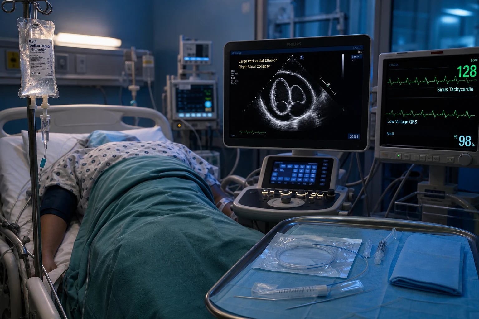
Cardiac tamponade
Fluid under pressure in the pericardial space compresses the heart, preventing the ventricular filling.[1]
Clinical:[1]
- Beck's triad (the classic): hypotension, distended neck veins (elevated JVP), muffled heart sounds.
- Pulsus paradoxus — an exaggerated fall in the SBP (over 10 mmHg) on inspiration (the increased venous return to the right heart in inspiration, with the fixed total cardiac volume from the compressing pericardium, causes the interventricular septum to shift left, reducing the LV filling and the stroke volume).
- Tachycardia, tachypnoea, a narrow pulse pressure.
Echo:[1]
- A pericardial effusion (an echo-free space around the heart).
- RA collapse (in early diastole) and RV collapse (in late diastole) — the chambers are compressed by the pericardial fluid.
- A plethoric IVC (dilated, non-collapsing — the right-sided pressures are high).
- A swinging heart (the heart moves freely within the effusion).
Management:[1]
- Immediate pericardiocentesis (echo-guided; the subxiphoid approach). This is life-saving.
- A fluid challenge (500 mL crystalloid) buys time by increasing the filling pressure against the compressing pericardium.
- Do NOT give diuretics or vasodilators — the filling pressure is needed against the pericardial compression.
Acute pericarditis
Inflammation of the pericardium, most commonly viral (coxsackie, echovirus) or idiopathic. Other causes: bacterial (TB, pneumococcal), uraemic, post-MI (early — the first few days; or Dressler syndrome — weeks to months, an autoimmune response to the necrotic myocardium), autoimmune (SLE, rheumatoid), neoplastic, radiation, drug (hydralazine, procainamide, isoniazid).[1]
Clinical:[1]
- Pleuritic chest pain — sharp, worse on inspiration and lying flat, better on sitting forward.
- A pericardial friction rub (a scratchy, leathery sound — best heard with the diaphragm at the left sternal border with the patient sitting forward).
- A low-grade fever, a flu-like prodrome.
ECG:[1]
- Diffuse, concave ST elevation (across multiple leads — unlike the focal, convex ST elevation of a STEMI).
- PR depression (in the same leads).
- May progress through stages (ST elevation, then normalisation, then T-wave inversion, then resolution).
- A pericardial effusion may cause low-voltage QRS and electrical alternans (the heart swinging within the effusion).
Treatment:[1]
- NSAIDs (ibuprofen, aspirin — for the pain and the inflammation).
- Colchicine (reduces the recurrence rate).
- Corticosteroids — reserved for the NSAID-resistant or the autoimmune case; avoided if possible because they increase the recurrence rate.
- Treat the underlying cause (antibiotics for the bacterial, dialysis for the uraemic).
Constrictive pericarditis
A thickened, fibrotic, sometimes calcified pericardium that restricts the cardiac filling.[1]
Causes: viral (the commonest in the developed world), TB (the commonest in the developing world), post-cardiac surgery, radiation, idiopathic.[1]
Clinical:[1]
- Right heart failure — raised JVP (with a prominent Y descent), hepatomegaly, ascites, peripheral oedema.
- The Kussmaul sign — the JVP rises on inspiration (the inverse of the normal fall; the constricted right heart cannot accommodate the increased venous return).
- A pericardial knock — an early diastolic sound (from the abrupt cessation of the filling when the rapidly filling ventricle hits the constricted pericardium).
- Pericardial calcification on the CXR or CT.
Echo:[1]
- A thickened pericardium.
- A restrictive filling pattern.
- A septal bounce (the interventricular septum shifts left on inspiration, from the increased RV filling against the constricted pericardium — the same mechanism as the pulsus paradoxus in tamponade).
- Respiratory variation of the mitral and tricuspid inflow.
Haemodynamics (cardiac catheterisation):[1]
- Equalisation of the diastolic pressures — the RA, the RV diastolic, the PA diastolic, and the PCWP all equalise at about 15-20 mmHg.
- The dip-and-plateau (the square-root sign) in the RV pressure trace.
- The Kussmaul sign (the RA pressure rises on inspiration).
Treatment:[1]
- Pericardiectomy — the surgical removal of the pericardium, the definitive treatment.
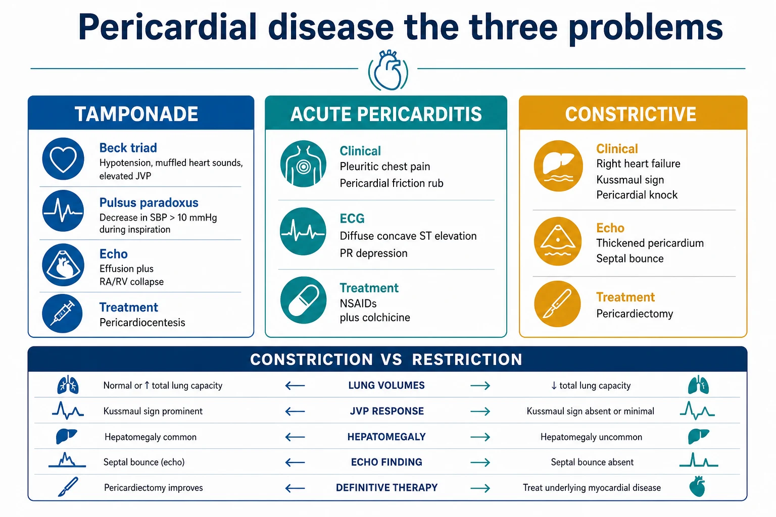
Constriction vs restrictive cardiomyopathy
Both present with right heart failure and equalised diastolic pressures. The distinction is critical because the treatment is different (pericardiectomy for constriction; medical for restriction):[1]
- Constriction: a thickened pericardium on CT/MRI; a normal LV thickness; a prominent Y descent; the septal bounce on echo; the equalised diastolic pressures with the dip-and-plateau.
- Restriction (amyloid, sarcoid, eosinophilic): a normal pericardium; a thick LV (and often a thick RV); a prominent X descent; no septal bounce.
- The cardiac MRI and the CT (showing the pericardial thickness and calcification) and the cardiac catheterisation (the haemodynamic pattern) help distinguish.[1]
Red flags
Cardiac tamponade — detailed pathophysiology
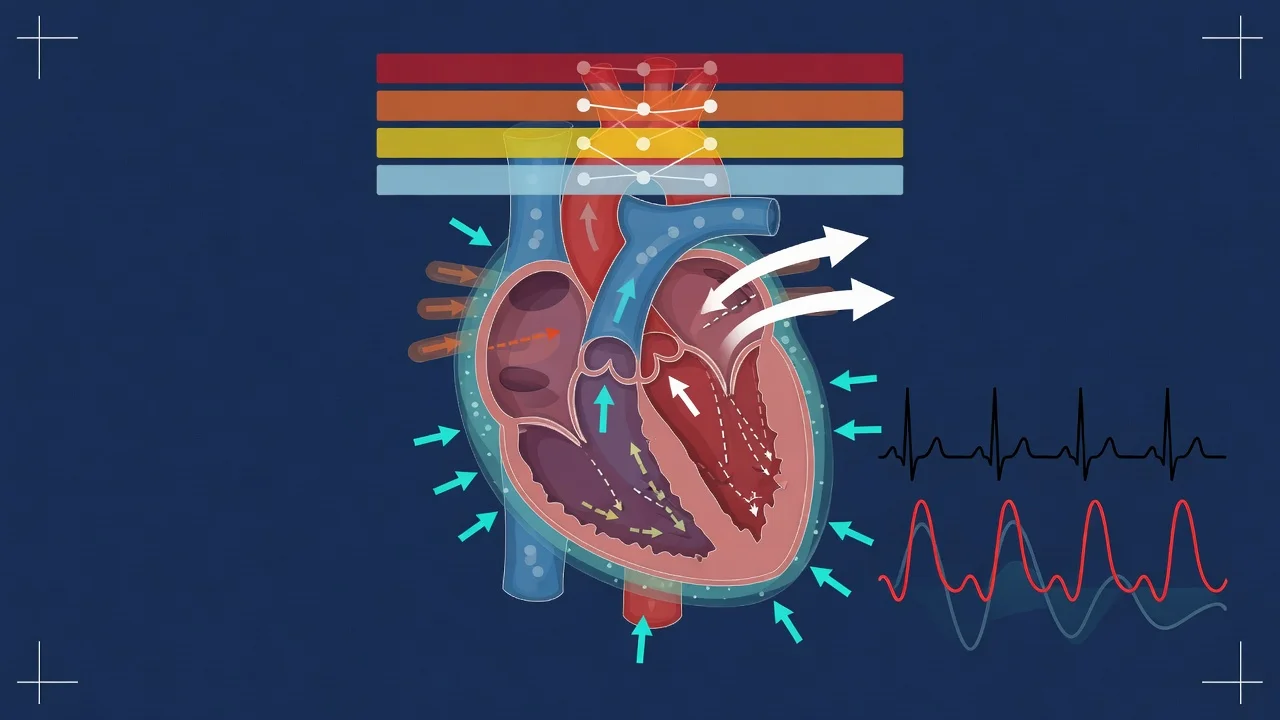
The pericardium is a relatively non-compliant fibrous sac. The acute addition of even modest fluid volumes overwhelms the pericardial reserve volume (typically only 80–200 mL can accumulate before pressure rises steeply), after which the pericardial pressure–volume curve rises almost vertically — every additional millilitre of effusion is transmitted directly as intrapericardial pressure, compressing the thin-walled chambers (RA and RV) and impairing diastolic filling. The stroke volume falls, the heart compensates with tachycardia and augmented adrenergic tone, and once compensation is exhausted the cardiac output collapses → cardiogenic shock → PEA arrest.[1]
Critically, the haemodynamic insult depends on (rate of accumulation × pericardial compliance), not absolute volume:
- A rapidly accumulating effusion (trauma, aortic dissection, cardiac rupture, iatrogenic post-procedure) of 150–250 mL can cause life-threatening tamponade.
- A slowly accumulating effusion (malignancy, uraemia, hypothyroid, chronic viral) of 1–2 L may cause only minimal symptoms, because the pericardium stretches over weeks.[1]
Beck's triad — the anatomy of the classic
The triad of Claude Beck (1935) describes the surgical (acute haemorrhagic) tamponade phenotype, and is clinically insensitive (present in only ~10–40% of medical tamponade):[1]
| Triad element | Mechanism | Caveat |
|---|---|---|
| Hypotension | Compressed ventricles → reduced stroke volume → reduced cardiac output → hypotension, narrow pulse pressure | May be relatively preserved in early/compensated tamponade due to tachycardia and vasoconstriction |
| Raised JVP / distended neck veins | Pericardial pressure transmitted back to RA and venous system → raised systemic venous pressure; a steep X descent is preserved (systolic atrial emptying still occurs), but the Y descent is attenuated or absent (early diastolic ventricular filling is blocked) | Absent in the hypovolaemic / low-pressure tamponade variant |
| Muffled heart sounds | Fluid insulates the stethoscope from the myocardium | Unreliable — depends on body habitus and fluid acoustic properties |
Pulsus paradoxus — the most useful bedside sign
Definition: an inspiratory fall in systolic BP > 10 mmHg during quiet (not forced) breathing. It is the most sensitive clinical marker of tamponade (≈ 70–80% sensitivity in medical series) but is not specific — severe asthma/COPD, tension pneumothorax, massive PE, hypovolaemic shock and anaphylaxis also produce it.[1]
Mechanism — ventricular interdependence:[1]
- Inspiration → intrathoracic pressure becomes more negative → increased systemic venous return to the right heart.
- The RV fills and expands — but the pericardium is fixed/non-compliant (filled by the effusion), so RV expansion can only occur by shifting the interventricular septum leftward into the LV cavity.
- LV filling and compliance fall → LV stroke volume falls → SBP drops >10 mmHg.
Bedside measurement technique:
- Inflate the BP cuff above the systolic pressure.
- Slowly deflate (~2 mmHg/s) while the patient breathes normally.
- Note the pressure at which Korotkoff sounds appear only in expiration (first threshold).
- Continue deflating; note the pressure at which sounds appear in both inspiration and expiration (second threshold).
- The difference between the two thresholds = pulsus paradoxus. >10 mmHg is abnormal; >20–25 mmHg suggests severe tamponade. [1]
Severe tamponade may produce a pulsus paradoxus that is palpable as a weak or absent pulse on inspiration, and the radial pulse may disappear altogether. [1]
Echocardiographic criteria — sensitivity and specificity
Echo is the diagnostic gold standard and is mandatory before pericardiocentesis whenever the patient is not in extremis.[1]
Echocardiographic features of tamponade — diagnostic yield
| Echo finding | Sensitivity | Specificity | Mechanism / interpretation |
|---|---|---|---|
| Pericardial effusion | (prerequisite) | — | Echo-free space; but effusion alone ≠ tamponade. A large chronic effusion may be haemodynamically silent; a small loculated effusion over the RA can tamponade. |
| RA collapse (in early diastole) | ~60–70% | ~80–90% | The thin RA is the first chamber to buckle under pericardial pressure; collapse lasting > 1/3 of the cardiac cycle is highly specific for tamponade. |
| RV collapse (in early diastole) | ~60–90% | ~85–95% | More specific than RA collapse but less sensitive; absent in pulmonary hypertension / RV hypertrophy (the RV wall resists collapse despite tamponade). |
| IVC plethora (dilated, non-collapsing, diameter > 2.1 cm with < 50% inspiratory collapse) | ~95% | ~80% | The most sensitive single finding; reflects elevated right-sided filling pressures. A collapsible IVC effectively excludes tamponade. |
| Respiratory variation in inflow (mitral E-wave velocity fall > 25% or tricuspid E-wave rise > 40% on inspiration) | ~70–85% | ~85–90% | The Doppler correlate of ventricular interdependence — the same mechanism as pulsus paradoxus. |
| Swinging heart | low | high | The heart oscillates freely in a large effusion → electrical alternans on ECG. Pathognomonic of a large effusion, usually pre-tamponade. |
In regional / loculated tamponade (post-cardiac surgery clot behind the RA, post-trauma) the classic signs may be absent — only the compressed chamber shows collapse, IVC may be non-plethoric, and a high index of suspicion with focused echo (or even transoesophageal echo in the postoperative patient) is essential. [1]
Causes of tamponade
Causes of cardiac tamponade by category — ICU context
| Category | Specific causes | ICU pearl |
|---|---|---|
| Malignancy (commonest overall) | Lung, breast, lymphoma, melanoma, leukaemia | Often recurrent; cytology positive in 50–85%. Needs pericardial window/sclerotherapy to prevent reaccumulation. |
| Iatrogenic / post-procedural | Post-CABG/valve (clot), post-PCI (perforation), post-pacemaker/ICD lead, post-EPS ablation, post-CVC malposition | Sudden stop in chest-tube drainage + collapse post-surgery = surgical clot tamponade — pericardiocentesis won't drain solid clot → urgent re-exploration. |
| Infective | Viral (coxsackie, echovirus, HIV), bacterial (pneumococcus, staph, TB), fungal | TB commonest cause in endemic regions; purulent pericarditis needs surgical drainage + IV antibiotics. |
| Aortic dissection | Type A retrograde haemopericardium | Haemorrhagic effusion in a hypertensive patient = think dissection — do NOT blindly pericardiocentese; needs emergency CT then surgery. |
| Trauma | Penetrating (stab, iatrogenic), blunt (deceleration, steering wheel) | Often acute small-volume tamponade — high risk of PEA arrest. Emergent pericardiocentesis / ED thoracotomy. |
| Post-MI | Free-wall rupture (days 1–5), Dressler syndrome (weeks–months) | Free-wall rupture is usually fatal; pseudoaneurysm leak can present as subacute tamponade. |
| Uraemia / metabolic | ESKD, myxoedema, severe hypothyroid | Myxoedematous effusion is slow and rarely tamponades; uraemic effusion responds to dialysis. |
| Connective tissue / autoimmune | SLE, rheumatoid, scleroderma, vasculitis | Often part of a serositis picture; treat the underlying disease. |
| Radiation / drug | Thoracic radiotherapy (late), hydralazine, procainamide, isoniazid | Drug-induced is lupus-like; resolves on withdrawal. |
Low-pressure tamponade
A clinically important variant: tamponade physiology without a markedly raised JVP, occurring in patients with concomitant hypovolaemia (e.g. haemorrhage, diuretics, dialysis). The intrapericardial pressure is only mildly elevated but still exceeds the depressed right-sided filling pressure, so the chamber collapse and haemodynamic compromise occur with a normal or flat JVP. IVC plethora may be absent, RA collapse may be subtle, and the diagnosis rests on the effusion + chamber collapse + respiratory variation + a clear clinical picture of shock. Treatment is volume loading then pericardiocentesis — diuretics are catastrophic.[1]
Pericardiocentesis — emergent vs echo-guided
Emergent (blind/resuscitative) vs echo-guided pericardiocentesis
| Feature | Emergent / resuscitative | Echo-guided (elective / urgent) |
|---|---|---|
| When | Cardiac arrest / PEA / profound shock — no time for echo | Haemodynamically compromised but perfusing; effusion confirmed on echo |
| Approach | Subxiphoid (classical) — 1 cm below and to the left of the xiphisternal junction, aim at the left shoulder at 30°, aspirate while advancing | Site chosen by echo — point of maximum fluid depth + shortest skin-to-fluid distance (often apical or subcostal) |
| Guidance | Landmark + ECG lead on needle (ST elevation on needle tip contact with RV = withdraw) | Real-time ultrasound throughout — visualise needle tip in pericardial space; agitated saline confirms position |
| Technique | 18G needle → aspirate fluid → Seldinger wire → dilator → pigtail catheter | Identical Seldinger technique, but wire/catheter position confirmed on echo |
| Complications | RV/RA laceration, coronary/laceration, pneumothorax, liver injury, mediastinal infection — higher | Same complications but 3–4× lower major complication rate (≈1–2% vs 5–10% blind) |
| Indwelling drain | Yes — leave pigtail for continued drainage | Yes — same management |
The Mayo Clinic series of 1,127 echo-guided pericardiocenteses reported a major complication rate of ~1.2% (laceration of a chamber, pneumothorax requiring chest tube, infection) and a clinical success rate of 97% — establishing echo guidance as the standard of care whenever the patient can wait for it.[4]
Echo-guided pericardiocentesis — the Seldinger technique, step by step
- PREPARE — patient supine, head of bed 30–45° (fluid pools posteriorly/dependently); ECG, BP, SpO₂ monitoring; large-bore IV access; correct any coagulopathy (INR < 1.5, platelets > 50). Bedside clotting, group & save.
- IMAGE — subcostal + parasternal long + apical 4-chamber views. Measure maximum effusion depth, skin-to-fluid distance, RA/RV collapse, IVC. Mark the entry site (pen on skin) at the point of greatest depth AND shortest trajectory, free of liver/lung.
- ASEPTIC — full sterile prep, drape, gown, gloves. 1% lidocaine local infiltration along the planned track down to the pericardium.
- NEEDLE ENTRY — 18G needle at the marked site, angling toward the effusion under real-time echo. Agitated saline in the syringe — once you enter the pericardial space, inject: microbubbles opacify the pericardial sac on echo, confirming intrapericardial position.
- WIRE — pass the J-tipped guidewire through the needle into the sac; confirm position on echo before removing the needle.
- DILATE & CATHETERISE — small skin nick; serial dilators; insert a 6–8 Fr multi-side-hole pigtail catheter over the wire. Remove wire.
- ASPIRATE & SEND — aspirate all accessible fluid; send for: cell count, protein, LDH, glucose, Gram stain, culture (including TB/AFB), cytology, triglycerides (chylopericardium), and a heparinised tube if dissection suspected.
- SECURE & DRAIN — suture the catheter in place; connect to a closed drainage bag. Document post-procedure echo (chamber re-expansion, resolution of RA/RV collapse, IVC collapsibility).
- DRAIN MANAGEMENT — flush q8h with 10 mL saline to prevent blockage. Continue until output < 25–50 mL/24 h AND no reaccumulation on serial echo (typically 24–72 h).
- REMOVAL — gentle traction with a Valsalva manoeuvre; occlusive dressing; observe for reaccumulation with a follow-up echo at 24 h and again at 1–2 weeks.
Post-pericardiocentesis complications to watch: recurrent tamponade (reaccumulation — drain malignant effusions and consider a window); coronary / chamber laceration (acute deterioration with haemopericardium); acute pulmonary oedema (rare, from sudden decompression and increased venous return — 'tamponade-removal pulmonary oedema'); pneumothorax. A repeat echo at 24 h is mandatory. [1]
Acute pericarditis — detailed management
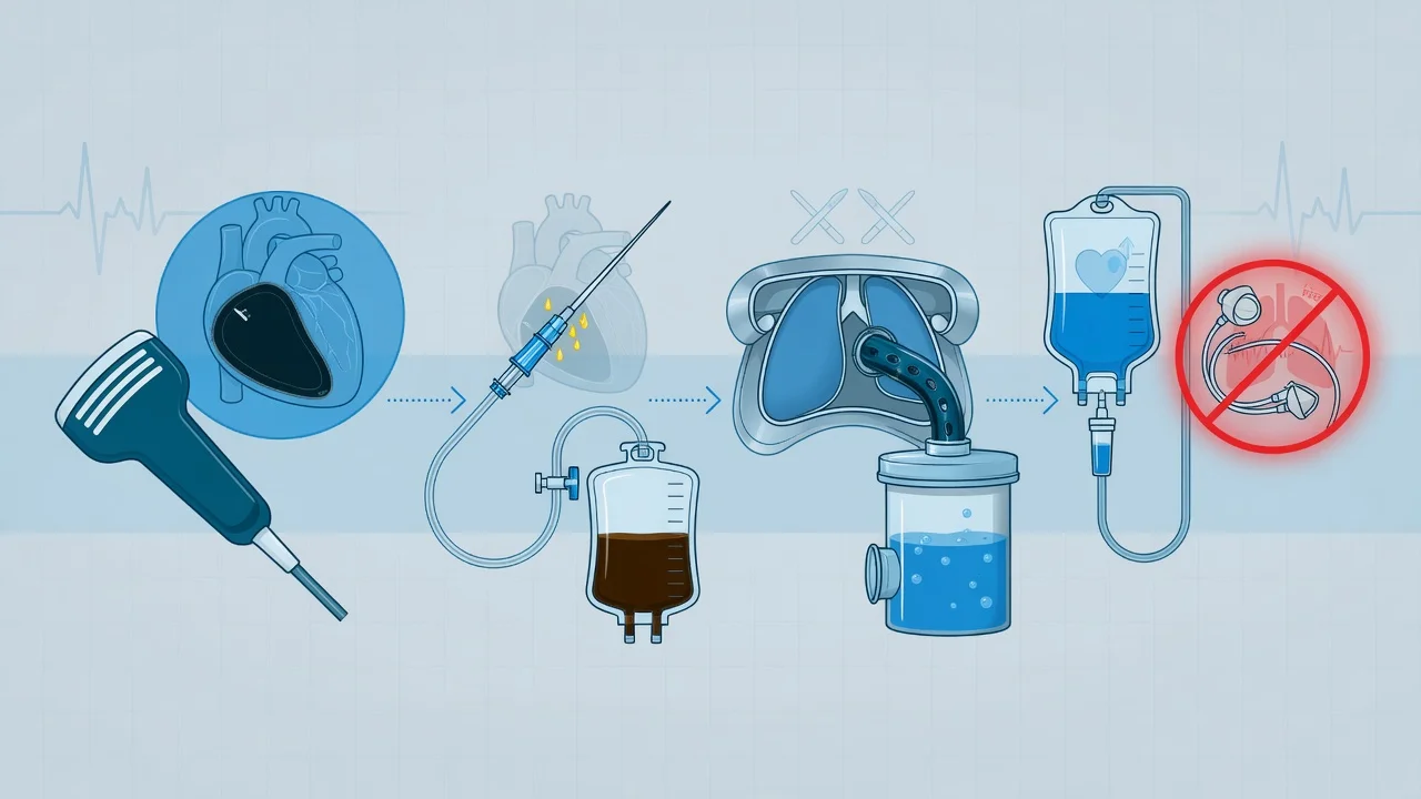
ECG stages of pericarditis
The ECG evolves in four stages over days to weeks:[1]
ECG stages of acute pericarditis
| Stage | Timing | ECG appearance |
|---|---|---|
| I (acute) | Days–weeks | Diffuse concave-up ST elevation (I, II, III, aVL, aVF, V3–V6); PR depression in the same leads; PR elevation in aVR (reciprocal). No reciprocal ST depression (unlike STEMI). |
| II | Days–weeks | ST elevation and PR deviation normalise; T waves begin to flatten. |
| III | Days–weeks | Diffuse T-wave inversion appears (after ST has returned to baseline). |
| IV | Weeks–months | T waves normalise; ECG returns to baseline (or T-wave inversion may persist indefinitely). |
Pericarditis vs STEMI vs early repolarisation — ECG differentiation: [1]
Pericarditis vs STEMI vs early repolarisation — ECG discriminators
| Feature | Acute pericarditis | STEMI | Early repolarisation |
|---|---|---|---|
| ST morphology | Concave-up, diffuse | Convex-up (tombstone), focal (anatomical territory) | Concave-up, diffuse, V4 most |
| Leads involved | I, II, III, aVL, aVF, V3–V6 | Territory of occluded artery | Inferolateral, sparing aVR |
| Reciprocal ST depression | Absent (except aVR) | Present (e.g. II/III reciprocal) | Absent |
| PR changes | PR depression (aVR PR elevation) — diagnostic | None | Usually none |
| T wave | Inverts after ST normalises | Inverts while ST still elevated | Tall peaked T, never inverts |
| Q waves | Absent | May develop pathologic Q | Absent |
| Evolution | Slow, over days–weeks | Rapid (hours) with treatment | Static over years |
| Spodick's sign | Downsloping TP segment / PR slope — supportive | Absent | Absent |
Troponin in pericarditis
Troponin is elevated in 20–50% of acute pericarditis cases (especially the young, viral, with ST elevation) — it reflects concomitant myopericarditis (epicardial myocardial inflammation), not coronary occlusion. A raised troponin does not mandate coronary angiography in a young patient with classic pericarditic features; it does mandate the diagnosis of myopericarditis and a period of cardiac monitoring. Troponin typically peaks early and resolves within 1–2 weeks; a persistent rise or a regional wall motion abnormality on echo should prompt coronary imaging.[3]
The ICAP trial — colchicine for acute pericarditis
ICAP — colchicine for acute pericarditis (Imazio NEJM 2013; PMID 23992557)
Design
Multicentre, double-blind, randomised placebo-controlled trial; 240 adults with a first episode of acute pericarditis
Intervention
Colchicine 1.0–2.0 mg on day 1 then 0.5–1.0 mg/day × 3 months, on top of standard NSAID/aspirin therapy
Primary outcome
Recurrence of pericarditis at 18 months
Key result
Colchicine reduced recurrence (16.7% vs 37.5%; NNT ≈ 5) AND reduced symptom persistence at 72 h (19.2% vs 39.7%)
Harms
Gastrointestinal (diarrhoea) in ~7%; no serious adverse events; well tolerated
Clinical bottom line
Colchicine is now FIRST-LINE for acute pericarditis (with aspirin/NSAIDs) — first trial to prove it reduces recurrence. ESC 2015 guidelines adopted it as class I.
COPPS-2 — colchicine for postpericardiotomy syndrome (Imazio JAMA 2014; PMID 25172965)
Design
Multicentre, double-blind RCT; 360 adults after cardiac surgery
Intervention
Colchicine 1 mg then 0.5 mg bid × 1 month postoperatively, vs placebo
Primary outcome
Composite of postpericardiotomy syndrome at 12 months
Key result
No significant reduction in primary endpoint (intention-to-treat), but a per-protocol analysis showed benefit and a significant reduction in postoperative atrial fibrillation. GI discontinuation in ~7%.
Clinical bottom line
Colchicine prophylaxis is reasonable after cardiac surgery in patients who tolerate it; check renal/hepatic function first.
Treatment of acute pericarditis — evidence-based protocol
Acute pericarditis — stepwise treatment (ESC 2015)
- FIRST LINE — aspirin or NSAID + colchicine. Aspirin 750–1000 mg every 6–8 h (or ibuprofen 600 mg q8h) for 7–14 days until pain resolves, then taper over 1–2 weeks. PLUS colchicine 0.5 mg bd (1 mg if >70 kg) for 3 months (level I evidence — ICAP). Treat the activity-limiting symptom, not the ECG.
- ADJUNCT — gastroprotection (PPI), and physical activity restriction (avoid competitive sport until symptom-free and CRP normal — usually 3 months).
- AVOID CORTICOSTEROIDS in the first instance — they are the single strongest risk factor for recurrent pericarditis (odds ratio ~4). Use only if NSAIDs are contraindicated (renal failure, active GI bleed, pregnancy) or the cause is autoimmune (SLE, rheumatoid). If unavoidable, use low-dose prednisolone 0.2–0.5 mg/kg/day (NOT 1 mg/kg) and taper slowly.
- RATE / SYMPTOM CONTROL — esmolol or a β-blocker may be useful adjuncts in the tachycardic, hypertensive patient with myopericarditis and chest discomfort; for the dysrhythmic ICU patient on background β-blockade, esmolol (titratable, ultra-short half-life) is preferred for rate control while the primary anti-inflammatory regimen takes effect.
- TREAT THE CAUSE — antibiotics for bacterial; dialysis for uraemic; anticoagulation review for drug-induced; immunosuppression for autoimmune; TB therapy for tuberculous.
- WATCH FOR RECURRENCE — 15–30% recur. Define recurrence and escalate (colchicine ± an NSAID; only then consider steroids/interleukin-1 blockade).
Recurrent pericarditis — the escalation ladder
Definition: recurrence of pericarditic pain + ≥ 1 of (new friction rub, ECG changes, echo effusion, CRP rise) after a symptom-free interval of ≥ 4–6 weeks.[3]
Treatment escalation:
- Re-introduce NSAID/aspirin at full dose + optimise colchicine (continue ≥ 6 months).
- Anakinra (recombinant IL-1 receptor antagonist) 1–2 mg/kg/day (max 100 mg/day) — the IRAP registry showed corticosteroid-sparing and colchicine-resistant pericarditis responds to anakinra, allowing steroid taper in most patients.[9]
- Rilonacept (IL-1α/β trap) — subcutaneous weekly; the RHAPSODY pivotal trial demonstrated a transition to rilonacept monotherapy in patients with recurrent pericarditis, with rapid pain relief and low recurrence.[11]
- Tocilizumab / canakinumab — for refractory disease; centres of expertise only.
- Pericardiectomy — last resort, in expert centres, for truly refractory, colchicine/IL-1-blocker-resistant cases.
Constrictive pericarditis — detailed
Constriction is the chronic end-state of pericardial inflammation: the pericardium becomes thickened, fibrotic and often calcified, encasing the heart in a rigid shell that resists filling. The cardinal consequence is equalisation of diastolic pressures across all four chambers, with filling that is rapid early in diastole (because the early filling pressures are high) then abruptly arrested when the volume hits the rigid pericardium — the dip-and-plateau (or square-root) configuration.[5]
Clinical signs — the high-yield quartet
- Kussmaul's sign — a rise in the JVP on inspiration (the inverse of the normal fall). The constricted right heart cannot expand to accept the increased venous return, so the venous column backs up. (Distinguished from tamponade, where the JVP is elevated but still falls on inspiration.) Kussmaul's sign is also seen in severe RV failure, tricuspid stenosis and severe pulmonary hypertension.
- Pericardial knock — a high-pitched early-diastolic sound (~60–120 ms after S2), best heard at the left sternal border. It is the acoustic correlate of the abrupt halt to filling — blood rapidly entering the ventricle in early diastole is suddenly arrested by the rigid pericardium.
- Pericardial calcification on CXR/CT — seen in ~25–50% (especially TB and radiation). Calcification on the lateral CXR along the atrioventricular groove and anterior pericardium is highly suggestive. A normal pericardium on CT effectively excludes constriction (sensitivity ~95%, false negatives in early/transient disease).
- Right heart failure phenotype — raised JVP with a prominent Y descent (rapid early diastolic collapse, then re-rise), hepatomegaly, ascites (often disproportionate to peripheral oedema), pleural effusions, cachexia from protein-losing enteropathy in advanced cases. [1]
Echocardiography of constriction
Echo features of constrictive pericarditis
| Finding | Mechanism | Diagnostic value |
|---|---|---|
| Pericardial thickening (often best on TEE or CT/MRI) | Fibrosis/calcification | Sensitive when seen; CT/MRI are preferred modalities |
| Septal bounce / shudder | On inspiration, increased RV filling shifts the septum leftward (ventricular interdependence) | Highly characteristic; the echo correlate of pulsus paradoxus in constriction |
| Respiratory variation in mitral/tricuspid inflow (>25% mitral E-wave fall on inspiration; >40% tricuspid rise) | Ventricular interdependence | High diagnostic yield; increase Valsalva/inspiration provocation |
| Restrictive LV filling (E-wave dominant, deceleration < 160 ms) | The rigid pericardium fixes total heart volume — early filling is rapid then halted | Reflects the dip-and-plateau |
| Tissue Doppler: preserved e′ (> 8 cm/s) at the medial mitral annulus | Myocardium itself is NORMAL — only the pericardium is diseased | The key discriminator from restrictive cardiomyopathy, where e′ is reduced (amyloid/sarcoid infiltrate the myocardium) |
| Annulus reversus (lateral mitral e′ > medial e′ on tissue Doppler) | The tethered lateral annulus moves less than the free medial annulus | Highly specific for constriction |
| Hepatic vein flow reversal on expiration | The constricted RV cannot accept the inspiratory surge | Sensitive Doppler sign |
BNP and the discriminator from restrictive cardiomyopathy
BNP/NT-proBNP is substantially lower in constriction than in restrictive cardiomyopathy (RCM). In RCM (amyloid, sarcoid, eosinophilic) the diseased, infiltrated myocardium is stretched and produces high natriuretic peptide; in constriction the myocardium is normal, so the BNP is often only mildly elevated despite dramatic right-heart-failure signs. A markedly raised BNP in a patient with right heart failure and equalised diastolic pressures points away from constriction and toward RCM.[6]
Cardiac catheterisation — the haemodynamic signature
Catheterisation findings in constriction
| Measurement | Finding | Mechanism |
|---|---|---|
| RA / RV diastolic / PA diastolic / PCWP | All equalise at 15–20 mmHg (within 5 mmHg of each other) | All four chambers share the rigid pericardial constraint |
| RA waveform | Prominent X and Y descents; M- or W-shaped; Kussmaul sign (rise on inspiration) | Rapid atrial emptying then abrupt arrest |
| RV waveform | Dip-and-plateau (square-root sign); RV systolic usually < 55 mmHg; RV diastolic > 1/3 of RV systolic | Rapid early filling then abrupt halt |
| LV–RV discordance with respiration | LV and RV systolic pressures shift out of phase on inspiration (ventricular interdependence) | The single best haemodynamic discriminator from RCM, where LV and RV stay in phase |
Transient and effusive-constrictive pericarditis
Transient constriction — occurs in ~7–10% after an acute pericarditic episode (often viral or post-cardiotomy). The pericardium is inflamed and stiff but not yet fibrotic, and the constriction resolves over 2–3 months with anti-inflammatory therapy (NSAIDs + colchicine ± a short steroid taper). A trial of medical therapy for 2–3 months is reasonable before committing to pericardiectomy, unless the patient is decompensating.[8]
Effusive-constrictive pericarditis — a hybrid syndrome: a pericardial effusion with tamponade-like physiology plus underlying constrictive visceral pericardium. The diagnosis is made at pericardiocentesis: after draining the effusion, the intrapericardial pressure falls to zero but the RA pressure remains elevated — proving there is constrictive epicardium/visceral pericardium beneath the effusion. Management: pericardiocentesis (to relieve the effusion) followed by pericardiectomy. Common in malignancy, TB, radiation, uraemia, post-surgery.[8]
Pericardiectomy — definitive treatment
Pericardiectomy is the only definitive treatment for chronic constrictive pericarditis. Key points:
- Timing — once the diagnosis is firm and the patient is symptomatic; not in the acute/transient phase (give anti-inflammatories first).
- Technique — radical (wide) pericardiectomy via median sternotomy on cardiopulmonary bypass; remove the parietal AND visceral pericardium (visceral pericardiectomy / epicardiectomy) to beyond both phrenic nerves.
- Mortality — perioperative mortality 5–15% in modern series; higher in radiation-induced disease (calcification adherent to coronaries and myocardium) and in advanced preoperative disease.
- Outcomes — the Mayo Clinic series (Ling 1999, et seq.) and the Spanish series (Porta-Sánchez 2015) demonstrate that ~80–90% of survivors have marked symptomatic improvement at 5 years, with 10-year survival of ~70–80% (worse in radiation and renal failure).[5][8]
- Post-pericardiectomy diastolic dysfunction is common (the myocardium has been chronically underfilled) and resolves over months.
Tamponade vs constriction vs restrictive cardiomyopathy — the master table
Tamponade vs Constrictive Pericarditis vs Restrictive Cardiomyopathy — definitive differentiation
| Feature | Tamponade | Constriction | Restriction (RCM) |
|---|---|---|---|
| Pathology | Pericardial fluid under pressure | Thickened, fibrotic ± calcified pericardium | Diseased, infiltrated myocardium (amyloid, sarcoid, eosinophilic); pericardium normal |
| Onset | Acute (hours–days) | Chronic (months–years) | Chronic (months–years) |
| Pulsus paradoxus | Prominent (>10 mmHg) | Often mild | Absent |
| JVP waveform | Prominent X descent, absent/attenuated Y descent | Prominent X and Y descents (M-shape) | Prominent X and Y descents |
| Kussmaul sign | Absent | Present | Variable (may be present in advanced RCM) |
| Pericardial knock | Absent | Present (early diastolic) | Absent (S3 gallop instead) |
| Pericardial calcification | Absent | Present (~25–50%) | Absent |
| Echo | Effusion + RA/RV collapse + IVC plethora + respiratory inflow variation | Septal bounce + respiratory inflow variation + preserved e′ + annulus reversus | Thick LV/RV + reduced e′ + no respiratory variation + no septal bounce |
| BNP | Variable | Low / mildly raised (normal myocardium) | Markedly elevated (diseased myocardium) |
| CT/MRI pericardium | Fluid; normal thickness | Thickened (> 2 mm) ± calcification | Normal thickness |
| Cardiac MRI | Diffuse effusion; no late gadolinium enhancement of pericardium | Pericardial thickening ± late enhancement; annulus reversus on tissue Doppler | Myocardial thickening; subendocardial/ diffuse late enhancement (amyloid) |
| Catheterisation — equalised diastolic pressures | Often present | Present (within 5 mmHg) | Present, but the LV-PCWP gradient persists (PCWP > RV diastolic by > 5 mmHg) |
| Pulmonary hypertension | Absent | Mild (PA systolic usually < 55 mmHg) | Often severe (PA systolic > 60 mmHg) |
| Ventricular interdependence (LV–RV discordance) | Mild | Marked (out of phase with respiration) | Absent (in phase) |
| Treatment | Pericardiocentesis | Pericardiectomy | Medical (diuretics, treatment of amyloid/sarcoid) |
The single most useful ICU discriminator: tissue Doppler mitral annular e′ velocity. Preserved e′ (> 8 cm/s) → constriction (myocardium is normal). Reduced e′ (< 8 cm/s) → restrictive cardiomyopathy (myocardium is infiltrated). Add BNP (low in constriction, high in RCM) and pericardial thickness on CT (thick in constriction, normal in RCM) and the diagnosis is robust. [1]
The 'Ten Commandments' of the 2015 ESC Pericardial Disease Guidelines
A useful exam distillation of Adler 2015 / Charron 2015:[1][10]
- Diagnosis of acute pericarditis requires ≥ 2 of: typical chest pain; pericardial friction rub; ECG changes (diffuse ST elevation / PR depression); new or worsening pericardial effusion.
- Triaged first-line workup: ECG, echo, CXR, routine bloods (CRP, troponin, renal), and Tuberculin/IGRA, ANA, HIV, thyroid when the cause is not obvious.
- Echo is mandatory — to look for effusion/tamponade, ventricular function, and signs of constriction.
- First-line therapy = aspirin or NSAID + colchicine (3 months); avoid steroids first-line.
- Activity restriction during the acute phase and for at least 3 months for athletes.
- Search for and treat the cause (TB, autoimmune, bacterial, neoplastic, uraemic).
- Corticosteroids — only when NSAIDs are contraindicated or failed; low dose (0.2–0.5 mg/kg/day), taper slowly.
- Recurrent pericarditis — multi-targeted therapy (NSAID + colchicine + steroid-sparing IL-1 blockade).
- Tamponade — urgent echo-guided pericardiocentesis; treat the cause.
- Constriction — confirm with multimodality imaging (echo + CT/MRI + catheterisation); pericardiectomy is curative; try anti-inflammatories first because transient constriction is common and reversible. [1]
Trial cards — constriction
Constrictive pericarditis in the modern era — Mayo Clinic (Ling Circulation 1999; PMID 10500037)
Study
Retrospective cohort of 135 patients undergoing pericardiectomy at Mayo Clinic, 1985–1995
Population
Predominantly idiopathic/viral (42%), post-cardiac surgery (21%), radiation (12%), post-infective (11%) — TB now uncommon in developed world
Key findings
Preoperative NYHA IV class and radiation aetiology predicted worse outcome; perioperative mortality 6%; 7-year survival 82%; 83% of survivors improved by ≥ 1 NYHA class
Clinical bottom line
Pericardiectomy offers excellent symptom relief and long-term survival in non-radiation constriction; radiation and advanced preoperative disease carry the worst prognosis.
Constrictive pericarditis — modern aetiology and outcomes (Porta-Sánchez Rev Esp Cardiol 2015; PMID 25936614)
Study
Single-centre 35-year Spanish cohort, 263 patients with constrictive pericarditis
Aetiologic shift
Idiopathic/viral now dominant (~50%); TB declined (~15%); iatrogenic post-surgical rising
Survival after pericardiectomy
Overall perioperative mortality ~6%; 5-year survival 78%; worse survival in radiation and renal failure
Clinical bottom line
Confirms the secular trend away from TB toward idiopathic/surgical causes; pericardiectomy remains the definitive treatment with good long-term outcomes.
Echo-guided pericardiocentesis — Mayo 21-year series (Tsang Mayo Clin Proc 2002; PMID 12004992)
Study
Consecutive 1,127 echo-guided therapeutic pericardiocenteses (1979–2000)
Indications
Malignancy (33%), post-procedural (i.e. catheter, surgery — 22%), idiopathic/viral (13%), uraemic, infective
Efficacy
Clinical success in 97%; drainage catheter left in for a mean of 2.4 days
Major complications
1.2% (chamber laceration requiring surgery, pneumothorax requiring chest tube, death in 0.2%)
Clinical bottom line
Echo-guided pericardiocentesis is safe and effective — the standard of care whenever the patient can tolerate the time for ultrasound guidance.
SAQ — Constrictive pericarditis vs cardiac tamponade vs restrictive cardiomyopathy
10 minutes · 10 marks
A 58-year-old man presents with four months of progressive exertional dyspnoea, abdominal swelling and ankle oedema. Eighteen months ago he was treated conservatively for an episode of presumed viral pericarditis. On examination the JVP is raised to the angle of the jaw at 45 degrees with a prominent Y descent and a Kussmaul sign (the venous column RISES on inspiration); an early diastolic pericardial knock is audible at the left sternal edge; there is hepatomegaly, ascites and bilateral pitting ankle oedema; the lung fields are clear. BP 104/78, HR 96 sinus. Echocardiography shows a thickened pericardium with a septal bounce, preserved medial mitral annular e-prime velocity of 10 cm/s with annulus reversus (lateral e-prime greater than medial), and hepatic vein flow reversal on expiration; there is no pericardial effusion and no chamber collapse. CT shows pericardial thickening of 5 mm with patchy calcification. NT-proBNP is mildly elevated at 180 ng/L. The registrar asks whether the raised venous pressure could instead represent cardiac tamponade or restrictive cardiomyopathy.
SAQ — Effusive-constrictive pericarditis: the RA pressure that does not fall after the tap
10 minutes · 10 marks
A 66-year-old woman with metastatic breast cancer presents with two weeks of progressive dyspnoea, presyncope and a raised JVP. On examination BP 92/64, HR 118 sinus, JVP raised to the jaw with a prominent X descent and a faint Y descent, clear lung fields, and pulsus paradoxus of 14 mmHg. Bedside echo shows a 2.2 cm circumferential pericardial effusion with right atrial collapse lasting one-third of the cardiac cycle, right ventricular diastolic collapse, and a plethoric non-collapsing IVC. At echo-guided pericardiocentesis 900 mL of serosanguinous fluid is drained; intrapericardial pressure falls to near zero, but the right atrial pressure remains elevated at 16 mmHg and the JVP does not descend. Cytology is positive for adenocarcinoma.
Clinical pearls
[1]Flow steps — the ICU resident's pericardial playbook
Tachycardic, hypotensive ICU patient with a pericardial effusion — what to do in 5 minutes
- RECOGNISE — hypotension + tachycardia + raised JVP (or distended neck veins) + pulsus paradoxus + an effusion on focused echo with RA/RV collapse + plethoric IVC = tamponade. Call for senior help and the pericardiocentesis tray now.
- RESUSCITATE WHILE PREPARING THE TAP — give a 500 mL crystalloid bolus to maintain preload against the pericardial constraint (buys time); do NOT give diuretics, vasodilators or inotropes (the myocardium is not failing — the filling is). Avoid intubation/PPV unless pericardiocentesis is imminent.
- GET AN ECHO — choose the entry site — point of maximum effusion depth and shortest skin-to-fluid distance; subxiphoid, apical or parasternal. Confirm RA/RV collapse, IVC plethora, swinging heart, respiratory inflow variation.
- PERICARDIOCENTESIS (Seldinger, echo-guided) — sterile prep, local, 18G needle under real-time echo, agitated saline to confirm position, J-wire, dilate, pigtail catheter. Aspirate all accessible fluid; send the full panel.
- POST-TAP CARE — leave the pigtail drain on free drainage; flush q8h; serial echo at 1, 6 and 24 h to confirm re-expansion and exclude reaccumulation. Investigate and treat the cause (malignancy → cytology + oncology + consider a window; dissection → CT aortogram + CT surgery; uraemic → dialysis; TB → quadruple therapy; post-procedural → expectant).
- RECURRENT TAMPONADE — if malignant or recurrent, leave the drain longer and arrange a pericardial window (subxiphoid surgical or VATS) ± intrapericardial sclerotherapy (doxycycline/bleomycin) to obliterate the pericardial space.
Suspected constrictive pericarditis — diagnostic pathway
- CLINICAL SUSPICION — chronic right heart failure with raised JVP, prominent Y descent, Kussmaul's sign, pericardial knock, ascites disproportionate to oedema; a history of prior pericarditis, TB, radiation or cardiac surgery.
- ECHOCARDIOGRAPHY — look for septal bounce, respiratory variation in mitral/tricuspid inflow, preserved e′ + annulus reversus on tissue Doppler, hepatic vein flow reversal on expiration. (NB: pericardial thickness is better assessed on CT/MRI.)
- BNP — low/normal supports constriction; markedly raised points to restrictive cardiomyopathy.
- CT or MRI — pericardial thickness > 2 mm ± calcification supports constriction; rule out myocardial infiltration (amyloid: subendocardial late enhancement, LV thickening, low voltages on ECG).
- CARDIAC CATHETERISATION — equalisation of diastolic pressures (RA/RVd/PAd/PCWP within 5 mmHg), dip-and-plateau, Kussmaul's sign, and LV–RV discordance with respiration (the best haemodynamic discriminator from RCM).
- DECIDE — confirmed chronic constriction → refer for pericardiectomy (radical, parietal + visceral, on bypass). If uncertain or early/transient → trial of anti-inflammatory therapy (NSAID + colchicine ± low-dose steroid taper) for 2–3 months and reassess.
Additional red flags
References
- [1]Adler Y, Charron P, Imazio M, et al. 2015 ESC Guidelines for the diagnosis and management of pericardial diseases: The Task Force for the Diagnosis and Management of Pericardial Diseases of the European Society of Cardiology (ESC)Endorsed by: The European Association for Cardio-Thoracic Surgery (EACTS) Eur Heart J, 2015.PMID 26320112
- [2]Imazio M, Brucato A, Cemin R, et al. A randomized trial of colchicine for acute pericarditis N Engl J Med, 2013.PMID 23992557
- [3]Imazio M, Gaita F, LeWinter M. Evaluation and Treatment of Pericarditis: A Systematic Review JAMA, 2015.PMID 26461998
- [4]Tsang TS, Enriquez-Sarano M, Freeman WK, et al. Consecutive 1127 therapeutic echocardiographically guided pericardiocenteses: clinical profile, practice patterns, and outcomes spanning 21 years Mayo Clin Proc, 2002.PMID 12004992
- [5]Ling LH, Oh JK, Schaff HV, et al. Constrictive pericarditis in the modern era: evolving clinical spectrum and impact on outcome after pericardiectomy Circulation, 1999.PMID 10500037
- [6]Reddy PR, Dieter RS, Das P, et al. Utility of BNP in differentiating constrictive pericarditis from restrictive cardiomyopathy in patients with renal insufficiency J Card Fail, 2007.PMID 17923360
- [7]Imazio M, Brucato A, Ferrazzi P, et al. Colchicine for prevention of postpericardiotomy syndrome and postoperative atrial fibrillation: the COPPS-2 randomized clinical trial JAMA, 2014.PMID 25172965
- [8]Porta-Sánchez A, Sagristà-Sauleda J, Ferreira-González I, et al. Constrictive Pericarditis: Etiologic Spectrum, Patterns of Clinical Presentation, Prognostic Factors, and Long-term Follow-up Rev Esp Cardiol (Engl Ed), 2015.PMID 25936614
- [9]Imazio M, Andreis A, De Ferrari GM, et al. Anakinra for corticosteroid-dependent and colchicine-resistant pericarditis: The IRAP (International Registry of Anakinra for Pericarditis) study Eur J Prev Cardiol, 2020.PMID 31610707
- [10]Charron P, Adler Y, Imazio M, et al. 'Ten Commandments' of 2015 ESC Guidelines for diagnosis and management of pericardial diseases Eur Heart J, 2015.PMID 26866075
- [11]Brucato A, Wheeler A, Luis SA, et al. Transition to rilonacept monotherapy from oral therapies in patients with recurrent pericarditis Heart, 2023.PMID 36316102