ICU · Perioperative critical care
Perioperative Critical Care
Also known as Perioperative medicine · High-risk surgery · Preoperative optimisation · Postoperative complications · Enhanced recovery · Goal-directed therapy · MINS · ERAS
Perioperative critical care is the optimisation of the high-risk surgical patient before, during, and after the operation — the preoperative risk assessment (the cardiopulmonary exercise testing, the POSSUM/ACS-NSQIP scoring, the smoking cessation, the anaemia correction, the glycaemic control), the enhanced recovery after surgery (ERAS) bundle, the intraoperative goal-directed therapy (the cardiac output, the fluid responsiveness, the stroke-volume optimisation, the oxygen delivery), the postoperative monitoring (the HDU/ICU), and the system-by-system complications (the cardiac — the AF and the myocardial injury after noncardiac surgery [MINS]; the respiratory — the atelectasis, the pneumonia, the PE; the renal — the AKI; the GI — the ileus and the anastomotic leak; the wound — the infection and the dehiscence), the goal-directed fluid, the multimodal opioid-sparing analgesia, and the postoperative nausea and vomiting.
On this page & tools
Your progress
Saved locally on this device.
8 MCQs with explanations
Target exams
Red flags
Overview & definition
Perioperative critical care is the optimisation of the high-risk surgical patient across the continuum — the preoperative risk assessment and the optimisation, the intraoperative monitoring and the goal-directed therapy, and the postoperative care and the enhanced recovery. The high-risk patient (the age, the comorbidity, the complex surgery) has a 5 to 10 per cent mortality, and the ICU is the place where the complications are anticipated and managed.[1][1]
The perioperative period is the single largest source of preventable hospital mortality worldwide — an estimated 4.2 million deaths per year globally within 30 days of surgery, equivalent to the third leading cause of death. Most perioperative deaths are NOT intraoperative (the anaesthetic mortality is now <1 in 100,000 in fit patients); they occur POSTOPERATIVELY, from a failure to rescue — the high-risk patient who deteriorates on the ward without the recognition or the escalation. The ICU team's role is to anticipate the complications, monitor the high-risk patient in the HDU/ICU, and rescue early.[1]
The high-risk surgical patient
The high-risk surgical patient is identified by the patient factors (the age over 65, the ASA grade 3 to 5, the limited exercise capacity, the comorbidities — the cardiac, the respiratory, the renal, the diabetes), the surgical factors (the emergency, the major, the long duration, the large blood loss), and the test factors (the low albumin, the anaemia, the impaired renal function, the abnormal ECG). The perioperative mortality is concentrated in this group — 80 per cent of the deaths occur in 12 per cent of the patients (the high-risk).[1]
High-risk surgical patient — the factors that define the high-risk group
| Domain | The factors |
|---|---|
| Patient | Age >65; ASA 3-5; poor exercise capacity (the inability to climb one flight of stairs); the comorbidity (the cardiac failure, the COPD, the CKD, the diabetes, the cirrhosis); the frailty; the obesity (the OSA); the smoking |
| Surgical | The emergency (the 3 to 5 × mortality vs elective); the major (the intraperitoneal, the intrathoracic, the vascular); the long duration (>2 hours); the large blood loss (>1 L); the aortic surgery |
| Test | The low albumin (<30 g/L); the anaemia (Hb <100 g/L); the eGFR <60; the abnormal ECG; the low functional capacity on CPET |
| Score | The POSSUM/P-POSSUM predicted mortality >10%; the ACS-NSQIP serious complication risk >10% |
The critical concept is the failure to rescue — the high-risk patient develops the complication (the hypotension, the tachycardia, the oliguria, the hypoxia) on the ward, but it is not recognised or escalated in time. The HDU or the ICU admission for the high-risk patient is the single most effective intervention to reduce the perioperative mortality — the monitored bed where the deterioration is caught early.[1][1]
The preoperative assessment and the optimisation
The cardiopulmonary exercise testing (CPET)
The cardiopulmonary exercise testing (the CPET) is the gold-standard preoperative risk assessment — it measures the VO2 max (the peak oxygen consumption) during the graded exercise, and it integrates the cardiac, the respiratory, and the muscular reserve into the single functional number. The CPET is the objective measure of the the physiological reserve — the capacity to meet the surgical stress (the 30 to 50 per cent rise in the oxygen demand postoperatively).[6]
The CPET variables and the thresholds for the high risk:
- The anaerobic threshold (AT) < 11 mL/kg/min — the point at which the lactate begins to rise (the onset of the anaerobic metabolism). The AT below 11 predicts the high perioperative mortality.
- The peak VO2 < 15 mL/kg/min (or < 75 per cent predicted) — the maximum oxygen consumption. Below 15 predicts the high risk; below 10 suggests the surgery is prohibitive.
- The VE/VCO2 slope > 34 — the ventilatory equivalent for the carbon dioxide (a marker of the pulmonary vascular disease and the heart failure). Above 34 predicts the high risk. [1]
The CPET guides the decision: the proceed (the normal — the ward), the optimise (the borderline — the prehabilitation, the HDU), or the reconsider (the very abnormal — the alternative procedure, the palliation).[6]
The scoring systems
The scoring systems estimate the perioperative mortality from the patient and the surgical factors, and they guide the discussion and the planning.[1][8]
The perioperative risk scoring systems
| Score | The basis | The use |
|---|---|---|
| POSSUM | 12 physiological + 6 operative factors (the physiological and operative severity score for the enumeration of morbidity and mortality) | The risk-adjusted surgical audit (Copeland 1991). The original POSSUM OVER-predicts the mortality in the low-risk group. |
| P-POSSUM | The Portsmouth modification — a recalibrated logistic equation | The more accurate prediction for the mortality (the elective and the emergency). |
| V-POSSUM | The vascular modification | For the vascular surgery (the AAA, the carotid). |
| SORT | The Surgical Outcome Risk Tool — 5 variables (the urgency, the ASA, the cancer, the age, the grade) | The simple, the validated, the bedside — the UK. |
| ACS-NSQIP | The American College of Surgeons — the national surgical quality improvement program — the multivariable model from >1 million cases | The online surgical risk calculator (the mortality + the 8 complications) — the most widely used and the best calibrated. |
| Lee Revised Cardiac Risk Index (RCRI) | 6 cardiac variables (the high-risk surgery, the ischaemic heart disease, the heart failure, the stroke/TIA, the insulin-dependent diabetes, the CKD) | The cardiac risk prediction for the non-cardiac surgery. |
The ACS-NSQIP surgical risk calculator (the Cohen 2016 evaluation) is the current standard — it gives the patient-specific, the procedure-specific risk of the mortality, the serious complication, and the 8 individual complications (the pneumonia, the cardiac, the SSI, the UTI, the VTE, the renal failure, the readmission, the return to theatre). It is used for the shared decision-making and the HDU/ICU planning.[8]
The preoperative optimisation: the four pillars
1. The smoking cessation. The smoking doubles the perioperative complication rate (the wound infection, the pneumonia, the atelectasis, the delayed healing). The cessation 4 weeks before the surgery reduces the wound and the pulmonary complications (the Møller 2002 randomised trial — the intensive intervention 6-8 weeks pre-op reduced the complications from 41 to 21 per cent). The short-acting nicotine replacement is used perioperatively. Even the brief intervention (the advice + the NRT) at the pre-admission clinic is effective.[7]
2. The anaemia correction. The preoperative anaemia (Hb < 130 g/L men, < 120 g/L women) is associated with the increased mortality, the transfusion, and the AKI. The cause is identified and treated before the elective surgery: the iron deficiency (the oral or the IV iron — the IV ferric carboxymaltose 1 g in the 2 weeks pre-op if the oral is ineffective or the time is short), the B12/folate deficiency, and the anaemia of chronic disease. The goal is the Hb > 130 g/L before the elective surgery — the patient blood management principle (the reduce the bleeding, the optimise the erythropoiesis, the restrictive transfusion).[1]
3. The glycaemic control. The perioperative hyperglycaemia (the glucose > 10 mmol/L) increases the wound infection, the mortality, and the length of the stay. The HbA1c > 8.5 per cent warrants the deferral of the elective surgery (the optimisation). The day of the surgery: the long-acting insulin is reduced (the 75-80 per cent of the basal), the short-acting is held, and the variable-rate intravenous insulin (the sliding scale or the GKI) is used for the major surgery or the poor control. The target glucose is 6 to 10 mmol/L (the avoid the hypoglycaemia and the severe hyperglycaemia).[1]
4. The cardiopulmonary optimisation (the prehabilitation). The prehabilitation — the structured exercise, the inspiratory muscle training, and the nutrition in the 4 to 8 weeks before the surgery — improves the functional capacity and the reduces the pulmonary complications in the high-risk patient (the thoracic and the upper GI surgery). The medications are optimised: the cardiac (the heart failure — the optimise the GDMT), the respiratory (the bronchodilators, the steroids, the pulmonary rehab), and the continue the chronic cardiac medications (the beta-blocker, the statin — DO NOT stop).[1][1]
The medication management before the surgery
The perioperative medication management — the stop, the continue, the bridge
| Medication | The action |
|---|---|
| Aspirin (primary prevention) | STOP 7 days (no benefit, bleeding risk) |
| Aspirin (secondary prevention) | CONTINUE for the cardiac stent <1 year; discuss with the cardiology for the >1 year |
| Clopidogrel/ticagrelor | STOP 5-7 days (the bridging with the aspirin ± heparin for the high thrombotic risk) |
| Warfarin | STOP 5 days (bridge with the LMWH if the high thrombotic risk — the mechanical valve, the AF with the prior stroke) |
| DOAC | STOP 24-48 h (longer for the apixaban/rivaroxaban in the renal impairment) |
| Beta-blocker | CONTINUE (do NOT stop — the POISE: the NEW initiation causes harm, but the continuation is safe and the withdrawal is dangerous) |
| Statins | CONTINUE (the pleiotropic, the anti-inflammatory, the reduce the MINS) |
| ACEi/ARB | STOP the morning of the surgery (the hypotension under the anaesthesia) — resume post-op |
| Metformin | STOP the day of the surgery (the lactic acidosis — esp if the contrast) |
| SGLT2 inhibitor | STOP 3-4 days (the euglycaemic DKA risk under the anaesthesia) |
| GLP-1 agonist | HOLD (the delayed gastric emptying — the aspiration risk — the semaglutide 1 week) |
The enhanced recovery after surgery (ERAS)
The ERAS is the bundle of the evidence-based perioperative interventions that reduce the surgical stress, accelerate the recovery, and shorten the stay: the preoperative counselling, the avoidance of the prolonged fasting (the carbohydrate drink up to 2 hours before), the minimally invasive surgery, the goal-directed fluid, the opioid-sparing analgesia, the early mobilisation, and the early enteral nutrition. The ERAS reduces the complication rate (by ~50 per cent), the length of the stay (by 2-3 days) and the cost.[1][1]
The ERAS bundle — the elements (the 20+ evidence-based interventions): [1]
The ERAS pathway — the preoperative, the intraoperative, the postoperative
- PREOPERATIVE — (a) Counselling (the set the expectations, the patient is the active participant). (b) The smoking and the alcohol cessation (>4 weeks; the Møller evidence). (c) The anaemia and the nutrition optimisation (the patient blood management). (d) The AVOID the prolonged fasting — the solids until 6 h and the CLEAR fluids until 2 h before (the evidence-based; the old 'NPO from midnight' is obsolete). (e) The carbohydrate loading (the 400 mL the night before + 200 mL 2 h before — the maltes coffee / the pre-op carb drink — reduces the insulin resistance, the thirst, the anxiety). (f) The thromboprophylaxis (the LMWH 12 h before — the mechanical + the pharmacological). (g) The antibiotic prophylaxis (the within 60 min of the incision — the cefazolin ± metronidazole).
- INTRAOPERATIVE — (a) The minimally invasive surgery (the laparoscopic or the robotic — the less the surgical stress). (b) The SHORT-acting anaesthetic (the propofol, the sevoflurane, the remifentanil — the rapid emergence). (c) The AVOID the long-acting opioids (the morphine — the ileus, the PONV). (d) The goal-directed fluid therapy (the stroke volume optimisation — the avoid the overload and the dehydration). (e) The normothermia (the forced-air warmer — the <36°C increases the wound infection and the bleeding). (f) The PONV prophylaxis (the dexamethasone, the ondansetron — the high-risk patients). (g) The normoglycaemia (the 6-10 mmol/L). (h) The surgical-site infection bundle (the chlorhexidine, the hair clip not shave, the wound protector).
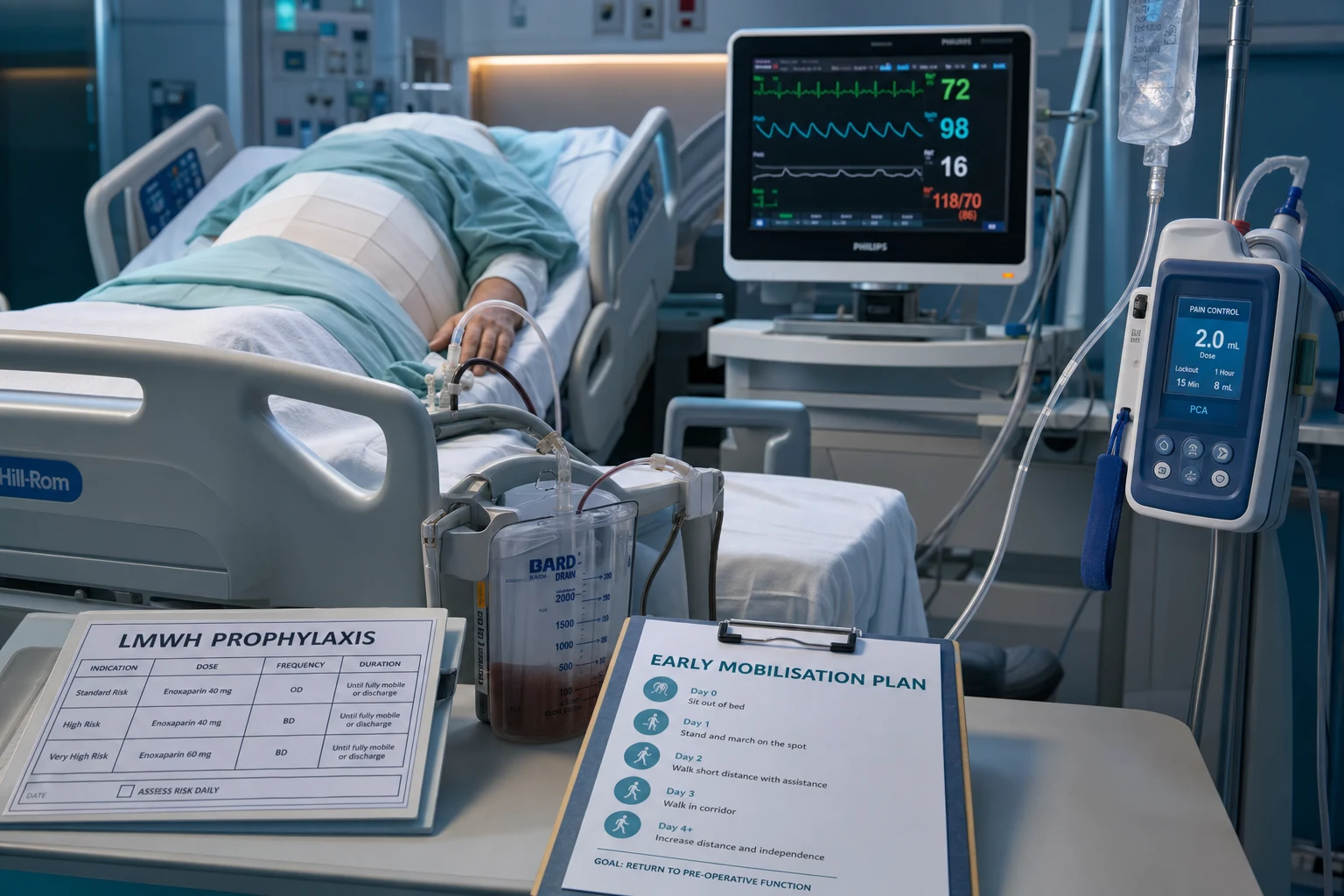
- POSTOPERATIVE — (a) The EARLY MOBILISATION (the day of the surgery or the day 1 — the out of bed, the walk — the prevent the atelectasis, the DVT, the deconditioning). (b) The EARLY ENTERERAL NUTRITION (the within 24 h — the gut works — the less the ileus, the less the infection). (c) The OPIOID-SPARING ANALGESIA (the multimodal — the paracetamol, the NSAID, the regional — the minimise the morphine). (d) The AVOID the NG tube and the drains (the routine NG and the drains delay the recovery — remove early). (e) The AVOID the routine IV fluids (the switch to the oral — the prevent the overload). (f) The early removal of the catheter and the lines. (g) The gum chewing (the reduce the ileus). (h) The EARLY DISCHARGE CRITERIA (the oral intake, the pain controlled, the mobilising, the afebrile).
The ERAS reduces the length of stay and the complications — but the adherence is the key (the full adherence to all the elements gives the maximum benefit; the partial adherence is less effective). The ERAS is the multidisciplinary pathway — the surgeon, the anaesthetist, the ICU team, the nurses, the dietitians, the physiotherapists.[1][1]
The intraoperative goal-directed therapy (GDT)
The goal-directed therapy (the GDT) is the optimisation of the oxygen delivery during the surgery — the cardiac output, the fluid responsiveness, the haemoglobin, and the oxygen delivery are monitored and targeted.[1]
The GDT uses the advanced monitors (the oesophageal Doppler, the arterial pulse-contour analysis, the transpulmonary thermodilution, the echocardiography) to guide the fluid (the stroke volume optimisation — the bolus until the stroke volume no longer rises, the fluid responsiveness test), the inotrope (if the output is low despite the fluid) and the transfusion (the restrictive threshold). The target is the supranormal or the normal oxygen delivery, guided by the individual patient's baseline.[1][1]
The OPTIMISE trial (Pearse 2014, JAMA): the multicentre RCT of the cardiac-output-guided GDT (the oesophageal Doppler + the fluid + the inotrope) in the 734 high-risk patients after the major gastrointestinal surgery. The primary outcome (the mortality + the complications at 30 days) was NOT significantly reduced in the intention-to-treat (the trend to the benefit), but the per-protocol analysis and the meta-analysis (the OPTIMISE + 38 trials) confirmed the reduction in the complications. The GDT remains the recommended practice for the high-risk surgery.[2]
The postoperative complications — by the system
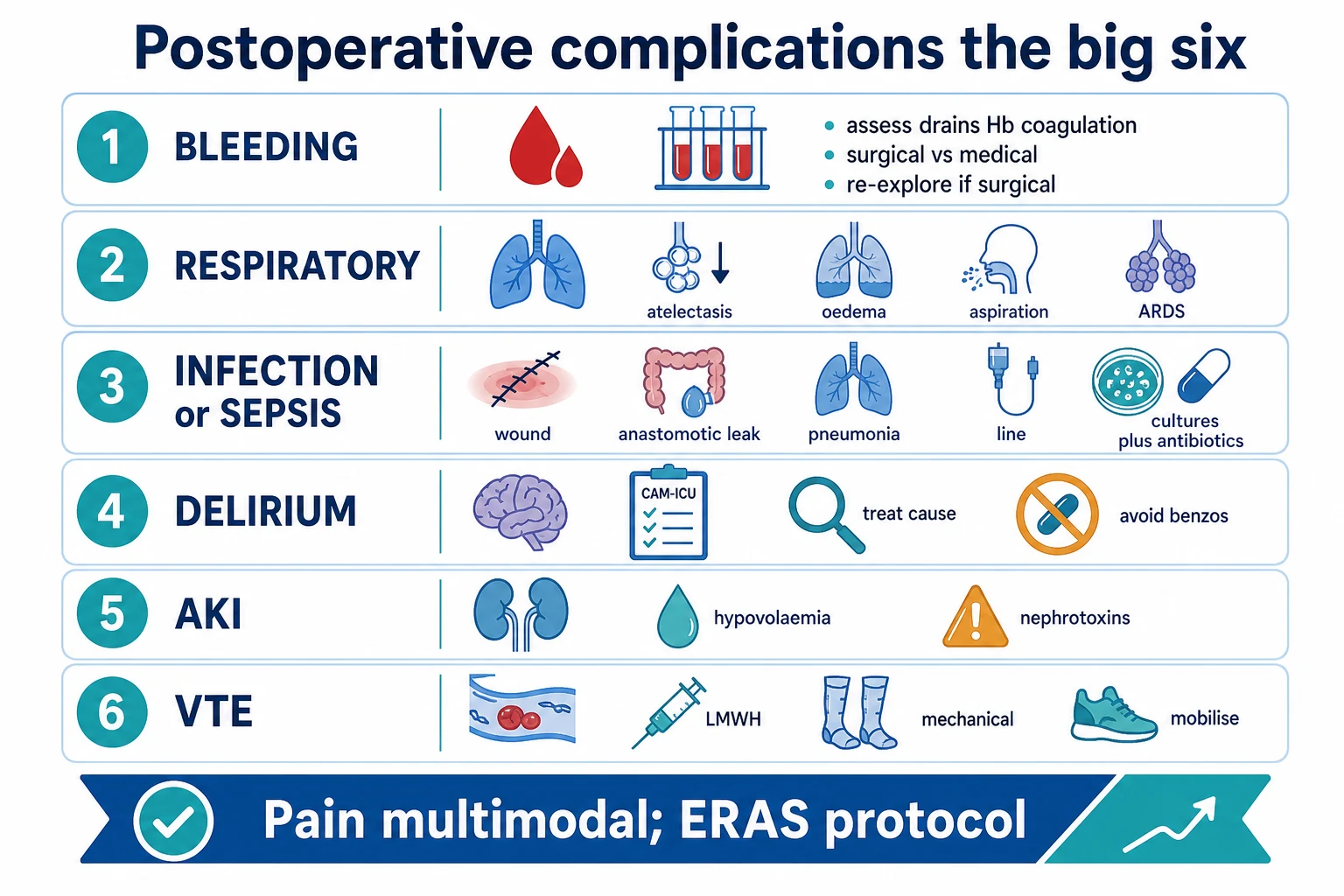
The postoperative complications are the leading cause of the perioperative mortality and the morbidity. The high-risk patient is monitored in the HDU/ICU, and the complications are anticipated and managed early. The complications are best considered by the system.[1]
1. The cardiac complications — the AF and the MINS
The postoperative atrial fibrillation (POAF): the commonest postoperative arrhythmia (the 10 to 40 per cent after the thoracic or the cardiac surgery; the 3 to 5 per cent after the major non-cardiac). It peaks at the day 2 to 3 (the sympathetic surge, the fluid shifts, the inflammation, the electrolyte derangement — the magnesium, the potassium). The management: the rate control (the beta-blocker — the metoprolol, the bisoprolol; the diltiazem if the beta-blocker contraindicated), the rhythm control if the unstable (the DC cardioversion; the amiodarone for the chemical cardioversion), the anticoagulation assessed (the CHA2DS2-VASc — but the early POAF may not need the long-term anticoagulation if it is self-limiting; the bleeding risk from the surgery). The correct the potassium (>4.0) and the magnesium (>0.8).[1]
The myocardial injury after the noncardiac surgery (MINS): the VISION study (Devereaux 2017, JAMA) — the postoperative high-sensitivity troponin rise is the COMMON, the under-recognised, and the prognostic. The MINS is defined as the troponin rise without the ischaemic feature (the peak ≥ the 99th percentile or the rise/fall) in the first 30 days after the noncardiac surgery. The MINS occurs in the 8 per cent of the patients over 45 (the 18 per cent in the vascular), and it carries the 30-day mortality of 10 per cent (the 3 × the mortality without the MINS). The MINS is usually the type 2 MI (the supply-demand mismatch — the tachycardia, the hypotension, the hypoxia, the anaemia, the vasospasm) rather than the type 1 (the plaque rupture).[5]
MINS — the type 1 vs the type 2 (the management differs)
| Feature | The type 1 MI (the plaque rupture) | The type 2 / MINS (the supply-demand mismatch) |
|---|---|---|
| The mechanism | The atherothrombosis — the plaque rupture, the thrombus | The supply-demand mismatch — the tachycardia, the hypotension, the hypoxia, the anaemia |
| The frequency | The minority of the postoperative troponin rises | The MAJORITY (the 80 per cent) of the postoperative troponin rises |
| The management | The aspirin, the statin, the revascularisation if the ongoing ischaemia | The CORRECT THE TRIGGER (the rate, the BP, the SpO2, the Hb); the aspirin; the statin. NOT the immediate DAPT + anticoag (the bleeding risk from the surgery). |
| The key | The cardiology; the angiography if the ongoing ischaemia | The fix the haemodynamics; the avoid the antithrombotic overload in the fresh post-op patient |
The troponin is measured routinely in the high-risk patient (the vascular, the age over 65, the prior cardiac disease) on the day 1 and the day 2 post-op. The rise is NOT ignored — it predicts the mortality, and the patient gets the aspirin and the statin and the cardiology follow-up.[3][5]
The POISE trial (Devereaux 2008, Lancet) — the perioperative beta-blockade paradox: the randomised 8351 patients to the metoprolol succinate vs the placebo before the non-cardiac surgery. The result: the metoprolol REDUCED the perioperative MI (and the cardiac arrest) but INCREASED the stroke and the overall MORTALITY (the hypotension and the bradycardia from the beta-blockade). The lesson: the ROUTINE initiation of the beta-blockade before the surgery is NOT recommended — but the CONTINUATION of the already-started beta-blocker is essential (the withdrawal is dangerous), and the cautious initiation (the slow titration, the avoid the hypotension) is reasonable for the very high cardiac risk.[3]
2. The respiratory complications — the atelectasis, the pneumonia, the PE
The respiratory complications are the commonest postoperative complication (the 5 to 40 per cent after the major abdominal or the thoracic). They are driven by the anaesthesia (the reduced FRC, the reduced ciliary clearance), the surgical site (the upper abdominal and the thoracic — the splinting), the pain (the splinting — the poor cough), the immobility, and the smoking. [1]
The atelectasis: the collapse of the dependent lung segments (the most common, the first 24 hours). The management: the early mobilisation, the incentive spirometry, the deep breathing, the CPAP or the NIV (the recruit the collapsed alveoli), the good analgesia (the enable the deep breathing — the regional, the epidural).[1]
The postoperative pneumonia: the aspiration, the atelectasis, the poor clearance. The prophylaxis: the head-up 30 degrees, the oral hygiene (the chlorhexidine), the smoking cessation, the early mobilisation, the incentive spirometry. The treatment: the antibiotics (the community-acquired pattern — the ceftriaxone; the hospital-acquired — the broader), the physiotherapy, the oxygen, the NIV if the type 2 respiratory failure.[1]
The pulmonary embolism: the DVT (the immobility, the surgery — the hypercoagulable state) → the PE. The prophylaxis: the LMWH (the enoxaparin 40 mg SC daily — start within 12 h, the mechanical SCDs, the early mobilisation). The high-risk surgery (the orthopaedic, the oncology, the pelvic) gets the extended prophylaxis (the 4 weeks). The treatment: the therapeutic anticoagulation (the LMWH, the DOAC); the thrombolysis if the massive PE (the haemodynamic instability); the surgical embolectomy if the contraindication to the thrombolysis. The filter (the IVC) if the absolute contraindication to the anticoagulation (the recent surgery, the bleeding).[1][1]
3. The renal complications — the acute kidney injury (AKI)
The perioperative AKI occurs in the 5 to 10 per cent of the major surgery (the 20 to 30 per cent after the cardiac surgery with the CPB). It is driven by the the pre-renal (the hypotension, the hypovolaemia, the bleeding), the the nephrotoxic (the contrast, the NSAIDs, the antibiotics — the vancomycin, the aminoglycosides), and the the obstructive (the catheter blockage, the surgical). The Kheterpal risk index (the 2009) predicts the AKI from the age, the ASA, the male, the emergency, the comorbidity (the diabetes, the CHF, the ascites, the hypertension), and the surgery type.[10]
The prevention: the avoid the hypotension (the goal-directed fluid, the vasopressor titration), the avoid the nephrotoxins (the hold the NSAIDs, the ACEi, the metformin; the minimise the contrast — the iso-osmolar, the lowest dose), the maintain the intravascular volume, the monitor the urine output (the >0.5 mL/kg/h), the electrolyte management (the potassium, the phosphate, the acid-base). The KDIGO stage 2 or 3 (the creatinine ×2, the oliguria) warrants the ICU, the renal dosing of the medications, and the consideration of the RRT.[1][10]
4. The GI complications — the ileus and the anastomotic leak
The postoperative ileus: the temporary paralysis of the bowel (the 2 to 4 days expected after the abdominal surgery). The PROLONGED ileus (>4 days) is driven by the opioids, the electrolyte derangement (the potassium, the magnesium), the immobility, the inflammation, and the haematoma. The management: the minimise the opioid (the opioid-sparing, the regional, the epidural — the MASTER trial), the correct the electrolytes, the early mobilisation, the gum chewing, the early enteral nutrition (the ERAS principle), and the rule out the mechanical obstruction and the leak (the CT).[9]
The anastomotic leak: the life-threatening complication of the bowel surgery (the colorectal — the 3 to 15 per cent; the oesophageal — the 5 to 20 per cent). It presents at the day 5 to 7 with the fever, the tachycardia, the abdominal pain, the ileus, the pelvic/abdominal collection, and the sepsis. The diagnosis: the the CT with the contrast (the contrast extravasation, the collection, the gas). The management: the antibiotics (the broad — the piperacillin-tazobactam), the source control (the radiological drainage or the surgical re-operation — the stoma, the resection), and the ICU support (the sepsis resuscitation). The mortality is the 5 to 15 per cent (the delay is the harm).[1]
5. The wound complications — the infection and the dehiscence
The surgical site infection (SSI): the commonest postoperative infection (the 2 to 5 per cent of the clean, the 10 to 20 per cent of the contaminated). The prophylaxis: the antibiotic within 60 min of the incision (the cefazolin; the re-dose if >4 h or the >1.5 L blood loss), the normothermia, the glycaemic control (the 6-10 mmol/L), the hair clip not shave, the chlorhexidine, the wound protector, the sterile technique. The management: the open the wound, the debride, the culture, the antibiotics (the gram-positive — the flucloxacacin; the broad — the piperacillin-tazobactam if the deep or the sepsis).[1]
The wound dehiscence: the fascial failure (the deep dehiscence — the surgical emergency — the evisceration) or the superficial (the skin only). The risk: the obesity, the diabetes, the malnutrition, the steroids, the high intra-abdominal pressure, the poor surgical technique, the cough (the COPD). The management: the deep dehiscence (the emergency surgery — the re-suture ± the mesh, the manage the intra-abdominal pressure), the superficial (the vacuum dressing, the secondary intention).[1]
The postoperative fluid management — the goal-directed
The postoperative fluid management is the balance of the two errors: the dehydration (the hypovolaemia — the AKI, the hypotension, the wound perfusion) and the overload (the hyperchloraemia, the oedema — the ileus, the pulmonary oedema, the wound healing). The traditional 'the 3 L of the crystalloid a day' is obsolete and harmful (the fluid overload increases the complications).[1][2]
The goal-directed fluid therapy (GDFT): the individualised, the dynamic, the stroke-volume-guided approach. [1]
The goal-directed fluid therapy — the stroke volume optimisation algorithm
- BASELINE — (a) Connect the advanced monitor (the arterial line + the pulse-contour analysis [LiDCO, FloTrac], the oesophageal Doppler, the transpulmonary thermodilution [PiCCO], or the echocardiography). (b) Establish the baseline stroke volume (SV) and the cardiac output (CO). (c) Establish the fluid responsiveness (the passive leg raise, the SV variation with the respiration >12 per cent, the IVC collapsibility >50 per cent).
- THE FLUID CHALLENGE (the stroke volume optimisation) — (a) Give the 200 mL balanced crystalloid (the Hartmann, the Plasma-Lyte — NOT the 0.9 per cent saline — the hyperchloraemia) bolus over 5-10 min. (b) Measure the SV. (c) If the SV RISES by >10 per cent — the patient is the fluid-responsive — repeat the bolus. (d) If the SV does NOT rise (the <10 per cent) — the patient is NOT fluid-responsive — STOP the fluid. (e) Continue until the SV plateaus (the Frank-Starling curve flattened).
- THE VASOPRESSOR — if the BP is low (the MAP <65) but the SV is optimised (the NOT fluid-responsive), start the vasopressor (the noradrenaline — the vasoconstriction, the raise the MAP). The vasoplegia is common in the postoperative patient (the inflammation, the anaesthetic effect).
- THE INOTROPE — if the CO is low (the SV low, the SVR high — the low contractility), start the inotrope (the dobutamine, the milrinone, the adrenaline) — guided by the echocardiography (the low EF, the regional wall motion). The mixed venous saturation (the >65 per cent) confirms the adequate the oxygen delivery.
- THE TRANSFUSION — the restrictive threshold (the TRICC — the Hb 70-80 g/L; the >80 if the cardiac ischaemia or the MINS). The single-unit transfusion with the reassessment.
- THE MAINTENANCE — once the optimised, switch to the low-rate maintenance (the 1-2 mL/kg/h of the balanced crystalloid) and the early transition to the oral intake (the ERAS). The AVOID the routine the saline (the hyperchloraemic acidosis, the AKI).
The balanced crystalloid vs the saline: the SMART and the SALT-ED trials (the 2018) — the balanced crystalloid (the Hartmann, the Plasma-Lyte) reduces the AKI and the mortality vs the 0.9 per cent saline (the hyperchloraemia causes the renal vasoconstriction). The balanced crystalloid is the perioperative standard.[4]
The postoperative analgesia — the multimodal opioid-sparing
The postoperative pain is universal and the poorly-controlled pain drives the complications (the splinting → the atelectasis and the pneumonia; the immobility → the DVT; the sympathetic surge → the cardiac). The modern approach is the multimodal opioid-sparing analgesia — the combination of the non-opioid medications and the regional techniques to minimise the opioid (the morphine — the ileus, the PONV, the sedation, the delirium).[1][9]
The multimodal postoperative analgesia — the layers
| The agent | The mechanism | The notes |
|---|---|---|
| Paracetamol (1 g QID IV/PO) | The COX-3, the central | The baseline — the safe, the opioid-sparing (the 20-30 per cent). The avoid the >4 g/day in the low-weight or the liver disease. |
| NSAID (the ibuprofen, the diclofenac, the ketorolac) | The COX-1/2 inhibition | The opioid-sparing (the 20-40 per cent). The CAUTION: the AKI, the bleeding, the anastomotic leak (the controversial — the COX-2 impairs the healing). The avoid in the renal impairment and the high-bleeding surgery. The short course (the 3-5 days). |
| Gabapentinoid (the gabapentin, the pregabalin) | The calcium channel, the neuropathic | The opioid-sparing but the sedation and the delirium in the elderly — the reduced use recently. |
| Ketamine (the 0.1-0.3 mg/kg/h) | The NMDA antagonist | The opioid-tolerant, the chronic pain, the major surgery. The sub-anaesthetic dose — the opioid-sparing, the prevents the hyperalgesia. |
| Lidocaine infusion (the 1-2 mg/kg/h) | The systemic local anaesthetic | The anti-inflammatory, the opioid-sparing, the reduces the ileus. The major abdominal. |
| Alpha-2 agonist (the dexmedetomidine, the clonidine) | The central alpha-2 | The opioid-sparing, the analgesic, the sedative. The avoid the bradycardia and the hypotension. |
| Regional (the wound infiltration, the TAP block, the fascial plane) | The local anaesthetic | The abdominal wall, the thoracic. The avoid the opioid. |
| Epidural (the local anaesthetic ± the opioid) | The neuraxial | The GOLD standard for the thoracic and the upper abdominal — the best analgesia, the opioid-sparing, the reduces the pulmonary complications. The MASTER trial. |
| Opioid (the morphine, the fentanyl, the oxycodone) | The mu receptor | The RESCUE only — the minimise. The PCA (the patient-controlled) if needed. |
The epidural analgesia: the local anaesthetic (the bupivacaine, the ropivacaine) ± the opioid (the fentanyl) via the thoracic or the lumbar epidural. It is the gold standard for the thoracic and the upper abdominal surgery — the superior analgesia, the opioid-sparing, the reduced pulmonary complications (the better the cough and the deep breathing), and the reduced the ileus (the sympathetic block). The MASTER trial (Rigg 2002, Lancet) — the epidural reduced the respiratory failure and the MI in the high-risk. The complications: the hypotension (the sympathetic block — the vasopressor), the epidural haematoma (the rare, the catastrophic — the CHECK the coagulation before the insertion and the removal), the infection, the nerve injury, the urinary retention.[9]
The transversus abdominis plane (TAP) block / the fascial plane blocks (the erector spinae, the quadratus lumborum): the ultrasound-guided local anaesthetic injection between the abdominal wall muscle layers. The opioid-sparing for the lower abdominal surgery (the colectomy, the caesarean, the hernia). The safer than the epidural (the no neuraxial risk), the simpler, and the suitable for the anticoagulated patient.[1]
The postoperative nausea and vomiting (PONV)
The PONV occurs in the 30 per cent of the surgical patients (the 70 to 80 per cent in the high-risk) and it drives the poor experience, the delayed discharge, the wound pain (the retching), and the (rarely) the oesophageal rupture (the Boerhaave).[11]
The Apfel simplified risk score — the 4 predictors (the 10, 20, 40, 60, 80 per cent risk for the 0, 1, 2, 3, 4 factors):
- The female sex
- The non-smoker
- The history of the motion sickness or the PONV
- The postoperative opioid [1]
The management — the multimodal prophylaxis (the high-risk get the 2-3 antiemetics from the different classes):
- The dexamethasone (8 mg IV at the induction — the anti-inflammatory, the long-acting)
- The 5-HT3 antagonist (the ondansetron 4 mg IV at the end of the surgery)
- The butyrophenone (the droperidol 0.625-1.25 mg IV — the dopamine antagonist)
- The NK1 antagonist (the aprepitant 40 mg PO — the for the very high-risk)
- The antihistamine / the anticholinergic (the promethazine, the scopolamine patch)
- The PROPofOL infusion (the TIVA — the reduces the PONV vs the volatile — the Apfel evidence)[11]
The treatment of the established PONV: the antiemetic from a DIFFERENT class than the prophylaxis (the rescue — the if had the ondansetron, give the droperidol or the promethazine). The avoid the repeated ondansetron (the no benefit, the QT prolongation).[11]
Management: the integrated perioperative approach
- Identify the high-risk patient — the CPET, the POSSUM/ACS-NSQIP scoring, the discussion.[6][8]
- Optimise before the surgery — the smoking cessation (>4 weeks), the anaemia (the iron), the glycaemic (the HbA1c <8.5%), the cardiopulmonary (the prehabilitation), the medication management (the continue the beta-blocker, the bridge the anticoagulant).[7]
- The ERAS bundle — the carbohydrate drink, the minimally invasive surgery, the goal-directed fluid, the opioid-sparing analgesia, the early mobilisation, the early nutrition.[1]
- The intraoperative GDT — the stroke volume optimisation, the oxygen delivery.[2]
- The postoperative HDU or the ICU for the high-risk.[1]
- The monitor and the manage the complications by the system — the cardiac (the troponin, the AF), the respiratory (the atelectasis, the pneumonia, the PE), the renal (the AKI), the GI (the ileus, the leak), the wound (the SSI).[5][10]
- The fluid management — the goal-directed, the balanced crystalloid, the avoid the overload.[2]
- The multimodal opioid-sparing analgesia — the regional/epidural, the paracetamol, the NSAID, the minimise the opioid.[9]
- The PONV prophylaxis — the dexamethasone, the ondansetron, the high-risk the 2-3 agents.[11]
Monitoring the perioperative patient
- The haemodynamics — the blood pressure, the heart rate, the urine output, the CVP (limited), the advanced monitors (the GDT — the stroke volume, the cardiac output, the SV variation).
- The respiratory — the saturation, the respiratory rate, the chest X-ray, the ABG if unwell, the incentive spirometry (the lung volumes).
- The cardiac — the ECG (the AF, the ischaemia), the TROPONIN (the MINS — the high-risk patient, the day 1 and 2), the echocardiography if the unexplained hypotension.
- The surgical — the wound, the drain output, the abdomen (the distension, the tenderness — the leak), the bowel function.
- The renal — the urine output, the creatinine trend, the electrolytes (the potassium, the phosphate, the acid-base).
- The delirium — the CAM-ICU each shift in the older patient.
- The glycaemic — the glucose (the 6-10 mmol/L), the sliding scale or the GKI.
- The temperature — the normothermia (>36°C).
- The analgesia — the pain score (the NRS, the CPOT for the intubated), the opioid consumption. [1]
Prognosis and outcomes
The perioperative mortality is concentrated in the high-risk group (5 to 10 per cent), and the morbidity (the complications, the prolonged stay, the readmission) is common. The enhanced recovery, the goal-directed therapy and the high-dependency monitoring reduce the complications and the stay. The MINS (the 8 per cent, the 10 per cent mortality) is the under-recognised killer — the troponin screening identifies the high-risk. The long-term follow-up (the cardiac, the functional, the quality of life) is the modern extension of the perioperative care.[1][5]
SAQs — fellowship exam practice
SAQ — Postoperative respiratory failure after Ivor-Lewis oesophagectomy
10 minutes · 10 marks
A 74-year-old man (BMI 26, current smoker 40 pack-years, FEV1 65 per cent predicted) is day 1 in HDU after an open Ivor-Lewis oesophagectomy for adenocarcinoma. A thoracic epidural is running bupivacaine 0.125 per cent at 6 mL/h. On 6 L oxygen via nasal spec, SpO2 is 90 per cent, RR 28 with shallow splinted breathing, T 37.9, HR 108, BP 104/62, and he cannot cough effectively. Arterial blood gas: pH 7.31, PaO2 58 mmHg, PaCO2 52 mmHg. CXR shows bilateral basal atelectasis with small bilateral pleural effusions. The nurse asks whether to escalate to non-invasive ventilation.
SAQ — Postoperative delirium after emergency hemicolectomy
10 minutes · 10 marks
A 78-year-old woman is day 2 after an emergency hemicolectomy for a perforated diverticulum. The nurses report she is acutely confused, pulling at her lines and trying to climb out of bed; she is intermittently drowsy, inattentive and disoriented. Pre-operatively she was independent with mild cognitive impairment (MMSE 24). Current medications are morphine PCA, ondansetron, metoclopramide, ceftriaxone and metronidazole; she received midazolam 2 mg pre-op. HR 112, BP 96/58, T 38.2, SpO2 94 per cent on 2 L, and urine output is 0.3 mL/kg/h over the last 4 hours. Na 130, glucose 9.4, lactate 2.6. You are asked to assess and manage.
Clinical pearls
Red flags
Prognosis — the evidence and the outcomes
Perioperative critical care — the landmark trials
POISE (Devereaux 2008, Lancet): perioperative metoprolol → reduced MI BUT increased stroke and mortality. Lesson: do NOT routinely initiate beta-blockade; continue the chronic.[3]
OPTIMISE (Pearse 2014, JAMA): cardiac-output-guided GDT in the high-risk GI surgery → trend to the reduced complications (the per-protocol + the meta-analysis positive). The GDT is the recommended practice.[2]
TRICC (Hébert 1999, NEJM): restrictive (Hb 70-80) = the liberal (Hb 100) for the mortality; the less the organ dysfunction. The restrictive transfusion is the standard.[4]
VISION (Devereaux 2017, JAMA): the MINS in the 8 per cent over 45 (the 18 per cent vascular), the 30-day mortality 10 per cent. The troponin screening identifies the high-risk.[5]
Møller (2002, Lancet): the pre-op smoking intervention (>4 weeks) → reduced the complications 41 to 21 per cent.[7]
MASTER (Rigg 2002, Lancet): the epidural in the major surgery → reduced the respiratory failure and the MI (the high-risk subgroup).[9]
SMART/SALT-ED (2018): the balanced crystalloid → reduced the AKI and the mortality vs the saline.[4]
Kheterpal (2009, Anesthesiology): the AKI risk index for the general surgery — the predicts the AKI from the patient and the surgical factors.[10]
Mortality: the high-risk surgical patient 5-10 per cent (the 80% of deaths in 12% of patients); the MINS 10 per cent at 30 days; the anastomotic leak 5-15 per cent; the perioperative AKI (the stage 3) 20-30 per cent.[1]
References
- [1]Copeland GP, Jones D, Walters M POSSUM: a scoring system for surgical audit Br J Surg, 1991.PMID 2021856
- [2]Pearse RM, Harrison DA, MacDonald N, et al. Effect of a perioperative, cardiac output-guided hemodynamic therapy algorithm on outcomes following major gastrointestinal surgery: a randomized clinical trial and systematic review JAMA, 2014.PMID 24842135
- [3]Devereaux PJ, Yang H, Yusuf S, et al. Effects of extended-release metoprolol succinate in patients undergoing non-cardiac surgery (POISE trial): a randomised controlled trial Lancet, 2008.PMID 18479744
- [4]Hebert PC, Wells G, Blajchman MA, et al. A multicenter, randomized, controlled clinical trial of transfusion requirements in critical care. Transfusion Requirements in Critical Care Investigators, Canadian Critical Care Trials Group N Engl J Med, 1999.PMID 9971864
- [5]Devereaux PJ, Biccard BM, Sigamani A, et al. Association of Postoperative High-Sensitivity Troponin Levels With Myocardial Injury and 30-Day Mortality Among Patients Undergoing Noncardiac Surgery JAMA, 2017.PMID 28444280
- [6]Older P, Hall A, Hader R Preoperative cardiopulmonary risk assessment by cardiopulmonary exercise testing Crit Care Resusc, 2000.PMID 16599898
- [7]Moller AM, Villebro N, Pedersen T, Tonnesen H Effect of preoperative smoking intervention on postoperative complications: a randomised clinical trial Lancet, 2002.PMID 11809253
- [8]Cohen ME, Liu Y, Ko CY, Hall BL Evaluation and Enhancement of Calibration in the American College of Surgeons NSQIP Surgical Risk Calculator J Am Coll Surg, 2016.PMID 27212006
- [9]Rigg JR, Jamrozik K, Myles PS, et al. Epidural anaesthesia and analgesia and outcome of major surgery: a randomised trial Lancet, 2002.PMID 11965272
- [10]Kheterpal S, Tremper KK, Heung M, et al. Development and validation of an acute kidney injury risk index for patients undergoing general surgery: results from a national data set Anesthesiology, 2009.PMID 19212261
- [11]Apfel CC, Kranke P, Katz MH, et al. Prospective application of a simplified risk score to prevent postoperative nausea and vomiting Can J Anaesth, 2005.PMID 15872125