ICU · Monitoring / ultrasound
Point-of-Care Ultrasound (POCUS) — Lung, Abdominal & Vascular
Also known as Point-of-care ultrasound · POCUS · Lung ultrasound · B-lines · FAST · eFAST · RUSH protocol · BLUE protocol · Compression ultrasonography · Lung point · Lung sliding
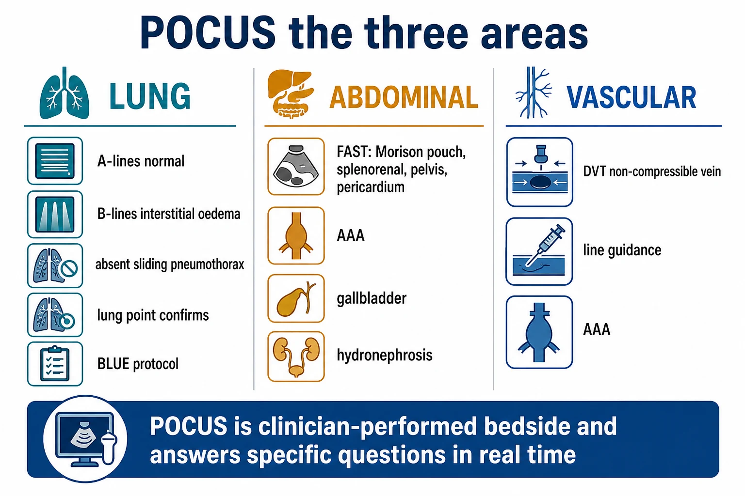
Point-of-care ultrasound (POCUS) is the clinician-performed, bedside ultrasound that answers specific clinical questions in real time, across three areas. Lung ultrasound: A-lines (normal), B-lines (interstitial syndrome — pulmonary oedema, ARDS, pneumonia), absent lung sliding (pneumothorax), the lung point (confirms pneumothorax), and the BLUE protocol for acute respiratory failure. Abdominal ultrasound: the FAST/eFAST (free fluid in trauma — Morison's pouch, splenorenal, pelvis, pericardium) and the non-trauma abdominal scan (AAA, gallbladder, hydronephrosis). Vascular ultrasound: compression ultrasonography for DVT (a non-compressible vein is a thrombus), ultrasound-guided line insertion, and AAA screening. POCUS integrates with the RUSH protocol (cardiac, lung, abdominal, vascular) for the bedside diagnosis of shock.
On this page & tools
Your progress
Saved locally on this device.
Target exams
Overview & definition
Point-of-care ultrasound (POCUS) is the clinician-performed, bedside ultrasound that answers specific clinical questions in real time — not a comprehensive radiology study. It covers three areas in the ICU: lung, abdominal, and vascular. Integrated with focused cardiac ultrasound (FOCUS/FATE, see the previous topic), it provides a rapid bedside assessment of the unstable patient (the RUSH protocol — Rapid Ultrasound in Shock).[1]
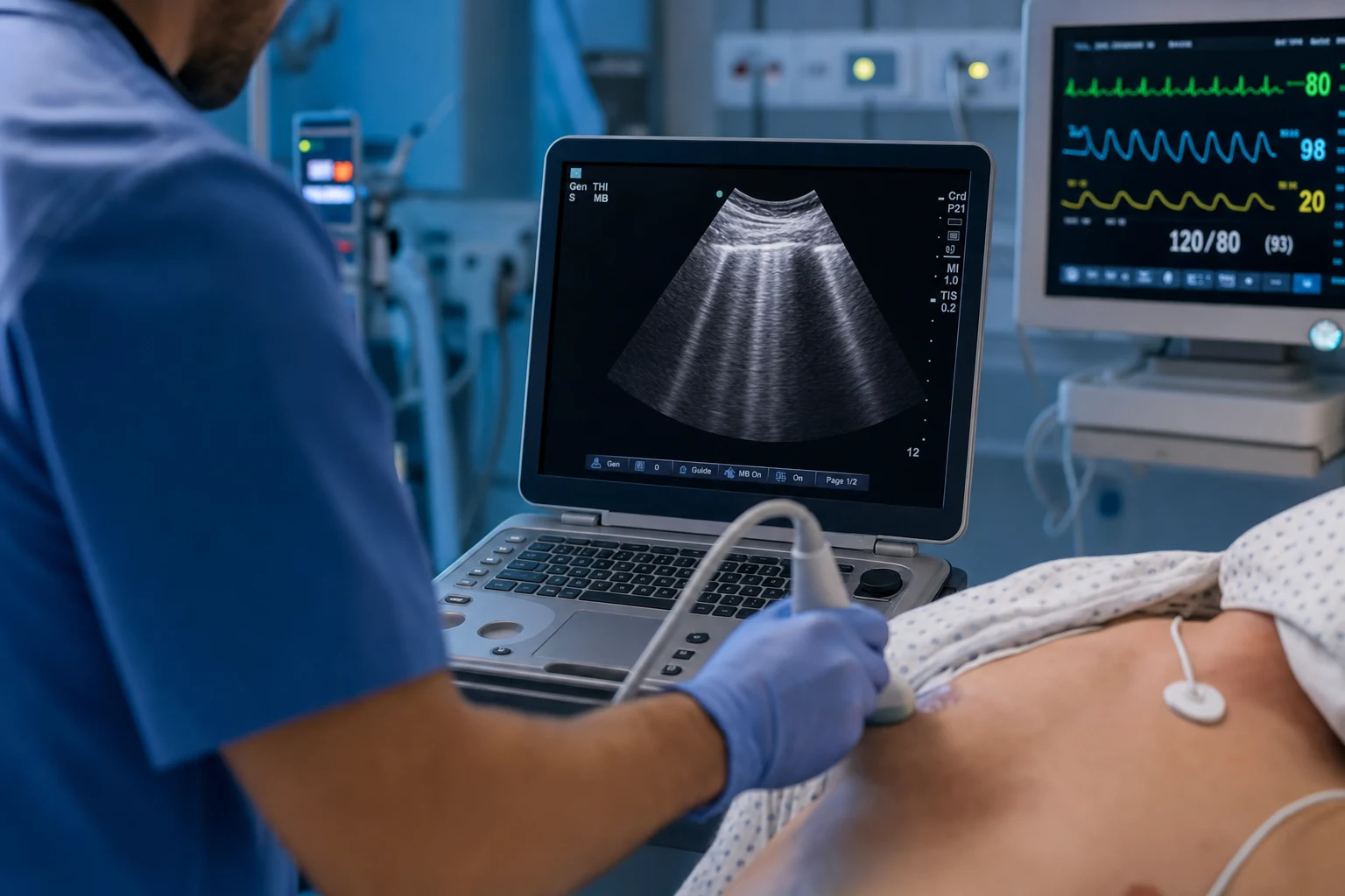
Lung ultrasound
The lung is normally air-filled (which blocks ultrasound), but the pleura and the interstitium produce characteristic artifacts:[1]
- A-lines — horizontal reverberation artifacts from the pleural line; the normal pattern. A normal lung shows A-lines with lung sliding.
- B-lines (comet-tail artifacts) — vertical, laser-like artifacts arising from the pleural line and moving with respiration. They indicate interstitial syndrome: fluid in the interlobular septa. Multiple diffuse B-lines suggest pulmonary oedema or ARDS; focal B-lines suggest pneumonia or atelectasis.
- Lung sliding — the normal shimmering of the pleural line with respiration (the visceral and parietal pleura sliding against each other). Absent lung sliding suggests a pneumothorax.
- The lung point — the transition point between the normal sliding lung and the non-sliding pneumothorax, visible as the lung intermittently enters the scan during inspiration. It confirms a pneumothorax with high specificity (though not always visible).
- Pleural effusion — an anechoic space above the diaphragm, with compressed lung floating in the fluid.
- Consolidation — tissue-like echo texture (hepatization) of the lung, with an air bronchogram (dynamic in pneumonia, static in atelectasis).[1][2]
The BLUE protocol (Lichtenstein) uses lung and venous ultrasound to diagnose the cause of acute respiratory failure: the lung pattern (A-profile, B-profile) combined with the DVT scan points to pulmonary oedema, pneumonia, pneumothorax, or PE.[1]
Abdominal ultrasound
The FAST (Focused Assessment with Sonography for Trauma):[1]
- Four views: the hepatorenal recess (Morison's pouch) — the most sensitive for free fluid; the splenorenal recess; the pelvis (the pouch of Douglas); and the pericardium.[5]
- Free fluid (anechoic) in any view = hemoperitoneum or hemopericardium in trauma.
- The eFAST (extended FAST) adds the bilateral thoracic views (for pneumothorax — absent lung sliding) and the IVC (for volume status).
Non-trauma abdominal POCUS:[1]
- Abdominal aortic aneurysm (AAA) — the aortic diameter (over 3 cm is aneurysmal).
- Gallbladder — stones, wall thickening (over 3 mm), a sonographic Murphy sign.[6]
- Hydronephrosis — an obstructed, dilated renal collecting system.
- Bladder volume and free fluid (ascites, perforation).
Vascular ultrasound
- DVT (deep vein thrombosis) — compression ultrasonography of the common femoral, the superficial femoral, and the popliteal veins. The criterion: a non-compressible vein is a thrombus (the normal vein collapses completely under the probe pressure; a vein with a clot does not). Highly sensitive and specific for proximal DVT.[1][8]
- Ultrasound-guided vascular access — real-time guidance for the internal jugular, subclavian, and femoral vein cannulation (reduces arterial puncture, pneumothorax, and the number of attempts; improves the success rate), and for the radial and femoral arterial lines.[8]
- AAA screening — the abdominal aorta diameter (above 3 cm is aneurysmal).
Red flags
Lung ultrasound in depth — probe, scanning, and the artefact vocabulary
Lung ultrasound works on a paradox: the aerated lung is the worst possible acoustic window (air reflects ~99 per cent of the ultrasound beam at the pleural line), yet the artefacts generated at the pleural interface carry diagnostic information that a chest X-ray and even a CT cannot deliver in real time at the bedside. The clinician reads a vocabulary of normal and pathological artefacts rather than imaging the lung substance directly.[2][13]
- Probe — a curvilinear (abdominal) or phased-array (cardiac) probe, low frequency (2–5 MHz), set to the lung preset (no harmonic imaging, no speckle reduction — these destroy the artefacts). A linear high-frequency probe is used when searching for the lung point or assessing pleural sliding in slim patients (M-mode for the seashore and barcode signs).
- Scanning zones — the BLUE-points: the upper and lower BLUE points on each hemithorax (hands placed palms down with the little finger on the clavicle — the upper point at the 3rd–4th intercostal space parasternally, the lower point at the mid-axillary line just above the diaphragm). Bilateral, two points per side = the standard four-zone scan; an eight-zone scan (adding anterior and lateral zones) increases sensitivity for focal disease (pneumonia, atelectasis).[1]
- The pleural line — the bright horizontal echogenic line just deep to the rib shadows (the bat sign: the two ribs are the wings, the pleural line the body). This is the reference line from which A-lines, B-lines, and lung sliding are assessed.
- M-mode — used to document lung sliding: the seashore sign (normal — a grainy "sandy" lung beneath a stratified "waves" chest wall) versus the barcode / stratosphere sign (absent sliding — parallel horizontal lines throughout). M-mode is a static confirmation of a dynamic finding; it does not substitute for seeing the sliding directly in 2D.[2]
The lung ultrasound artefact vocabulary
| Finding | Appearance | Mechanism | Significance |
|---|---|---|---|
| A-lines | Horizontal reverberation lines spaced at equal intervals deep to the pleural line | The beam reflects back and forth between the probe and the highly reflective pleural-air interface | Normal lung (aerated). The A-profile (A-lines with sliding) is the baseline against which pathology is defined |
| B-lines (comet-tails) | Vertical, laser-like, well-defined bright lines arising from the pleural line, erasing A-lines, moving with sliding, reaching the bottom of the screen | Reverberation from thickened, fluid-laden interlobular septa (<7 mm interlobular septal thickness) | Interstitial syndrome. Diffuse bilateral = pulmonary oedema / ARDS; focal = pneumonia / atelectasis / contusion |
| Lung sliding | A shimmering to-and-fro movement of the pleural line with respiration | The visceral and parietal pleura moving against each other | Present = pleural apposition (rules out PTX at that point). Absent = PTX, apnoea, pleurodesis, pneumonia, ARDS, main-stem intubation |
| Lung pulse | A fine cardiac-rate shimmering of the pleural line in an apnoeic patient | The heartbeat transmitted to the lung when there is no air (atelectasis / main-stem intubation) | Rules out PTX at the examined point and explains absent sliding that is NOT a pneumothorax |
| Lung point | The transition zone where sliding lung intermittently sweeps into a non-sliding field during inspiration | The visceral pleura edge entering the acoustic window at the periphery of a PTX | Pathognomonic for PTX (specificity ~100 per cent, sensitivity modest — not always visible) |
| Consolidation (hepatization) | Tissue-like echo-texture replacing the aerated lung, with hyperechoic punctate air bronchograms | The alveoli filled with fluid/pus/transudate become sonically transmissible | Pneumonia (dynamic air bronchograms), atelectasis (static), pulmonary infarct, contusion |
| Pleural effusion | An anechoic (or echoic/complex) space above the diaphragm, with floating/compressed lung (the jellyfish sign) | Fluid in the pleural space displacing the lung | Transudate (anechoic) vs exudate/haemothorax (echoic, septations). Volume estimable from the inter-pleural distance |
B-line semi-quantification — turning artefacts into a number
The number of B-lines in a single intercostal space correlates with the degree of extravascular lung water and, in pulmonary oedema, with the pulmonary artery occlusion pressure (PAOP). This is what allows lung ultrasound to be a quantitative, not just qualitative, tool.[3]
- Count — the number of distinct B-lines visible in a single longitudinal view between two ribs (0 = normal; 1–2 = mild; 3+ per zone = significant interstitial syndrome).
- Thresholds — Lichtenstein showed that a diffuse bilateral B-pattern (the B-profile) predicts a PAOP > 18 mmHg with high accuracy, whereas the A-profile predicts a PAOP < 18 mmHg. This underpins the FALLS-protocol (see below).[3][10]
- Waterfall sign — confluent B-lines that coalesce into a bright white sheet (severe interstitial oedema, alveolar oedema).
- Asymmetry matters — bilateral diffuse B-lines = hydrostatic or permeability oedema; unilateral or focal B-lines = pneumonia, atelectasis, contusion, or a unilateral effusion compressing adjacent lung.
B-profile vs A-profile — what the lung pattern predicts
| Lung profile | Additional finding | Predicted PAOP | Most likely diagnosis |
|---|---|---|---|
| A-profile (A-lines + sliding, bilateral) | — | <18 mmHg (low) | Normal lung, COPD/asthma, PE, or pulmonary embolism (A + DVT on venous scan) |
| B-profile (diffuse bilateral B-lines) | Decreased ejection fraction on FOCUS | >18 mmHg (high) | Cardiogenic pulmonary oedema |
| B-profile (diffuse bilateral B-lines) | Normal/hyperdynamic LV on FOCUS | Variable (often normal) | ARDS / non-cardiogenic oedema (permeability) — B-lines are irregular, sub-pleural consolidations common |
| A/B-profile (A-lines one side, B-lines other) | — | — | Pneumonia, focal atelectasis, or unilateral pathology — never seen in pure hydrostatic oedema |
| A-profile + absent sliding | Lung point | — | Pneumothorax |
| A-profile + DVT on venous scan | RV strain on FOCUS | — | Pulmonary embolism (the BLUE-protocol signature) |
The BLUE protocol — the decision tree for acute respiratory failure
The BLUE-protocol (Bedside Lung Ultrasound in Emergency) is Lichtenstein's validated algorithm for the bedside diagnosis of acute respiratory failure, using lung ultrasound at the BLUE-points plus a venous (DVT) scan, completed in under three minutes. In the derivation study it achieved a 90.5 per cent diagnostic accuracy immediately at the bedside, matching or exceeding the combination of history, examination, CXR, and blood tests.[1]
The BLUE protocol — step by step
- Scan the lungs at the BLUE-points (upper and lower, bilateral). Classify each side as A (A-lines with sliding) or B (diffuse B-lines), and check for lung sliding, the lung point, and consolidation/alveolar syndrome.[1]
- Define the lung profile — A-profile = bilateral A-lines with sliding; B-profile = bilateral diffuse B-lines; A/B-profile = A one side, B the other; absent sliding = suspected pneumothorax (search for the lung point).[1]
- Perform the venous (DVT) scan — compression ultrasonography of the femoral and popliteal veins on both sides. A non-compressible vein = DVT.[1]
- A-profile + DVT → pulmonary embolism (the A′ profile). The combination of dry lungs and a leg clot in a dyspnoeic patient is the BLUE signature of PE.
- A-profile + no DVT + posterior alveolar/interstitial syndrome (PLAPS) → pneumonia. The posterior lung base shows consolidation or a pleural effusion not seen in the anterior zones (dependent atelectasis/pneumonia).
- B-profile → pulmonary oedema. Diffuse bilateral B-lines with a low EF on FOCUS (cardiogenic). If the LV is normal, consider ARDS.
- A/B-profile → pneumonia (asymmetry is not oedema).
- Absent lung sliding + lung point → pneumothorax. If no lung point is found, consider main-stem intubation, apnoea, pleurodesis, or ARDS (all abolish sliding).
- A-profile + no DVT + no PLAPS → asthma or COPD exacerbation (the "naked" A-profile — dry lungs with no other finding, in a wheezing patient).
The BLUE-protocol profiles and their diagnoses
| Profile | Lung finding | Venous scan | FOCUS (added) | Diagnosis |
|---|---|---|---|---|
| A′ | A-lines + sliding | DVT present | RV strain | Pulmonary embolism |
| B | Diffuse bilateral B-lines | — | Low EF | Pulmonary oedema (cardiogenic) |
| A/B | A one side, B other | — | — | Pneumonia |
| A + PLAPS | A anterior + consolidation/effusion posterior | — | — | Pneumonia |
| A-no-flag | A-lines + sliding, no PLAPS | No DVT | Normal | Asthma / COPD |
| No sliding | Absent sliding | — | — | Pneumothorax (confirm with lung point) |
The FALLS-protocol — fluid therapy guided by lung ultrasound
The FALLS-protocol (Fluid Administration Limited by Lung Sonography) extends the BLUE logic into the shocked patient: it uses the appearance of B-lines as the endpoint of fluid resuscitation. The principle is that the lung is the most sensitive clinical indicator of fluid overload — B-lines appear before oxygen saturations fall and before crackles are heard.[10]
The FALLS-protocol — the sequence
- Establish the diagnosis of shock with the RUSH protocol (cardiac, lung, abdominal, vascular). If the cause is obstructive (tamponade, tension PTX) or cardiogenic (hypokinetic LV with B-lines), treat directly — do not give fluid.[10]
- If hypovolaemic/distributive shock suspected and lungs are A-profile (dry) → give fluid in 500 mL boluses, reassessing the lung bases after each bolus.[10]
- Watch for the transition A → B — the appearance of B-lines at the dependent lung zones signals that the pulmonary artery occlusion pressure has reached ~18 mmHg and the interstitium is flooding. This is the endpoint of fluid resuscitation.[10]
- If shock persists after the A→B transition → switch to vasopressors/inotropes — the patient is no longer fluid-responsive; further fluid will cause pulmonary oedema. The lung ultrasound has set the ceiling on fluid therapy.
Pneumothorax protocol — ruling it in and out
Lung ultrasound is more sensitive than a supine AP chest X-ray for pneumothorax (the supine CXR misses air that has risen to the anterior chest). The protocol is a stepwise search that uses the high negative predictive value of lung sliding to rule PTX out, and the lung point to rule it in.[4]
The lung ultrasound pneumothorax protocol
- Scan the anterior chest wall (2nd intercostal space, mid-clavicular line — the most dependent point for air in the supine patient, since air rises). Use M-mode to document the seashore (sliding) or barcode (no sliding) sign.[4]
- If lung sliding (or a lung pulse) is present → pneumothorax excluded at that point (negative predictive value ~99 per cent for occult PTX at the scanned zone).[4]
- If lung sliding is absent → move the probe laterally and posteriorly, searching for the lung point (the transition where the sliding lung sweeps back in during inspiration). Finding the lung point confirms the PTX and localises its edge (allows estimation of size: a lateral lung point = small, a medial/posterior point = large).[4]
- If the lung point is found, semi-quantify: a lung point at the mid-axillary line suggests a small (<15 per cent) PTX; a lung point posterior to the mid-axillary line suggests a larger PTX.[4]
- In the ventilated patient, a PTX with absent sliding is treated regardless of the lung point — positive-pressure ventilation can convert an occult PTX into a tension PTX rapidly. Insert a chest drain.[4]
Abdominal ultrasound in depth — the FAST/eFAST
The FAST (Focused Assessment with Sonography in Trauma) is the prototypical POCUS application: a rapid, bedside, clinician-performed scan for free intraperitoneal and intrapericardial fluid in the trauma patient. The eFAST extends it to the bilateral thoracic cavities (haemothorax, pneumothorax) and the IVC (volume status). A curvilinear or phased-array probe is used.[5]
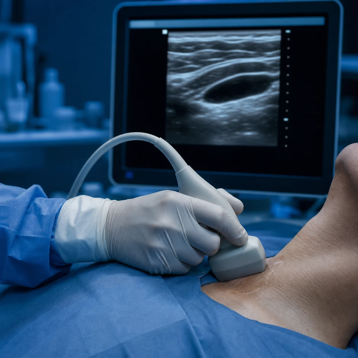
The eFAST — six views, step by step
- Morison's pouch (hepatorenal recess) — probe in the right mid-axillary line at the 7th–8th intercostal space, in coronal orientation. The most dependent part of the upper abdomen; the most sensitive single view for free fluid (detects ~250–500 mL). Anechoic (black) fluid between liver and kidney = haemoperitoneum.[5]
- Splenorenal recess — mirror image on the left (posterior axillary line, 6th–8th intercostal space). The spleen is smaller and higher than the liver; blood also collects in the subphrenic space. Left-sided injuries may be missed if only Morison's is scanned.[5]
- Pelvis (pouch of Douglas / rectovesicular pouch) — probe just superior to the symphysis pubis, aimed into the pelvis (full bladder as an acoustic window). The most dependent part of the peritoneum in the supine patient; a small volume of fluid pools here first.[5]
- Subxiphoid pericardial — probe just below the xiphoid, aimed toward the left shoulder, using the liver as a window. Fluid (anechoic) in the pericardial sac = haemopericardium (a penetrating cardiac injury until proven otherwise).[5]
- Right and left thorax / costophrenic angles — probe in the posterior axillary line at the base. An anechoic stripe above the brightly echogenic diaphragm = haemothorax; above the diaphragm on the thoracic side, look for lung sliding (absent = pneumothorax).[5]
- (Optional) IVC — subxiphoid sagittal, to estimate volume status (see the IVC section below).
The eFAST views — what each detects, sensitivity, and the trap
| View | Probe position | Detects | Sensitivity / trap |
|---|---|---|---|
| Morison's pouch | R mid-axillary, 7th–8th ICS, coronal | Free intraperitoneal fluid (haemoperitoneum) | Most sensitive single view (~250–500 mL detectable). Bowel gas and obesity reduce sensitivity |
| Splenorenal / subphrenic | L posterior axillary, 6th–8th ICS | Left upper quadrant blood | Often missed — always scan the left side; spleen injuries bleed into the subphrenic space |
| Pelvis | Suprapubic, aimed caudad | Dependent pelvic fluid | The most dependent space; small volumes pool here. A full bladder aids the window |
| Subxiphoid pericardium | Subxiphoid, toward L shoulder | Haemopericardium / tamponade | Penetrating cardiac injury until proven otherwise. Parasternal windows if subxiphoid is poor |
| Thoracic (bilateral) | Posterior axillary line, base | Haemothorax, pneumothorax | Anechoic above diaphragm = haemothorax; absent lung sliding = PTX. Replaces the missed supine CXR PTX |
The gallbladder — cholecystitis at the bedside
Acute cholecystitis is a common ICU and emergency diagnosis, and right-upper-quadrant ultrasound is the first-line imaging test. POCUS of the gallbladder has a sensitivity of ~80–90 per cent for stones and can establish the bedside diagnosis of acute calculous cholecystitis when the full sonographic Murphy's triad is present.[6]
The gallbladder POCUS — five findings
- Find the gallbladder — curvilinear probe in the right subcostal area, aimed toward the right shoulder (the "sweep"). Have the patient take a deep breath to bring the liver and gallbladder down under the costal margin. The gallbladder is an anechoic (black) pear-shaped structure attached to the undersurface of the liver (the gallbladder fossa).[6]
- Identify gallstones — bright, echogenic foci within the gallbladder that cast a clean acoustic shadow (the stone blocks the beam). Stones are gravity-dependent — roll the patient to confirm mobility. A stone impacted in the gallbladder neck or cystic duct is the cause of acute cholecystitis. The WES sign (Wall, Echo, Shadow) — a single echogenic line with shadowing — is a gallbladder packed with stones (and at risk of empyema).[6]
- Measure the gallbladder wall thickness — the normal wall is < 3 mm. A wall > 3 mm (measured in the anterior, non-dependent wall, with the gallbladder not over-distended) is oedema from inflammation. A wall > 4.5 mm has the highest positive likelihood ratio for cholecystitis.[6]
- Check for a sonographic Murphy's sign — maximal tenderness when the probe presses directly over the sonographically-identified gallbladder (more specific than the clinical Murphy's sign). The combination of stones + wall thickening + sonographic Murphy's sign has a positive predictive value > 90 per cent for acute cholecystitis.[6]
- Look for complications — pericholecystic fluid (anechoic rim around the gallbladder — perforation/empyema), intramural gas (emphysematous cholecystitis — a surgical emergency), and a distended gallbladder (> 10 cm long, > 4 cm wide) with a hydropic, tender gallbladder (hydrops / distal obstruction).
The sonographic features of acute calculous cholecystitis
| Feature | Normal | Acute cholecystitis | Notes |
|---|---|---|---|
| Gallstones | Absent | Present (impacted in neck/cystic duct) | A stone must be present to call it calculous cholecystitis. Acalculous cholecystitis (ICU) shows no stone but wall thickening and distension |
| Wall thickness | <3 mm | >3 mm (often 4–6 mm) | Measure the anterior non-dependent wall, GB not over-distended. A globally thick, striated, oedematous wall suggests severe/inflammation |
| Sonographic Murphy's sign | Negative | Positive (focal maximal tenderness over the GB) | PPV of the triad (stones + wall + Murphy) >90 per cent |
| GB size | Normal (fasted <10 cm) | Distended / hydropic (>10 cm × 4 cm) | Suggests cystic duct obstruction |
| Pericholecystic fluid | Absent | Present | Perforation, empyema — surgical urgency |
The IVC — volume status and fluid responsiveness
The inferior vena cava is the bedside surrogate for the right atrial pressure and, with caveats, for fluid responsiveness. The collapsibility index (spontaneously breathing) and the distensibility index (ventilated) translate the IVC's respiratory variation into a number. Crucially, the static IVC is a poor predictor of fluid responsiveness in the equivocal range — dynamic tests (passive leg raise with a flow-based readout like LVOT VTI) are superior.[9]
- Collapsibility index (CI), spontaneous breather — CI = (Dmax − Dmin) / Dmax × 100 per cent, measured 2 cm from the RA–IVC junction in the subcostal sagittal plane. A CI > 50 per cent with a small (<1.5 cm) IVC suggests low RA pressure and likely fluid responsiveness; a CI < 20 per cent with a large (>2.5 cm) IVC suggests high RA pressure and fluid is unlikely to help.[9]
- Distensibility index (DI), ventilated patient — positive pressure distends (rather than collapses) the IVC on inspiration. DI = (Dmax − Dmin) / Dmin × 100 per cent; a DI > 18 per cent predicts fluid responsiveness (sensitivity ~80–90 per cent in deeply sedated, passively ventilated patients with a regular rhythm).[9]
- Caveats — the static IVC is unreliable in the spontaneously breathing patient making strong inspiratory efforts (high negative intrathoracic pressure collapses even a normal IVC), in abdominal hypertension (extrinsic compression distends the IVC regardless of volume), and in arrhythmia. In the equivocal range (CI 20–50 per cent, diameter 1.5–2.5 cm), do not rely on the IVC — use a dynamic test.[9]
IVC interpretation — collapsibility (spontaneous) vs distensibility (ventilated)
| Patient type | Measurement | Threshold | Predicts |
|---|---|---|---|
| Spontaneously breathing | Collapsibility index = (Dmax − Dmin)/Dmax | >50 per cent (with small IVC <1.5 cm) | Likely fluid-responsive |
| Spontaneously breathing | Collapsibility index | <20 per cent (with large IVC >2.5 cm) | Not fluid-responsive — high RA pressure |
| Mechanically ventilated | Distensibility index = (Dmax − Dmin)/Dmin | >18 per cent | Likely fluid-responsive (deeply sedated, regular rhythm) |
| Mechanically ventilated | Distensibility index | <18 per cent | Not fluid-responsive |
| Equivocal (any) | Passive leg raise + LVOT VTI / carotid flow | >10–15 per cent rise in VTI | Gold-standard non-invasive test for fluid responsiveness |
The passive leg raise (PLR) with lung/cardiac ultrasound — the gold-standard bedside fluid test
- Position the patient semi-recumbent at 45 degrees, calm, head supported, no stimulation (avoid sympathetic activation). The PLR is a reversible, endogenous ~300 mL fluid bolus from the legs.[9]
- Baseline — measure a flow-based parameter: the LVOT VTI (apical 5-chamber, PW Doppler), the carotid artery flow, or (proxy) the IVC diameter/collapsibility. Record it.
- Tilt the patient flat and raise the legs to 45 degrees for 60–90 seconds — this auto-transfuses ~300 mL of venous blood from the legs into the thorax, mimicking a fluid bolus.
- Re-measure the parameter — an increase in LVOT VTI (or carotid flow) of >10–15 per cent predicts a positive response to a 500 mL crystalloid bolus (sensitivity and specificity ~85–90 per cent).
- Return the patient to the semi-recumbent position — the effect reverses within minutes. If positive, give fluid; if negative, the patient will not benefit from further fluid — consider vasopressors/inotropes.[9]
Vascular ultrasound in depth — DVT compression ultrasonography
Compression ultrasonography is the bedside standard for proximal DVT. The principle is simple and powerful: a normal vein collapses completely under probe pressure (apposed walls); a vein containing a thrombus does not compress. No Doppler is required for the diagnosis — non-compressibility IS the diagnostic criterion (the "Doppler" you may add — colour flow, augmentation, respiratory variability — is confirmatory, not primary).[8]
Two-point and extended compression ultrasonography for DVT
- Probe — linear high-frequency probe (5–10 MHz), patient supine, leg slightly externally rotated and flexed. Transverse orientation with the vein and artery side by side.[8]
- Identify the common femoral vein (CFV) and the saphenofemoral junction at the groin crease — the CFV is medial to the common femoral artery (the "Mickey Mouse sign": the head is the CFV, the two ears are the CFV and SFV bifurcation). Apply firm pressure until the artery begins to deform — a normal vein collapses fully first.[8]
- Move down the leg, compressing every 1–2 cm — the proximal superficial femoral vein (SFV), to the adductor canal, then the popliteal vein behind the knee (probe in the popliteal fossa, posterior approach). The two-point technique compresses only the CFV and popliteal vein (rapid, misses isolated SFV and calf clot); the whole-leg (extended) technique compresses the entire deep system down to the calf (slower, detects calf DVT).[8]
- Interpret — full compressibility at all points = no proximal DVT (negative). Non-compressibility of a segment = DVT (the lumen remains open, often with echogenic clot visible). The artery should still pulsate; a non-compressible vein with a normally pulsating artery confirms the diagnosis.[8]
- If the scan is negative but the Wells score is high (or D-dimer elevated) — repeat in 5–7 days (to catch a propagating calf clot) OR proceed to whole-leg imaging / D-dimer. A single negative whole-leg scan safely excludes DVT.[8]
Two-point vs whole-leg compression ultrasonography for DVT
| Feature | Two-point (CFV + popliteal) | Whole-leg (extended) |
|---|---|---|
| Veins compressed | Common femoral + popliteal only | CFV, SFV, popliteal, tibial, peroneal, muscular calf veins |
| Time | 2–4 minutes | 8–15 minutes |
| Sensitivity for proximal DVT | High (~95 per cent) | High (~96–99 per cent) |
| Detects isolated SFV / calf DVT | No — missed | Yes |
| Negative scan management | Repeat in 5–7 days if Wells high / D-dimer up | No repeat needed — single negative scan safely excludes |
| Best for | Rapid bedside POCUS in the ICU/ED | Definitive radiology department study; high pre-test probability |
AAA screening and bedside aortic measurement
The abdominal aortic aneurysm (AAA) is a silent killer that can be detected by a 30-second bedside ultrasound: an aortic diameter > 3 cm is aneurysmal. Bedside POCUS of the aorta is part of the RUSH protocol (any unexplained shock — rule out a ruptured AAA) and is the basis of the one-off screening programme for older men (the MASS study showed a ~40 per cent reduction in AAA mortality with ultrasound screening).[7][11]
Bedside aortic ultrasound — measuring the AAA
- Probe — curvilinear, abdominal preset. Patient supine. Begin in the transverse plane just below the xiphoid.[7]
- Identify the aorta in transverse — the pulsatile, thick-walled vessel anterior to the spine, just left of midline (the IVC is to the right, thin-walled, with respiratory variation). Trace it from the diaphragm to the bifurcation (at L4, ~2 cm below the umbilicus).[7]
- Measure the maximal antero-posterior diameter from outer wall to outer wall, in transverse, at the proximal, mid, and distal aorta. > 3 cm = aneurysmal. Most AAAs are infrarenal. Also measure in the longitudinal (sagittal) plane to confirm and to show the craniocaudad extent.[7]
- Identify the renal arteries and the bifurcation — to determine whether the AAA is infrarenal (most are) and to plan repair. Measure the distance from the lowest renal artery to the aneurysm sac (the "neck").
- Look for rupture — in the shocked patient with an AAA, look for retroperitoneal fluid/haematoma (complex free fluid, loss of the psoas shadow). A ruptured AAA is a surgical emergency — call the vascular surgeon and theatre immediately; do not delay for a CT if the patient is unstable.[7]
AAA diameter and management thresholds
| Diameter (men) | Risk | Management |
|---|---|---|
| <3 cm | Normal | No aneurysm |
| 3.0–4.4 cm (small) | Low rupture risk | Surveillance ultrasound at 2–3 year intervals |
| 4.5–5.4 cm (medium) | Moderate | Surveillance annually; optimise BP, stop smoking |
| 5.5–6.9 cm (large) | Significant rupture risk (~3–10 per cent/year) | Surgical/endovascular assessment for elective repair |
| >5.5 cm | — | Offer elective repair (open or EVAR) — this is the intervention threshold |
| Symptomatic / ruptured (any size) | Emergency | Immediate vascular surgery — theatre, cross-clamp/EVAR. Mortality of rupture ~80 per cent |
Ultrasound-guided central venous catheter (CVC) insertion
Real-time ultrasound guidance for central venous cannulation is now the standard of care — it reduces the number of attempts, the failure rate, arterial puncture, pneumothorax, and haematoma, and increases the first-pass success rate. The Randolph meta-analysis established the benefit for the internal jugular vein; expert consensus extends it to the subclavian and femoral veins and to arterial lines.[8]
Ultrasound-guided CVC insertion — the standard technique
- Identify the target vein in transverse and longitudinal — confirm patency (compressible, no clot), the relationship to the artery, and the depth. The internal jugular (IJ) is lateral/anterior to the carotid; the common femoral is medial to the femoral artery; the subclavian is posterior to the clavicle (challenging to see).[8]
- Choose the in-plane (long-axis) or out-of-plane (short-axis) approach. Out-of-plane (transverse) — the vein is a circle, the needle appears as a bright dot; easier to identify the vessel, but the needle tip is not always seen (risk of posterior wall puncture). In-plane (long-axis) — the vein is a tube, the entire needle and tip are seen; more precise but technically harder and requires a stable hand. The tip must always be visualised.[8]
- Sterile prep and drape, with a sterile probe cover and sterile gel. Confirm the view through the cover before draping.
- Advance the needle under real-time vision toward the vein — watch the tip (the bright dot in out-of-plane, or the whole needle in in-plane). "Walk down" the needle to the tip. A tissue movement precedes the tip arrival. Aspirate as you advance.[8]
- Flash of venous blood → thread the guidewire (Seldinger) under vision (see the wire in the vein). Confirm with ultrasound (longitudinal: the wire is a bright line within the vein; or transverse with the probe turned).[8]
- Dilate and insert the catheter over the wire, then scan to confirm the catheter is in the vein and there is no pneumothorax (IJ/subclavian — lung sliding at the ipsilateral apex).[8]
In-plane vs out-of-plane ultrasound-guided CVC insertion
| Feature | Out-of-plane (short-axis) | In-plane (long-axis) |
|---|---|---|
| Vein appearance | Circle (cross-section) | Tube (long axis) |
| Needle appearance | A bright dot (the tip, when correctly placed) | The entire needle and tip visible |
| Tip visualisation | Intermittent — must "walk down" to keep the tip in view; posterior-wall puncture risk | Continuous — the tip is seen throughout |
| Ease | Easier for the beginner; better vessel identification | Harder — needs a steady, aligned hand along the beam plane |
| Complication profile | Higher posterior-wall puncture and inadvertent arterial cannulation if the tip is lost | Lower, because the tip is always seen, but a small misalignment can take the tip out of plane |
| Recommendation | Standard for the IJ and femoral in most units | Preferred by experts for the subclavian and for small/deep veins where precision matters |
Integration — the RUSH protocol for undifferentiated shock
The RUSH protocol (Rapid Ultrasound in Shock and Hypotension) sequences the cardiac (FOCUS), lung, abdominal, and vascular POCUS into a single bedside examination that categorises the shock and identifies the immediately treatable causes. It pairs the haemodynamic answer (the pump) with the volume answer (the tank — IVC, lungs) and the vascular answer (the pipes — aorta, DVT).[11][1]
The RUSH protocol — pump, tank, pipes
| Component | What to scan | What it answers |
|---|---|---|
| The pump (cardiac) | FOCUS/FATE — PLAX, PSAX, A4C, subcostal | Is the LV failing (cardiogenic)? Is the RV dilated (PE)? Is there tamponade (effusion + collapse)? |
| The tank (volume) | Lung (A vs B lines), IVC (collapsibility/distensibility), FAST (free fluid) | Dry or wet? Fluid-responsive? Is there haemorrhage (positive FAST)? |
| The pipes (vascular) | Aorta (AAA), DVT compression (PE source), IJ/femoral for lines | Is there a ruptured AAA? A clot in the leg (PE)? Where to put the line? |
The RUSH protocol — the bedside sequence for the shocked patient
- The pump — perform FOCUS: is there an effusion with collapse (tamponade)? Is the LV dilated and hypokinetic (cardiogenic) or small and hyperdynamic (hypovolaemic/distributive)? Is the RV dilated with a D-shaped septum (PE / acute cor pulmonale)?[11]
- The tank — volume status — scan the lungs (A-lines = dry, B-lines = wet) and the IVC (small collapsing = responsive, plethoric fixed = high RA pressure). Perform a FAST for free fluid (haemorrhage — intra-abdominal bleed, ruptured ectopic, traumatic haemoperitoneum).[11]
- The pipes — scan the aorta for an AAA (ruptured AAA in the older shocked patient), and the leg veins for a DVT (the source of a massive PE). Identify a route for vascular access.[11]
- Synthesise — combine the pump, tank, and pipes findings to categorise the shock: hypovolaemic (empty tank, hyperdynamic pump, dry lungs), cardiogenic (failing pump, wet lungs), obstructive (tamponade, PE, tension PTX), distributive (normal pump, variable tank, often vasodilated).[11]
POCUS vs formal imaging — when each is right
This is the central question for the exam and for practice. POCUS and formal radiology studies answer different questions: POCUS answers a focused bedside question in real time; formal imaging answers a comprehensive, characterising question that requires an expert and time.[1][13]
POCUS vs formal imaging — the comparison the exam wants
| Domain | POCUS (clinician-performed, bedside) | Formal imaging (radiology: US, CT, MRI) |
|---|---|---|
| Operator | The treating intensivist/ED physician (after structured training) | A sonographer/radiologist |
| Question | Focused, binary, real-time: "Is there a PTX? Free fluid? A clot? B-lines?" | Comprehensive, characterising: "What is the AAA anatomy? Is this mass malignant?" |
| Timing | Seconds to minutes, at the bedside, repeated serially | Minutes to hours, requires transport, single time point |
| Patient transport | None — done on the unstable patient in the resuscitation bay/ICU | Often requires moving the critically ill patient (risk of transport-related deterioration) |
| Image quality / detail | Lower; focused; may be limited by body habitus, dressings, gas | Higher; full characterisation; multiplanar; contrast/doppler as needed |
| Documentation | Limited stills/clips; focused report; operator-dependent | Full annotated study; structured radiology report; archived |
| Radiation | None (ultrasound) | CT/fluoroscopy involve ionising radiation |
| Cost / availability | Low cost; available 24/7 at the bedside | Higher cost; may be limited by scanner/staff availability |
| Best for | Resuscitation, serial monitoring, procedural guidance, answering the immediate question | Definitive diagnosis, staging, complex anatomy, surgical/interventional planning |
| Cannot do | Quantify to standard (exact EF, valve gradients), characterise masses, see behind gas/bone | Be at the bedside in real time; be repeated every hour by the treating clinician |
Modality by clinical question — which to choose
| Clinical question | First-line | Definitive / confirmatory |
|---|---|---|
| Pneumothorax (trauma/ICU) | Lung ultrasound (absent sliding + lung point) — more sensitive than supine CXR | CT chest (gold standard; if lung US equivocal) |
| Pulmonary oedema | Lung US (B-profile) + FOCUS (LV function) | CXR (alveolar oedema); formal echo for the cause |
| Pleural effusion | Lung US (anechoic above diaphragm) + volume estimate | CT chest (characterise; loculated/complex) |
| Free intraperitoneal fluid (trauma) | eFAST (Morison, splenorenal, pelvis, pericardium) | CT with IV contrast (injury characterisation) |
| Acute cholecystitis | RUQ POCUS (stones, wall, Murphy) | Formal RUQ US; HIDA scan if US equivocal |
| DVT | Two-point or whole-leg compression US | Formal Doppler US (whole leg) |
| AAA | Bedside aortic US (>3 cm) | CT angiogram (anatomy, rupture, EVAR planning) |
| Tamponade | FOCUS (effusion + RA/RV collapse) | Formal echo; CT if localised/post-surgical |
| PE | FOCUS (RV strain) + lung US (A-profile) + DVT scan (venous) | CT pulmonary angiogram (gold standard) |
| Aortic dissection | FOCUS (aortic root dilatation — limited) | CT angiography or TOE — do not rely on POCUS |
Exam practice — SAQs
SAQ — Acute respiratory failure: applying the BLUE protocol at the bedside
10 minutes · 10 marks
A 72-year-old man is admitted to the ICU with acute, severe dyspnoea of 4 hours duration. He has a background of ischaemic cardiomyopathy (ejection fraction 30 per cent), severe COPD, and a long-haul flight 5 days ago. RR 34, SpO2 86 per cent on 15 L via non-rebreather, BP 96/58, HR 118, JVP elevated 6 cm. Examination reveals bibasal crackles and mild expiratory wheeze; the chest X-ray shows bilateral infiltrates but is deemed non-diagnostic. Arterial blood gas: pH 7.30, PaO2 54 mmHg, PaCO2 34 mmHg, lactate 2.8 mmol/L. The consultant asks you to perform bedside lung ultrasound to narrow the differential.
SAQ — Lung ultrasound B-lines: cardiogenic versus non-cardiogenic pulmonary oedema
10 minutes · 10 marks
A 58-year-old woman with severe influenza A pneumonia is intubated and ventilated for ARDS (PaO2/FiO2 110). On ICU day 3, bedside lung ultrasound shows bilateral, irregular, coarse B-lines with sub-pleural consolidations and clearly spared areas; there is no significant pleural effusion. The nurse suggests that these B-lines mean she is fluid overloaded and asks whether diuresis will improve her oxygenation. FOCUS shows a small, hyperdynamic left ventricle.
Clinical pearls
Trial cards and key evidence
The BLUE protocol (Lichtenstein & Mezière, Chest 2008, PMID 18403664)
Source
Prospective derivation study of lung and venous ultrasound in 260 patients with acute respiratory failure, in a medical ICU
Accuracy
90.5 per cent diagnostic accuracy immediately at the bedside — within minutes, using only lung ultrasound at the BLUE-points plus a venous (DVT) scan
Profiles
A′ (A-profile + DVT) = PE; B-profile = pulmonary oedema; A/B or A+PLAPS = pneumonia; naked A = asthma/COPD; absent sliding + lung point = pneumothorax
Implication
Lung ultrasound + a venous scan can diagnose the cause of acute respiratory failure at the bedside faster and as accurately as the conventional workup
International lung ultrasound recommendations (Volpicelli et al, Intensive Care Med 2012, PMID 22392031)
Source
International consensus (24 experts) on point-of-care lung ultrasound technique, artefact definitions, and clinical applications
Standardisation
Defines the artefact vocabulary (A-lines, B-lines, lung sliding, lung point, consolidation, pleural effusion), the scanning zones, and the M-mode signs (seashore, barcode)
Applications
Standardises lung US for pneumothorax, interstitial syndrome, consolidation/pneumonia, pleural effusion, and integration with cardiac POCUS
Implication
The reference for what lung ultrasound can and cannot claim, and the technical standard against which competence is assessed
A-lines, B-lines and the PAOP (Lichtenstein et al, Chest 2009, PMID 19809049)
Source
Study correlating the lung ultrasound profile with the pulmonary artery occlusion pressure in critically ill patients
Finding
The A-profile (bilateral A-lines) predicts a PAOP <18 mmHg; the B-profile (diffuse B-lines) predicts a PAOP >18 mmHg
Implication
Lung ultrasound is a non-invasive surrogate for the wedge pressure — the basis of the FALLS-protocol (fluid therapy limited by the appearance of B-lines)
IVC respiratory variability and fluid responsiveness (Airapetian et al, Critical Care 2015, PMID 26563768)
Source
Prospective study of IVC collapsibility as a predictor of fluid responsiveness in 116 spontaneously breathing ICU patients
Finding
The static IVC collapsibility index was a poor predictor of fluid responsiveness in spontaneous breathers; only very high collapsibility (with a small IVC) was reliably fluid-responsive
Implication
Confirms that the static IVC is unreliable in the equivocal range — a dynamic test (passive leg raise with a flow-based readout) is needed for fluid responsiveness in spontaneously breathing patients
Ultrasound guidance for CVCs — meta-analysis (Randolph et al, Critical Care Medicine 1996, PMID 8968276)
Source
Landmark meta-analysis of randomised trials of ultrasound-guided central venous catheter placement
Finding
Ultrasound guidance reduced failed catheter placements, the number of attempts, and mechanical complications (arterial puncture, pneumothorax, haematoma); improved first-pass success
Implication
Established ultrasound guidance as the standard of care for internal jugular cannulation; the benefit extends to subclavian, femoral, and arterial lines by expert consensus
MASS — AAA screening trial (Multicentre Aneurysm Screening Study, Lancet 2002, PMID 12443589)
Source
Population-based randomised controlled trial of one-off abdominal aortic ultrasound screening in men aged 65–74
Finding
Screening reduced AAA-related mortality by ~40 per cent over 4 years; the benefit persisted at long-term follow-up
Implication
Established the one-off ultrasound screening programme for men aged 65–74 — the aorta >3 cm is aneurysmal; >5.5 cm is the intervention threshold
Pitfalls and limitations
Common POCUS pitfalls and how to avoid them
| Pitfall | What goes wrong | How to avoid |
|---|---|---|
| Calling Z/E-lines B-lines | Over-calling interstitial syndrome in emphysema/subcutaneous emphysema | Apply the strict B-line criteria (arise from pleura, well-defined, erase A-lines, reach the bottom) |
| Reading the B-profile without FOCUS | Cannot separate cardiogenic from ARDS oedema | Always pair lung US with a cardiac echo (LV function) |
| Using the spontaneous-breather IVC thresholds on a ventilated patient | The ventilated IVC distends on inspiration — collapsibility thresholds are wrong | Use the distensibility index (Dmax − Dmin)/Dmin; threshold >18 per cent |
| Trusting the static IVC in the equivocal range | Predicts fluid responsiveness poorly | Use a passive leg raise with LVOT VTI / carotid flow |
| Calling the IVC the aorta (or vice versa) | The aorta is thick-walled, pulsatile, crosses midline; the IVC is thin-walled, enters the RA, has respiratory variation | Trace the vessel: the IVC enters the right atrium; the aorta does not |
| Missing the left side on FAST | Splenorenal blood is missed if only Morison's is scanned | Always scan both upper quadrants; the spleen bleeds into the subphrenic space |
| Discharging a high-risk patient on a negative two-point DVT scan | Misses isolated SFV or calf clot that propagates | Repeat in 5–7 days, or do a whole-leg study / D-dimer if Wells is high |
| Confident false-negative for PTX on POCUS | A complete PTX has no lung point (retracted lung) | Use lung sliding (present = no PTX) as the rule-out; if absent and the patient is ventilated, drain |
| Cannulating a thrombosed vein | A non-compressible IJ/femoral vein is unusable and thrombosed | Confirm patency/compressibility before every CVC |
| Over-calling cholecystitis from wall thickening alone | A non-fasted, over-distended GB has a falsely thick wall; cirrhosis/hypoalbuminaemia thicken the wall | Measure the anterior non-dependent wall with the GB not over-distended; require the full triad |
Summary
Point-of-care ultrasound (POCUS) is the clinician-performed, bedside ultrasound that answers focused, real-time questions across lung, abdominal, and vascular domains, integrated with focused cardiac ultrasound (FOCUS/FATE) into the RUSH protocol for undifferentiated shock. Lung ultrasound reads an artefact vocabulary — A-lines (normal), B-lines (interstitial syndrome), lung sliding (present = no PTX), the lung point (confirms PTX), consolidation, and effusion — and the BLUE protocol uses the lung profile plus a venous scan to diagnose the cause of acute respiratory failure (A′ = PE, B = oedema, A/B or A+PLAPS = pneumonia, naked A = asthma/COPD). The FALLS-protocol uses the A→B transition to set the ceiling on fluid therapy. Abdominal ultrasound performs the FAST/eFAST (Morison's pouch is the most sensitive view for free fluid; a negative FAST does not exclude injury), the gallbladder (stones + wall >3 mm + sonographic Murphy = acute cholecystitis), and the aorta (>3 cm = AAA; >5.5 cm = repair threshold). Vascular ultrasound performs compression ultrasonography for DVT (a non-compressible vein is a thrombus), ultrasound-guided CVC insertion (in-plane shows the whole needle, out-of-plane shows a dot — always see the tip), and AAA screening. The IVC estimates volume status (collapsibility for spontaneous breathers, distensibility >18 per cent for the ventilated), but a dynamic test (passive leg raise + LVOT VTI) is the gold standard for fluid responsiveness. POCUS rules findings IN more reliably than it rules them OUT — escalate to formal imaging when exclusion matters.[1][1][2][8][13]
References
- [1]Lichtenstein DA, Mezière GA. Relevance of lung ultrasound in the diagnosis of acute respiratory failure: the BLUE protocol Chest, 2008.PMID 18403664
- [2]Volpicelli G, Elbarbary M, Blaivas M, et al. International evidence-based recommendations for point-of-care lung ultrasound Intensive Care Med, 2012.PMID 22392031
- [3]Lichtenstein DA, Mezière GA, Lagoueyte JF, Biderman P, Goldstein I, Gepner A. A-lines and B-lines: lung ultrasound as a bedside tool for predicting pulmonary artery occlusion pressure in the critically ill Chest, 2009.PMID 19809049
- [4]Lichtenstein DA. The lung point, still a sign specific to pneumothorax Intensive Care Med, 2019.PMID 31399778
- [5]Quinn AC, Sinert R. What is the utility of the Focused Assessment with Sonography in Trauma (FAST) exam in penetrating torso trauma? Injury, 2011.PMID 20701908
- [6]Trowbridge RL, Rutkowski NK, Shojania KG. Does this patient have acute cholecystitis? JAMA, 2003.PMID 12503981
- [7]The Multicentre Aneurysm Screening Study Group. The Multicentre Aneurysm Screening Study (MASS) into the effect of abdominal aortic aneurysm screening on mortality in men: a randomised controlled trial Lancet, 2002.PMID 12443589
- [8]Randolph AG, Cook DJ, Gonzales CA, Pribble CG. Ultrasound guidance for placement of central venous catheters: a meta-analysis of the literature Crit Care Med, 1996.PMID 8968276
- [9]Airapetian N, Maizel J, Alyamani O, et al. Does inferior vena cava respiratory variability predict fluid responsiveness in spontaneously breathing patients? Crit Care, 2015.PMID 26563768
- [10]Lichtenstein DA. Fluid administration limited by lung sonography: the place of lung ultrasound in assessment of acute circulatory failure (the FALLS-protocol) Expert Rev Respir Med, 2012.PMID 22455488
- [11]Alalwad AA, Goyal M, Gaieski DF, Kim W. Accuracy of bedside emergency physician performed ultrasound in diagnosing different causes of acute abdominal pain: a prospective study Clin Imaging, 2015.PMID 25667065
- [12]Reissig A, Copetti R, Mathis G, et al. Lung ultrasound in community-acquired pneumonia and in interstitial lung diseases Respiration, 2014.PMID 24481027
- [13]Volpicelli G, Lamorte A, Tarricone V. Common pitfalls in point-of-care ultrasound: a practical guide for emergency and critical care physicians Crit Ultrasound J, 2016.PMID 27783380