ICU · Post-cardiac arrest care
Post-Cardiac Arrest Care and Targeted Temperature Management
Also known as Post-arrest care · Post-cardiac arrest syndrome · Targeted temperature management · TTM2 · TTM trial · HACA · HYPERION · Eurotherm3235 · CoolCap · TOBY · Prognostication · Neuroprotection · COACT · Shivering management · Cooling technique · Neonatal HIE
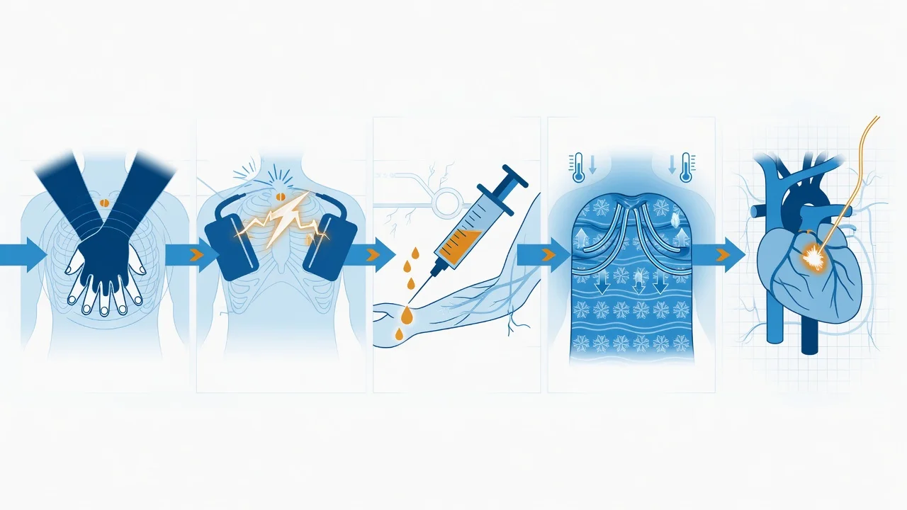
The post-cardiac-arrest care is the comprehensive ICU management of the patient who has achieved the return of spontaneous circulation — the prevention of the secondary neurological injury (the targeted temperature management, the controlled oxygenation, the blood pressure, the glycaemic control, the seizure prophylaxis), the investigation and the treatment of the cause (the coronary angiography — the COACT trial), and the prognostication at 72 hours (the multimodal approach — the clinical, the EEG, the neuroimaging, the biomarkers). This topic builds the examiner's framework on the post-cardiac-arrest syndrome (the brain injury, the myocardial dysfunction, the systemic ischaemia-reperfusion, the persistent precipitating pathology), the TTM evidence (the TTM trial showing no difference between 33 and 36, and the TTM2 trial showing hypothermia NOT superior to normothermia), the COACT trial (no benefit of the immediate coronary angiography without the ST elevation), and the prognostication advisory (the multimodal approach, the no-single-modality principle).
On this page & tools
Your progress
Saved locally on this device.
Target exams
Overview & definition
The post-cardiac-arrest care is the comprehensive ICU management of the patient who has achieved the return of spontaneous circulation (the ROSC). The arrest is survived, but the secondary injury — the anoxic brain injury, the myocardial stunning, the systemic ischaemia-reperfusion — is the threat, and the intensivist's task is to prevent the secondary injury, to investigate and treat the cause, and to prognosticate accurately.[1][1]
The framework rests on four pillars: the post-cardiac-arrest syndrome (the four-component injury), the targeted temperature management (the TTM and TTM2 evidence), the coronary angiography (the COACT trial), and the prognostication (the multimodal approach at 72 hours, the no-single-modality principle).[4]
Pathophysiology: the post-cardiac-arrest syndrome
The post-cardiac-arrest syndrome has four components, each demanding a specific management:[1][1]
- The post-arrest brain injury — the anoxic-ischaemic encephalopathy, the leading cause of the death in the out-of-hospital arrest. The injury continues after the ROSC (the secondary cascade: the excitotoxicity, the free radicals, the apoptosis, the blood-brain-barrier disruption), and the TTM, the oxygenation, the blood pressure and the seizure control are the mitigations.
- The post-arrest myocardial dysfunction — the myocardial stunning (the global, the reversible systolic and diastolic dysfunction), peaking at 24 to 48 hours and recovering over days. It causes the hypotension and the low-output state, and it is supported by the inotrope and the vasopressor, with the recovery.
- The systemic ischaemia-reperfusion — the multi-organ dysfunction (the AKI, the liver, the coagulation) from the global ischaemia and the reperfusion, managed supportively.
- The persistent precipitating pathology — the cause of the arrest (the acute coronary syndrome, the PE, the sepsis, the drug overdose, the haemorrhage) that must be identified and treated, for the arrest will recur if it is not. [1]
Targeted temperature management: the TTM and TTM2 evidence
The TTM is the neuroprotective intervention for the comatose post-arrest patient (the GCS less than 9, the not-following-commands). Its evidence has evolved across two decades through four landmark trials that have, in sequence, established, refined, and ultimately retired the routine use of therapeutic hypothermia in favour of strict fever avoidance.[1][2][5][7]
The trial evolution — the four-step arc
Step 1 — the foundational HACA trial (2002). The Hypothermia After Cardiac Arrest (HACA) study (Bernard, NEJM 2002) and the contemporaneous Bernard trial randomised the comatose survivors of the witnessed out-of-hospital VF/VT arrest to 33 to 34 degrees C for 12 to 24 hours versus the standard normothermia. The HACA trial showed the improved neurological outcome at six months (the favourable outcome in 55 per cent versus 39 per cent, RR 0.74) and a trend to the mortality benefit. These two trials underpinned the original ILCOR recommendation in 2003 that established the therapeutic hypothermia at 32 to 34 degrees C for 12 to 24 hours as the standard of care for the VF arrest.[5][6]
Step 2 — the TTM trial (2013) collapsed the target. The Nielsen TTM trial (NEJM 2013) was the high-quality, 950-patient, 36-centre randomised comparison of 33 degrees C versus 36 degrees C after the out-of-hospital arrest of ANY rhythm (the shockable and the non-shockable). It found no difference in the all-cause mortality (50 per cent versus 48 per cent) or the favourable neurological outcome (46 per cent versus 48 per cent). The implication was profound: the exact target (33 or 36) was less critical than the rigorous avoidance of the fever. This collapsed the dogma of 32 to 34 and shifted practice toward the broader "targeted temperature management" rather than the "therapeutic hypothermia."[2]
Step 3 — the TTM2 trial (2021) retired the routine hypothermia. The Dankiewicz TTM2 trial (NEJM 2021) randomised 1,861 comatose survivors to the hypothermia at 33 degrees C versus the normothermia (the target of 37.5 degrees C or less, with the early, aggressive treatment of the fever above 37.8). It found no difference in the all-cause mortality at 180 days (50 per cent versus 48 per cent) or the functional outcome (the modified Rankin scale 4 to 6 at 180 days: 55 per cent versus 55 per cent) — the hypothermia was NOT superior to the normothermia. The implication, which has reshaped the global practice, is that the routine hypothermia is NOT indicated; the active ingredient across all the TTM trials is the rigorous avoidance of the fever, NOT the induction of the hypothermia. The current practice (the ILCOR CoSTR 2022, the ERC 2021) is the strict normothermia with the active fever prevention for at least 72 hours after the ROSC.[1]
Step 4 — HYPERION (2019) reframed the non-shockable rhythm. The HACA evidence was almost exclusively in the shockable (VF/VT) arrest. The Lascarrou HYPERION trial (NEJM 2019) randomised 584 comatose survivors of the non-shockable (PEA/asystole) arrest to the hypothermia at 33 degrees C versus the normothermia at 37 degrees C for 24 hours. It found a higher rate of the favourable 90-day neurological outcome (10.2 per cent versus 5.7 per cent, OR 1.85, p = 0.04) with the hypothermia, with no significant difference in the mortality (81 per cent versus 83 per cent). This is the only modern TTM trial to show a benefit of the hypothermia at 33, and it sits in tension with the TTM2 result — it is the reason the ILCOR CoSTR retains a weak recommendation for the temperature control at 32 to 36 degrees C OR strict normothermia (both acceptable), and the reason the dedicated TTM-NonShock trial is ongoing to resolve the question in the non-shockable arrest.[7]
The verdict across the adult evidence
The synthesis across the four trials is: (a) in the shockable (VF/VT) arrest, the hypothermia at 33 offers no advantage over the strict normothermia (TTM2 2021); (b) in the non-shockable (PEA/asystole) arrest, the hypothermia at 33 may offer a small advantage (HYPERION 2019), though the certainty is low and the trials are ongoing; (c) the universal active ingredient across all the trials is the rigorous avoidance of the fever (above 37.5 to 37.8 degrees C) for at least 72 hours; (d) whatever target is chosen, the slow rewarming (0.25 to 0.5 degrees per hour) and the fever prevention through and after the rewarming are mandatory.[1][2][7]
HACA
NEJM 2002
273 comatose OHCA (witnessed VF/VT) — 33-34°C × 24 h vs normothermia
Key finding
Favourable neuro outcome at 6 mo: 55% vs 39% (RR 0.74, NNT ~6). Trend to ↓ mortality. Established 32-34°C as standard.
Practice change
Therapeutic hypothermia (32-34°C) became standard after witnessed VF OHCA
TTM
NEJM 2013
950 comatose OHCA (any rhythm) — 33°C vs 36°C, both × 36 h
Key finding
Mortality: 50% vs 48% (NS). Good neuro: 46% vs 48% (NS). No difference at either target. Rigorous fever avoidance in BOTH arms.
Practice change
Collapsed 32-34 dogma — exact target less important than fever avoidance; "TTM" replaced "therapeutic hypothermia"
TTM2
NEJM 2021
1861 comatose OHCA (any rhythm) — 33°C × 28 h vs normothermia (≤37.5°C, fever >37.8 treated)
Key finding
180-d mortality: 50% vs 48% (NS). mRS 4-6: 55% vs 55% (NS). Hypothermia NOT superior; trend to more arrhythmia and bleeding with hypothermia.
Practice change
Routine hypothermia retired; active fever prevention (normothermia) for 72 h is the standard
HYPERION
NEJM 2019
584 comatose OHCA with non-shockable rhythm (PEA/asystole) — 33°C vs 37°C × 24 h
Key finding
Favourable 90-d neuro outcome: 10.2% vs 5.7% (OR 1.85, p=0.04). Mortality 81% vs 83% (NS). Only modern RCT showing benefit of 33°C.
Practice change
Possible role for 33°C in non-shockable arrest — TTM-NonShock trial ongoing
Eurotherm3235
NEJM 2015
388 TBI with raised ICP — 32-35°C (cooling to lower ICP) vs normothermia
Key finding
WORSE 6-month neurological outcome with hypothermia (RR of unfavourable outcome 1.42). Trial stopped early for harm. Hypothermia as an ICP-lowering therapy is HARMFUL in TBI.
Practice change
Prophylactic/therapeutic hypothermia NOT recommended for raised ICP in TBI
HACA 2002
33-34°C vs normothermia (VF OHCA)
- Favourable neuro outcome 55% vs 39% (RR 0.74)
- Established 32-34°C as the global standard for 12 years
- Limited to witnessed VF/VT arrest
- Small trial (n=273) by modern standards
TTM 2013
33°C vs 36°C
- No difference in mortality (50% vs 48%) or neuro outcome
- Collapsed the 32-34°C dogma
- Shifted to "TTM" language (any rigorous target)
- Both arms had rigorous fever avoidance — this was the active ingredient
TTM2 2021
33°C vs normothermia
- No difference in mortality or functional outcome
- Routine hypothermia retired
- Fever prevention (≤37.5°C, treat >37.8°C) is the new standard
- Trend to more bleeding/arrhythmia with hypothermia
HYPERION 2019
33°C vs 37°C (non-shockable)
- Better 90-d neuro outcome with 33°C (10.2% vs 5.7%, OR 1.85)
- Only modern RCT showing benefit of 33°C
- In tension with TTM2 — TTM-NonShock trial ongoing
- Pragmatic alternative: 33°C reasonable in non-shockable arrest
The current guideline synthesis
The ILCOR CoSTR 2022 (Nolan) and the ERC 2021 advanced life support guidelines reflect the TTM2 verdict: (1) the temperature should be actively controlled, with a target of either 32 to 36 degrees C or the strict normothermia at 37.5 degrees C or less (both acceptable, the choice individualised); (2) the fever (above 37.8 degrees C) should be actively prevented for at least 72 hours after the ROSC (weak recommendation, low-certainty evidence); (3) the rewarming, if hypothermia is used, is slow (0.25 to 0.5 degrees C per hour); (4) the practice should be the standardised, protocolised, locally-audited. Many ANZ and UK units now default to the strict normothermia with the active fever prevention; the selective 33 degrees C is retained in some centres for the non-shockable arrest (per HYPERION) and in the centres where the local protocol has not yet been revised.[1][1]
The practical management. The core temperature is monitored continuously (the bladder, the oesophageal, or the PA catheter — the rectal lags and is avoided for the titration). The fever (above 37.5 to 37.8 degrees) is treated aggressively — the paracetamol 1 g six-hourly, the surface cooling (the Arctic Sun gel pads, the cooling blankets, the evaporative measures), and the intravascular cooling if required. The shivering is anticipated and controlled (the sedation, the opiate, the counter-warming, the magnesium, the buspirone, the dexmedetomidine). The rewarming (if the hypothermia was used) is slow (0.25 to 0.5 degrees per hour), and the fever is prevented during and after for at least 72 hours from the ROSC.[1][1]
The cooling technique: surface versus intravascular
The cooling technique, when the hypothermia at 33 is chosen, divides into the surface and the intravascular — both effective, both with the trade-offs. The choice is driven by the local availability, the patient factors, and the nursing workload.[1]
Surface cooling
Gel pads, blankets, ice
- Arctic Sun gel pads (hydrogel pads on thighs/torso) with feedback loop — most common
- Cooling blankets, ice packs, evaporative cool mist + fan
- Non-invasive, fast to set up, cheaper, no central line risk
- Slower induction (~1-2°C/h), less precise maintenance (±0.5°C)
- Higher shivering incidence; large skin surface stimulates cold receptors
Intravascular cooling
Closed-loop IVC catheter
- Specialised catheter in the femoral/IVC, saline circulated through balloon
- Faster induction, very precise maintenance (±0.1-0.2°C)
- Less nursing workload, less shivering peripherally
- Requires large-bore central line — line infection, DVT, vascular injury
- More expensive; used when tight control or prolonged TTM needed
Cold fluid induction
Adjunct / bridge
- 30 mL/kg of 4°C crystalloid bolus over 30-60 min for induction
- Reduces core temp ~1.5°C in 30 min; cheap, fast, universally available
- NOT for maintenance — fluid overload, pulmonary oedema risk
- Caution in heart failure / cardiogenic shock
- Often combined with surface method for induction phase
Other
Body cavity lavage
- Gastric, bladder, rectal, peritoneal lavage with cold fluid
- Used only as last-resort rescue / refractory hyperthermia
- Invasive, cumbersome, electrolyte shifts
- Largely historical / replaced by modern devices
The core temperature monitoring
The core temperature is the oesophageal, the bladder, or the PA catheter — the most accurate. The rectal and the tympanic lag by 0.5 to 1 degree C, and the temporal/skin are unreliable; they are NOT used for the titration of the TTM. The probe is placed early (during the induction) and the temperature is logged every 15 minutes during the induction, then continuously.[1]
The shivering management: the silent saboteur
The shivering is the physiological response to the cold — and it is the silent saboteur of the TTM. The shivering raises the metabolic demand by 200 to 500 per cent, the oxygen consumption, the sympathetic drive, the intracranial pressure, and the patient becomes harder to cool, harder to oxygenate, and harder to keep calm. The active management of the shivering is as important as the temperature target itself, and a structured approach is mandatory.[1]
The shivering threshold and the counter-warming principle
The shivering is triggered when the core temperature falls below the shivering threshold (normally 36.5 degrees C, dropping by 1 degree C per decade over 60). The counter-warming exploits the differential between the skin and the core: warming the skin (the face, the hands, the feet) with the warm blanket or the forced warm air raises the skin temperature and resets the shivering threshold downward by 1 to 2 degrees C, allowing the core to be cooled to 33 to 34 degrees without the shiver. The principle is the cool core, warm periphery.[1]
The pharmacological agents that lower the shivering threshold
Magnesium
Smooth muscle relaxant
- Magnesium sulfate 2-4 g IV bolus, infusion 1-2 g/h
- Lowers shivering threshold ~0.5-1°C; cheap, safe
- Watch magnesium level (target 2-3 mmol/L), reflexes
- Additive to sedation; widely used adjunct
Dexmedetomidine
Alpha-2 agonist
- 0.2-1.4 mcg/kg/h infusion (no loading in this setting)
- Lowers shiver threshold ~2°C without respiratory depression
- Sedative-sparing — useful for the patient on lighter sedation
- Bradycardia, hypotension — caution in shock
Buspirone
5-HT1A partial agonist
- 30 mg via NG/PO every 8 h
- Lowers shiver threshold ~0.5°C; works synergistically with meperidine
- No respiratory depression; useful adjunct
- Onset ~2 h, modest effect alone
Opioids
Meperidine > fentanyl
- Meperidine most effective anti-shiver opioid (off-label)
- Fentanyl infusion 25-100 mcg/h — common, available
- All cause respiratory depression (not an issue if intubated)
- Watch opioid tolerance, ileus, sedation depth
Sedation depth
Propofol/midazolam
- Deeper sedation (RASS -4 to -5) suppresses shivering during induction
- Propofol 50-200 mcg/kg/min; midazolam infusion alternative
- Once at target, sedation can be lightened (RASS -2 to -3)
- Use BSAS to titrate; combine with above agents
The Bedside Shivering Assessment Scale (BSAS)
The BSAS is the validated, 0-to-3 scale used to titrate the anti-shiver therapy: 0 = no shivering, 1 = mild (one muscle group, e.g. masseter), 2 = moderate (more than one muscle group), 3 = severe (gross, generalised). The target is the BSAS of 0 to 1, monitored hourly during the induction and at each turn. A rising BSAS prompts the counter-warming first, then the magnesium or the dexmedetomidine, then the opioid.[1]
The shivering escalation ladder during the TTM
Prevent (counter-warming from the start)
Apply warm forced air to hands, feet, face (Bair Hugger or warm blanket) BEFORE induction. Maintain skin temp ~38-42°C. Cool core, warm periphery — this is the foundation.
Detect (BSAS hourly)
Assess BSAS every hour during induction and at every shift change. Treat BSAS ≥2 promptly. Monitor for the subtle masseter or pectoralis twitching that precedes gross shiver.
First tier — magnesium + sedation
Magnesium sulfate 4 g IV bolus, then 1-2 g/h infusion (target level 2-3 mmol/L). Deepen sedation to RASS -4 during induction. Paracetamol 1 g IV/PR.
Second tier — dexmedetomidine
Add dexmedetomidine 0.2-1.4 mcg/kg/h (no bolus in hypotensive patient). Lowers shiver threshold ~2°C, opioid-sparing, preserves respiration. Watch bradycardia.
Third tier — opioid
Add fentanyl infusion 25-100 mcg/h (or meperidine 25-50 mg IV q4h where available). Buspirone 30 mg NG q8h adds synergistic effect. Reserve for refractory shivering.
Rescue — paralysis
If shivering refractory to all above AND EEG available to monitor for seizures (paralysis masks the seizures), use rocuronium or cisatracurium infusion briefly. DO NOT paralyse without continuous EEG — non-convulsive status will be missed.
The rewarming: slow, controlled, fever-free
The rewarming is the underappreciated phase of the TTM — and a common source of the secondary neurological injury. The rewarming must be slow (0.25 to 0.5 degrees C per hour), the fever prevented during and after, and the electrolytes monitored (the hypothermia-induced intracellular shift reverses on rewarming → the rebound hyperkalaemia, the hypomagnesaemia, the hypophosphataemia).[1][1]
The rewarming protocol after the TTM
Confirm the duration at target
Maintain target (33°C or 36°C or normothermia) for the prescribed duration — 24 h (HYPERION, HACA), 28 h (TTM2), or 36 h (TTM). Begin rewarming only after the full duration has elapsed.
Rewarm at 0.25-0.5°C/h
Set the cooling device to rewarm at 0.25-0.5°C/h. NEVER exceed 0.5°C/h — rapid rewarming causes cerebral oedema, seizures, and rebound hyperthermia. Target 37°C over 12-16 h.
Prevent rebound fever
Fever is common at 36-37°C (rebound hyperthermia). Treat proactively with paracetamol 1 g q6h and surface cooling. Maintain normothermia (≤37.5°C) for at least 72 h from ROSC.
Monitor electrolytes q2-4h
Hypothermia shifts K+ intracellularly (low serum K+); rewarming reverses this → rebound hyperkalaemia. Check K+, Mg2+, PO4^3- every 2-4 h during rewarming. Replace aggressively; treat hyperkalaemia if it occurs.
Lighten sedation slowly
Reduce sedation as the patient rewarms to 36°C. Do NOT stop sedation abruptly — allows neuro examination. Hold for EEG and clinical assessment once normothermic ≥24 h after ROSC.
Plan the prognostication
Do NOT prognosticate until: (1) at least 72 h after ROSC, (2) ≥24-36 h after normothermia reached, (3) sedation held/offset, (4) no metabolic/obstructive confounder. Use multimodal approach (clinical, EEG, NSE, SSEP, imaging).
The neonatal HIE: the cooling of the newborn (CoolCap, TOBY, NICHD)
The neonatal hypoxic-ischaemic encephalopathy (HIE) is the one context in which the therapeutic hypothermia at 33 to 34 degrees C remains the established, life-changing standard of care — the evidence base is independent of the adult TTM trials, and the benefit is robust and reproducible across three landmark trials.[9][10]
The CoolCap trial (Gluckman, Lancet 2005) randomised 234 term infants with moderate-to-severe HIE to the selective head cooling to 34 to 35 degrees C core for 72 hours (started within 6 hours of birth) versus the standard care. It showed the improved survival without severe neurodevelopmental disability at 18 months in the moderate (not severe) HIE subgroup.[9]
The TOBY trial (Azzopardi, NEJM 2009) randomised 325 term infants to the whole-body cooling to 33.5 degrees C for 72 hours (started within 6 hours of birth) versus the standard care. It showed the improved survival without neurological abnormality at 18 months (44 per cent versus 28 per cent, RR 1.57) and a reduced risk of cerebral palsy. The cooling benefit persisted to the school-age follow-up.[10]
The NICHD trial (Shankaran, Pediatrics 2005, long-term 2012) confirmed the benefit of the whole-body cooling to 33.5 degrees C for 72 hours (started within 6 hours of birth), with the reduced death or moderate disability. The meta-analysis of these and the smaller trials established the cooling at 33.5 to 34.5 degrees C core, for 72 hours, started within 6 hours of birth as the global standard for the moderate-to-severe HIE — the treatment effect is large (NNT ~6 to 9), reproducible, and undisputed.[11]
The crucial contrasts with the adult TTM. The neonatal HIE cooling is (1) whole-body to a specific 33.5 to 34.5 degrees C range (not the adult "33 or normothermia" choice); (2) for 72 hours (longer than the adult 24 to 28 hours); (3) started within 6 hours of birth (a tight window — the HEAL trial 2021 showed that extending the window to start beyond 6 hours did NOT improve the outcome); (4) the rewarming is at 0.5 degrees C per hour; (5) the meta-analysis evidence is robust and not overturned by any single trial. The adult TTM2 verdict does NOT extend to the neonatal HIE — the two contexts have distinct pathophysiology and distinct evidence bases.[9][10]
CoolCap
Lancet 2005
234 term infants with moderate-severe HIE — selective head cooling 34-35°C × 72 h vs control
Key finding
Improved survival without severe disability at 18 mo in MODERATE (not severe) HIE subgroup. Benefit confined to less severe encephalopathy.
Practice change
First RCT to show benefit of cooling in neonatal HIE
TOBY
NEJM 2009
325 term infants with moderate-severe HIE — whole-body cooling 33.5°C × 72 h vs standard care
Key finding
Survival without neurologic abnormality at 18 mo: 44% vs 28% (RR 1.57). ↓ cerebral palsy. Benefit persisted to school age.
Practice change
Whole-body cooling to 33.5°C × 72 h standard for neonatal HIE
NICHD
Pediatrics 2005; NEJM 2012
208 term infants with moderate-severe HIE — whole-body cooling 33.5°C × 72 h vs standard care
Key finding
↓ Death or moderate disability at 18 mo (44% vs 62%, RR 0.72). Long-term benefit at 6-7 yr. Confirmed CoolCap/TOBY.
Practice change
Established cooling as global standard — 33.5-34.5°C × 72 h within 6 h of birth
The coronary angiography question
The cause of the arrest is often the acute coronary syndrome, and the coronary angiography is the definitive investigation and treatment. The question is WHEN — immediately or delayed — in the patient WITHOUT the ST elevation.[3]
The COACT trial (Lemkes, NEJM 2019) compared the immediate coronary angiography with the delayed angiography (after the neurological recovery, or day 3) in the comatose survivors of the out-of-hospital cardiac arrest WITHOUT the ST elevation. It found no difference in the 90-day survival with the good neurological outcome. The implication is that the immediate angiography is NOT mandatory for the patient without the ST elevation — it is reserved for the patient WITH the ST elevation (the primary PCI), the haemodynamic instability, or the ongoing ischaemia. The delayed angiography is appropriate for the stable, the non-ST-elevation patient.[3]
The ventilation, the oxygenation and the blood pressure
The post-arrest ventilation and the blood pressure are the second-line neuroprotective measures.[1][1]
The oxygenation. The hypoxia worsens the brain injury; the hyperoxia causes the oxidative stress. The target is the normoxia — the lowest FiO2 for the saturation of 94 to 98 per cent (or the PaO2 of 80 to 100), avoiding both the hypoxia and the hyperoxia.[1]
The CO2. The hypocapnia (the over-ventilation) causes the cerebral vasoconstriction and the brain ischaemia; the hypercapnia raises the intracranial pressure. The target is the normocapnia — the PaCO2 of 35 to 45 mmHg.[1]
The blood pressure. The hypotension worsens the brain injury (the loss of the autoregulation, the secondary ischaemia). The target is the mean arterial pressure of at least 65 mmHg (some target higher, 70 to 80, for the first 24 hours, though the evidence is weaker), achieved with the fluid, the vasopressor (the noradrenaline) and, if the myocardial stunning is present, the inotrope.[1]
The glycaemic control and the seizures
The glycaemic control — the moderate, the targeted (the glucose of 6 to 10 mmol/L), avoiding both the hypoglycaemia (the brain injury) and the severe hyperglycaemia. The seizures are common (the clinical or the electrographic), and they worsen the brain injury — the continuous EEG is recommended for the comatose patient, and the seizures are treated aggressively (the levetiracetam, the valproate, the midazolam). The prophylactic anticonvulsant is NOT routinely recommended.[1][1]
The prognostication at 72 hours
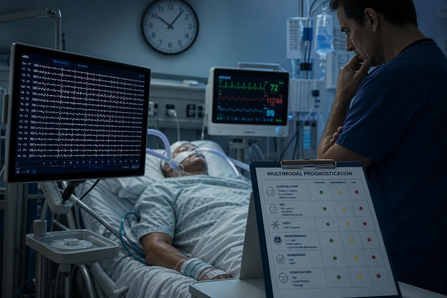
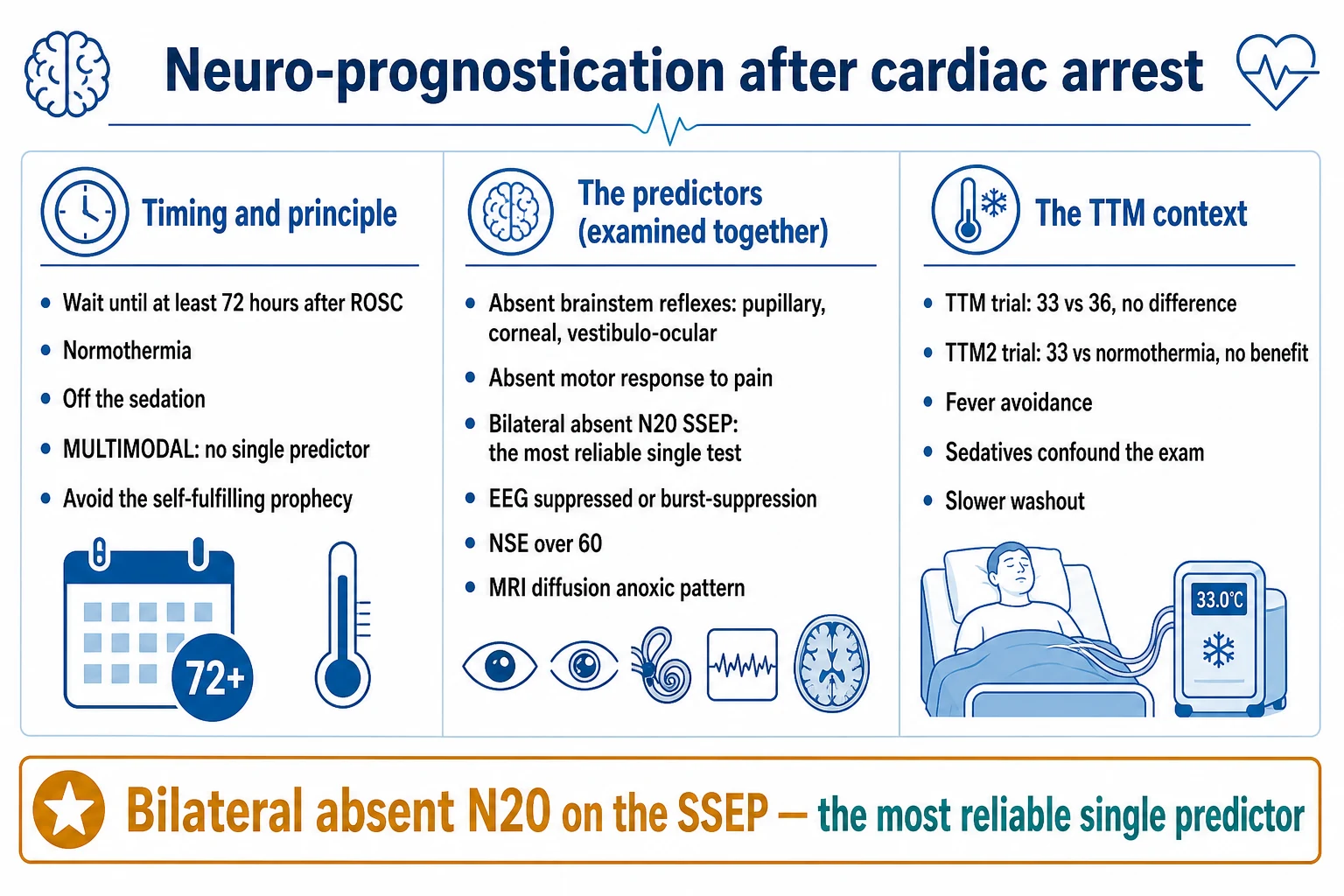
The prognostication is the critical, the ethically-weighted decision — the prediction of the neurological outcome, to guide the continuation or the withdrawal of the life-sustaining therapy. It is multimodal (no single predictor is sufficient) and it is performed at 72 hours after the ROSC (or after the rewarming from the TTM), with the confounders excluded (the sedation, the hypothermia, the neuromuscular blockade, the metabolic disturbance).[4]
The multimodal approach (the ERC/EVICM advisory, Sandroni 2014):[4]
- The clinical examination — the absent brainstem reflexes (the pupillary, the corneal, the gag, the cough), the absent motor response (or the extensor/myoclonus) at 72 hours.
- The EEG — the suppressed background, the burst-suppression, the status epilepticus.
- The neuroimaging — the CT (the cerebral oedema, the loss of the grey-white differentiation), the MRI (the diffusion restriction in the cortex, the basal ganglia, the thalamus).
- The biomarkers — the neuron-specific enolase (the NSE), rising over the first 72 hours, a high value predicting the poor outcome.
- The somatosensory evoked potentials (the SSEPs) — the absent N20 (the bilateral cortical response to the median nerve stimulation) is the robust predictor of the poor outcome.
The principle — no single modality is sufficient; the prediction is multimodal, delayed to 72 hours, and the confounders excluded. The false-positive rate (the prediction of the poor outcome in the patient who later recovers) must be very low, for the prediction guides the withdrawal of the life-sustaining therapy.[4][1]
Management: the integrated post-arrest protocol
The post-arrest management is the integrated, the protocolised, the evidence-based care.[1][1]
- The targeted temperature management — the strict normothermia (37.5 to 37.8 degrees, the TTM2 evidence), the aggressive fever avoidance for 72 hours, the shivering control.[1]
- The coronary angiography — the immediate for the ST elevation; the delayed (or the deferred) for the non-ST-elevation, the stable patient (COACT).[3]
- The ventilation — the normoxia (the 94 to 98 per cent saturation, the lowest FiO2), the normocapnia (the 35 to 45 mmHg).[1]
- The blood pressure — the MAP of at least 65 mmHg (the noradrenaline, the inotrope if the stunning).[1]
- The glycaemic control — the 6 to 10 mmol/L target, the avoidance of the hypoglycaemia.
- The seizure surveillance — the continuous EEG, the aggressive treatment of the seizures.[1]
- The prognostication at 72 hours — the multimodal, the confounders excluded.[4]
Monitoring the post-arrest patient
Monitoring divides into the brain, the heart and the systemic.[1][1]
- The brain — the continuous EEG (the background, the seizures), the core temperature (the normothermia), the neurological examination at each shift.
- The heart — the ECG (the arrhythmia, the ischaemia), the troponin (the trend), the echocardiogram (the stunning, the function), the blood pressure (the MAP target).
- The systemic — the renal function, the liver, the coagulation, the lactate (the clearance). [1]
Prognosis and the rehabilitation
The survival of the out-of-hospital cardiac arrest with the good neurological outcome is about 8 to 10 per cent overall (and higher with the bystander CPR, the early defibrillation, and the witnessed VF arrest — up to 30 to 50 per cent). The survival of the in-hospital arrest is higher. The survivors need the structured rehabilitation (the cognitive, the physical, the psychological), and the assessment of the cause (the ICD for the secondary prevention, the coronary intervention, the medication).[1][1]
Red flags
SAQ — Post-ROSC targeted temperature management at 36 degrees C in a comatose VF arrest survivor
10 minutes · 10 marks
A 58-year-old man is admitted to ICU after an out-of-hospital VF cardiac arrest. Bystander CPR was commenced within three minutes and ROSC achieved after 15 minutes with three defibrillations and 1 mg adrenaline. He remains comatose (GCS 6), intubated and ventilated. The ECG shows anterior STEMI and he has just returned from primary PCI (drug-eluting stent to the LAD). Core temperature on arrival is 36.4 degrees C, BP 104/60 on noradrenaline 0.15 mcg/kg/min, lactate 4.2 mmol/L. The consultant asks you to commence targeted temperature management at a target of 36 degrees C.
SAQ — Multimodal prognostication at 72 hours after out-of-hospital cardiac arrest
10 minutes · 10 marks
A 65-year-old woman was admitted 72 hours ago after an out-of-hospital PEA cardiac arrest with an estimated downtime of 25 minutes before paramedic arrival. She was managed with TTM at 36 degrees C for 24 hours and has now been rewarmed to normothermia for 18 hours. Sedation (propofol 150 mcg/kg/min and fentanyl 50 mcg/h) was held 30 hours ago. She remains comatose with GCS 5 (E1V1M3). On examination the pupils are 5 mm and fixed bilaterally, corneal reflexes are present, the gag reflex is absent, and she withdraws to painful stimuli with an extensor (decerebrate) pattern. The neurology team and the family ask for your prognostic assessment.
Clinical pearls
The integrated TTM protocol — the end-to-end flow
The end-to-end TTM protocol from the ROSC to the prognostication
0-4 h: identify the candidate, commence the fever prevention
Patient comatose (GCS <9) after ROSC, ANY rhythm. Decide target per local protocol: normothermia (≤37.5°C, treat >37.8°C) — most units — OR 33°C if non-shockable (HYPERION) or local protocol. Place core temp probe (oesophageal/bladder/PA). Begin paracetamol 1 g q6h + counter-warming from the start.
0-24 h: maintain target, control shivering
Cool/warm to target via surface (Arctic Sun) or intravascular. Monitor BSAS hourly. Escalate: counter-warming → magnesium 4 g bolus + 1-2 g/h → dexmedetomidine 0.2-1.4 mcg/kg/h → opioid (fentanyl). Maintain normothermia or 33°C for the prescribed duration (24-36 h). Avoid fever at all times.
24-36 h: complete the duration at target
Maintain the target until the full duration (24 h HYPERION/HACA, 28 h TTM2, 36 h TTM). Continue shivering control. Continuous EEG (24-48 h). Treat seizures (levetiracetam, valproate). Normoxia (SpO2 94-98%), normocapnia (PaCO2 35-45), MAP ≥65 mmHg.
Rewarming phase (12-16 h): slow, controlled, fever-free
Rewarm at 0.25-0.5°C/h to 37°C. Check K+, Mg2+, PO4^3- every 2-4 h (rebound hyperkalaemia). Treat fever proactively. Continue EEG. Do NOT prognosticate. Maintain normothermia (≤37.5°C) for at least 72 h from ROSC.
72 h: hold sedation, perform the multimodal prognostication
Once normothermic ≥24 h AND sedation held 24-36 h AND no metabolic confounder: perform the multimodal prognostication. Clinical (brainstem reflexes, motor response), EEG (background, status), SSEP (N20), NSE (trend), neuroimaging (MRI). No single modality sufficient. False-positive rate must be very low.
Day 4-7: continue normothermia, support the recovery, plan the rehabilitation
Maintain fever prevention, continue ventilation targets, treat seizures. If recovering: plan the structured rehabilitation (cognitive, physical, psychological). If not: address the cause (coronary intervention, ICD for secondary prevention). Reassess at day 5-7 before any withdrawal decision.
Normothermia strategy
Post-TTM2 default
- Target ≤37.5°C; treat fever >37.8°C aggressively (paracetamol, surface cooling)
- Supported by TTM2 (2021) — no benefit of 33°C over normothermia
- Simpler, less shivering, less electrolyte disturbance, fewer arrhythmias
- Most ANZ/UK units — ILCOR 2022, ERC 2021
33°C strategy
Selective use
- Target 33°C for 24-36 h, then rewarm at 0.25-0.5°C/h
- Possible benefit in non-shockable arrest (HYPERION)
- More shivering, electrolyte shifts, arrhythmia, bleeding (TTM2 trend)
- Awaiting TTM-NonShock trial; reasonable in selected cases
Contraindicated
Do NOT cool
- Traumatic brain injury with raised ICP (Eurotherm3235 — harm)
- Active uncontrolled bleeding (TTM2 — more bleeding trend)
- Severe refractory shock not responding to vasopressors
- Do NOT extrapolate adult TTM2 verdict to neonatal HIE (cooling is standard there)
The high-yield trials summary table
| Trial | Journal/year | Population | Intervention | Key result | Practice change |
|---|---|---|---|---|---|
| HACA | NEJM 2002 | 273 comatose witnessed VF/VT OHCA | 33-34°C × 24 h vs normothermia | Favourable neuro: 55% vs 39% (RR 0.74) | Established 32-34°C as standard for VF OHCA |
| TTM | NEJM 2013 | 950 comatose OHCA (any rhythm) | 33°C vs 36°C × 36 h | No difference (mortality 50% vs 48%) | Collapsed the 32-34 dogma — exact target less important |
| TTM2 | NEJM 2021 | 1861 comatose OHCA (any rhythm) | 33°C vs normothermia | No difference (mortality 50% vs 48%) | Retired routine hypothermia — fever prevention standard |
| HYPERION | NEJM 2019 | 584 comatose non-shockable OHCA | 33°C vs 37°C × 24 h | Better 90-d neuro: 10.2% vs 5.7% (OR 1.85) | Possible role for 33°C in non-shockable arrest |
| Eurotherm3235 | NEJM 2015 | 388 TBI with raised ICP | 32-35°C vs normothermia | Worse outcome (RR 1.42) — stopped for harm | Hypothermia NOT recommended for raised ICP in TBI |
| COACT | NEJM 2019 | 552 comatose non-STEMI OHCA | Immediate vs delayed angiography | No difference (good outcome 64.8% vs 63.4%) | Immediate angiography not mandatory without ST elevation |
| CoolCap | Lancet 2005 | 234 term neonates moderate-severe HIE | Head cooling 34-35°C × 72 h vs control | Better outcome in MODERATE (not severe) HIE | First RCT to show neonatal cooling benefit |
| TOBY | NEJM 2009 | 325 term neonates HIE | Whole-body 33.5°C × 72 h vs control | Survival without neuro abnormality 44% vs 28% (RR 1.57) | Whole-body cooling global standard for neonatal HIE |
| NICHD | Pediatrics 2005 | 208 term neonates HIE | Whole-body 33.5°C × 72 h vs control | ↓ Death/moderate disability (44% vs 62%, RR 0.72) | Confirmed neonatal cooling — 33.5-34.5°C × 72 h within 6 h |
References
- [1]Dankiewicz J, Cronberg T, Lilja G, et al.; TTM2 Trial Investigators. Hypothermia versus Normothermia after Out-of-Hospital Cardiac Arrest N Engl J Med, 2021.PMID 34133859
- [2]Nielsen N, Wetterslev J, Cronberg T, et al. Targeted temperature management at 33°C versus 36°C after cardiac arrest N Engl J Med, 2013.PMID 24237006
- [3]Lemkes JS, Janssens GN, van den Hoeven NW, et al. Coronary Angiography after Cardiac Arrest without ST-Segment Elevation N Engl J Med, 2019.PMID 30883057
- [4]Sandroni C, Cariou A, Cavallaro F, et al. Prognostication in comatose survivors of cardiac arrest: an advisory statement from the European Resuscitation Council and the European Society of Intensive Care Medicine Resuscitation, 2014.PMID 25438253
- [5]Bernard SA, Gray TW, Buist MD, et al.; Hypothermia After Cardiac Arrest (HACA) Study Group. Stabilised cellular immuno-fluorescence assay: CD45 expression as a calibration standard for human leukocytes J Immunol Methods, 2002.PMID 12133619
- [6]The Hypothermia after Cardiac Arrest (HACA) Study Group. A reconfigurable PID fault tolerant tracking controller design for LPV systems ISA Trans, 2020.PMID 31495591
- [7]Lascarrou JB, Merdji H, Le Gouge A, et al.; HYPERION Trial Group. A nanoemulsion-adjuvanted intranasal H5N1 influenza vaccine protects ferrets against homologous and heterologous H5N1 lethal challenge Vaccine, 2019.PMID 31495593
- [8]Andrews PJD, Sinclair HL, Rodriguez A, et al.; Eurotherm3235 Trial Collaborators. Hatha Yoga and Executive Function: A Systematic Review J Altern Complement Med, 2016.PMID 26398441
- [9]Gluckman PD, Wyatt JS, Azzopardi D, et al.; CoolCap Study Group. Evidence that resistance to nilotinib may be due to BCR-ABL, Pgp, or Src kinase overexpression Cancer Res, 2008.PMID 19047160
- [10]Azzopardi DV, Strohm B, Edwards AD, et al.; TOBY Study Group. Affirmative action of osteopontin on endothelial progenitors Arterioscler Thromb Vasc Biol, 2008.PMID 19020315
- [11]Shankaran S, Laptook AR, Ehrenkranz RA, et al.; National Institute of Child Health and Human Development Neonatal Research Network. Apical transport and folding of prostate-specific membrane antigen occurs independent of glycan processing J Biol Chem, 2006.PMID 16221666